1. Department of Dermatology, Razi Hospital, Tehran University of Medical Sciences, Tehran, Iran
2. Faculty of Medicine, The University of Queensland, Brisbane, Australia
3. Department of Dermatology, Ankara University, Medicine Faculty - Ankara, Turkey
4. Rheumatology Research Center, Shariati Hospital, Tehran University of Medical Sciences, Tehran, Iran
first study in this field and the findings may give way to further investigations.
Keywords: Behcet's disease; Dermatoscopy.
IntroductIon
Behcet’s disease (BD) is a multisystem vasculitis, with a relapsing remitting nature, most commonly presenting with mucocutaneous lesions such as oral and genital aphthous ulcers, erythema nodosum-like features, papulopustular lesions and pseudo-folliculitis as well as a variety of possible non-cutaneous features including uveitis, arthritis, vascular manifestations, epididymitis and neural and gastrointestinal disorders1-4. This rare
disease was originally endemic in geographic areas along the ancient silk road, including Iran; however due to migration it is now encountered worldwide5,6.
All of the clinical presentations of BD are nonspeci -fic. There are more than 16 sets of diagnostic/classifi-cation criteria for the disease, all of them based on clin-ical features of the disease7. However, there is no
specific laboratory test, the only available test being cu-taneous pathergy, a test which must be read by an ex-pert after 24-48hours8.
Recently dermatoscopy, a novel non-invasive diag-nostic technique, has been employed for the evaluation of pigmented and non-pigmented skin, nail apparatus and mucocutaneous lesions, including tumors as well as some inflammatory and infectious diseases. It ena -bles recognition of vascular structures and other sub-tle features that usually are not visible to the naked eye9.
It is not known if dermatoscopy improves the dia gnostic accuracy of BD in comparison with exami-nation with the unaided eye. We designed this study to determine whether if there is any characteristic dermatos co pic pattern in BD which might assist in the diagnosis.
Dermatoscopic and mucoscopic features
of lesions in patients with Behcet’s disease
Ghodsi SZ1, Bahrololoumi Bafruee N1, Chams Davatchi C1, Rosendahl C2, Akay BN3,
Davatchi F4, Chams H4, Shahram F4, Nadji A4, Akhlaghi M4, Faezi T4, Kavosi H4
ACTA REUMATOL PORT. 2019;44:225-231
AbstrAct
Objective: Behcet's disease (BD), as a vasculitis, can
af-fect small and large vessels. As dermatoscopy has been shown to improve the accuracy in diagnosis of various skin lesions especially vascular patterns, we set this study to find if there is any characteristic pattern in the dermatoscopy of Behcet’s mucocutaneous lesions.
Methods: This prospective cross-sectional
observa-tional study designed to evaluate dermatoscopic fea-tures of Behcet’s mucocutaneous lesions. Fifty six con-secutive patients presenting at the outpatient clinic of the BD Research Unit were included. If present, for each patient one oral, one skin and one genital lesion were evaluated by dermatoscopy. When indicated, pathergy test was performed according to a standard protocol and the results were evaluated by dermatoscopy.
Results: A total of 40 oral, 8 genital, 14 skin lesions and
14 pathergy tests were evaluated by dermatoscopy. While vascular component was the most prominent feature in oral aphthae, this component was less promi-nent in genital lesions. Dot vessels were the most com-mon form of vessels in both oral and skin lesions. All the oral lesions were characterized by a central white structureless area. Skin lesions were characterized by a red structureless background. In pathergy tests, nega-tive pricks showed absence of specific features while positive pricks were characterized by a structureless background in pink, purple or red. No obvious vascu-lar component was detected in any of the pricks.
Conclusion: It seems that these findings have no
pAtIents And Methods
Between January and September 2016, 56 consecutive patients presenting at the outpatient clinic of the BD Research Unit (Rheumatology Research Center, Shariati Hospital affiliated to Tehran University of Medical Sciences, Tehran, Iran) were recruited. They all fulfilled BD criteria as defined by the original In-ternational Criteria for Behcet’s Disease (ICBD) and each of them had at least one mucocutaneous lesion at the time of their initial visit10-11. Patients using topical
treatment less than one month prior to recruitment were excluded.
All patients were examined in a multidisciplinary clinic by physicians who were experts in the diagno-sis and management of BD, including rheumatologists, ophthalmologists, and dermatologists.
For each patient one oral, one skin, and one genital lesion were evaluated dermatoscopically if present. If there was more than one lesion of each type, the most recently developed lesion was evaluated. If pathergy testing was indicated it was performed according to the following protocol: three intradermal pricks were performed on the forearm with a needle, the first prick being done with a 21-gauge needle, the second with a 25-gauge needle, and the third with a 25 gauge needle with the injection of one to two drops of normal saline. The test was read 24 hours later by an expert derma-tologist. A positive pathergy test was defined by an ery-thematous papule > 2 mm in diameter surrounded by a large area of erythema or a pustule forming at the site of the needle prick 12-14. The pathergy test reactions
were also evaluated by dermatoscopy after they had first been determined as either positive or negative.
Capture of dermatoscopic images was performed using a DermLite DL3N (San Juan Capistrano, CA, USA) in polarizing mode. All dermatoscopy images were obtained by a single physician (N.B.B).
In order to check the reproducibility of the findings, all of the dermatoscopy images were evaluated by two experts (B.N.A and C.R) and one of the experts (C.R) rechecked all of the images three months later.
For all continuous variables, mean and standard de-viation were calculated but for categorical variables, frequencies were reported.
The study protocol was reviewed and approved by the Dermatologic Review Board Committee and the Ethics Review Committee of Tehran University of Med-ical Sciences. All patients provided written informed consent.
results
Fifty-six patients that fulfilled the original ICBD crite-ria (mean score of 5.31 ± 1.06) and had at least one mucocutaneous lesion were included. The mean age of the patients was 36.8 ±10.9 years and the majority were male (62.5%). Ocular involvement and arthritis were detected in 30 (53.6%) and six (10.7%) patients, respectively. A total of 40 oral, 8 genital, 14 skin le-sions, and 14 pathergy tests were dermatoscopically evaluated. Some of the dermatoscopic patterns ob-served are shown in Figures 1-6. The baseline charac-teristics of the patients according to the site of the le-sions are provided in Table I.
orAl Aphthous ulcers:
All of the 40 oral lesions were characterized by a cen-tral white structureless area. Apart from the white structureless area the vascular component was the most prominent feature. The vessels were mostly dis-tributed at the periphery (75%) being mainly in dot form (67.5%) (Figures 1-4). In nine cases (22.5%) no obvious vascular component was detected (Table II).
GenItAl Aphthous ulcers:
All the eight genital ulcers were characterized by a cen-tral white structureless area. The vascular component was a less prominent feature compared to oral lesions and vessels were distributed at periphery in 50% of the cases. Dot and curved were the most common ves-sels structure in the genital lesions there being 50% in each category (Table II). In three cases (37.5%) no ob-vious vascular pattern was detected. The fiber sign (Figure 5), a dermatoscopic sign associated with ul-ceration was prevalent in these lesions (62.5%).
skIn lesIons:
Overall, skin lesions (n=14, including erythema no-dosum n=5, pseudo-folliculitis n=7, thrombophlebitis n=1, and erythematous edematous plaque n=1) were characterized by a red structureless area with a vascu-lar pattern only seen in four cases (28.6%). These le-sions showed an absence of any specific features (Table II, Figure 6).
pAtherGy tests:
For 14 patients pathergy testing was performed with three prick tests as described. In all of these patients at least one of the prick tests was positive (in five cases all three were positive, in five cases two were positive,
and in four cases one was positive). All of the 42 prick tests were evaluated dermatoscopically after reading. No obvious vascular pattern was detected at any of these sites.
All positive prick test sites were characterized by a structureless pink, purple or red area, and a central yel-low clod was seen in nine (31%). Negative prick tests did not display any specific features showing only a central red dot surrounded by a very small structure-less pink area (Table III, Figure 7).
dIscussIon
Behcet’s disease is a vasculitis which can affect vessels of all types and sizes. Micro- and macro-vascular
in-FIGure 1.Oral aphthous ulcer: a central white structureless area surrounded by a white collarette (arrow) as well as coiled
(circle) and dotted (rectangle) vessels FIGure 4.Oral aphthous ulcer: a white structureless area
surrounded by serpentine (circle), curved (arrows), dotted (pentagon) and looped (rectangle) vessels
FIGure 5.Genital ulcer: a yellow structureless area with fiber sign (rectangle) as a clue to ulceration. The minimal peripheral structureless pink area is not associated with any vascular pattern presumably the stratum corneum of genital skin obscures vessel structures and thus vascular patterns.
FIGure 6.Papulopustular lesion: a central yellow clod (rectangle) centered on a pink structureless area without any visible vessel pattern
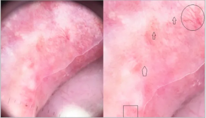
FIGure 2.Oral aphthous ulcer: a central white structureless area surrounded by polymorphic vessels including dotted (arrows), looped (circle) and curved (rectangle) vessels
FIGure 3.Oral aphthous ulcer: a central white structureless area surrounded by dotted (rectangle) and linear (circle) vessels
fold capillaroscopy (NFC) was used in previous stu -dies16-18. These studies have shown NFC abnormalities
with different frequencies and different patterns in pa-tients with BD. These differences between published studies may be due to different magnification (3.2× to 200×) employed for capillaroscopy, different exclusion volvement is always seen and correlates with the cli
-nical presentations15. The inflammatory vessel lesions
are the histologic and pathogenic substrate of the dis-ease and are responsible for the clinical patterns that are the main clues in the diagnostic clinical criteria of BD15.
For in vivo evaluation of microcirculation in BD,
nail-tAble I. pAtIents’ deMoGrAphIc chArActerIstIcs AccordInG to the sIte oF the lesIons Site of the lesions
Total Genital (n=8) Oral (n=40) Skin (n=14) Pathergy (n=14) Sex (male) 35 (62.5%) 6 (85%) 23 (57.5%) 11 (78.6%) 8 (57.1%) Age 36.8±10.9 34.1±6.3 36.8±10.0 40.6±13.5 34.9±10.2 n=7 n=31 n=5 n=10 HLA HLA-B27 3 (7.9%) 2 (40%) 3 (9.7%) 1 (14.3%) 0 (0%) HLA-B5 25 (65.8%) 3 (60%) 20 (65.4%) 6 (85.7%) 6 (42.9%) HLA-B51 20 (52.6%) 1 (20%) 16 (51.6%) 5 (71.4%) 6 (42.9%) Ocular involvement 30 (53.6%) 4 (50%) 18 (45%) 8 (57.1%) 8 (57.1%) Arthritis 6 (10.7%) 2 (25%) 2 (5%) 3 (21.4%) 1 (7.1%)
tAble II. Frequency oF derMAtoscopIc crIterIA In orAl, skIn, GenItAl lesIons And pAtherGy test Pathergy test Total Oral lesions Skin lesions Genital lesions (n=42) (n=104) (n=40) (n=14) (n=8) Negative Positive Vessel Distribution peripheral 36 (34.6%) 30 (75%) 2 (14.3%) 4 (50%) 0 (0%) 0 (0%) central 9 (8.7%) 6 (15%) 2 (14.3%) 1 (12.5%) 0 (0%) 0 (0%) Shape dot 34 (32.7%) 27 (67.5%) 3 (21.4%) 4 (50%) 0 (0%) 0 (0%) linear 17 (16.3%) 15 (37.5%) 0 (0%) 2 (25%) 0 (0%) 0 (0%) serpentine 6% (5.8) 4 (10%) 1 (7.1%) 1 (12.5%) 0 (0%) 0 (0%) coiled 4 (3.8) 3 (7.5%) 0 (0%) 1 (12.5%) 0 (0%) 0 (0%) looped 1 (1%) 1 (2.5%) 0 (0%) 0 (0%) 0 (0%) 0 (0%) curved 11 (10.6) 7 (17.5) 0 (0%) 4 (50%) 0 (0%) 0 (0%)
Central white structureless 48 (46.1%) 40 (100%) 0 (0%) 8 (100%) 0 (0%) 0 (0%) Collarette scale 4 (3.8%) 1 (2.5%) 1 (7.1%) 2 (25%) 0 (0%) 0 (0%) Structureless collarette 1 (1%) 0 (0%) 0 (0%) 1 (12.5%) 0 (0%) 0 (0%)
Fiber sign 7 (6.7%) 2 (5%) 0 (0%) 5 (62.5%) 0 (0%) 0 (0%)
Red structureless halo 21 (20.2%) 18 (45%) 1 (7.1%) 2 (25%) 0 (0%) 0 (0%)
Crust 3 (2.9%) 0 (0%) 2 (14.3%) 1 (12.5%) 0 (0%) 0 (0%)
Red structureless area 50 (48.1%) 0 (0%) 13 (92.9 %) 1 (12.5%) 7 (53.8%) 29 (100%) Central yellow clod 9 (8.6%) 0 (0%) 0 (0%) 0 (0%) 0 (0%) 9 (31%) Central red dot 32 (30.8%) 0 (0%) 0 (0%) 0 (0%) 13 (100%) 19 (65.5%)
criteria, and varying definitions of abnormality or be-cause of the ethnic variations between patients.
Pastorelli et al.,18used videocapillaroscopy at 200
times magnification to evaluate microcirculation, not only in hand and foot nail folds but also in gingival margins, labial mucosa, and conjunctiva. They found videocapillaroscopy to be useful in diagnosis,
follow-up, and response to treatment of BD by showing the systemic extension of the vascular inflammatory pro-cess and the alteration of vessel structure, especially in peripheral and conjunctival areas.
A number of different devices with different designs and magnifying power have been used for capil-laroscopy. A total magnification of 12× to 14× was sug-gested to provide optimal conditions for evaluating the overall pattern of the capillary bed19. On the other
hand, it was reported that capillaroscopy findings ob-tained with a dermatoscope are comparable with those described for other instruments19, 20. Dermatoscopy
ena bles the assessment of many lesions in a short time and is accessible in many clinics with the magnifica-tion power typically of 10 times. As dermatoscopy has been shown to improve diagnostic accuracy for various skin lesions, we anticipated that it might improve the diagnostic accuracy for BD lesions.
Scherrer et al.,21assessed dermatoscopic and
histo-logic characteristics of pathergy tests in 23 patients. Dermatoscopy showed needle marks and mild erythe-ma in the negative tests (two patients) and erytheerythe-ma- tous papule/pustule surrounded by a larger erythema-tous and/or edemaerythema-tous area in the positive cases. In this study they concluded that dermatoscopy was a use-ful tool specially to examine the inflammatory aspects of the small lesions (less than 2mm) resulting from pathergy testing21. In our study, 14 patients underwent
pathergy testing which was performed with three se -parate prick tests on each patient. In all the patients at least one of the prick tests was positive. All the 42 prick tests were evaluated dermatoscopically after being read. No obvious vascular pattern was detected in any of the prick tests. All positive prick tests were characterized by a structureless pink area as well as a central yellow clod in nine cases (31%). Negative prick tests showed tAble III. Frequency oF derMAtoscopIc FIndInGs In pAtherGy tests (42 prIcks)
Total (n=42) Negative Pathergy (n=13) Positive pathergy (n=29)
Vessels 0 (0%) 0 (0%) 0 (0%)
Central yellow clod 9 (21.4%) 0 (0%) 9 (31%)
Central hemorrhagic crust 32 (76.2%) 13 (100%) 19 (65.5%)
Brown dot 1 (2.4%) 0 (0%) 1 (3.4%)
Pink structureless 22 (52.4%) 5 (38.5%) 17 (58.6%)
Purple structureless 7 (16.4%) 2 (15.4%) 5 (17.2%)
Red structureless 7 (16.4%) 0 (0%) 7 (24.1%)
Structureless in total 36 (85.7%) 7 (46.1%) 29 (100%)
FIGure 7.Pathergy Test: A: Close-up image of pathergy test with 3 pricks applied as described in the text, with the prick sites variously labelled B, C and D. B: Dermatoscopic view of the prick test B shown in the first image with a negative result manifested by a central red dot and minimal surrounding erythema. C: Dermoscopic view of the prick test C shown in the first image with a positive result manifested by a central yellow clod and a wide area of surrounding erythema. D: Dermoscopic view of prick test D shown in the first image with a negative result manifested by a red dot and minimal surrounding erythema as in image “B”.
an absence of specific features except a central red dot surrounded by a very small structureless pink area. This finding was similar to that that made by Scherrer
et al. The pink structureless area surrounding the prick
site would reasonably correlate with the reactive hyperemia which is the basis of pathergy testing, the lar -ger area covered by this hyperemia defining a positive result. It is possible that further studies comparing novices to experienced readers of pathergy tests may show that dermatoscopy may help physicians with less experience, to diagnose a positive pathergy test with more confidence.
Currently dermatoscopy is widely used for the exa -mination of skin lesions but its utility for evaluating mu-cous membranes has only had limited investigation in a few studies on mucosal melanomas. Dermatoscopic features of lesions located on the mucous membranes are expected to differ compared to skin lesions22. Our
study showed that a central white structureless area with a peripheral vascular pattern was the most prominent feature in oral lesions, the vascular pattern being less prominent in genital lesions. Dot form was the most common vessel in both lesions. The dermatoscopic cen-tral white structureless area seen with mucosal lesions demonstrate the clinically evident ulceration and the surrounding vascular pattern would reasonably demon-strate dilated dermal papillary vessels consistent with reactive hyperemia in response to the ulceration. It is likely that the stratum corneum of genital skin obscures vessel structures and thus vascular patterns.
The main limitation of our study was the absence of any control group. A small number of patients and limi -tations to skin and genital lesions is another reason for our results to be interpreted with caution. Finally, we did not take into account the duration of the disease in our cases, this being something which may affect the pattern of lesions due to factors of disease progression or medical therapy.
In conclusion, while a central structureless white area and a peripheral vascular pattern were the most prominent features in oral lesions, this vascular pattern was less prominent in genital lesions. Dotted vessels were the most common form in both oral and skin le-sions. Skin lesions were characterized by a red struc-tureless area. In pathergy tests, positive prick tests were characterized by a large structureless pink area with no vascular component detected. We discovered no spe-cific clues for the diagnosis of BD but our study is the first study in this field and further investigation may be warranted.
correspondence to Negar Bahrololoumi Bafruee
Razi Hospital, Vahdat Islami Sq, 1199663911 Tehran, Iran. E-mail: [email protected]
reFerences
1. Faezi ST, Paragomi P, Shahram F, Shams H, Shams-Davatchi C, Ghodsi Z, et al. Clinical features of Behcet's disease in patients without oral aphthosis. Mod Rheumatol. 2014; 24: 637-639. 2. Mat MC, Sevim A, Fresko İ, Tüzün Y. Behçet’s disease as a
sys-temic disease. Clin Dermatol. 2014; 32: 435-442.
3. Davatchi F, Shahram F, Chams C, Nadji HC. Behçet’s disease. Acta Med Iran. 2005; 43: 233-42.
4. Davatchi F, Shahram F, Chams-Davatchi C, Shams H, Nadji A, Akhlaghi M, et al. Behcet’s disease in Iran: analysis of 6500 cas-es. International Int J Rheum Dis. 2010;13: 367-373. 5. Keino H, Okada AA. Behcet’s disease: global epidemiology of an
Old Silk Road disease. Br J Ophthalmol. 2007; 91: 1573-1574. 6. Davatchi F, Shahram F, Chams-Davatchi C, Shams H, Nadji A, Akhlaghi M, et al. Behcet’s disease: from East to West. Clin Rheumatol. 2010; 29: 823-833.
7. Davatchi F. Diagnosis/Classification Criteria for Behcet's Di sease. Patholog Res Int. 2012; 2012:607921.
8. Davatchi F, Sadeghi Abdollahi B, Chams-Davatchi C, Shahram F, Ghodsi Z, Nadji A, et al. Impact of the positive pathergy test on the performance of classification/diagnosis criteria for Be-hcet’s disease. Mod Rheumatol. 2013; 23: 125-132.
9. Zalaudek I, Argenziano G, Di Stefani A, Ferrara G, Marghoob AA, Hofmann-Wellenhof R, et al. Dermoscopy in general der-matology. Derder-matology. 2006; 212: 7-18.
10. Davatchi F, Assaad‐Khalil S, Calamia KT, Crook JE, Sadeghi‐Abdollahi B, Schirmer M, et al. The International Cri-teria for Behçet's Disease (ICBD): a collaborative study of 27 countries on the sensitivity and specificity of the new criteria. J Eur Acad Dermatol Venereol. 2014; 28: 338-347.
11. Davatchi F, Sadeghi Abdollahi B, Shahram F, Nadji A, Chams-Davatchi C, Shams H, et al. Validation of the International Cri-teria for Behçet’s disease (ICBD) in Iran. Int J Rheum Dis. 2010; 13: 55-60.
12. Antonieta Rios Scherrer M, Porto Fonseca de Castro L, Barreto Rocha V, Pacheco L. The dermatoscopy in the skin pathergy testing: case series in patients with suspected Behçet's Disease. Rev Bras Reumatol. 201; 54: 494-498.
13. Varol A, Seifert O, Anderson CD. The skin pathergy test: in-nately useful?. Arch Dermatol Res. 2010; 302:155-168. 14. Ozden MG, Bek Y, Aydin F, Senturk N, Canturk T, Turanli AY.
Different application techniques of pathergy testing among der-matologists. J Eur Acad Dermatol Venereol. 2010; 24:1240-1242.
15. Fei Y, Li X, Lin S, Song X, Wu Q, Zhu Y, et al. Major vascular involvement in Behcet’s disease: a retrospective study of 796 pa-tients. Clin Rheumatol. 2013; 32: 845-852.
16. Aytekin S, Yuksel EP, Aydin F, Senturk N, Ozden MG, Canturk T, et al. Nailfold capillaroscopy in Behcet disease, performed using videodermoscopy. Clin Exp Dermatol. 2014; 39: 443--447.
17. Alan S, Balkarlı A, Tuna S, Özkan Ü, Temel Ş, Özhan N, et al. The nailfold videocapillaroscopy findings of Behçet's syndrome. Dermatol Sin. 2016; 34: 74-77.
18. Pastorelli M, Pasqui AL, Puccetti L, Beermann U, Biagi F, Camarri A, et al. Quantitative Evaluation of Microvessels in Beh
-çet’s Disease. In: Adamantiades-Beh-çet’s Disease: Springer. 2004; 427-433.
19. Bergman R, Sharony L, Schapira D, Nahir MA, Balbir-Gurman A. The handheld dermatoscope as a nail-fold capillaroscopic instrument. Arch Dermatol. 2003; 139: 1027-1030.
20. Dogan S, Akdogan A, Atakan N. Nailfold capillaroscopy in sys-temic sclerosis: Is there any difference between videocapil-laroscopy and dermatoscopy? Skin Res Technol. 2013; 19: 446--449.
21. Antonieta Rios Scherrer M, Porto Fonseca de Castro L, Barreto Rocha V, Pacheco L.. The dermatoscopy in the skin pathergy testing: case series in patients with suspected Behçet's Disease. Rev Bras Reumatol. 2014; 54: 494-498.
22. Olszewska M, Banka A, Gorska R, Warszawik O. Dermoscopy of pigmented oral lesions. J Dermatol Case Rep. 2008; 2: 43.