SUBGRUPO DE MICROBIOLOGIA
CELL‐HOST INTERACTION: ASSESSING THE ROLE OF
HELPER FACTORS IN HIV‐1 REPLICATION
Sylvie Ferreira Rato
Tese de Doutoramento em Farmácia (Microbiologia), apresentada à
Universidade de Lisboa através da Faculdade de Farmácia
Orientador: Prof. Doutor João Gonçalves
LISBOA
2010
Sylvie Ferreira Rato was financially supported by a PhD fellowship (SFRH/BD/24129/2005) from Fundação para a Ciência e Tecnologia (FCT), Lisboa, Portugal. This work was supported by the grant PTDC/SAU‐MII/65346/2006 from FCT, Lisboa, Portugal.
De acordo com o disposto no ponto 1 do artigo nº 40 do Regulamento de Estudos Pós‐Graduados da Universidade de Lisboa, deliberação nº 961/2003, publicado em Diário da República ‐ II série nº 153 – 5 de Julho de 2003, a autora desta dissertação declara que participou na concepção e execução do trabalho experimental, interpretação dos resultados obtidos e redacção dos manuscriptos.
The Research work described in the present thesis was performed from January of 2006 until July of 2010 under de supervision of Prof. Doutor João Gonçalves at the URIA/CPM of the Faculdade de Farmácia da Universidade de Lisboa, Lisbon, Portugal
The results described in this thesis were included in manuscripts already published or in preparation:
Rato S., Maia S., Brito PM., Resende L., Pereira CF., Moita C., Freitas RP., Moniz‐Pereira, J., Hacohen N., Moita L.F., Goncalves J. “Novel HIV‐1 knockdown targets identified by a restricted long‐term shRNA‐based screen in Jurkat T‐cells.” Plos One. 2010 Feb 17;5(2):e9276.
Rato S., Maia, S., Brito P.M., Resende L., Ramos A., Pereira C.F., Moita L.F., Goncalves, J. “Novel host factors essential for HIV‐1 replication: their importance in HIV‐1 life cycle”. Work presented in the Centennial Retroviruses Meeting, Praha, Czech Republic, April 29‐ May 4. Bologna: MEDIMOND, International Proceedings (accepted for publication).
Rato S., Maia, S., Brito P.M., Resende L., Ramos A., Cardoso J. Simões, P, Moita L.F., Goncalves, J. “DNA‐PKcs, an Helper factor in HIV‐LTR driven Transcription”. Manuscript in
preparation
Aos meus Pais
vii
Preface
“The greatest grand challenge for any scientist is discovering how to prevent the spread of HIV and finding the cure or an effective vaccine for AIDS.”
Philip Emeagwali
The first recognized cases of AIDS, although not yet identified, occurred in the USA in the early 1980s. A number of homosexual men in New York and California suddenly began to develop rare opportunistic infections, such as pneumocystis pneumonia and Kaposis sarcoma, and cancers that seemed stubbornly resistant to any treatment. From these occurrences, a number of theories were developed included infection with cytomegalovirus, the use of amyl nitrite or butyl nitrate "poppers", and "immune overload", but it was of common sense that these facts were due to a common syndrome.
In January 1983, Montagnier group obtained a lymph node biopsy from a young homosexual man with a lymphadenopathy in the neck. They analysed the lymph node, dissociated it into single cells, and cultured the T lymphocytes with interleukin‐2 and antiserum to human interferon. Surprisingly, Françoise Barré‐Sinoussi found traces of reverse transcriptase viral enzyme in the supernatant of the lymphocyte culture, indicating the presence of a retrovirus. However, after testing if the viral proteins in the supernatant were recognized by antibodies against HTLV (the only type of retrovirus known at the time, discovered by Gallo´s group), the
viii
labelled viral supernatant did not precipitated with the HTLV antibodies, but could be precipitated with a protein of the patient’s own serum. This protein with a molecular mass of about 25 kD seemed to be the equivalent to the p24 protein of HTLV‐1. However they were not able to isolate the virus from the lymphocytes. Afterwards, several blood samples from other infected patients were analysed in Montagnier laboratory and other laboratories, and the first publications came out at the end of 1983 were Montagnier and Gallo’s groups identified a new retrovirus as the causative agent of AIDS (Barre‐Sinoussi et al., 1983; Gallo et
al., 1983). Gallo claimed that the virus they isolated from an AIDS patient was strikingly similar in shape to other HTLVs and designated by HTLV‐III. In contrast, Montagnier's group demonstrated that the isolated virus was distinct from HTLV‐1 and named it LAV from lymphadenopathy‐associated virus (LAV). HIV was chosen as a compromise between the two claims (LAV and HTLV‐III).
Soon after the identification of AIDS in humans, similar symptoms were identified in captive colonies of Asian rhesus macaques in USA primate centres. The sera from these animals showed cross‐reactivity to HIV‐1 antigens, which led to the identification of the related lentivirus, termed simian immunodeficiency virus (SIV) (Daniel et al., 1985). Subsequently, it was isolated a virus from West African AIDS patients from Guinea‐Bissau and Cape Verde, more closely related to SIV than HIV‐1. This virus, referred to as lymphadenopathy‐associated virus type 2 was then denominated as HIV‐2 (Clavel et al., 1986).
From 1983 until nowadays, the scientific achievements in HIV research have been outstanding. Thousands of scientist articles have been published and a great knowledge was achieved at diverse areas as etiology, molecular virology, natural history, epidemiology and virus pathogenesis. Moreover, the identification of infected patients through blood tests and the development of anti‐retroviral drugs have helped the infected patients to be better controlled and having a prolonged life. Nevertheless, despite the effort, a cure for HIV‐1 has not been achieved and AIDS pandemic continues to be a reality, being the underdeveloped countries the most affected.
Therefore it is imperative to continue to study HIV‐1 infection giving close attention to the mechanisms underlying the complex battle between HIV‐1 and its host. It is important to develop new antiviral strategies and new therapies that can be effective against HIV‐1.
ix With these goals in mind, I have performed my PhD research, focused on the interplay between HIV and its host. The presented thesis is divided in five chapters: Chapter 1 provides a general introduction and review of HIV‐1 biology and the cellular proteins and pathways known to be involved in HIV‐1 infection; Chapter 2 describes an iterative shRNA screen in Jurkat T cells that we have developed to identify helper factors for HIV‐1 infection. Here it is described the screen itself and the identification and validation of newly cellular proteins essential for HIV‐1 replication; Chapter 3 presents the characterization of the identified proteins function during the major steps of HIV‐1 life cycle; Chapter 4 focus on the characterization of DNA‐PKcs function in HIV‐1 replication, compared with CIB2, identified in the shRNA screen; and Chapter 5 integrates our overall findings in a general conclusion. With this thesis we hope to contribute to a better knowledge of HIV‐1 infection, through the study of mechanisms involved in the interaction between HIV‐1 and its cell host and ultimately by providing evidences for new therapeutic targets against HIV‐1.
x
xi
Abstract
Much progress has been made in the past twenty‐seven years to understand the complexity of human immunodeficiency virus type 1 (HIV‐1) infection and to develop an efficient strategy that can eliminate the virus from its host. Despite all efforts this strategy has not been achieved mainly due to the emergence of resistant viruses and to the persistence of latently infected viral reservoirs. Hence, it is crucial to identify novel drug targets and new therapeutic strategies to combat the acquired immunodeficiency disease syndrome (AIDS). It is conceivable that the use of cellular proteins as antiviral targets instead of viral proteins could be a good alternative strategy, once cellular proteins are less variable than those from viral origin, avoiding effectiveness fluctuations of drug efficacy. Therefore it is important to understand HIV‐1 and host interaction, by studying the exploitation of the cellular machinery by HIV‐1.
The work described in this thesis was focused on the interaction between HIV‐1 and its host cell and on the discovery of new co‐factors for HIV‐1 infection. We have developed an iterative shRNA screen in Jurkat T CD4+ cells to identify co‐factors for HIV‐1 infection, focusing on kinases and phosphatases, a very druggable class of proteins. With this innovative screen we were able to identify 14 new cellular proteins essential for HIV‐1 replication in T lymphocytes but that do not interfere with cell survival. We have identified two phosphatases, PTPN9 and PTPRE; five kinases, PRKD1, MAP3K2, MAPK9, SGK and STK24; one hypothetical kinase‐binding‐protein, CIB2; two phosphatase‐binding‐proteins, PPFIA2 and
xii
PPFIBP1; and four other proteins with diverse functions, RAD23B, EZH2, WT1 and ELA1. The role of these proteins in HIV‐1 replication was validated through replication assays with T cell lines‐expressing‐shRNA for each gene in study and the role of the identified co‐factors in different steps of the HIV‐1 life cycle was then evaluated. It was verified that none of the studied proteins have a relevant role in HIV‐1 proviral integration. Instead, all proteins seemed to play an important role before viral integration, in an early step of HIV‐1 life cycle. Moreover, our results indicate that PRKD1, MAP3K2, MAPK9, RAD23B, EZH2, PPFIA2, PPFIBP1, WT1 and STK24 have an additional effect on HIV‐1 LTR transcription.
The identification of CIB2, a hypothetical DNA‐PKcs binding protein, in the initial screen led us to study the cellular protein DNA‐PKcs and its importance in HIV‐1 replication. We assessed its role in different steps of HIV‐1 life cycle and verified that DNA‐PKcs is essential for HIV‐1 replication. Our results indicated that this co‐factor does not have a role in the early steps of HIV‐1 life cycle until viral cDNA integration but it is crucial to HIV‐1‐LTR driven transcription, having dramatic effects in the expression of Tat viral protein levels.
This study brings new insights for the complex interplay of HIV‐1/host cell, showing additional knowledge on cellular proteins and pathways that are essential for HIV‐1 replication but non‐important for cell viability and opens new possibilities for antiviral strategies. Keywords: HIV‐1; shRNA; kinases; phosphatases; DNA‐PKcs.
xiii
Resumo
Desde a descoberta em 1983 do vírus da imunodeficiência humana (VIH) como o agente etiológico da síndrome da imunodeficiência adquirida (SIDA), que tem sido feito um enorme esforço para compreender a complexidade da infecção com o VIH e desenvolver estratégias eficientes que consigam neutralizar o vírus no seu hospedeiro. Actualmente 33,4 milhões de pessoas vivem infectadas com o VIH, tendo sido descritos 2,7 milhões de novos casos em 2008.
O VIH é um retrovírus do género lentivírus, sendo conhecidos dois tipos de VIH, o vírus da imunodeficiência humana tipo 1 (VIH‐1) e de tipo 2 (VIH‐2), ambos identificados como responsáveis pela SIDA. No entanto o VIH‐1 é o mais infeccioso e prevalente na população em geral.
A terapia antiviral actualmente aceite pela comunidade científica e médica, HAART (de Highly
Active AntiRetroviral Treatment), embora consiga aumentar a esperança de vida dos
indivíduos infectados e diminuir a carga viral, não consegue eliminar o VIH‐1 do seu hospedeiro. Uma das principais razões para a persistência da infecção é o aparecimento de mutantes de resistência nos indivíduos infectados devido à baixa fidelidade da enzima viral transcriptase reversa, que gera mutações durante a replicação do genoma viral. Assim sendo, é peremptório o estudo e desenvolvimento de novas terapias antivirais para combater o VIH/SIDA.
xiv
O uso de proteínas celulares como alvos terapêuticos poderá ser uma estratégia alternativa para uma terapia antiviral mais abrangente. As proteínas celulares são muito menos variáveis comparativamente com as proteínas virais, diminuindo assim a probabilidade do aparecimento de mutantes de resistência. Deste modo, as proteínas celulares são consideradas um alvo mais fácil para o desenvolvimento de fármacos antivirais. Por esta razão é imperativo estudar a interacção do VIH‐1 com o hospedeiro, compreendendo melhor as estratégias usadas pelo vírus para explorar as vias bioquímicas celulares para seu próprio benefício.
Duas das famílias de proteínas celulares consideradas como bons alvos para o desenvolvimento de fármacos são as cinases e as fosfatases. Estas proteínas devido à sua actividade catalítica e à sua capacidade de controlar as cascatas de sinalização na célula são um promissor alvo terapêutico. Estas proteínas são actualmente usadas como alvos terapêuticos para diversas doenças, como o cancro, sendo comercializados vários inibidores químicos.
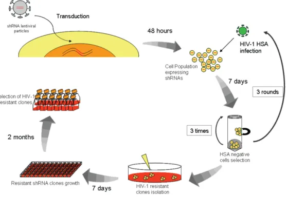
Neste contexto o trabalho desenvolvido nesta tese focou‐se na identificação e estudo de proteínas celulares importantes para a replicação do VIH‐1. Para isso, desenvolvemos um método inovador baseado numa biblioteca de “short hairpin RNAs” (shRNA) que tem como alvo principal as cinases e fosfatases. As moléculas shRNAs, são um tipo de RNA de interferência (RNAi), que quando transcritas numa célula activam a maquinaria do RNAi e inibem especificamente o RNA menssageiro celular alvo. O nosso trabalho baseou‐se numa biblioteca de shRNAs clonada em plasmídeos lentivirais. Estes vectores permitiram a produção de lentivírus que expressam os shRNAs e que após transdução levam à integração do shRNA no genoma da célula, tornando a sua expressão constitutiva. Neste trabalho foi utilizada a linha celular Jurkat, uma linha celular imortalizada de linfócitos T de modo a mimetizar o hospedeiro natural do VIH‐1. Durante o processo, as células Jurkat previamente transduzidas com os shRNA lentivirais foram infectadas com VIH‐1 e, as resistentes à infecção, foram recuperadas e isoladas. Deste modo, com este procedimento obtivemos células que devido à expressão do shRNA adquiriram resistência à infecção com VIH‐1. Após sequenciação dos alvos dos shRNA nas células isoladas, conseguimos identificar 14 proteínas celulares essenciais para a replicação do VIH‐1 em linfócitos T, que não tinham sido descritas anteriormente. Estas proteínas embora essenciais para o VIH‐1 não interferem com a
xv sobrevivência da célula. Dentro deste grupo identificámos duas fosfatases, PTPN9 e PTPRE; cinco cinases, PRKD1, MAP3K2, MAPK9, SGK e STK24; uma proteína que hipoteticamente se liga a uma cinase, CIB2; duas proteínas de ligação a fosfatases, PPFIA2 e PPFIBP1; e outras quatro proteínas, RAD23B, EZH2, WT1 e ELA1 de diferentes funções na célula. As proteínas indentificadas estão envolvidas em várias vias de sinalização celular, nomeadamente nas vias de sinalização das MAPK/JNK, MAPK/ERK e NFκB.
Após a identificação das 14 novas proteínas celulares essenciais para a replicação do VIH‐1 em linfócitos T procedemos à confirmação da sua importância na replicação do VIH‐1 com a constituição de linhas celulares Jurkat a expressarem shRNAs contra cada proteína em estudo. Observámos que a regulação negativa das proteínas por parte dos shRNAs leva a uma drástica inibição da replicação do VIH‐1 sem afectar a viabilidade celular. Posteriormente, continuamos o estudo com ensaios de replicação para avaliar o efeito das proteínas identificadas no ciclo de vida do vírus. Nestes ensaios observou‐se que a inibição da replicação do VIH‐1 nos shRNA clones é progressiva e acumulativa ao longo do tempo. Ao analisarmos especificamente algumas das principais etapas do ciclo de vida do VIH‐1, observámos que as proteínas em estudo não têm uma função importante aquando da integração do genoma viral no cromossoma do hospedeiro, tendo uma função anterior, numa fase inicial do ciclo de vida.
Além das fases iniciais de entrada e integração, estudámos também o papel destas proteínas na transcrição do provírus sob o controlo do promotor viral, LTR. Os nossos resultados indicam que as proteínas PRKD1, MAP3K2, MAPK9, RAD23B, EZH2, PPFIA2, PPFIBP1, WT1 e STK24 além de serem importantes logo numa fase inicial do ciclo de replicação, também são importantes para a transcrição dos RNAs virais.
Numa fase final do trabalho, avaliámos a função da cinase celular, DNA‐PKcs, durante a replicação do VIH‐1, devido à identificação da proteína CIB2 na selecção inicial. CIB2 é uma proteína muito pouco conhecida e com grande homologia com CIB1, outra proteína celular descrita como parceira de interacção com DNA‐PKcs. DNA‐PKcs é uma proteína muito importante para a célula desempenhando diversas funções, sendo a mais crucial na reparação não homóloga de DNA após quebras na cadeia dupla de DNA. DNA‐PKcs já tinha sido anteriormente descrito como um factor celular importante na infecção do VIH‐1 mas a sua função é bastante controversa. No nosso trabalho estudámos o efeito da regulação negativa
xvi
de DNA‐PKcs na replicação do VIH‐1. Para isso constituímos, linhas celulares Jurkat com uma cassete de shRNA contra DNA‐PKcs e avaliámos a replicação viral. Os nossos resultados indicam que a inibição de DNA‐PKcs faz com que as células sejam resistentes à replicação com o VIH‐1 indicando a importância desta proteína para a replicação viral. A função de DNA‐PKcs no ciclo de vida do VIH‐1 também foi avaliada e verificámos que, contrariamente a CIB2, DNA‐ PKcs não possui um papel nem na entrada do vírus na célula nem na integração do genoma viral no cromossoma da célula hospedeira. A importância de DNA‐PKcs na replicação do VIH‐1 parece ocorrer aquando da transcrição dos RNAs virais a partir do promotor viral LTR, afectando significativamente os níveis transcripcionais da proteína viral Tat.
Em resumo, o trabalho desenvolvido nesta tese contribuiu de forma significativa para um melhor conhecimento da complexa interligação entre o VIH‐1 e a célula hospedeira, uma vez que foram identificados novos factores celulares de ajuda para VIH‐1 e que nunca tinham sido directamente relacionados com o vírus. Estas proteínas demonstraram ser importantes para o vírus mas inócuas para a célula. A identificação e caracterização destas proteínas no contexto da replicação do VIH‐1 juntamente com o estudo funcional de DNA‐PKcs contribuiram para uma melhor compreensão dos mecanismos virais de sobrevivência e replicação, na célula hospedeira. Estes estudos além de contribuirem para um aumento de conhecimento nesta área podem levar também ao desenvolvimento de novas terapias antivirais mais abrangentes, tendo como alvos as proteínas celulares identificadas. Palavras‐chave: VIH‐1; shRNA; cinases; fosfatases, DNA‐PKcs.
xvii
Acknowledgments (Agradecimentos)
As minhas primeiras palavras de agradecimento são dirigidas ao Professor Doutor João Gonçalves, orientador desta tese. Agradeço o facto de ter acreditado e apostado em mim; por tudo o que me ensinou; pelas longas conversas e debate de ideias; por me ter feito evoluir a nível científico e ter‐me ajudado a chegar até aqui. E acima de tudo pela amizade, ajuda, compreensão e apoio pessoal. Um sincero muito obrigada!
À Faculdade de Farmácia da Universidade de Lisboa e à Unidade de Retrovírus e Infecções Associadas que tão bem me acolheram. Em especial, ao Professor Doutor José Moniz Pereira por me ter proporcionado a realização deste trabalho e ter disponibilizado os meios necessários para a execução do trabalho experimental. A todos os meus colegas da Microbiologia, pela boa disposição e camaradagem. Um especial obrigada ao Pedro Borrego e à Cheila Rocha por tornarem as horas infindáveis que passámos no P3 parecerem mais curtas e bastante mais divertidas. À Professora Doutora Madalena Pimentel, por todas as suas palavras de apoio e pela ajuda. Obrigada também por ter possibilitado a escrita desta tese no seu gabinete, foi uma ajuda crucial. Ao Professor João Vital, pela sua constante preocupação e pelas suas histórias fantásticas que sempre me presenteou. À Filipa, um muito obrigada, pelo companheirismo e amizade e pela companhia de muitos fins‐de‐semanas no CPM. À Maria João Catalão, pela
xviii
grande amiga que te tornaste, pelo apoio e por tudo o que partilhámos neste anos de Doutoramento.
A todos os meus colegas e amigos do laboratório. A todos os que já lá trabalharam (que já são alguns) e aos presentes. À Sofia Coelho, Maria João Leite, Ana Fonseca, Sofia Côrte‐Real, Iris, Claudia e Rita Nogueira um beijinho grande e obrigada por estarem lá. Ao Fred pela tua paciência, apoio e boa disposição que sempre me presentiaste. Ao Acil pelas conversas sábias e ajuda que sempre te prontificaste a dar. À Leonor por toda a ajuda e tantas horas de P3 dedicadas a este trabalho. À Carina pela grande amiga que te tornaste, pela ajuda incondicional e pela alegria e boa disposição que deixaste sempre no laboratório. À Sara, obrigada por tudo o que me ensinaste, pela tua força gigante, pelo apoio, por todas aquelas horas de trabalho em conjunto; pela grande amiga que te tornaste; À Andreia, pela companhia, ajuda “informática” e pela partilha nos tempos de colega de bancada; à Inês Soeiro e Lídia pelos anos de companheirismo que partilhámos neste laboratório; Ao Luís, em especial pela ajuda informática e ao carregador porque sem ele esta tese não estaria escrita; Ao André, por toda a ajuda prestada para este trabalho e pela tua boa disposição contagiante; À Ana Catarina Santos, à Ana Catarina Santos (sem enganos) e à Cátia por toda a boa disposição e pelo companheirismo; À Mariana, por todos estes anos de amizade; por me teres ensinado a base de tudo, sem ela não estaria aqui; por seres uma boa amiga e confidente; e pela ajuda na correcção da tese. À Soraia, pela grande amiga que te tornaste, por me teres aturado tantas e tantas vezes, pelos fins de semanas passados no CPM, um especial obrigada. À Paula por seres como és, por me teres ensinado tanto; pela ajuda preciosa na correcção da tese; mas fundamentalmente pela grande amiga que te tornaste, pelo apoio e presença constantes.
E a todos os que passaram pelo Laboratório e que de uma maneira ou de outra me ajudaram nesta etapa da minha vida, muito obrigada!
Gostaria também de agradecer aos restantes colegas dos outros grupos de investigação do CPM. À Professora Doutora Cecília Rodrigues e a todos os membros do seu grupo. “Cecilianos” obrigada pela vossa constante boa disposição, pela ajuda e por estarem sempre presentes.
xix Obrigada à Professora Doutora Elsa Rodrigues e à Professora Doutora Maria João Gama pela partilha, troca de ideias e apoio. Agradeço, a todos os membros do seu grupo, em especial à Maria por te teres tornado uma grande amiga, pela alegria e apoio e por todos os bons momentos que passámos dentro e fora do CPM. E claro, à Inês, por tudo, pela grande amizade já de tantos anos, pela partilha de tanta e tanta coisa, pela ajuda e dicas preciosas para muitos trabalhos, pela tua alegria e boa disposição contagiantes; resumindo por seres uma excelente amiga!
No Instituto de Medicina Molecular, ao Doutor João Barata e ao seu grupo pela partilha de reagentes e debate de ideias. Ao Doutor Luís Moita e ao seu grupo pela biblioteca. Ao Professor Doutor João Ferreira e à Joana Cardoso pela ajuda, pela disponibilização do seu laboratório e pela partilha de reagentes. À Doutora Ana Espada de Sousa e ao Rui Soares por me disponibilizarem o seu laboratório e pela ajuda em técnicas essenciais. E à Unidade de Citometria de Fluxo por tudo o que me ensinaram e pela ajuda crucial para este trabalho.
A todos os meus amigos, muitos deles descritos acima, mostrando que tantas horas de trabalho foram partilhadas com grandes amigos: Inês Milagre, Paula, Soraia, Sara, Mariana, Carina, Maria João Catalão, Maria, um beijinho grande. À Isabel, ao Hugo, à Sofia, à Tânia, à Ana Rita e a todo o grupo de amigos da faculdade. Obrigada pelo apoio constante e amizade, por me ouvirem e aturarem, por tudo! Adoro‐vos! À família Reis, por me acolherem como se fosse vossa. Por todo o amor, apoio e preocupação. Paulinha, um grande, grande beijinho e muito obrigada por seres como és! À minha família, avós, tios, por todo o apoio e preocupação. Um especial obrigada à minha Tia Lena pelo apoio incessante, pela ajuda e por toda a força dada, principalmente nesta última fase. E por último, aos meus pais, sem eles nunca teria chegado aqui. Obrigada por me terem feito o que sou hoje. Obrigada por acreditarem, pela paciência, por se esforçarem para que eu
xx
conseguisse ter o que fosse preciso para chegar até aqui, por tentarem compreender. Obrigada pelo amor incondicional. Gosto muito de vocês! A TODOS, MUITO, MUITO OBRIGADA!
xxi
Abbreviations
Reagents and Techniques
CPRG ChloroPhenolRed‐β‐D‐Galactopyranoside DMEM Dulbecco’s modified Eagle’s Medium BLAST Basic Local Alignment Search Tool ECL Enhanced ChemiLuminescence reagent ELISA Enzyme‐Linked ImunoSorbent Assay MOI Multiplicity Of Infection FACS Fluorescence Activated Cell Sorting FBS Fetal Bovine Serum HEK293T Human Embryonic Kidney 293T cells PBS Phosphate Buffer Saline PCR Polymerase Chain Reaction RIPA Radio‐Immunoprecipitation Assay buffer RPMI Roswell Park Memorial Institute medium qPCR quantitative Real Time PCR SDS‐PAGE Sodium Dodecyl Sulfate PolyAcrylamide Gel ElectrophoresisGeneral
μg microgram β‐TrCP β‐Transducin repeat‐Containing Protein AGO2 ArGOnaute 2 AIDS Acquire Immunodeficiency Syndrome AIP1/ALIX Actin Interacting Protein 1 AP Adaptor Protein APJ Apelin receptor APOBEC APOlipoprotein B mRNA Editing enzyme, Catalytic polypeptide‐likexxii APOBEC3B APOlipoprotein B mRNA Editing enzyme, Catalytic polypeptide‐like 3B APOBEC3C APOlipoprotein B mRNA Editing enzyme, Catalytic polypeptide‐like 3C APOBEC3F APOlipoprotein B mRNA Editing enzyme, Catalytic polypeptide‐like 3F APOBEC3G APOlipoprotein B mRNA Editing enzyme, Catalytic polypeptide‐like 3G ARP Actin Related Protein ATM Ataxia Telangiectasia Mutated ATR Ataxia Telangiectasia and Rad3 related BAF Barrier‐to–Autointegration Factor BCA2 Breast cancer‐associated gene 2 CA Capsid CBP CREB Binding Protein CCR2 C‐C chemokine Receptor type 2 CCR3 C‐C chemokine Receptor type 3 CCR5 C‐C chemokine Receptor type 5 CCR8 C‐C chemokine Receptor type 8 CCR9 C‐C chemokine Receptor type 9 CD28 Cluster of Differentiation 28 CD3 Cluster of Differentiation 3 CD4 Cluster of Differentiation 4 CD45 Cluster of Differentiation 45 CDK9 Cyclin Dependent Kinase 9 cDNA complementary DNA ChemR23 G protein‐coupled receptor CMKLR1 CIB1 Calcium and Integrin Binding family 1 CIB2 Calcium and Integrin Binding family 2 CMV CytoMegaloVirus CRM1/XPO1 Exportin 1 Cul5 Cullin 5 CXCR1 C‐X‐C chemokine Receptor type 1 CXCR4 C‐X‐C chemokine Receptor type 4 CXCR6 C‐X‐C chemokine Receptor type 6 CXCR7/RDC1 C‐X‐C chemokine Receptor type 7 CypA Cyclophilin A DC Dendritic Cell
DC‐SIGN Dendritic Cell‐Specific Intercellular adhesion molecule‐3‐Grabbing Non‐ integrin DDX1 DeaD boX protein RNA helicase 1 DDX3 DeaD boX protein RNA helicase 3 DNA DeoxyriboNucleic Acid DNA‐PK DNA‐dependent Protein Kinase DNA‐PKcs DNA‐dependent Protein Kinase catalytic subunit dsRNA double‐stranded RNA EED Embryonic Ectoderm Development protein EF Elongation factor EGFP Enhanced Green Fluorescent Protein ELA1 ELAstase 1 EloB Elongin B EloC Elongin C endo‐siRNA/esiRNA endogenous siRNA
xxiii Env Envelop polyprotein ER Endoplasmic Reticulum ERK 1 Extracellular signal‐Regulated Kinase 1 ERK 2 Extracellular signal‐Regulated Kinase 2 ERK 5 Extracellular signal‐Regulated Kinase 5 ESCORT EndoSomal‐associated COmplex Required for Transport EZH2 Enhancer of Zeste Homolog 2 FIV Feline Immunodeficiency Virus Fv1 Friend Virus susceptibility‐1 protein Gag Group specific antigen polyprotein GAPDH GlycerAldehyde 3‐Phosphate DeHydrogenase GR Glucocorticoid Receptor h hour HAART Highly Active Antiretroviral Therapy HAT Histone AcetylTransferase HIV Human Immunodeficiency Virus HIV‐1 Human Immunodeficiency Virus type 1 HIV‐2 Human Immunodeficiency Virus type 2 HLA Human leukocyte Antigen HLE Human Leukocyte Elastase HMG High‐Mobility Group HMG I (Y)/HMGA1 Chromosomal protein A1 HRP HorseRadish Peroxidase HRP2 Hepatoma‐derived growth factor Related Protein 2 HSA Heat Stable Antigen HSP60 Heat shock Protein 60 hTRIM5α human TRIM5α IL‐1 InterLeukine 1 IL‐7 InterLeukine 7 IN Integrase IRES Internal Ribosome Entry Site JAK Janus Kinase JNK Jun N‐terminal Kinase kD kiloDalton LAP2 α Lamina‐Associated Polypeptide 2 alpha LEDGF Lens Epithelium‐Derived Growth Factor LFA‐1 Lymphocyte function‐associated Antigen 1 lhRNA long harpin RNA LTR Long Terminal Repeat MA MAtrix MAP3K2/MEKK2 Mitogen Activated Protein Kinase Kinase 2 MAPK Mitogen‐Activated Protein Kinase MAPK9 Mitogen Activated Protein Kinase 9 MEK MAP kinase kinase MHC I Major Histocompatibility Complex class I MHC II Major Histocompatibility Complex class II min minute miRNA microRNA ml millilitre
xxiv
MLV Murine Leukemia Virus
mM miliMolar
mRNA messenger RNA
NC NucleoCapsid
NCBI National Center for Biotechnology Information NDR1 Nuclear Dbf2‐Related kinase 1 NDR2 Nuclear Dbf2‐Related kinase 2 Nef NEgative Regulatory Factor NES Nuclear Export Signal NFκB Nuclear Factor‐Kappa B NFAT Nuclear Factor of Activated T‐cells Ng nanogram NHEJ Non‐Homologous End Joining NIAID National Institute of Allergy and Infectious Diseases NLS Nuclear Localization Signal nm nanometer NPC Nuclear Pore Channel PACS‐1 Phosphofurin Acidic Cluster Sorting protein 1 PAK2 p21 Activated Kinase 2 PARP‐1 Poly [ADP‐Ribose] Polymerase 1 PBS Primer Binding Site PCAF P300/CBP‐Associated Factor PCE Post‐transcriptional Control Element PI3K PhosphatidylInositol‐3‐Kinase PIC Post‐Integration Complex PIP2 PhosphatidylInositol 4,5‐bisPhosphate PIP3 PhosphatidylInositol (3,4,5)‐trisPhosphate piRNA PIWI‐interacting RNA PKB Protein Kinase B PKC Protein Kinase C PKR Protein Kinase R Pol Polymerase PP1 Protein Phosphatase 1 PP6 Protein Phosphatase 6
PPFIA2 Protein tyrosine Phosphatase, receptor type F polypeptide Interacting (liprin) Alpha 2 PPFIBP1 PTPRF Interacting protein, Binding Protein 1 (liprin beta 1) PR PRotease PRKD1 Protein Kinase D1 PtdIns(3,4)P2 PhosphatidylInositol 3,4‐bisPhosphate P‐TEFb positive Transcription Elongation Factor b PTEN Phosphatase TENsin homolog PTPN9 Protein Tyrosine Phosphatase Non Receptor type 9 PTPRE Protein Tyrosine Phosphatase Receptor type E PTPRF Protein tyrosine Phosphatase, Receptor type F Rab/hRIP human Rev‐Interacting Protein RAD23A RAD23 homolog A(S. Cerevisiae) RAD23B RAD23 homolog B(S. Cerevisiae) ra‐siRNA repeat‐associated siRNA
xxv RBX1 RING‐BoX protein 1 Ref1 Redox factor‐1 Rev Regulator of virus protein expression RHA RNA Helicase A rhTRIM5α rhesus macaque TRIM5α RISC RNA‐Induced Silencing Complex RNA RiboNucleic Acid RNAi RNA interference RNAPol II RNA Polymerase II RNase H RiboNuclease enzyme H RRE Rev Responsive Element RT Reverse Transcriptase RTC Reverse Transcription Complex SAM68 Src‐Associated in Mitosis, 68 kDa SCID Severe Combined ImmunoDeficiency scnRNA scan RNA SGK Serum/Glucocorticoid regulated Kinase SHIP Src Homology 2 domaincontaining Inositol‐5‐Phosphatase shRNA short hairpin RNA siRNA small interfering RNA SIV Simian Immunodeficiency Virus SIVagm SIV from african green monkeys SIVmac SIV from macaques SKIP Splicing‐associated c‐SKi‐Interacting Protein SKP1 S‐phase‐Kinase‐associated Protein 1 SP1 Specificity Protein 1 STAT Signal Transducers and Activator of Transcription STK24 Serine/Threonine Kinase 24 SU SUrface TAR TransActivation Response ta‐siRNA trans‐acting siRNA Tat Transcriptional transactivator protein TF Transcription Factor TFIID Transcription Factor II D TM TransMembrane tncRNA tiny non‐conding RNA TNF‐α Tumor Necrosis Factor‐alpha TRBP1 TAR RNA Binding Protein 1 TRBP2 TAR RNA Binding Protein 2 TRC The RNAi Consortium TRIM5α Tripartite Motif Protein 5 alpha tRNA transference RNA TSG101 Tumor Susceptibility Gene 101 protein U3 Unique 3´ sequence U5 Unique 5´sequence UNAIDS United Nations Joint Programme on HIV/AIDS UNG2 Uracil DNA glycosilase 2 Vif Viral infectivity factor Vpr Viral protein R
xxvi Vpu Viral protein U VSV‐G Glicoprotein G from Vesicular Stomatitis Virus WT1 Wilms Tumor 1 XRCC4 X‐Ray Repair Cross‐Complementing protein 4
xxvii
Table of Contents
Preface vii Abstract xi Resumo xiii Acknowledgements xvii Abbreviations xxi Table of Contents xxvii Chapter 1: General Introduction 1 1.1. Human Immunodeficency Virus and Acquired Immunodeficiency Syndrome (AIDS) 3 1.2. HIV‐1 taxonomy 4 1.3. HIV‐1 structure 4 1.4. HIV‐1 genome 5 1.5. HIV‐1 Life Cycle 7 1.5.1 Early phase 8 1.5.1.1. Virus entry 8 1.5.1.2. Post‐entry: Uncoating and Reverse Transcription 9 1.5.1.3. Nucleus Import and Integration 10 1.5.2. Late Phase 11 1.5.2.1. HIV‐1 LTR Transcription 11
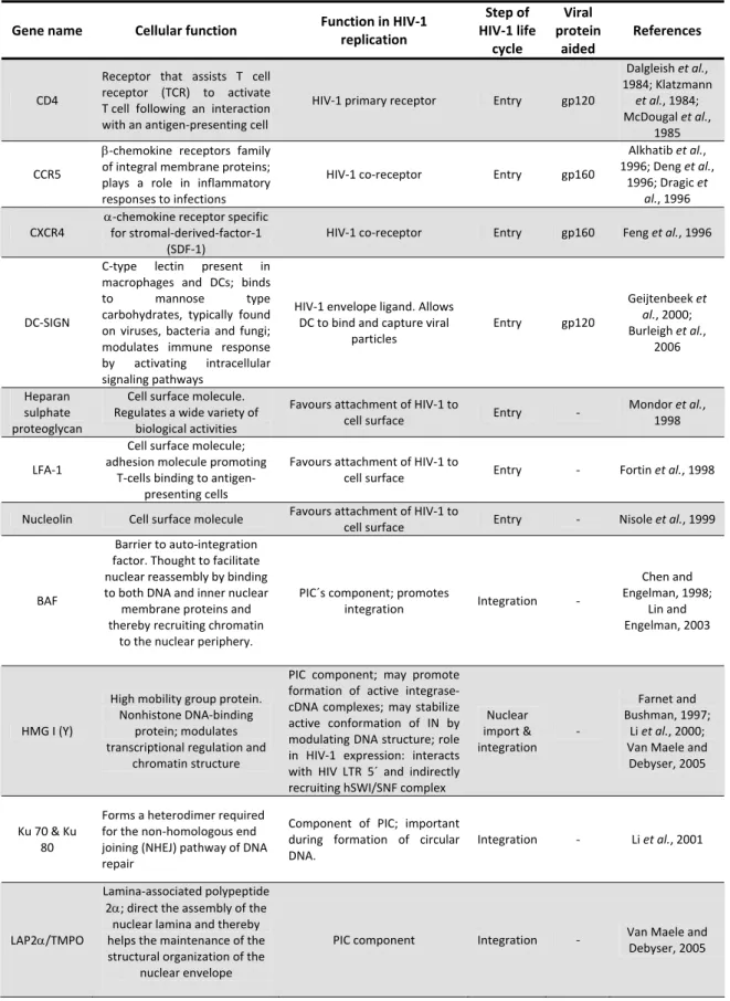
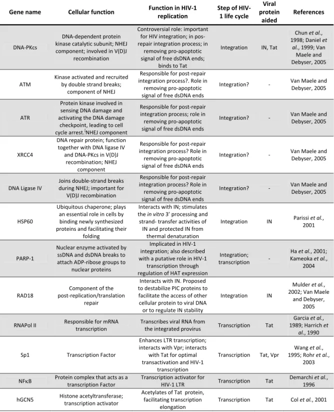
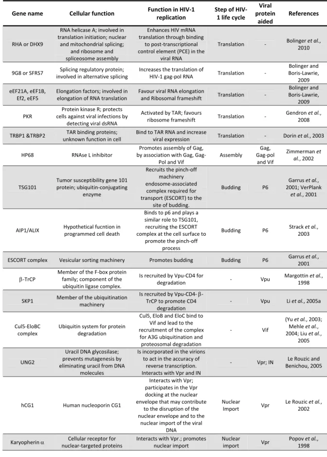
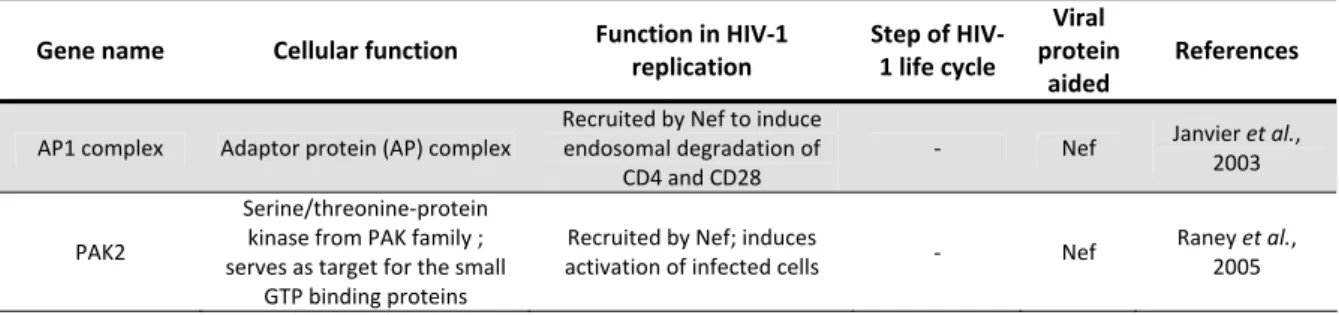
xxviii 1.5.2.2. Gene expression, Assembly and Budding 12 1.5.2.3. Maturation 13 1.6. Host factors for HIV‐1 replication 13 1.6.1 Restriction Factors 14 1.6.1.1. Fv1 15 1.6.1.2. TRIM family 15 1.6.1.3. APOBEC family 17 1.6.1.4. Tetherin 18 1.6.2 Helper Factors 19 1.6.2.1. Role in HIV‐1 entry 25 1.6.2.2. Role in HIV‐1 Pos‐entry until Nucleus import 26 1.6.2.3. Role in HIV‐1 Integration 26 1.6.2.4. Role in HIV‐1 Transcription 28 1.6.2.5. Role in HIV‐1 mRNA Processing and Nuclear export 29 1.6.2.6. Role in HIV‐1 Translation 30 1.6.2.7. Role in HIV‐1 Assembly and Budding 30 1.6.2.8. Role of Helper factors towards viral accessory proteins 31 1.6.2.9. Role of Kinases/Phosphatases and cell signaling in HIV‐1 Infection 32 1.6.2.10. Genome‐wide screenings for identification of Helper factor for HIV‐1 37 1.7. RNA Interference (RNAi) 38 1.7.1. RNAi and HIV‐1 41 Aims 45 Chapter 2: Development of a Long‐Term Iterative shRNA Screen in Jurkat T‐cells to identify kinases/phosphatases important for HIV‐1 replication 47 2.1. Abstract 49 2.2. Introduction 50 2.3. Material and Methods 52 2.3.1. Cell Lines and Culture Conditions 52 2.3.2. Viral Production 52 2.3.3. Infection Assays 52 2.3.4. Lentiviral shRNA Library Screen 53
xxix 2.3.5. shRNA Sequence Identification 53 2.3.6. Development of individual shRNA Jurkat cell clones 54 2.3.7. Quantitative Real‐Time PCR analysis 54 2.3.8. Immunoblotting 56 2.3.9. Assessment of cell viability 57 2.3.10. Bioinformatic analysis 57 2.3.11. Statistical analysis 57 2.4. Results 57 2.4.1. shRNA screening to isolate HIV‐1 resistant Jurkat T‐cells 58 2.4.2. Identification of the shRNAs targets 59 2.4.3. Identified host‐proteins are essential for HIV‐1 replication 70 2.5. Discussion 78 2.6. Acknowledgments 82 Chapter 3: Discovery of Novel Helper Factors essential for HIV‐1 entry and/or HIV‐LTR driven Transcription 83 3.1. Abstract 85 3.2. Introduction 86 3.3. Material and Methods 88 3.3.1. Cell Lines and culture conditions 88 3.3.2. Viral production 88 3.3.3. Infection assays 88 3.3.4. Immunoblotting 88
3.3.5. EGFP‐encoding lentiviral particles production and shRNA clones transduction 89 3.3.6. Flow Cytometry analysis 89 3.3.7. Transient transfection assays 90 3.4. Results 93 3.4.1. Efficient knockdown is of HIV‐1 replication is maintained overtime 91
3.4.2. Knockdown of host‐proteins does not affect integration but rather affect an early step in HIV‐1 replication 97
3.4.3. Knockdown of some host‐proteins affects HIV‐1 LTR driven Replication 98
xxx 3.5. Discussion 100 3.6. Acknowledgments 103 Chapter 4: DNA‐PKcs, an Helper factor in HIV‐LTR driven Transcription 105 4.1. Abstract 107 4.2. Introduction 108 4.3. Material and Methods 112 4.3.1. Cell Lines and culture conditions 112 4.3.2. shRNA Jurkat cell lines construction 112 4.3.3. Viral Production and infection assays 112 4.3.4. Immunoblotting 113 4.3.5. Quantitative Real‐Time PCR analysis 113 4.3.6. EGFP‐encoding Lentiviral Particles Production and shRNA Clones transduction and Flow Cytometry analysis 114 4.3.7. Transient transfection assays 114 4.3.8. Assessment of cell viability 115 4.3.9. Statistical analysis 115 4.4. Results 116 4.4.1. DNA‐PKcs clones constructions efficiently knockdown DNA‐PKcs using shRNAs 116 4.4.2. DNA‐PKcs protein is crucial for HIV‐1 replication 117 4.4.3. DNA‐PKcs‐knockdown affects HIV‐1 replication without influencing host‐cell viability 118 4.4.4. shDNA‐PKcs do not affect integration or early steps in HIV‐1 Replication 119 4.4.5. DNA‐PKcs has acts as co‐factor in HIV‐1‐LTR driven transcription 120 4.4.6. shDNA‐PKcs affects expression of viral RNA transcripts 121 4.5. Discussion 122 4.6. Acknowledgments 125 Chapter 5: Concluding Remarks 127 References 135
xxxi
Figures
Figure 1.1. HIV‐1 structure 4 Figure 1.2. HIV‐1 genome 5 Figure 1.3. Schematic representation of HIV‐1 life cycle 8 Figure.1.4. Host‐restriction factors for HIV‐1 and their viral antagonists 16 Figure 1.5. MAPK pathway 33 Figure 1.6. PI3K/Akt signaling 35 Figure 1.7. Mechanism of RNA interference in mammalian cells 39 Figure 2.1. shRNA screen in Jurkat cells 58 Figure 2.2. Resistance of shRNA clones to HIV‐1 replication 60 Figure 2.3. Representation of shRNA sequence cassette 61 Figure 2.4. Biochemical relationships between identified proteins Network 1 64 Figure 2.5. Biochemical relationships between identified proteins Network 2 65 Figure 2.6. Molecular and cellular functions of identified genes. 66 Figure 2.7. Representation of the different canonical pathways wherein the identified genes are present 67 Figure 2.8. Resistance of shRNA Jurkat clones to HIV‐1 replication 71 Figure 2.9. mRNA knockdown in individual clones stably expressing shRNA 74 Figure 2.10. shRNA clones are resistant to HIV‐1 replication 77 Figure 3.1. Neutralization of HIV‐1 replication by shRNAs is cumulative over time 92 Figure 3.2. Monitoring HIV‐HSA infection in shRNA Jurkat clones. 94 Figure 3.3. CD4 surface expression in Jurkat shRNA clones during HIV‐1 95 Figure 3.4. Knockdown of host‐proteins do not affect integration but affect an early step in HIV‐1 replication 97 Figure 3.5. HIV‐1 LTR‐driven transcription is affected by host‐proteins 98 Figure 4.1. DNA‐PKcs is efficiently knockdown efficiency in shRNA clones 116 Figure 4.2. DNA‐PKcs knockdown inhibit HIV‐1 replication 117 Figure 4.3. shRNA clones viability 118Figure 4.4. Knockdown of DNA‐PKcs do not affect integration or early step during HIV‐1 life cycle 119 Figure 4.5. DNA‐PKcs is important for HIV‐1 LTR‐driven transcription 120 Figure 4.6. DNA‐PKcs knockdown affect late LTR‐driven transcription 121
xxxii
Figure 5.1. Schematic Representation of the studied proteins and their function in the HIV‐1 life cycle 132
Tables
Table 1.1. Highlighted Helper factors for HIV‐1 replication 20 Table 2.1. shRNA sequences used for each gene in study 55 Table 2.2. Oligonucleotide sequence for target‐gene cDNA amplification by qPCR 56 Table 2.3. Proteins identified in the shRNA screen important for HIV‐1 replication 62 Table 2.4. Sequences identified from the 30 clones analysed 63 Table 2.5. Canonical Pathways involving identified genes 68 Table 2.6. Genes identified in this screen as important for HIV‐1 replication 81 Table 4.1. Oligonucleotides used for qPCR 1141
General Introduction
CHAPTER 1
2
3
1.1. Human Immunodeficiency Virus (HIV) and Acquired
Immunodeficiency Syndrome (AIDS)
Human immunodeficiency virus (HIV) is the causative agent of the progressive disease acquired immunodeficiency syndrome (AIDS) (Barre‐Sinoussi et al., 1983; Gallo et al., 1983), one of the leading causes of death worldwide (UNAIDS, 2008). At the present, approximately 33.4 million of people live with HIV, with 2.7 million new cases of HIV infection described in 2008 (UNAIDS, 2009). The natural HIV infection results in a continuous process where progressive depletion of cluster differentiation 4 (CD4)+ T cells and immune dysfunctions occur. This leads to a final stage of the disease where individuals are more susceptible to opportunistic infections or malignancies that constitute the clinically defined AIDS (Fauci et
al., 1985; Phillips et al., 1991). There are two types of human immunodeficiency virus
described, type 1 (HIV‐1) (Barre‐Sinoussi et al., 1983; Gallo et al., 1983) and type 2 (HIV‐2) (Clavel et al., 1986). Both types are known to cause AIDS, although HIV‐2 represents a significant minority off all HIV infections. Furthermore, HIV‐2 is characterized by a slow disease progression and transmission, a lower plasma viral load and a low rate of CD4‐T cell decline (Marlink et al., 1994; Whittle et al., 1994; Jaffar et al., 1997).
Since HIV discovery until our present days many efforts have been made to counteract AIDS. Several drugs were developed and efficient HIV therapies were settled. The current antiviral therapy accepted by scientific and medical community is the highly active anti‐retroviral therapy (HAART) and consists of the administration of a combination of antiviral drugs that act against HIV proteins or viral RNA (De Clercq, 2009). HAART is able to reduce viral loads to almost undetectable levels. Still it has several drawbacks such as toxicity and the appearance of viral resistant mutants. Despite the substantial progress in the development of these anti‐ retroviral drugs, eradication of HIV‐1 and consequently the cure of AIDS has still not been achieved. Therefore, HIV‐1 research is still a very active field of research, where the study of HIV‐1 biology and more precisely the interaction between the virus and its host cell can provide new insights for the development of new antiviral therapeutic strategies.
4
1.2. HIV‐1 taxonomy
HIV‐1 belongs to the genus Lentivirus of the Retroviridae family. Retroviruses are characterized by their single strand positive RNA diploid genome and by the virion polymerase that is capable of RNA‐directed DNA synthesis (reverse transcription) generating a DNA intermediate that is integrated in the host genome (Bishop, 1978; Coffin, 1997).
Lentiviruses differ from other retroviruses mainly due to their long incubation period before manifestations of clinical illness. Moreover, lentiviruses share a common morphogenesis and morphology, a tropism for macrophages, extensive genetic and antigenic variability, and additional regulatory genes not found in the other groups of retroviruses (described below) (Narayan and Clements, 1989; Coffin, 1997).
Phylogenetic studies have distinguished 3 major groups among HIV‐1 isolates: group M (Major), group O (Outlier) and group N (non‐M, non‐O). M group includes more than 90 % of HIV/AIDS cases and it is further subdivided into eleven clades (from A to K) (Geretti, 2006). In 2009, a newly‐analyzed HIV sequence was reported and a new group, Group P, was proposed (Plantier et al., 2009).
1.3. HIV‐1 structure
HIV‐1 mature virions are spherical shaped, with a diameter of approximately 100 nm. Like the other lentiviruses, HIV‐1 has an outer membrane and an inner membrane. The outer viral membrane is derived from the host cell membrane, which consist of a lipid bilayer where the viral surface (gp120 or SU) glycoprotein is anchored via interaction with the viral transmembrane (gp41 or TM) glycoprotein (Turner and Summers, 1999; Wanget al., 2000). The outer viral membrane is also
constituted by cellular proteins derived from
host cell, namely the histocompatibility antigens, actin and ubiquitin (Arthur et al., 1992). The
Figure 1.1. HIV‐1 structure (Elsevier Science :
5 viral inner membrane is composed by several matrix proteins (p17 or MA) that involve the conical capsid core, formed by the capsid protein (p24 or CA). Subsequently, the capsid core encompasses two RNA double strand molecules of approximately 9 Kb, which are stabilized as a ribonucleoprotein complex. The ribonucleoprotein complex is formed by several copies of the nucleocapsid protein (p7 or NC) and by the viral enzymes integrase (p31 or IN), reverse transcriptase (p66/p51 or RT) and protease (p11 or PR). The viral proteins, negative regulatory factor (Nef), virion infectivity factor (Vif) and viral protein R (Vpr) are also incorporated in the viral particle (reviewed in Turner and Summers, 1999; Hoffmann et al., 2007) (Figure 1.1).
1.4. HIV‐1 genome
HIV‐1 carries its genetic information in double strand RNA molecules that are synthesized by the host DNA‐dependent RNA polymerase II (RNAPol II). Similarly to eukaryotic cellular mRNAs, HIV‐1 RNA has post‐translational modifications such as 5´ cap and 3´poly A tract (Whitcomb and Hughes, 1992). During HIV‐1 life cycle, RNA molecules are reverse transcribed by RT to viral complementary DNA (cDNA) and then integrated into the host chromosome. Conceptually, HIV genome organization is discussed in terms of viral DNA after integration (i.e. provirus). Provirus representation places viral promoter, RNA start site and polyadenylation site in the same positions as they are typically found in host genes (Coffin, 1997).
HIV‐1 genome, common to all replication‐competent retroviruses, has three major genes,
gag, for group specific antigen polyprotein (coding for structural proteins), pol, for polymerase
(coding for viral enzymes) and env, for envelop (coding for envelope glycoproteins) flanked by long terminal repeats (LTR) sequences (figure 1.2) (Frankel and Young, 1998; Wang et al., 2000). LTR regions are composed by short directed repeat sequence (R) flanked by unique 5´ (U5) and 3´ (U3) sequences (U5‐R‐U3). These regions are formed after duplication of U3 e U5
Figure 1.2. HIV‐1 genome (adapted by permission from Macmillan Publishers Ltd: [Nat Rev Immunol], Robinson,
6
in both ends during reverse transcription (Whitcomb and Hughes, 1992). LTR sequence contains functional regions essential for HIV‐1 transcription regulation, such as the transactivation response (TAR) element, the basal promoter and the core enhancer (Pereira et
al., 2000).
HIV‐1 genes expression is mediated by viral proteins through regulatory mechanisms with overlapping reading frames and through alternative messenger RNA (mRNA) splicing (Wang et
al., 2000). The gag gene encodes a precursor protein of 55 kDa (p55Gag) that during virus maturation is cleaved by the viral protease into the structural proteins matrix, capsid and nucleocapsid, and into the peptides p1, p2 and p6. The pol gene codifies three enzymes: protease, reverse transcriptase and integrase that are essential during HIV‐1 life cycle (discussed bellow). These proteins are originated from a fusion polyprotein precursor P160GagPol that is synthesized through a rare frameshift event that occurs during P55Gag translation. The env gene encodes the polyprotein Env precursor gp160, which is cleaved by a cellular protease originating the surface (gp120) and the transmembrane (gp41) glycoproteins (Frankel and Young, 1998) (figure 1.2).
Differently from other retroviruses, HIV‐1 codifies for six additional proteins: two regulatory proteins, transcriptional transactivator (Tat) and regulator of virus protein expression (Rev); and four accessory proteins Nef, Vif, Vpr and viral protein U (Vpu) (Frankel and Young, 1998). The regulatory proteins are essential for virus replication by controlling HIV‐1 expression in host cells. On the other hand, the so called “accessory proteins” have this designation since they are often dispensable for virus replication in vitro. However, these proteins play essential roles in virus persistence, spread and pathogenesis in vivo and carry out many important functions during HIV‐1 life cycle by interacting with cellular proteins (Li et al., 2005a)
Tat is a multifunctional protein that acts mainly as a transactivating protein inducing a variety of effects through modulation of cellular and viral genes expression levels. Tat functions include chromatin remodelling, induction of phosphorylation of RNAPol II, transactivation of viral genes and binding to specific structures of HIV‐1 mRNA (reviewed in Romani et al., 2010).
Rev is a regulator of viral mRNA production that binds to rev responsive element (RRE) in viral RNA and facilitates the nuclear export of the single spliced viral RNAs (Hope, 1999).
7 Vif is essential for in vivo infectivity and pathogenesis. Its main function is to counteract the innate antiretroviral defences mediated by cytidines deaminases such as the apoliprotein B mRNA editing enzyme, catalytic polypeptide‐like 3G (APOBEC3G) (Sheehy et al., 2002) and the apoliprotein B‐editing catalytic polypeptide 3F (APOBEC3F) (Liddament et al., 2004; Wiegand
et al., 2004; Zheng et al., 2004) by different mechanisms (discussed below and reviewed in
Henriet et al., 2009).
Nef, Vpr and Vpu act as multiple‐functional proteins in regulating HIV‐1 infectivity and pathogenesis. Nef induces downregulation of CD4, major histocompatibility complex class I (MHC I) and II (MHC II), human leukocyte antigen (HLA) and cluster differentiation 28 (CD28) from HIV‐1 surface of infected cells; enhances virion infectivity; stimulates viral replication; and modulates T cell activation state of its cell host (reviewed in Jere et al., 2010). Vpr modulates transcription of the virus genome, promotes nuclear transport of the HIV‐1 pre‐integration complex (PIC), facilitates reverse transcription, causes G2 cell cycle arrest, induces apoptosis, induces defects in mitosis, suppresses immune activation and balances HIV‐1 mutation rate (reviewed in Romani and Engelbrecht, 2009). Vpu is responsible for CD4 degradation, induction of apoptosis, enhancement of viral particle release and downregulation of MHC I and MHC II (reviewed in Nomaguchi et al., 2008). More recently, it was reported that Vpu counteracts the cellular protein Tetherin, that specifically inhibits virion release from the host cells (Neil et al., 2008).
1.5. HIV‐1 life cycle
HIV‐1 life cycle, commonly to all retroviruses, is divided into two distinct phases: 1) an early phase that begins with the recognition of the target cell by the infectious virion and involves all following steps until integration of the genomic DNA into the chromosome of the host cell; 2) a late phase, which begins with the expression of the integrated proviral genome, and involves all processes up to and including virus budding and maturation of the progeny virions (Turner and Summers, 1999) (represented in Figure 1.3). All these processes depend not only on viral proteins but also on the host cell machinery, which is exploited by the virus during its replication.
8
1.5.1 Early phase
1.5.1.1. Virus entry
HIV‐1 life cycle begins when an infectious particle encounters its cell host. HIV host cells are mostly helper T cells, monocytes and macrophages. Other cells, such as Langerhans, follicular dendritic, glial, and certain colon tumor cell lines, are susceptible to HIV‐1 infection (Vaishnav and Wong‐Staal, 1991; Weiss, 2002). When a typical helper T cell is infected, virion attaches to cell surface through a high‐affinity interaction between the viral glycoprotein gp120 and the primary cell receptor CD4 (Dalgleish et al., 1984; Klatzmann et al., 1984; McDougal et al., 1985). This binding causes structural alterations in gp120 enhancing its affinity to a
Figure 1.3. Schematic representation of HIV‐1 life cycle. See main text for detailed description. Reprinted by
9 coreceptor, that trigger fusion of the viral envelope to the cellular membrane (reviewed in Melikyan, 2008). HIV‐1 co‐receptors were identified such as G protein‐coupled receptor superfamily of seven‐transmembrane domains proteins (Berger et al., 1999). The two major co‐receptors for HIV‐1 infection in vivo are C‐ X‐C chemokine receptor type 4 (CXCR4) (Feng et
al., 1996) and C‐C chemokine Receptor type 5 (CCR5) (Alkhatib et al., 1996; Deng et al., 1996;
Dragic et al., 1996). Based on the CD4/chemokine receptors paradigm, HIV‐1 strains were classified as R5 viruses (isolates that uses only CCR5 co‐receptor), X4 viruses (isolates that use only the CXCR4 coreceptor) and R5X4 viruses (isolates that use both co‐receptors (Berger et
al., 1998). The coreceptor binding triggers conformational changes in gp120 and gp41 viral
glycoproteins, namely the exposure of the gp41 fusion peptide and consequently insertion into the target cell membrane (Doms and Moore, 2000). This process leads to the fusion of the viral envelope with the target cell membrane and to the delivery of viral core into the cytoplasm of the target cell.
1.5.1.2. Post‐entry: Uncoating and Reverse Transcription
Immediately after release of the viral core into the cell host cytoplasm, HIV‐1 undergoes a partial and progressive process of uncoating, where viral core is rearranged into a new structure, the reverse transcription complex (RTC) (Lehmann‐Che and Saib, 2004). The RTCs are large dense complexes that contain a different viral protein composition from the original viral core, like MA and RT; and several cellular host proteins necessary for reverse transcription, translocation and forward processes, such as actin, barrier‐to‐autointegration factor (BAF) and lens epithelium‐derived growth factor (LEDGF/p75) (McDonald et al., 2002; Warrilow and Harrich, 2007). Exposure of RTC to a significant concentration of deoxyribonucleotides in the cytoplasm is thought to trigger the initiation of reverse transcription (Lehmann‐Che and Saib, 2004). Reverse transcription, although being a very complex process, is very well established (reviewed in Gotte et al., 1999). During this reaction, viral RNA genome is converted into a double‐stranded DNA molecule, by the action of the viral heterodimer RT protein (p51/p66). The p66 subunit contains both polymerase and RiboNuclease enzyme H (RNase H) enzymatic activities, essential for the reverse transcription process, while the role of p51 is mainly structural (Kohlstaedt et al., 1992).
![Figure 1.3. Schematic representation of HIV‐1 life cycle. See main text for detailed description. Reprinted by permission from Macmillan Publishers Ltd: [Nat Rev Drug Discov], Flexner, 2007, copyright (2007).](https://thumb-eu.123doks.com/thumbv2/123dok_br/18475617.899551/40.892.170.688.487.1076/schematic-representation-description-reprinted-permission-macmillan-publishers-copyright.webp)