Vol. 44, N. 4 : pp. 383 - 394, December, 2001
ISSN 1516-8913 Printed in Brazil BRAZILIAN ARCHIVES OF
BIOLOGY AND TECHNOLOGY
A N I N T E R N A T I O N A L J O U R N A L
Morphology and Anatomy of the Flowers of
Trichilia catigua
A. Juss.,
T. elegans
A. Juss. and
T. pallida
Sw. (Meliaceae)
Luiz A. de Souza
*; Ismar S. Moscheta; Káthia S. M. Mourão and Adriano Silvério
Universidade Estadual de Maringá, Departamento de Biologia, Avenida Colombo, 5790, 87020-900 Maringá - PR, Brazil
ABSTRACT
Morphological and structural features of flowers ofTrichilia catiguaA. Juss.,T.elegansA. Juss. andT.pallida Sw. (Meliaceae) are presented here with the purpose to stimulate future reproductive and preservation studies of these brazilian native species. Anthesis occurred from April to August inT. catigua, January to May inT.pallida and September to January in T. elegans. Sepals and petals presented a papillose and pilose epidermis and a parenchymatous mesophyll. Male flowers presented pistillodes with abortive ovules and a staminal tube with tetrasporangiate anthers. Anther wall had a papilose epidermis, a fibrous endothecium, two middle layers and a secretory tapetum. Female flowers presented antherodes and bi - or tricarpellate syncarpous pistils. Ovary had a simple structure with anatropous, bitegmic ovules. Style was hollow or solid and the stigma was constituted by uni -or bicelullar trichomes. Strictly dioecious specimens were not found in the study area.
Key words:Trichilia, Meliaceae, floral morphology and anatomy, flowering
*Author for correspondence
INTRODUCTION
Trichilia P. Browne, belonging to the Meliaceae family, accounts for about 70 species distributed throughout Tropical America. The species frequently present unisexual (bisexual to a lesser degree) flowers in dioecious plants and there are even records of polygamous plants (Pennington, 1981; Klein, 1984). In the forest remnants in and near Maringá, Paraná State, Brazil, three species ofTrichiliaoccur: T. catiguaA. Juss (catiguá),T. elegans A. Juss. (pau-de-ervilha) and T. pallida
Sw. (baga-de-morcego). These species have a wide distribution in South and Central America. The first two are more abundant in southern Brazil and the last one is dispersed throughout Brazil and be very common in the southeastern gallery forests (Pennington, 1981; Klein, 1984).
The flowers of Trichilia species are not well known in their anatomical aspects. However, anatomical features such as ovaries and ovules in
T. grandifoliaOliv.,T. elegansA. Juss.,T. pallida
Sw. and T. catigua A. Juss. are described by Boesewinkel (1981), Beltrati (1984), Beltrati and Brunini (1988) and Moscheta (1995), respectively. Morphological descriptions of flowers are limited to the works of Pennington (1981) and Puentes et al. (1993). Studies about floral biology include works of Styles (1972), Opler and Bawa (1978) and Opler (1983).
the situation is more critical, with less than 1% of the forest remnants located in units of conservation and on the islands in the Upper Paraná River which are subject to flooding (Ipardes, 1992; Campos, 1997).
With the purpose of understanding the differentiated behavior of Trichilia plants (dioecious, monoecious and polygamous) recorded in the literature and stimulating future reproductive and preservation studies on Paraná’s forests, morphological and anatomical features of the flowers of Trichilia catigua A. Juss., T. elegansA. Juss., and T. pallidaSw. are presented and discussed.
MATERIALS AND METHODS
The “Horto Florestal Dr. Luiz TeIxeira Mendes”, where the species were observed and studied, is situated in the city of Maringá, Paraná State, Brazil between the meridians 51° 30' and 54°W and the parallels 22° 30' and 24° 30' S, with an average altitude of 556 meters. In this forest remnant, there are shrubs and small trees of
Trichilia elegans A. Juss., and small trees of T. catigua A. Juss. and T. pallida Sw., whose vouchers were deposited in the Herbarium of the Universidade Estadual de Maringá, under numbers HUM 5111, HUM 2210 and HUM 197, respectively.
The flowering time for the species was followed in at least five previously marked specimens between April 1999 and June 2000 by means of weekly observations. During this period, flowers were collected before and after the anthesis, immediately analyzed in the laboratory and fixed in FAA 50 and FPA 50 (Johansen, 1940). The anatomical study of the collected botanical material was sectioned by microtome techniques in paraffin wax (Johansen, 1940) or methacrylate (Ruetze and Schmitt, 1986). Erlich hematoxylin and safranin were used for staining (Johansen, 1940). In addition, temporary and semi-permanent slides obtained from hand sections and stained with safranin and astra blue were made. The following stains were used for specific color tests: phloroglucinol and sulfuric acid, Sudan IV and IKI for lignins, lipids and starch, respectively (Johansen, 1940; Berlyn and Miksche, 1976).
RESULTS
Morphology
The flowering time for the species can be long, as inT. catigua,where the floral buds can stay on the plant for five to six months before the anthesis process begins. In T. pallida and T. elegans the floral buds stay on the plants for about two months. The anthesis occurred from April to August in T. catigua, from January to May in T. pallida and from September to January in T. elegans.
The flowers of the species were minute,
approximately three millimeters long in
T.
elegans,
about four millimeters in
T. catigua
and a little more than five millimeters in
T.
pallida
. These flowers occurred in axillary
panicles in the three species. However, the
inflorescence in
T. catigua
was congested and
in
T. pallida
small racemes with short axes
can occur. The flowers presented a short
pedicel, at whose base occurred a reduced
bract and two bracteoles with the same shape,
but smaller dimensions. They were still
dichlamydeous, actinomorphic and unisexual
(Figures 1 and 2).
The synsepalous green calyx had four sepals inT. catigua and T. pallida (Figure 21) and five in T. elegans.InT. catiguathe sepals were more pilose and reduced. The corolla presented four separate petals (Figure 21), except inT. elegans, which had five separate petals, white to yellow in color and lanceolate (Figures 3 to 5) to ovate in shape. The petals of T. elegans presented only one central vein, which branched out into secondary veins of smaller caliber (Figure 4). InT. catiguathe petals showed two main veins, which emited several branches (Figure 3). In T. pallida, several veins started from the petal base. The central vein presented larger caliber. Eventually, these veins branched out (Figures 5 and 7).
female flowers, but the anthers lacked pollen (Figures 1 and 20).
The gynoecium in female flowers presented a syncarpous pistil. The conical ovary (Figure 1) had three carpels and locules, two ovules per locule with axile placentation, short style and prominent
stigma. Some variations were also found in the studied species: inT. elegansthe gynoecium might have present two or three carpels and there were many shaggy trichomes in the ovary ofT. pallida. In male flowers, the gynoecium was represented by a pistillode with abortive ovules (Figure 2).
Figure 1–5. 1–2 -Female flower ofT. elegansand male floral bud ofT. pallidain longitudinal section, respectively;
3–5: Cleared petals ofT. catigua, T. elegans andT. pallida, respectively. (ad=antherode; an=anther; ne=nectary; nv=vein; ov=ovary; pd=pistillode; pe=petal; se=sepal; sg=stigma; st=staminal tube; sy=style; vb=vascular bundle)
Nectaries were present only in T. elegans and T. pallida(Figure 2). The nectaries inT. elegans, not always present, occurred as a disk-like structure
Anatomy
The sepals of the species had a single, papillose epidermis (Figure 6), with unicellular trichomes of thin extremity in T. catigua and T. pallida. The parenchyma cells of the mesophyll showed thin walls and vary in size and shape. There were also secretory cavities and wide secretory cells (Figure 6). Vascularization was made by a central collateral vascular strand, next to others with small dimensions.
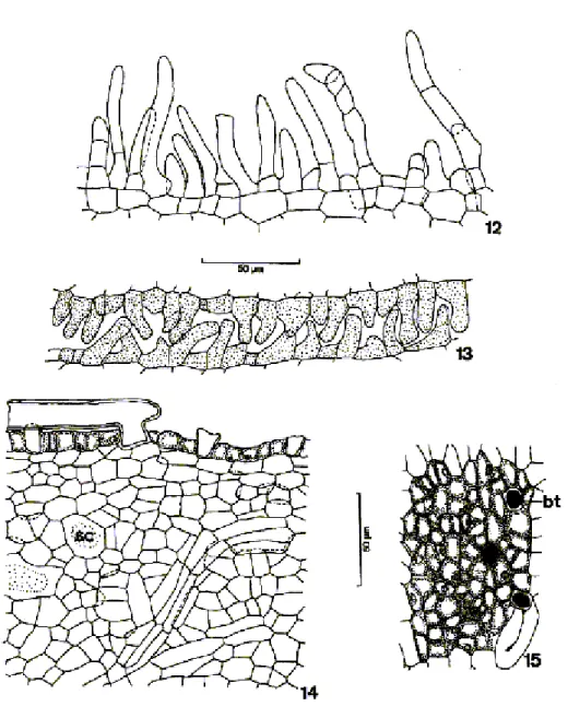
The petals had a uniseriate papillose epidermis, with trichomes, stomata (Figures 7 to 13) and cuticle, which could also be ornamented (Figure 11). The papils occurred frequently in the epidermis of the three species and showed thick cellular walls (Figures 8 to 11). The trichomes could be uni or pluricelullar, with thin or thick cellular walls and pointed, or more or less rounded extremities (Figures 10 and 12). They were common in the petal edges. In the floral buds there was a perfect adjustment among the trichome epidermes with contiguous petals (Figure 13). The mesophyll was parenchymatic and constituted by cells with thin walls and by secretory cells and cavities. This region had few layers in T. catigua
and T. elegans (Figure 9) and many in T. pallida
(Figure 8). The presence of calcium oxalate druses was notable in the parenchyma cells of T. catigua
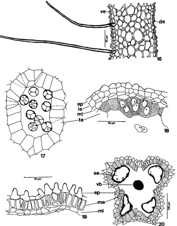
and T. elegans. There was a central collateral vascular strand and others of small dimensions immersed in the mesophyll (Figure 7).The nectaries were constituted by a single epidermis with thin-walled cells and non-glandular unicellular trichomes with relatively thick walls (Figure 15), and by a secretory parenchyma where the phloematic vascular tissue and secretory cells and cavities stood out (Figure 14).
The male flowers of T. catigua and T. elegans
presented androecium constituted by a staminal tube with a unilayered epidermis and prolonged non-glandular unicellular trichomes. The epidermal cells were papillose and showed a relatively thick and ornamented outer periclinal wall. Below the epidermis there was a homogeneous parenchyma with thick-walled cells (Figure16). It presented secretory cells and cavities and vascular bundles; one for each anther (Figure 21). The vascular bundles of the tube could be collateral or concentric. The androecium ofT. pallidaalso had a staminal tube, but differed from the other two species by nectaries and papils
at the base and trichomes in the proximities of the anthers; however, in T. pallida the papils did not show a thick or ornamented wall. The wall of the immature anther presented a single papillose epidermis, endothecium with thin-walled cells, slightly prismatic in shape, two middle layers and a secretory tapetum with binucleate cells (Figure 18). In the mature anther, the endothecium cells acquired thickenings on the anticlinal walls and on the inner periclinal wall. The middle layers and the tapetum began a disintegration process, were flattened and crushed (Figure 19). The pollen grains remained united in tetrahedron-type tetrads (Figure 17).
In female flowers, the antherodes without pollen had a papillose epidermis at maturity and an endothecium with parietal thickenings. The middle layers and the tapetum disintegrated in this phase (Figure 20). The only pistil in female flowers had an ovary with a uniseriate outer epidermis, which showed stomata and a cuticle. The epidermal cells varied from tabulate to prismatic in shape. The epidermis was glabrate and papillose inT. elegans
and pilose in T. catigua and T. pallida. The trichomes, very abundant in these species, were non-glandular, unicellular, presented thick cellular walls and thin extremities. In the multilayered parenchyma mesophyll there were secretory cells and cavities, notably numerous in T. elegans. A single and glabrate epidermis covered the ovarian cavity and presented cells tabulate to slightly prismatic in shape. Both the subepidermal layers (Figure 30) as the ventral epidermis of T. elegans
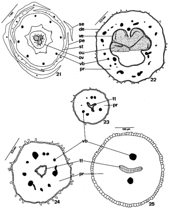
and T. pallida could undergo periclinal divisions in the preanthesis stage. Ovarian vascularization was made by differentiated vascular bundles or by lateral, ventral or marginal and dorsal procambial bundles, in accordance with carpel number. The septs, which separated the locules, were interrupted in the ovary apex and joined the locules to each other (Figures 21 and 22).
The pistil still had a short style with an epidermis similar to that of the ovary, which covered the parenchyma tissue, where there were vascular bundles and central transmitting tissue (Figures 23 to 25).
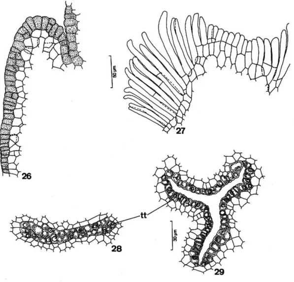
the epidermis and the secretory subepidermal layer which were arranged in the central region of the style as three small edges (Figures 23, 24 and 29) or two in T. elegans specimens (Figures 25 and 28).
The style in T. catigua andT. pallida, represented by a small rift, was hollow (Figures 23 and 29). In
T. elegans, however, a central rift occured in the middle region of the style only, therefore presenting a solid nature in the base and in the apex (Figures 25 and 28).
Figure 6–11.6 -Detail of the floral bud sepal of T. elegansin cross-section; 7-8: Diagram and detail of the floral bud petal of T. pallida in cross-section;9: Detail of the petal of T. elegansin cross-section;10-11: Detail of the adaxial and abaxial surface of the petal epidermis of T. pallida and T. elegans, in frontal view, respectively,. (eb=abaxial epidermis; ed=adaxial epidermis; pa=epidermal papillae; sc=secretory cell; vb=vascular bundle)
The transmitting tissue traversed the whole style and reached the apical region of the ovarian locule. The stigma presented unicellular trichomes, more frequent, and bicelullar; these trichomes came
Figure 21-25 -Diagrams of the cross-sections.21: Flower of theT. pallida;22: Upper portion of the ovary inT. catigua.23-24: Basal and apical portion of the hollow style inT. catigua;25: Solid style inT. elegans(de=dorsal epidermis; ve=ventral epidermis; ou=ovule; ov=ovary; pe=petal; pr=parenchyma; se=sepal; st=staminal tube; tt=transmitting tissue; vb=vascular bundle)
The ovules were anatropous and bitegmic and had several cellular layers between the nucellus epidermis and the embryo sac (Figures 31 and 32). The integuments were multi-layered and the number of layers varied from three to eight in the outer integument and three to seven in the inner integument (Figure 31). Two synergids of
prolonged contour were observed in the embryo sac, where its filiform apparatus and the two polar nuclei were notable (Figure 33).
Figure 26-29. 26-27 - Details of the epidermis in the young and mature stigma of T. pallida and T. catigua, respectively;28 – 29: Detail of the transmitting tissue of the solid and hollow style of T. elegansandT.catigua, respectively. (tt=transmitting tissue)
DISCUSSION
Pennington (1981) and Klein (1984) recorded for
Trichilia generally unisexual flowers in dioecious plants and less frequently bisexual and then polygamous plants. The flowers of T. catigua, T. elegansandT. pallidawere unisexual, but strictly dioecious specimens were not found in the studied area, all of them being monoecious to a greater or lesser degree. The nectary presence in Trichilia
species in the form of a thick ring around the ovary seemed to be common (Pennington, 1981), as verified in T. elegans and T. pallida; although
in the latter species it was fused to base of staminal tube.
The gynoecium structure is very important in the reproductive process of the studied species, from the arrival of the pollen on the stigma until the occurrence of the syngamy inside the ovule. The receptive stigma surface of the species constituted by unicellular or bicellular trichomes seems to fit in Group III of Heslop-Harrinson and Shivanna (1977). This group is characterized by papillae or trichomes of reduced or medium extension. The transmitting tissue of the style, where the pollen tube was developed, was mainly epidermal and limited to a small central cavity inT. catigua and
T. pallida; but it was solid at the base and apex of the T. elegans style. This meant that the pollen tube developed along the style on the transmitting tissue surface in the first two species or penetrated this tissue growing among the cells in T. elegans. When in the apex region of the ovary, the septs were interrupted, facilitating communication between the locules. This could favor eusyncarpy, in which the pollen tube could fertilize an ovule of any carpel -- a process verified in the Rutales, very physiologically important and with evolutionary consequences (Carr and Carr, 1961). Finally, the obturator was found to be in contact with the transmitting tissue, which facilitated the penetration of the pollen tube into the micropyle. The anatropous and bitegmic ovules seemed to be characteristic for Trichilia species, as recorded in the literature (Beltrati, 1984; Beltrati and Brunini, 1988, Moscheta, 1995 and Boesewinkel, 1981). According to Boesewinkel (1981), the ovule ofT. grandifolia Oliv. is crassinucellate. According to Davis (1966), the use of the terms crassinucellate and tenuinucellate have been generating a lot of confusion in the botanical literature. Therefore, she introduced pseudocrassinucellate. According to her, crassinucellate should be limited to the ovule, in which the archesporial cell divides forming a primary parietal cell which does or does not divide, while pseudocrassinucellate should be used for ovules, where no the parietal cells are formed; but the apical cells of the nucellar epidermis undergo periclinal divisions giving rise to a nucellar cap. Thus, an ontogenetic study of the nucellar development of the species would be
necessary to be able to affirm if the ovules are crassinucellate or pseudocrassinucellate.
The synergids of the studied species presented their respective filiform apparatus well-defined at the basal end of the cell. Kapil and Bhatnagar (1981) present several filiform apparatus shapes for several Angiosperm species and attribute several functions to this structure, such as absorption, nutrient synthesis and transport, glandular nature and an important role in the penetration and effusion of the pollen tube, with the consequent diffusion of spermatic cells to the egg and the central cell (polar nuclei).
In several taxa one of the synergids can survive after the fertilization, becoming hypertrophic, polyploid or multinucleate and to exercising a haustorial function - absorbing, storing or secreting nutrients necessary for the zygote and the young pro embryo (Kapil and Bhatnagar, 1981). It seemed that in T. catigua the two synergids degenerated. This is confirmed by the analysis of Moscheta (1995).
RESUMO
REFERENCES
Aegthe, C. (1951), Uber die physiologische Herkunft des Pflanzennektars. Ber. schweiz. bot. Ges. 61, 240-277.
Beltrati, C. M. (1984), Morfologia e anatomia das sementes de Trichilia elegans A. Juss. (Meliaceae).
Naturalia9, 35-42.
Beltrati, C. M. and Brunini, J. (1988), Morfologia, anatomia e desenvolvimento das sementes e plântulas deTrichilia pallidaSwartz (Meliaceae).R. bras. Biol.
48, 673-681.
Berlyn, G. P. and Miksche, J. P. (1976), Botanical Microtechnique and Cytochemistry. The Iowa State University Press, Ames, Iowa.
Boesewinkel, F. D. (1981), Development of the seed of
Trichilia grandifolia Oliv. (Meliaceae). Acta Bot. Neerl.30, 459-464.
Campos, J. B. (1997), Análise dos desflorestamentos, estrutura dos fragmentos florestais e avaliação do banco de sementes do solo da ilha de Porto Rico na planície de inundação do Alto Rio Paraná, Brasil. Tese de doutorado, Universidade Estadual de Maringá, Maringá, Brasil.
Carr, S. G. M. and Carr, D. J. (1961), The functional significance of syncarpy. Phytomorphology 11(3), 249-256.
Davis, G. L. (1966), Systematic Embryology of the Angiosperms.John Wiley and Sons, New York Heslop-Harrinson, Y. and Shivanna, K. R. (1977), The
receptive surface of angiosperm stigma.Ann. Bot.41, 1233-1258.
Ipardes (1992), Diagnóstico para a implantação de políticas para o setor florestal no Paraná. Fundação Ipardes, Curitiba.
Johansen, D. A. (1940), Plant Microtechnique.
McGraw-Hill, New York.
Kapil, R. N. and Bhatnagar, A. K. (1981), Ultrastructure and biology of female gametophyte in flowering plants.Int. Rev. Cytol.70, 291-341.
Klein, R. M. (1984), As plantas Meliáceas. In-Flora Ilustrada Catarinense,ed. R. Reitz. Herbário Barbosa Rodrigues, Itajái, Santa Catarina, 9-65.
Moscheta, I. S. (1995), Morfologia e desenvolvimento dos frutos, sementes e plântulas de Cabralea canjerana(Vell.) Mart.,Guarea kunthianaA. Juss. e
Trichilia catiguaA. Juss. (Meliaceae – Melioideae). Tese de doutorado, Universidade Estadual Paulista, Rio Claro, Brasil.
Opler, P. A. (1983), Nectar production in a tropical ecosystem. In – The Biology of Nectaries,B. Bentley and T. Elias. Columbia University Press, New York, 259p.
Opler, P. and Bawa, K. S. (1978), Sex ratios in tropical forest trees.Evolution,32, 812-821.
Pennington, T. D. (1981), A monograph of neotropical Meliaceae.Flora Neotr.,28, 1-449.
Puentes, D. A., Hernandes-de-Armas, J. and Lopez-Almiral, A. (1993), Phenology and floral structure of
Trichilia havanensis Jacq. (Meliaceae). Ann. Miss. bot. Gdn. 80, 862-869.
Ruetze, M. and Schmitt, V. (1986), Glykol-methacrylat (GMA) als Einbettungssystem in histologische Untersuchungen von Koniferen-Nadeln. Eur. J. Forest Path.16, 321-324.
Styles, B. T. (1972), The flower biology of the Meliaceae and its bearing on tree breeding. Silvae Genet.,21, 175-182.