Dissertation presented to obtain the Ph.D degree in Biology
Instituto de Tecnologia Química e Biológica António Xavier | Universidade Nova de LisboaInsert here an image
with rounded corners
Oeiras,
December, 2016
Filipa Baltazar da Costa Vaz
recognition by PGRP-SA and PGRP-LC
Filipa Baltazar da Costa Vaz
Dissertation presented to obtain the Ph.D degree in Biology
Instituto de Tecnologia Química e Biológica António Xavier | Universidade Nova de LisboaOeiras, December, 2016
evade cellular and humoral
responses mediated through
peptidoglycan recognition by
Title
Bacteria present mechanisms to evade cellular and humoral
responses mediated through peptidoglycan recognition by
PGRP-SA and PGRP-LC
Cover image
Temporal Color Code of Drosophila melanogaster
GFP-Moesin macrophages phagocytosing Staphylococcus aureus
NCTC 8325-4 strain.
Second Edition
March 2017
Author
Filipa Baltazar da Costa Vaz
All Rights Reserved
Printed in the UK
It seems that in Science great discoveries started with “Eureka!”, “This is interesting…” and similar expressions that translate excitement and curiosity. The first major discovery I did that put me on the track to some of the findings that I present in this thesis started with an expression that translates annoyance. As soon as my supervisor stepped into the lab, I followed him to his office and said “Sergio, I did something wrong”. After I told him the results of my last pull-down assay, without blinking, he looked at me and said “You didn`t do anything wrong, that makes total sense”. As I stared at him from his office door waiting for him to follow me to my bench, he added “I mean, I don´t think you did anything wrong in the experimental procedure”. For not doubting my results, when I am pretty sure everyone else would (and some did), I am profoundly grateful. This is something that even after all these years I had never expressed. It turns out that the following steps were the hardest. Sergio demanded me several experimental controls with repeats. After long and hard hours of work, I had ended any doubts that what I was observing was merely a result that was only reproducible in certain specific conditions. Naturally, I am thankful to Sergio for giving me the opportunity to do my PhD, for all the excellent conditions that he provided me and to allow me to pursue my sometimes (I admit it) crazy and out-of-the-box ideas, without any judgement. Also, I deeply appreciate that he gave me the freedom to perform experiments on my own initiative which sometimes were a little bit out of the aims of the work, just because… Just because I thought something was fun and wanted to try it! At his own terms and ways, with the interaction and working dynamics that I developed with him, I was able to learn from him how to build elegant experiments that give results which are convincing and explicit to others and to anticipate variables that can
not thrive, no matter how good or smart you may be. I hope in the future to continue developing such an exciting work (at least exciting for me) and Sergio certainly has a major impact in helping me to one day become (hopefully) a successful scientist.
Mariana Pinho, “the Lady next door”, was the best neighbour I could have encountered. Her outsider-insider role in my work throughout all these years proved to be valuable in several moments. With Mariana I learnt to stay focused on a certain topic, to not allow myself diverge from the “main road”, whilst keeping in mind all the observations that I would eventually need to interpret my data and/or to get “unstuck” from a puzzling data. I learnt that the success of a project relies in a sharp and objective mind where at all times it is key to determine very well what I aim to get with a certain experiment. In sum, she showed me that my time is valuable so I might as well spend it properly going straight to point. However, I admit that I still occasionally indulge myself in experiments just for fun and mere curiosity… I guess some things never really change!
I am very grateful to Petros because he invested in me, in my work, he believed in my data and he received me as a member of his Lab. Without him, probably I would have never been able to finish this work the way I did and without his investment in my study of the macrophages I would probably have never been able to find the role of the lytic activity of PGRP-SA. Thank you very much for everything! The faith and enthusiasm that you deposited in me was something that I had never been given before. It was indeed a great experience to go through. I will certainly remember this for my future and I hope to be able to one day provide similar experience to others.
To Maria João Gravato-Nobre, you are a sunshine in the Department of Biochemistry, a great friend and you opened me the doors
had the secret goal to prove the lytic activity of PGRP-SA. I remember your words “I saw and felt that you were living a moment that many people work for many years in Science and never live.” I am glad you could put words into that moment, because even today I still cannot quite describe it. Thank you!
To Dr. Jonathan Hodgkin, thank you for being so welcoming by letting me use your lab space and trusting in me without knowing me at all, that I would be able to do the zymopgraphy experiments for Maria`s paper. Thank you for reading my thesis and all the support for my next adventure!
Richard Parton, the microscopy guru, thank you very much for your time, help and sharing the knowledge with me. In particular, thank you for the patience and giving me the mini-courses on “microscopy for dummies”!
To everybody from the Micron Unit – David, Andrew, Vick and Ian – thanks for your help, good mood and fast responses in solving all the problems. David, thank you for letting me steal your teas and tea pot (I replaced some teas anyway) and I am sorry that I made go crazy looking for you tea pot in every single place of the entire building when it was right next door…. I really thought you had figured by then that I stole your tea pot every night for my shifts! Andrew, thank you for the help, chats and cookies! Vick thanks for sharing the knowledge and help and also for the great times we had!
To Dr. Alberto Baena, I have to give a big thanks for the help in the analysis of the behaviour of the macrophages and the type of cell death. Your help was crucial for me to be able to interpret and analyse my data. Also, I am very thankful for your interest in reading my thesis and for the
Prof. Carlos Romão, I cannot thank you enough for your patience, support and help. At the end of the day, I feel that I was lucky to have met someone like you, who stands by good and positive values and makes a difference in this little world of the Academia. Thank you for making this day possible and helping me to conclude a personal/professional journey that I started when I was 15 years old.
To Karina Xavier, I have to thank for your constant interest in my work to make sure that my work would reach this day in the best possible way. You surprised me in a positive way by reminding me that there are people who still care about the Science for what it is. Even if it is not their field of work and they will not get a direct and evident advantage for themselves at the end of that day, they still care and show genuine interest. Also, thank you for showing me that some people still care about people and remembering that a PhD student is a person, not a slave.
Teresa Baptista da Silva, you are an angel walking in the ITQB corridors! Not only you are fast, efficient and do everything right, but also you have a great spirit and lovely presence. I feel good whenever you are around. I always do everything myself to make sure that if something fails I can spot every step and figure out where I went wrong. However, with you, I trusted all my buffers and media in a way that I am not sure that in the future I will trust someone else.
To Rupal and Ilias, many big thanks for the friendship, flyroom moments, pub, allotment and I could go on… Rupal, I was really glad that we met, I know the feeling is mutual – you changed my out of the work PhD moments! To my 22 year-old drama queen, Maria, you are the best kiddo, sorry for being away lately, but I will now be cheesy as you like me to be “you were always on my mind!”.
moments and for being supportive to my nocturne Lab life. Maca, you will always have a special place, I will never forget your help and friendship in our very own quirky and special way. I am trying to find words to express how important you were and are. I guess you will find them in a letter I will write to you when we are old! António, how can I say thanks for joining your brain and rational mind with mines. The trust we shared with each other in this untrusty world of ours is something quite uncommon! The many hours in the park during our breaks and incubation times, thinking how to thrive and analyse every little thing from every possible perspective and come up with the most elegant solutions, albeit chaotic for others! Nuria, your objective mind in solving out some issues and the way you make it look so easy still surprises me today. Thank you for being there. Ola, I immensely admire your strength and I am happy to know you are in a good place now. I really appreciate that you call me really often and do not complain that I have not been very available for you in the past months! Thank you sweets! I miss you all a lot!
I have to thank everyone, I am not mentioning names because I will surely forget someone, that walked with me at night to the Lab and waited for me while I counted my flies. Also, for keeping food aside for me during the dinners because I was running more than late in the Lab and remembering to keep aside a cheeky beer or bottle of wine for me!
Thanks to everybody from the Department of Biochemistry for giving me food! Maybe you do not know it, but I am sure at some point you “gave” me food… Thank you! Food aside, I have to thank the great vibes from Peggy, Vick, Eze, Justin, Josep, Boris, David, Marko, Sacha, Darragh, Davis Lab, Barr Lab, Castello Lab, Hodgkin Lab, Woollard Lab!
From ITQB: Teresa Ferreira, Joana Wilton, Vânia Dias, Mariana Palma, Rita Santos, Nazua, Andreia Matos, Liliana Tomé, Mário Soromenho,
Teresa, Joana and Vânia, for their support and friendship. To Trish Reed I would like to express that I acknowledge that in the midst of everything, I am aware that you were the only person (or should I say one of the few persons) of the labs who truly believed in me and apart from the sheep mentality opinions, I do remember that you were the only person who guided me in the bench when I simply asked you “do you have this protocol?”. You did not had to do that, it was not your job, it was not your responsibility – thanks! James Yates, I believe that under a different context we would have done a great team, just the two of us! Although always with a joking smile, I know that you had the capacity to appreciate my creativity and out-of-the-box ideas. I also feel that underneath it all, I had a special secret place in the hierarchy of the Labs for you. To all the people who questioned my work and methods, a big thanks. You contributed to set my mind and my goals to become the scientist I am today. To the ones that every single day help us and make our life easy – Economato and all the people from the 1st and 2nd floor! Your great vibes and constant help with a smile is perhaps taken for granted too often. Thank you all for everything and keep up the good work!
Tom, even after all the hell that I stood by your side, I think it was worthwhile, that you are worthwhile. You have some kind of magic that can still make me smile no matter how bad things are. I am very proud of you and I truly wish that you do not give up on yourself now! I hope you find what you are looking for and overcome all of the suffering and pain. I am really thankful for your companionship, altruistic attitude towards me and for giving me the opportunity to help and make a big impact in the life of an exquisite person like you. I would do it all over again and help you crawl out of the black hole. Please, do not make all of our efforts go to waste!
programmed to unconditionally love their children, albeit few exceptions. Thus, having two people that are not programmed to love you unconditionally but made the conscious choice to do so, it puts me perhaps in the top ranking of the “luckiest persons” ever. I owe them and myself everything that I am and have accomplished so far. You are the most important people in my Life, words cannot express how and what I feel. I have tried it for many years and it seems that I have to keep trying. I wish you better than the best that this world can give. I love you and I always will. Thank you, thank you, thank you! To Cristina and Mamã, I am deeply grateful for you Kindness and Patience. Massive thanks to both of you!
I would like to acknowledge Fundação para a Ciência e Tecnologia for my PhD fellowship (SFRH/BD/78748/2011) and EMBO (ASTF 645-2014) for my short-term fellowship. In addition, I want to thank ITQB-UNL for the excellent working conditions and environment. I feel very lucky to have done my PhD at ITQB. Also, I have to thank the Department of Biochemistry, University of Oxford, for giving me the opportunity to prolong my stay, which was fundamental for my work, and to provide such great working and access conditions to academic visitors.
Abbreviations and Acronyms 1
Summary/Sumário 5
Thesis at a glance 13
Chapter I. The Bacteria and the Host 15
The Bacterial Cell Wall 18
Peptidoglycan composition and Host recognition 28 The Drosophila melanogaster Immune System 39
Final remarks and thesis overview 47
References 50
Chapter II. Recognition of peptidoglycan determines efficient antibacterial responses 63 Summary 65 Introduction 66 Results 76 Discussion 97 Conclusions 109
Materials and Methods 112
Summary 129
Introduction 130
Results 146
Discussion 166
Conclusions 177
Materials and Methods 179
References 190
Chapter IV. PGRP-SA is involved in bacterial clearance 201
Summary 203
Introduction 204
Results 213
Discussion 231
Conclusions 244
Materials and Methods 247
References 250
Chapter V. Bacteria present mechanisms that evade cellular and humoral responses mediated through peptidoglycan recognition by PGRP-SA and PGRP-LC
Chapter I. The Bacteria and the Host 15
Figure 1. Schematic representation of the Gram-negative and Gram-positive bacterial CWs.
22
Figure 2. Schematic representation of PGN monomeric species and polymeric PGN.
30
Figure 3. Modifications in the PGN glycan strands. 32 Figure 4. Differences between the amino acids L-Lys, DAP and amiDAP.
37
Figure 5. Summary of the similarities across the NF-κB signalling pathways.
46
Chapter II. Recognition of peptidoglycan determines efficient antibacterial responses
63
Figure 1. Representation of the PGN hydrolytic activities in an S.
aureus PGN model.
67
Figure 2. Chromosomal distribution of the bacterial PGN-BD proteins associated with autolytic activity in NCTC 8325.
70
Figure 3. Cleavage sites of the S. aureus autolysins. 70 Figure 4. Representation of the PGN-BD domains from each of the 19 proteins identified.
72
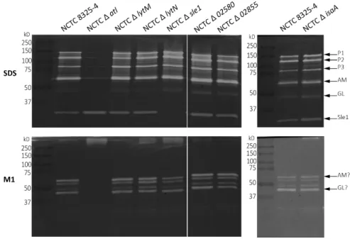
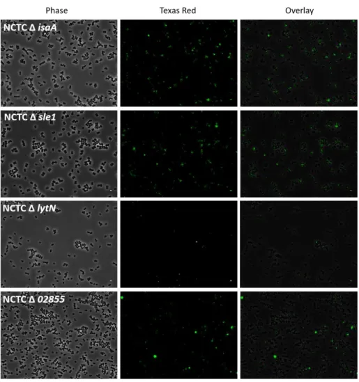
Figure 5. Zymography analysis of SDS crude autolytic enzyme extracts of S. aureus strains.
Figure 7. Co-precipitation of live cells with mCherry_PGRP-SA. 79 Figure 8. Co-precipitation of live cells in exponential and stationary growth phase with mCherry_PGRP-SA.
82
Figure 9. Survival curves of y,w flies infected with the parental strain and the autolysins mutants.
83
Figure 10. Survival curves for y,w flies. 84 Figure 11. Survival curves of semmelweis flies infected with the parental strain and less virulent mutants.
85
Figure 12. Survival curves upon infection with NCTC 8325-4. 86 Figure 13. Survival curves upon infection with NCTC Δ atl. 86 Figure 14. Survival curves upon infection with NCTC Δ lytM. 87 Figure 15. Survival curves upon infection with NCTC Δ isaA. 87 Figure 16. Survival curves upon infection with NCTC Δ 02855. 88 Figure 17. Survival curves upon infection with NCTC Δ sle1. 89 Figure 18. Co-precipitation of live cells with mCherry_PGRP-SA. 90 Figure 19. Survival curves of y,w and semmelweis flies by NCTC Δ
atl Δ sle1 and its parental strains.
91
Figure 20. Induction of drosomycin at 16h p.i. in y,w flies. 92 Figure 21. Haemolytic activity in sheep blood. 94
Figure 1. Schematic summary of the activation of the TOLL pathway by PGN recognition.
134
Figure 2. Schematic summary of the activation of the IMD pathway.
138
Figure 3. Schematic representation of the cell wall of a Gram-positive wild-type vs. a WTA mutant bacteria.
145
Figure 4. Co-precipitation of live cells with mCherry_PGRP-SA and mCherry_PGRP-LC.
147
Figure 5. GFP-PGRP-LCx binding to live bacterial cells. 149 Figure 6. Co-precipitation of PGN with rPGRP-SA and rPGRP-LC. 151 Figure 7. Survival data for B. subtilis. 155 Figure 8. Survival curves for B. subtilis Δ tagO infections. 157 Figure 9. Survival curves for S. aureus infections. 159 Figure 10. Survival curves for S. aureus Δ tagO infections. 161 Figure 11. Survival curves of semmelweis/PGRP-LCΔE12 flies. 164 Figure 12. Survival curves upon PBS injection. 165 Figure 13. Muropeptide profiles of pure PGNs. 169 Figure 14. Sugar composition of pure CWs pre-incubated with HCl. 170
Chapter IV. PGRP-SA is involved in bacterial clearance 201
Figure 3. Snapshots of semmelweis-GFP-Moesin macrophages infected with bacteria.
218
Figure 4. Snapshots of semmelweis-GFP-Moesin macrophages complemented with mCherry_PGRP-SA infected with bacteria.
220
Figure 5. Snapshots of seml/PGRP-LCΔE12-GFP-Moesin
macrophages infected with bacteria.
223
Figure 6. Snapshots of seml/PGRP-LCΔE12-GFP-Moesin
macrophages complemented with mCherry_PGRP-SA infected with bacteria.
225
Figure 7. Co-precipitation of live bacterial cells with mCherry_PGRP-SA at pH 3.
227
Figure 8. Co-precipitation of live S. aureus cells with mCherry_PGRP-SA at different pH.
228
Figure 9. Zymography assay of the lytic activity of mCherry_PGRP-SA at pH 3.
229
Figure 10. Zymography assay of the lytic activity of rPGRP-SA at pH 6.
230
Figure 11. Haemolytic expression pattern in sheep blood. 234 Figure 12. Phagocytosis-apoptosis process. 238
Chapter II. Recognition of peptidoglycan determines efficient antibacterial responses
63
Table 1. List of strains used in this study, plasmids and primers for cloning.
112
Chapter III. PGRP-SA and PGRP-LC recognise both Lys and DAP residues of the peptidoglycan in vivo
127
Table 1. Summary of the statistical analysis of the survival data. 165
Chapter IV. PGRP-SA is involved in bacterial clearance 201
Table 1. Types of Programmed Cell Death. 208 Table 2. Phagocytosis-clearance pathways. 236
Chapter IV. PGRP-SA is involved in bacterial clearance 201
Video 1. Macrophages attached to the microscope slide in Schneider media or infected with bacteria.
212
Video 2. GFP-Moesin macrophages infected with S. aureus bacteria.
214
Video 3. GFP-Moesin macrophages infected with S. aureus Δ tagO bacteria.
214
Video 4. GFP-Moesin macrophages infected with B. subtilis bacteria.
214
Video 5. GFP-Moesin macrophages infected with B. subtilis Δ tagO bacteria.
214
Video 6. GFP-Moesin macrophages in Schneider media. 214 Video 7. GFP-Moesin macrophages infected with heat-killed S.
aureus cells.
214
Video 8. semmelweis-GFP-Moesin macrophages infected with S.
aureus bacteria.
217
Video 9. semmelweis-GFP-Moesin macrophages infected with S.
aureus Δ tagO bacteria.
217
Video 10. semmelweis-GFP-Moesin macrophages infected with B.
subtilis bacteria.
217
Video 11. semmelweis-GFP-Moesin macrophages infected with B.
subtilis Δ tagO bacteria.
217
Video 12. semmelweis-GFP-Moesin macrophages in Schneider media.
Video 14. semmelweis-GFP-Moesin macrophages infected with S.
aureus Δ tagO bacteria rescued with mCherry_PGRP-SA.
219
Video 15. semmelweis-GFP-Moesin macrophages infected with B.
subtilis bacteria rescued with mCherry_PGRP-SA.
219
Video 16. semmelweis-GFP-Moesin macrophages infected with B.
subtilis Δ tagO bacteria rescued with mCherry_PGRP-SA.
219
Video 17. semmelweis-GFP-Moesin macrophages in Schneider media rescued with mCherry_PGRP-SA.
219
Video 18. seml/PGRP-LCΔE12-GFP-Moesin macrophages infected
with S. aureus bacteria.
222
Video 19. seml/PGRP-LCΔE12-GFP-Moesin macrophages infected
with S. aureus Δ tagO bacteria.
222
Video 20. seml/PGRP-LCΔE12-GFP-Moesin macrophages infected
with B. subtilis bacteria.
222
Video 21. seml/PGRP-LCΔE12-GFP-Moesin macrophages infected
with B. subtilis Δ tagO bacteria.
222
Video 22. seml/PGRP-LCΔE12-GFP-Moesin macrophages in
Schneider media.
222
Video 23. seml/PGRP-LCΔE12-GFP-Moesin macrophages infected
with S. aureus bacteria rescued with mCherry_PGRP-SA.
224
Video 24. seml/PGRP-LCΔE12-GFP-Moesin macrophages infected
with S. aureus Δ tagO bacteria rescued with mCherry_PGRP-SA.
224
Video 25. seml/PGRP-LCΔE12-GFP-Moesin macrophages infected
with B. subtilis bacteria rescued with mCherry_PGRP-SA.
224
Video 26. seml/PGRP-LCΔE12-GFP-Moesin macrophages infected
with B. subtilis Δ tagO bacteria rescued with mCherry_PGRP-SA.
1
List of abbreviations and acronyms
µL microlitro
Ala Alanine
AMP Antimicrobial peptide CFU Colony Forming Unit
CHAP cysteine, histidine dependent amidohydrolase/peptidase domain
ChBD Choline binding domain CM Cytoplasmic membrane
CW Cell wall
Cys Cysteine
DAP meso-diaminopimelic acid
DD Death domains
dIAP-2 Drosophila Inhibitor of apoptosis-2 Dif Dorsal-related immunity factor
DREDD Apical caspase death-related Ced-3/Nedd2-like protein Dscam Down syndrome cell adhesion molecule
dSR-CI Drosophila scavenger receptor class C, type I
ERM Ezrin, radixin and moesin
FADD The Drosophila homologue of FAS-associated death-domain protein
Fig. Figure
GFP Green Fluorescent Protein GlcNAc N-acetylglucosamine Glu Glutamic acid
Gly Glycine
2 GroP Glycerol phosphate
h Hour
HF Hydrofluoric acid
HPAEC-PAD High Performance Anion Exchange Chromatography coupled with Pulsed Amperometric Detection
HPLC High Performance Liquid Chromatography IIS Innate Immune System
IKK IkB kinase
IM Inner Membrane
IMD Immune deficiency pathway
IS Immune System
kDa Kilodalton
LA Luria Agar
LB Luria Broth
LPS Lipopolysaccharide LTA Lipoteichoic Acids
Lys L-Lysine
LysM Lysin motif
LYZ2 Lysozyme subfamily 2
M Molar
M1 Mutanolysin
MAMP Microbe Associated Molecular Patterns ManNAc N-acetylmannosamine
MDP muramyl-dipeptide
mg miligram
mL mililitro
ModSP Modular Serine Protease
3 MurNAc N-acetylmuramic acid
MurNAcLAA N-acetylmuramoyl-L-alanine amidase
MYD88 Myeloid differentiation primary response protein 88 N/A Not applicable
NF-κB Nuclear factor kappa-light-chain-enhancer of activated B cells
NimC4/SIMU Nimrod C4/Six-microns-under
Nod2 Nucleotide-binding oligomerization domain-containing protein 2
ns Not significant
O/N overnight
OD Optical density
OM Outer Membrane
OMP Outer Membrane Protein
PAMP Pathogen Associated Molecular Patterns PBPs Penicillin Binding Proteins
PCD Programmed Cell Death PGN Peptidoglycan
PGN-BLD PGN-Binding/Lytic domain
PGRPs Peptidoglycan Recognition Proteins PPO Prophenoloxidase Cascade
PRR Pattern-recognition receptors
PSH Persephone
RboP Ribitol phosphate RHD Rel Homology domain
RHIM RIP homotypic interaction domain RIP Receptor-interacting protein ROS Reactive Oxygen Species
4
seml semmelweis
SH3 Src homology 3 SH3b Bacterial SH3
SLT transglycosylases SLT domain 1 SPE Spatzle processing enzyme
Spz Spatzle
TAB2 TAK1-associated binding protein 2
TAK1 Transforming growth factor -β- activated kinase 1 TCT tracheal cytotoxin
TF Transcription factor
TGL transglycosylases-like domain TIR Toll/interleukin-1 receptor TLR Toll-like Receptor
TNF tumour necrosis factor
TRIF TIR domain-containing adaptor protein inducing IFNβ TSA Tryptic Soy Agar
TSB Tryptic Soy Broth WTA Wall Teichoic Acids
5
Summary
The Host presents different innate immune components to fight bacterial infections, most of which are evolutionary conserved strategies. Conversely, Bacteria present numerous mechanisms of virulence and evasion, transversal to different bacterial species, that confer resistance or subvert the activity of the Host components. This thesis presents a study on how the Host perceives the Bacteria and reacts to them and how the Bacteria protect themselves from those responses.
Peptidoglycan (PGN) is a ubiquitous cell surface bacterial polymer. It is composed of glycan chains cross-linked through peptide bridges. The residue at the third position of the peptide moiety shows the highest variability among the different PGNs. Nevertheless, most bacteria possess either a lysine (Lys) or a meso-diaminopimelic acid (DAP) residue.
The Host components that act on the PGN, range from enzymes that degrade it to receptors that upon recognition initiate downstream signalling cascades leading to the production of antimicrobial peptides. The Peptidoglycan Recognition Proteins (PGRPs) are evolutionary conserved innate immune receptors in Invertebrates and Vertebrates, including Humans. In Drosophila melanogaster, PGN is the main bacterial component that triggers an immune response and it is recognised by the PGRPs. According to the current model of bacterial recognition, the PGRPs possess discriminatory ability between Lys- and DAP- type PGN. PGRP-SA,
in vivo, binds exclusively to Lys-type bacteria, whereas PGRP-LC
specifically binds to DAP-type bacteria.
Previous work from the Host Labs1 has shown that bacteria
1Filipe Lab, Laboratory of Bacterial Cell Surfaces and Pathogenesis, Instituto de Tecnologia
Química e Biológica António Xavier, Universidade Nova de Lisboa.
Ligoxygakis Lab, Laboratory of Cell Biology, Development and Genetics, Department of Biochemistry, University of Oxford.
6 present components at the cell surface that impair detection of the PGN and thus compromise host survival [1], [2]2. The wall teichoic acids (WTA) are phosphate-rich polymers of the Gram-positive cell wall that exert stereo hindrance by shielding the PGN from PGRP-SA [1]. Atl is an autolysin that cleaves PGN in order to sustain cell growth and division. In the absence of this enzyme, the bacteria accumulate cell surface exposed PGN which is recognised by PGRP-SA and allows the survival of the Fly [2]. From the Bacteria side, I aimed to understand: 1) whether WTA are a transversal bacterial evasion mechanism that impair PGN recognition by Host receptors; 2) whether other autolysins could impair the binding of PGN by PGRP-SA and contribute to evasion. From the Host side, I aimed to ascertain the discriminatory ability of PGRP-SA and PGRP-LC towards the Lys and DAP residues. Why are two major immune receptors limited in bacterial recognition due to the discrimination of two residues? As the work evolved, the data suggested that PGRP-SA could engage in cellular responses. Thus, the final goal of my work was to evaluate the role of PGRP-SA in phagocytosis.
As an overall, the work here presented asks for a re-evaluation of the current model of bacterial recognition and confirms the importance of cellular responses as a first line of defence, followed by the prolonged and protective humoral responses. In addition, I show that recognition of PGN is paramount for the triggering of efficient responses by both the cellular and humoral responses.
I show that PGRP-SA has access to PGN fragments that are
2
[1] M. L. Atilano, J. Yates, M. Glittenberg, S. R. Filipe, and P. Ligoxygakis, “Wall teichoic acids of staphylococcus aureus limit recognition by the drosophila peptidoglycan recognition protein-SA to promote pathogenicity,” PLoS Pathog., vol. 7, no. 12, 2011. [2] M. L. Atilano, P. M. Pereira, F. Vaz, M. J. Catalão, P. Reed, I. R. Grilo, R. G. Sobral, P.
Ligoxygakis, M. G. Pinho, and S. R. Filipe, “Bacterial autolysins trim cell surface peptidoglycan to prevent detection by the drosophila innate immune system,” Elife, vol. 2014, no. 3, pp. 1–23, 2014.
7 temporarily surface exposed during the bacterial cell division. Thus autolysins do play a role in the impairment for PGN detection. If their activity is compromised, there is accumulation of PGN at the cell surface. Particularly, Atl and Sle1 have a great impact in cell division. Besides Atl, Sle1 also contributes to impair PGN detection. In the absence of Sle1, there is the accumulation of newly synthesised PGN fragments at the septum which are recognised by PGRP-SA. However, the amount of PGN that is recognised is fundamental for the triggering of a potent immune response. Therefore, paradoxically, Sle1 does not seem to contribute for virulence regarding PGN recognition. I propose that it is the sum of the autolysins activity that can properly impair the detection of PGN during cell division, albeit the Atl protein presents a more preponderant role
I show that PGRP-SA and PGRP-LC are promiscuous for the Lys and DAP residues of the PGN and both participate in the immune responses upon infection. When the PGN is detected, both PGRPs act as opsonins and engage in the phagocytic and clearance processes. In particular, PGRP-SA has optimal binding ability and presents lytic activity at low pH, indicating that it may act as an enzyme inside the phagolysosomes. After phagocytosis, the macrophages seem to present two ways of clearance: exocytosis or phagocytosis-apoptosis. Through induction of apoptosis, the macrophages may be able to incorporate the internalised bacteria in the apoptotic bodies which will be phagocytosed. The bacterial destruction can then be achieved through a new phagocytosis process. This strategy of phagocytosis-apoptosis is likely to occur until complete bacterial clearance.
Concomitantly, the clearance is dependent on the degradation of the PGN, which is compromised by the WTA. Therefore, the WTA are a dual strategy of immune evasion. At a first level, they impair the primary recognition by both PGRPs which is crucial to trigger the phagocytic processes (and also the induction of antimicrobial peptides). At a second
8 level, they impair the processes of clearance by impairing the activity of these PGRPs (and perhaps by other Host components).
I propose that it is the accessibility of the PGRPs through the cell wall that determines the PGN recognition and not the discrimination between the DAP and Lys residues. Furthermore, I propose that the WTA of the positive bacteria and the outer membrane of the Gram-negative bacteria, are conserved bacterial immune evasion strategies towards the recognition and activity of PGN host receptors. In addition, I propose that the macrophages present two pathways of clearance: phagocytosis-exocytosis and phagocytosis-apoptosis. The activation of either pathway is in turn dependent on the bacterial type and the factors that they possess that interfere with an efficient phagocytosis-clearance process.
9
Sumário
O Hospedeiro apresenta inúmeras estratégias de resposta inata contra a Bactéria. Estas estratégias estão presentes em diferentes Hospedeiros por serem conservadas ao longo da evolução das espécies. A Bactéria, por seu turno, apresenta diferentes mecanismos de virulência e de evasão que permitem a sobrevivência e o estabelecimento de uma infeção no Hospedeiro. Estas estratégias são transversais a diversas espécies de bactérias. A presente tese de doutoramento descreve um estudo sobre o modo como o Hospedeiro perceciona e reage à Bactéria e o modo com que esta se protege de tais respostas.
O peptidoglicano (PGN) é um polímero ubiquitário da superfície das bactérias. É composto por cadeias polissacarídeas que se interligam por pontes peptídicas. A terceira posição da cadeia peptídica alberga o aminoácido mais variável dentro dos vários tipos de PGNs. Ainda assim, na maioria dos casos, encontra-se ou uma Lisina (Lys) ou um ácido diaminopimélico (DAP).
O PGN é detetado por componentes do Hospedeiro que iniciam respostas imunitárias. Estes componentes compreendem quer enzimas que degradam o PGN, quer recetores que o reconhecem e ativam cascatas imunitárias que conduzem à expressão de péptidos antimicrobianos. As “Peptidoglycan Recognition Proteins” (PGRPs) são recetores evolutivamente conservados do Sistema Imunitário Inato de Invertebrados e Vertebrados, inclusive o Homem.
O PGN é o principal componente bacteriano que inicia respostas imunitárias na mosca Drosophila melanogaster. Segundo o modelo de reconhecimento do PGN em D. melanogaster, as PGRPs possuem especificidade para os aminoácidos Lys ou DAP. A PGRP-SA, in vivo, liga-se exclusivamente à Lys e a PGRP-LC ao DAP.
10 Os Grupos de Investigação que participaram no presente trabalho1, verificaram anteriormente que as bactérias apresentam componentes à sua superfície que impedem o reconhecimento do PGN com consequências na sobrevivência da mosca [1], [2]2. Os ácidos teicóicos da parede das bactérias Gram-positivas (WTA – “Wall teichoic acids”) são polímeros de fosfato que impedem o reconhecimento do PGN pela PGRP-SA por comporem uma camada que impede o acesso da proteína ao ligando [1]. A “major autolysin” Atl, cliva o PGN para promover a duplicação e a divisão da célula. Na ausência do Atl, a bactéria acumula à sua superfície fragmentos de PGN que são reconhecidos pela PGRP-SA o que resulta na sobrevivência da Mosca.
Pelo lado da bactéria, o meu trabalho de doutoramento teve dois objetivos: 1) avaliar se o efeito dos WTA é um mecanismo bacteriano conservado que permitem a evasão do reconhecimento do PGN; 2) se outras autolisinas impede o reconhecimento do PGN pela PGRP-SA como mecanismos de evasão. Concomitantemente, procurei averiguar a capacidade discriminatória da PGRP-SA e da PGRP-LC ao PGN. Por que motivo estes recetores centrais à resposta antibacteriana, encontram-se limitados na deteção devido à especificidade a uma molécula? Com o avançar do trabalho, os resultados obtidos levantaram a hipótese de que a PGRP-SA participa em respostas celulares. Com efeito, o trabalho culminou com o objetivo de avaliar a função da PGRP-SA no processo de fagocitose.
1Filipe Lab, Laboratory of Bacterial Cell Surfaces and Pathogenesis, Instituto de Tecnologia
Química e Biológica António Xavier, Universidade Nova de Lisboa.
Lygoxygakis Lab, Laboratory of Cell Biology, Development and Genetics, Department of Biochemistry, University of Oxford.
2
[1] M. L. Atilano, J. Yates, M. Glittenberg, S. R. Filipe, and P. Ligoxygakis, “Wall teichoic acids of staphylococcus aureus limit recognition by the drosophila peptidoglycan recognition protein-SA to promote pathogenicity,” PLoS Pathog., vol. 7, no. 12, 2011. [2] M. L. Atilano, P. M. Pereira, F. Vaz, M. J. Catalão, P. Reed, I. R. Grilo, R. G. Sobral, P.
Ligoxygakis, M. G. Pinho, and S. R. Filipe, “Bacterial autolysins trim cell surface peptidoglycan to prevent detection by the drosophila innate immune system,” Elife, vol. 2014, no. 3, pp. 1–23, 2014.
11 As conclusões do trabalho constante nesta tese pedem que a comunidade científica reconsidere o modelo atual de reconhecimento das bactérias e intensifica a importância das respostas imediatas celulares como primeira linha de defesa, seguidas pelas respostas prolongadas e protetoras humorais. O trabalho aqui presente demonstra ainda que o reconhecimento do PGN é crucial para a ativação de respostas imunitárias celulares e humorais eficientes contra as bactérias.
O meu trabalho demonstra que a PGRP-SA reconhece fragmentos de PGN que são temporariamente expostos à superfície durante a divisão das células. Por conseguinte, as autolisinas assumem um papel importante na protecção do reconhecimento desta molécula. Quando a actividade das autolisinas é comprometida de tal modo que a célula não consegue controlar a exposição do PGN durante a divisão, fragmentos de PGN recentemente sintetizados não são prontamente incorporados no polímero da macromolécula e por isso acumulam-se à superfície. Particularmente, a ausência do Atl e do Sle1 têm um forte impacto na divisão da bactéria. Com efeito, para além do Atl, também o Sle1 assume um papel importante contra o reconhecimento pela PGRP-SA. Na ausência do Sle1, há acumulação de fragmentos de PGN recentemente sintetizados na região septal que são reconhecidos pela PGRP-SA. Porém, a quantidade de PGN que é reconhecida revela-se crucial para a ativação de uma resposta eficiente. Por conseguinte, paradoxalmente, o Sle1 não contribui para a virulência da bactéria através da evasão ao reconhecimento do PGN. Por fim, eu proponho um modelo que se baseia no todo das atividades das autolisinas que eficientemente conseguem impedir que uma grande quantidade de PGN esteja acessível ao reconhecimento, sendo que o Atl assume um papel preponderante.
Eu demonstro que a PGRP-SA e a PGRP-LC reconhecem quer a Lys quer o DAP do PGN e que ambas participam nas respostas imunitárias contra os dois tipos de bactérias. Aquando da deteção do PGN, ambas as
12 PGRPs atuam como opsoninas e participam nos processos de fagocitose e degradação da bactéria. A PGRP-SA demonstrou ser uma potencial enzima lítica que atua em condições acídicas, indicando que deverá exercer atividade no fagolisosoma. Após a fagocitose, os resultados sugerem que os macrófagos seguem uma de duas estratégias para a degradação da bactéria: fagocitose-exocitose ou fagocitose-apoptose. Através da indução da apoptose, os macrófagos potencialmente podem conter as bactérias dentro dos corpos apoptóticos que serão por seu turno fagocitados. As bactérias serão eliminadas ao sofrerem uma nova fagocitose. É provável que este processo de fagocitose-apoptose se repita até se atingir a destruição total das bactérias.
Por outro lado, a degradação do PGN é essencial para a destruição da bactéria e os WTA comprometem e dificultam a clivagem do PGN. Assim sendo, os WTA atuam a dois níveis contra as defesas do Hospedeiro. Primariamente, eles impedem o reconhecimento pela PGRP-SA e PGRP-LC e, por conseguinte, a ativação do processo de fagocitose (e a indução da expressão de péptidos antimicrobianos). Quando o Hospedeiro ultrapassa esta barreira de reconhecimento, os WTA atuam contra a segunda linha de resposta que é a destruição da bactéria após fagocitose, ao impedirem a ação da PGRP-SA e PGRP-LC (e provavelmente de outros componentes).
Em suma, eu proponho um modelo de reconhecimento segundo o qual é a acessibilidade das PGRPs através da parece celular que determina o reconhecimento do PGN. Eu proponho que os WTA das bactérias Gram-positivas e a “outer membrane” das bactérias Gram-negativas são estratégias bacterianas conservadas de evasão ao Sistema Imunitário. Por fim, proponho que possivelmente os macrófagos apresentam duas vias de degradação de microrganismos: exocitose e fagocitose-apoptose. A ativação de uma das vias está por sua vez dependente do tipo de bactéria e de fatores que possuem que dificultam a eficiente destruição por parte dos macrófagos.
13
Thesis at a glance
Chapter I. The Bacteria and the Host
Are autolysins redundant in impairing access to surface exposed peptidoglycan by PGRP-SA?
Are PGRP-SA and PGRP-LC specific towards Lys and DAP residues of the peptidoglycan?
Are WTA a transversal Gram-positive mechanism to shield different peptidoglycans from different PGRPs?
Is PGRP-SA involved in phagocytosis?Chapter II. Recognition of peptidoglycan determines efficient
antibacterial responses
PGRP-SA recognises PGN that is temporarily exposed at the cell surface during cell division, which can be impaired by the temporal and spatial activity of the autolysins.
Sle1 appears to be crucial for cleavage of septal PGN to allow septum re-shaping upon splitting of the cells.
Recognition of peptidoglycan is key for efficient antibacterial responses.Chapter III. PGRP-SA and PGRP-LC recognise both Lys and DAP
residues of the peptidoglycan in vivo
In vitro assays showed promiscuity of the PGRPs towards Lys and DAP residues.
In vivo assays showed that the recognition of both PGN types by both PGRPs is paramount for host survival.
WTA impair recognition of both peptidoglycan types by both PGRPs.Chapter IV. PGRP-SA is involved in bacterial clearance
PGRP-SA and PGRP-LC participate in phagocytosis and clearance. PGRP-SA has lytic activity towards different peptidoglycans.
Apoptosis is a mechanism to promote clearance.Chapter V. Bacteria present mechanisms to evade cellular and
humoral responses mediated through peptidoglycan recognition by
PGRP-SA and PGRP-LC
The access through the cell wall to the peptidoglycan determines the detection by PGRPs.
PGRP-SA and PGRP-LC play dual roles in phagocytosis and humoral responses.
Bacteria possess conserved mechanisms that impair accessibility to the PGN.15
CHAPTER I
The Bacteria and the Host
The event of the first cell was paramount in shaping all the subsequent steps of Evolution of Life. The first cell was the first individual and self-sufficient structure that presented defined boundaries, raising the concepts of intra- and extracellular environment. Thus, it constituted the primordial individualized entity that clearly presented a “self” and therefore a “non-self” – the surrounding environment and other cells that were grown from it, identical but not the same individuality.
Colonial organisms evolved to multicellularity, which can be described as whole organisms that in their own self are composed of many non-self-individualities. Evolution of multicellularity culminated in organisms composed of interconnected systems and finally the idea of self and non-self was fully stablished and thus shaped the evolution of the Immune System (IS).
As an integrated system in an organism, like the other systems, the IS guarantees and maintains homeostasis, i.e. it helps to maintain a state of equilibrium in the organism. Particularly, the IS plays the role of protecting the organism against danger [1]. As the scientific knowledge grew, we became aware of the two branches of the IS – the Innate and the Adaptive. Whereas only the Vertebrates possess an Adaptive Immune System, the most basic mechanisms of an Innate Immune System (IIS) are found to some extent in almost all life forms across the Eukarya Domain. The IIS is a constant vigilant of danger signal and is the first line of defence in an organism.
The surveillance and detection of invading microorganisms relies on an evolutionary conserved system of receptors that are constitutively expressed, non-clonal and independent of immunological memory. They
16 are collectively known as Pattern-recognition receptors (PRR) and recognise specific structures in the microorganisms, i.e. they detect danger through discrimination of non-self [2]. Whether the detection of danger is followed by an immune response is dependent on the confirmation of the danger signalling, which is in turn dependent on how the microorganism is perceived. It is within these interactions that two concepts became implemented: a “Host organism”1 and a “pathogenic organism”2.
These evolutionary conserved PRR detect conserved microbial structures that are specific of a class of microbe and absent in the Host. These microbial structures are designated as Pathogen Associated Molecular Patterns (PAMP) [2], however nowadays they are also commonly referred to as Microbe Associated Molecular Patterns (MAMP), since these structures are not specific of pathogenic microorganisms [3], [4]. As PAMP are not present in the Host and are conserved in a class of microbes, it is the logical that they are crucial for the microbial life. Hence, the pathogens present various mechanisms to avoid the detection of their PAMP by PRR. The Host-pathogen interaction can be viewed as a battlefield that in extreme cases, upon failure of efficient recognition and response, can culminate with either the survival of the Host or of the pathogen.
As the definition of boundaries in a cell, the cell surface, was of extreme importance to evolution of Life, again we find that it is at the surface of the cells that most immune responses begin. Upon breaching of the Host surface, many of the first encounters of a PRR and a pathogen happen mainly at the level of the microbial cell surface. Thus, many PRR are specific for PAMP that are components of the outer surface of the pathogens – viruses, bacteria, fungi and parasites.
1An organism that harbours another organism to which provides nutrients and shelter. 2 An organism that can cause disease in a Host.
17 The basic mechanisms underlying Innate Immunity are conserved among Plants, Invertebrates and Vertebrates, including Humans [5]. Moreover, the virulence determinants and strategies that bacteria employ against Host defences are also conserved among a group of pathogens, such as Bacteria [6], [7]. Therefore, it is possible to study fundamental questions in one organism and extrapolate to others. Hence, the concept of model organisms became a foundation for studying processes and mechanisms both in Microbiology and Immunology.
The work presented in this thesis describes a study on Host-Bacteria interactions. As an overall, the work aimed to understand how a certain type of PRRs – the Peptidoglycan Recognition Proteins (PGRPs) – recognise a ubiquitous bacterial component, the peptidoglycan, and how Bacteria can prevent this recognition. The study of the bacterial factors that evade the IS was focused in the bacteria Staphylococcus aureus and
Bacillus subtilis and the in vivo studies of the PGRPs recognition were
conducted in the fruit fly Drosophila melanogaster. In this chapter, I address the current knowledge on the structure and biology of the bacterial surface, particularly the peptidoglycan, and a brief state of the art of the Drosophila immune responses. Finally, I present the goal of my PhD project and an overview of the contents of each chapter of the thesis.
18
The Bacterial Cell Wall
Bacteria present several molecules that “coat” and cover the outer surface of the cytoplasmic membrane. The arrangement of these “coating” molecules forms the cell wall or cell envelope. The cell wall (CW) is a complex and dynamic structure that protects the cell and helps in the adaptation to different environmental conditions. In broad and simple terms, the CW components can be divided in three types: 1) a ubiquitous bacterial component, the peptidoglycan; 2) components that are characteristic of either positives, negatives or of positive Corynebacteria; 3) components that may be present in both Gram-types but are often species- or strain-specific (capsular polysaccharides and S-layers).
The CW comprises many bacterial components that are in contact with the external environment which are determinant for infection. It comprises PAMP, virulence factors and components that function in immune evasion strategies. Indeed, most current models of bacterial recognition are based on how certain CW factors confer resistance to Host responses. Thus, the knowledge of the CW composition is crucial to understand how bacteria interact with the Host. In addition, peptidoglycan is a major PAMP and PRRs that specifically recognise it can be found from Plants to Humans. Moreover, along with PRR, the Host also produces lytic enzymes capable of degrading the peptidoglycan such as Lysozymes and produces antimicrobial peptides3 (AMPs). The PRR, the lysozyme proteins and the AMPs are the most conserved immune strategies found in almost all Animals.
3Innate Immunity cationic peptides. They are expressed in response to pathogen detection and
19 Most cell biology studies on bacteria rely on microscopy techniques that use specific staining methods. Any staining method is inherently dependent in either or both chemical and physical properties of the bacterial CW. The most widely used staining is the Gram stain, which was first described by Carl Friedlander in 1883 for detecting Streptococcus
pneumoniae in autopsy of lung tissues [8]4. It was his colleague, Christian Gram, who published it in 1884 and soon it started being gradually stablished as a routine stain in Hospital settings (Roux 1886) [8]5. The original procedure was subsequently improved and published as we know it today by Hucker in 1921 [9], [10]. It has been known for years that this differential stain which discriminates Gram-positive from Gram-negative bacteria, is a function of cell surface characteristics. However, only in the past decades have we achieved some understanding behind its mechanism [11], [12].
The first step of the Gram-stain is with the crystal violet, a basic positively charged dye that stains all bacterial surfaces (i.e. it stains negatively charged cells). The next step is an iodine-iodide mixture which serves as a mordant, so it forms a complex with the primary dye thereby fixing the dye to the cells. Through the use of a solvent, usually ethanol, some cell types lose the dye-mordant complex and the purple colour given by the crystal violet. Hence, any cell that retains the dye is designated Gram-positive and any cell that loses it is designated Gram-negative. For practical visualization, the final step includes a counterstaining with safranin that stains Gram-negative bacteria in red. It was long presumed that the CW of Gram-positive bacteria has physical properties regarding thickness and porosity that are able to retard the efflux of the dye-mordant complex and confer higher resistance to the solvents. Indeed, it is known
4Original reference: C. Friedlander, “Die Mikrokokken der Pneumonie,” Fortschr. Med., vol. 1,
pp. 715-733, 1883.
5 Original reference: G. Roux, “Sur un procede technique de diagnose des gonococci,” Arch. Gen.
20 today that these bacteria, in contrast to the Gram-negatives, possess a thick peptidoglycan layer, a macromolecule that covers the entire cell surface nearby the cytoplasmic membrane and that it accounts for most of the physical properties of the cells.
In this thesis, the description of the Gram-negative CW and peptidoglycan takes as a reference Escherichia coli, because it is the main bacterial model organism thus the most well studied, including in vivo Host responses. Regarding the Gram-positives, two model organisms are here referred which were used for the work presented in this thesis: Bacillus
subtilis and Staphylococcus aureus. B. subtilis is the major Gram-positive
model organism and the S. aureus` CW has been extensively studied because its pathogenicity is highly associated with the surface components.
The structure of the Gram-negative CW is relatively uniform among the bacterial species, whereas the Gram-positive bacteria show two types: the typical and what is commonly referred to as “Gram-positive CW” and the CWs of Corynebacterineae, that although also capable to retain the Gram dye, they have a distinct wall structure from the classical Gram-positive bacteria.
Cell Wall architecture – layered vs. mesh-like structure
The Gram-negative CW is structurally well defined by three distinct layers (Fig. 1): the external layer called the outer membrane (OM), the middle layer harbouring the peptidoglycan (PGN) and finally the cytoplasmic or inner membrane (IM). The thin PGN layer is enclosed by the OM and IM, such that it forms an outside cellular compartment, the periplasm. The OM and the periplasm are not found in the Gram-positive CW.
21 In contrast, the Gram-positive CW presents itself as a 3D mesh-like structure (Fig. 1). A very thick layer of PGN is decorated by wall components that apart from proteins, they are characteristic and exclusive to Gram-positives, such as the teichoic acids – wall teichoic acids (WTA) and Lipoteichoic acids (LTA). Since these walls do not possess an external membrane covering the PGN, there is no cellular compartment like a periplasm. It is presumed that the thickness of the PGN compensates for the lack of a protective OM [13]. Indeed, in Gram-positives the PGN accounts for 30-70% of total CW whereas it accounts for only 10% in Gram-negative bacteria [14]. In the same line of thought, it can also be presumed a protective role for the teichoic acids as they can represent over 60% of CW mass, they extend beyond the PGN layer thus concealing it and are great contributors for the wall structure and function in various processes [15].
Peptidoglycan – the common feature of every cell wall
The word “peptidoglycan” describes the chemical composition of the molecule. Historically, it has received many names and nowadays it is also commonly referred to as “murein”, that derives from the Latin murus meaning “wall” and was introduced in analogy to “protein” by Weidel and Petzer [16]. The PGN may be understood as somewhat equivalent to the Insects exoskeleton, since it is a rigid structure responsible for cell shape and integrity. However, it is simultaneously a flexible and dynamic structure as it is involved in growth and cell division, can reversibly expand in response to pressure changes and serves as a scaffold for anchoring CW components [17], [18].
22 Figure 1. Schematic representation of the Gram-negative and Gram-positive bacterial CWs. OM – Outer Membrane. CM – Cytoplasmic membrane / IM – Inner
Membrane. LPS – Lipopolysaccharide. OMP – Outer Membrane Protein. PGN – Peptidoglycan – composed of GlcNAc acetylglucosamine) and MurNAc (N-acetylmuramic acid) that form linear glycan strands linked through the stem peptides. WTA – Wall Teichoic Acids. LTA – Lipoteichoic Acids. ManNAc – N-acetylmannosamine. Glycerol-P – (two) Glycerol-Phosphates. The WTA repeating units are one of two types: glycerol or ribitol.
23 The PGN is exclusive and ubiquitous to Bacteria, albeit there are exceptions. PGN and its biosynthetic genes have not been detected in some bacteria: Mycoplasma spp., and Orientia tsutsugamushi [19]. The PGN has been detected in non-bacterial organisms, the glaucophytes algae, as a component of the photosynthetic organelles [20] and, its biosynthetic genes (but not PGN) have been found in two Plants and are presumed to participate in chloroplast division – Arabidopsis thaliana and
Physcomitrella patens [21].
The PGN macromolecule is formed by linear glycan strands of repeating disaccharide units of acetylglucosamine (GlcNAc) and N-acetylmuramic acid (MurNAc), linked to a peptide chain, the stem peptides, which cross-link one linear strand to another (Fig. 1). Hence, it is a large polymer that forms layers around the entire cell surface. The chemical structure of the PGN, described in the “Peptidoglycan composition and Host recognition” section, is quite similar in both Gram-positives and Gram-negatives. However, regarding the architecture there are two main differences between the CW types: the thickness of the layers and the presence/absence of certain wall-covalently linked polymers. The Gram-negative PGN is only a few nanometres thick, which can comprise only one to a few layers and the Gram-positive PGN is 30–100 nm thick because it is composed of many layers (Fig. 1) [22]. In addition, Gram-positives typically present surface polymers covalently attached to the PGN, such as the wall teichoic acids (WTA), that are absent in the Gram-negatives (Fig. 1). Although the thickness varies among these two bacterial types, the pores have similar average sizes [23]. It is estimated that globular, uncharged proteins with molecular weights of 22–24 kDa can penetrate the isolated, relaxed PGN [24]. Interestingly, the length of the strands does not correlate with the thickness. S. aureus that presents a very thick layer has short strands with an average length of about 18 disaccharide units [25], [26]. Both in Bacilli and E. coli, the average length varies within
24 strains and growth conditions [27] but it is estimated to be 50-250 disaccharide units in Bacilli [26], [28], [29] and up to 30 disaccharide units in E. coli [30].
Components of the Gram-negative cell wall
The Outer Membrane – LPS and OMPs
The OM is essential for the survival of E. coli and its only known function is to serve as a protective barrier. Like other biological membranes, the OM is a lipid bilayer but in contrast, it is asymmetrical: the inner leaflet is composed by phospholipids and the outer leaflet is composed by proteins, glycolipids and mainly of lipopolysaccharide (LPS) [31]. Both LPS and proteins participate in the selective permeability of the OM [32]. LPS forms an effective barrier for hydrophobic molecules, the protein porins limit diffusion of hydrophilic molecules >700 Daltons [32] whilst other type of proteins – the outer membrane proteins (OMPs) – allow the passive diffusion of small molecules such as disaccharides and amino acids. The OM is linked to the PGN as if it was stapled, through a lipoprotein called Lpp, or murein lipoprotein, or Braun’s lipoprotein, and it is the most abundant protein in E. coli [33].
LPS is the molecule responsible for the endotoxic shock associated with septicemia by Gram-negative bacteria thus it is also known as “endotoxin” [34]. It is composed of a lipid portion, the lipid A, which is anchored at the outer leaflet of the OM. The lipid A is linked to a polysaccharide core that connects to the outer surface polysaccharide chain, the O-polysaccharide or O-antigen, which carries the antigenic specificity and its composition varies among bacteria (Fig. 1) [34].
OM proteins can be divided into lipoproteins and β-barrel proteins. Whereas the latter are transmembrane proteins, lipoproteins are
25 not and seem to be embed in the inner leaflet by the lipid moiety. There are about 100 OM lipoproteins in E. coli, still the functions of most of them are unknown [35]. Nearly all of the integral transmembrane proteins assume a β-barrel conformation and are designated Outer Membrane Proteins (OMPs). Some OMPs, such as OmpA from E. coli, can be non-covalently linked to the PGN.
The Periplasm
The periplasm can sequester potentially harmful compounds thus it has been proposed as an evolutionary precursor of the lysosomes in eukaryotic cells [36]. It is densely packed with proteins such as enzymes involved in CW biosynthesis and periplasmic binding proteins that function in chemotaxis and transport of sugars and amino acids [37].
Components of the Gram-positive cell wall
The teichoic acids – WTA and LTA
The teichoic acids (TA) are long anionic polymers that are divided into Wall Teichoic Acids (WTA) and Lipoteichoic Acids (LTA). Although neither of them are essential, it is not possible to delete genes from both pathways because they are synthetic lethal and their presence appears to be crucial for proper CW architecture and integrity [38], [39].
The major distinguishing feature between LTA and WTA, is that the WTA are covalently attached to the PGN, whereas the LTA are anchored to the cytoplasmic membrane (Fig. 1 and Fig. 3) [18]. The WTA are attached via a phosphodiester linkage to the hydroxyl group at position 6 of MurNAc and they have been propose to extend perpendicularly through the PGN mesh [40], [41], into what has been characterized as a “fluffy” layer [18], [42], [43]. Generally, WTA are composed of a conserved
26 linkage unit to which is appended a chain of either polyribitol phosphates (polyRboP) like in S. aureus and some B. subtilis strains, or polyglycerol phosphates (polyGroP) as in most B. subtilis spp.. The RboP or GroP repeats are in turn commonly tailored with D-alanyl esters and glycosyl moieties. The nature and extent of these tailoring significantly affect the properties and functions of WTA as they introduce positive charges along the polymer backbone [18]. Accordingly, S. aureus strains lacking D-alanine esters are more susceptible to the cationic AMPs and to Host lytic enzymes [44]–[46]. The TA present several functions, some of which may be species-specific. They have been described as a “continuum of anionic charge” which seems to be of vital for the cell [18]. Because they are anionic they play major roles in cation homeostasis which in turn influence the rigidity and porosity of the CW. Indeed, the presence of covalently attached glycopolymers is a hallmark of the Gram-positive CW. Gram-positives lacking WTA like Micrococcus luteus present other polymers such as teichuronic acids (repeating units of the disaccharide N-acetylgalactosamine - D-glucuronic acid). Similarly, when unable to produce WTA, bacteria can compensate by producing other polymers instead. For instance, under phosphate limiting conditions Bacillus spp. can produce teichuronic acids [47].
Surface Proteins
The surface of the Gram-positive bacteria is decorated with a variety of proteins, some of which are analogous to proteins found in the periplasm [17]. They can be inserted in the CM, covalently attached or tightly associated with the PGN, or even bound to the TA [48]. Most of these proteins play major roles as virulence factors. In S. aureus the presence of surface proteins is highly dependent on environmental changes and growth conditions [49] and they have great impact in pathogenicity. Furthermore, S. aureus relies both in TA and in surface proteins, generally
27 called adhesins, to successfully stablish colonization on Host tissues [50]– [52]. In addition, many proteins have been implicated in iron acquisition, which is necessary for pathogenesis since it is a requisite for the function of many bacterial enzymes [53]–[55].
The Gram-positive cell wall of Corynebacterineae
The Family Corynebacterineae includes the major pathogens
Mycobacterium tuberculosis and Mycobacterium leprae and the complexity
of their walls substantially contributes to virulence. Similar to the typical Gram-positive wall, the PGN is composed of several layers and contains covalently attached glycopolymers, the arabinogalactan, which is covalently attached to mycolic acids [22]6. Both components are unique to these bacteria and the mycolic acids are responsible for their acid-fast resistance. Similar to the Gram-negative bacteria, they possess an OM, but in contrast it appears to be symmetrical. Moreover, the porin proteins seem to be structurally different from the typical OM porins of the Gram-negatives. In sum, these walls comprise features of both Gram-positive and Gram-negative bacteria. Accordingly, a genome-based phylogeny places them in between the two types [56].
6Original reference: D. E. Minnikin. Lipids: Complex lipids, their chemistry, biosynthesis and
roles, p. 95–184. In Ratledge C., Stanford J. (ed.). The biology of the mycobacteria, vol 1. Physiology, identification and classification. Academic Press, Inc., New York, NY. 1982.
28
Peptidoglycan composition and Host recognition
As the name intuitively suggests, PGN is a macromolecule composed of sugars and peptides where linear glycan strands are connected to one another, i.e. they are cross-linked by short peptide bridges, the stem peptides [57] (Fig. 2 and Fig. 3). The glycan strands are composed of repeating units of disaccharides made of GlcNAc residue linked to MurNAc residue through β-(1,4) glycosidic bonds. The MurNAc sugar is exclusively found in the Bacteria Domain (apart from the
glaucophytes algae), whereas GlcNAc composes the chitin present in the
Fungi CW and in the exoskeleton of Insects and Crustaceans. Regarding the stem peptides, they are pentapeptide chains that are covalently linked through the N-terminus to the lactyl group in the position 3 of the MurNAc residues (Fig. 2 and Fig. 3). The pentapeptide chain contains alternating L- and D- amino acids, being the latter a typical feature of PGN. Whereas the glycan backbone is highly conserved among bacteria, the peptide moiety shows a great degree of variability in composition [14]. Apart from the third position, the amino acids in all other positions of the pentapeptide are quite conserved among Gram-positive and Gram-negative bacteria. The amino acid at the third position is most commonly either L-Lysine (Lys) or meso-diaminopimelic acid (DAP) [14]. Generally, the Gram-negative bacteria like E. coli and the rod shape Gram-positives like Bacilli and Listeria spp. and the Gram-positive mycobacteria present a DAP-type PGN. In contrast, most of the other Gram-positive bacteria present a Lys-type PGN (Fig. 2). As for the stem peptides, typically, Gram-positive bacteria present several types of peptide bridges that link one stem peptide to another, in contrast to the Gram-negatives which have a direct cross-link. Thus, the composition and structure of PGN is quite uniform among negatives, but shows great variability among the Gram-positives [14]. Accordingly to being the most conserved PGN moiety,