STRATEGIES FOR T-CELL RECONSTITUTION:
INSIGHTS FROM HUMAN CLINICAL MODELS
Adriana Silva de Albuquerque
Tese orientada por:
Prof. Doutora Ana Espada de Sousa
DOUTORAMENTO EM CIÊNCIAS BIOMÉDICAS
ESPECIALIDADE DE CIÊNCIAS BIOPATOLÓGICAS
A impressão desta dissertação foi
aprovada pela Comissão Coordenadora
do Conselho Científico da Faculdade de
Medicina de Lisboa em reunião de 28 de
Setembro de 2010.
As opiniões expressas nesta
publicação são da exclusiva
responsabilidade do seu autor.
Dissertação apresentada à Faculdade
de Medicina da Universidade de
Lisboa, para obtenção do grau de
Doutor em Ciências Biomédicas.
A presente dissertação foi realizada na Unidade de Imunologia Clínica do Instituto de Medicina Molecular, Faculdade de Medicina da Universidade de Lisboa.
O trabalho aqui apresentado foi co-financiado pelo POCI 2010 e o FSE.
Bolsa de Doutoramento da Fundação para a Ciência e a Tecnologia
Aos meus pais
Às minhas irmãs
Acknowledgements ... i Abbreviations... iii Resumo ... vii Summary ... xiii
CHAPTER 1
Introduction ... 1
1.1 T cell physiology ...3 1.1.1 T cell generation...31.1.2 The naïve T cell compartment ...10
1.1.3 The mounting of specific immune responses ... ………..14
1.1.4 Memory-effector T cell subsets... ………15
1.1.5 Regulatory T cells ...19
1.1.6 Maintenance of T cell homeostasis... ………23
1.2 T cell deficiencies...27
1.2.1 Overview of immunodeficiencies ... ………27
1.2.2 Primary immunodeficiencies.. ………..28
1.2.2.1 Classification...………28
1.2.2.2 Primary immunodeficiencies investigated in this work...……….30
1.2.2.2.1 Defects mainly targeting T cells ...30
1.2.2.2.2 Defects mainly targeting other immune populations but also leading to T cell alterations ...37
1.2.3 Acquired immunodeficiencies...39
1.2.3.1 Classification...………39
1.2.3.2 HIV/AIDS...………40
1.3 Strategies for T cell reconstitution...46
1.3.2 Cytokine based therapies... 47
1.3.3 Hematopoietic stem cell transplantation………49
1.3.4 Thymic transplantation... 51
1.3.5 Gene therapy ... 53
References... 57
CHAPTER 2
Aim and Work Plan...81
CHAPTER 3
T cells in FOXN1 deficiency and kinetics of their recovery upon thymic
transplantation...87
3.1 First use of thymus transplantation therapy for FOXN1 deficiency (nude/SCID): A report of two cases... 88
Abstract... 89
Introduction ... 90
Materials and Method ... ………..……91
Results... 93
Discussion ... 104
References... 111
3.2 Deciphering the human FOXN1-deficiency phenotype through thymic transplantation ... 115
Abstract... 116
Results ans Discussion... 117
Methods... 128
CHAPTER 4
T cell loss and recovery in HIV/AIDS ... 135
Low CD4 T-cell counts despite low viremia: insights from the comparison of HIV-1 infected patients with discordant response to antiretroviral therapy and untreated advanced HIV-2 disease ...135
Abstract ...136
Introduction ...137
Patients and methods ...139
Results...143
Discussion ...151
References ...154
CHAPTER 5
An AIDS-like immunological picture in a primary defect of the phagocytic
oxidative burst... 161
An AIDS-like immunological profile in a phagocytic immunodeficiency: Chronic Granulomatous Disease...161
Abstract ...………162 Introduction ...………163 Case Report...164 Discussion ...168 References ...171
CHAPTER 6
Conclusions and Future Perspectives ... 173
CHAPTER 1
Introduction
Figure 1. Early stages of T cell development in the thymus ...5 Figure 2. T cell receptor excision circles ...8 Figure 3. Schematic representation of intrathymic proliferation occurring between late TN and early DP
thymocyte differentiation stages and its effects on TREC and sjTREC levels...…………10
Figure 4. Post-thymic proliferation of human naive CD4 T cells ………..13 Figure 5. Protein and gene defects in T cell development ad function. ………..…………31
CHAPTER 3
T cells in FOXN1 deficiency and kinetics of their recovery upon thymic
transplantation
3.1. First use of thymus transplantation therapy for Foxn1 deficiency (nude/SCID): A report of two cases.
Figure 1. T cells are oligoclonal in subject 1 prior to transplantation but are polyclonal after
transplantation in both subjects by flow cytometry...94
Figure 2. CD4 RNA spectratyping shows oligoclonality in subject 1 prior to transplantation and
polyclonality in both subjects after transplantation ...95
Figure 4. T cell subtype populations and PHA responses before and after thymus transplantation... ….99
3.2. Deciphering the human FOXN1-deficiency phenotype through thymic transplantation Figure 1. Peripheral T-cells in a patient with R255X FOXN1 mutation... 118
Figure 2. Expansion of circulating Treg-like (FoxP3+) cells in a patient with R255X FOXN1 mutation.. ... 119
Figure 3. Immunological reconstitution and recovery of the Treg compartment upon HLA-mismatched thymic transplantation in a patient with R255X FOXN1 mutation ... 123
Supplementary Figure 1. Reduced but functional CD8+ T-cell compartment after fully-mismatched HLA class I thymus transplantation ... 124
Figure 4. Persistence of DNαβ T-cells in a patient with R255X FOXN1 mutation upon thymic transplantation ... 126
CHAPTER 4
T cell loss and recovery in HIV/AIDS
Low CD4 T-cell counts despite low viremia: insights from the comparison of HIV-1 infected patients with discordant response to antiretroviral therapy and untreated advanced HIV-2 disease Figure 1. T-cell differentiation ... 145Figure 2. Circulating IL-7 levels... 146
Figure 3. IL-7Rexpression... 148
CHAPTER 5
An AIDS-like immunological picture in a primary defect of the phagocytic
oxidative burst
An AIDS-like immunological profile in a phagocytic immunodeficiency: Chronic Granulomatous Disease
CHAPTER 1
Introduction
Table I. Classification of primary immunodeficiencies ...30
CHAPTER 3
T cells in FOXN1 deficiency and kinetics of their recovery upon thymic
transplantation
3.1. First use of thymus transplantation therapy for Foxn1 deficiency (nude/SCID): A report of two cases. Table 1. Presenting immunophenotypes prior to transplantation...96Table 2. HLA typing of subjects and thymus donors ...97
Table 3. T cell proliferative responses to antigens ...100
Table 4. B cell function in subjects after stopping immunoglobulin replacement...……….102
CHAPTER 4
T cell loss and recovery in HIV/AIDS
Low CD4 T-cell counts despite low viremia: insights from the comparison of HIV-1 infected patients with discordant response to antiretroviral therapy and untreated advanced HIV-2 disease Table 1. Clinical and epidemiological features of the cohorts...140First, I want to express my gratitude to my supervisor Professora Doutora Ana Espada de Sousa, from Unidade de Imunologia Clínica, whose guidance, advice, ideas and scientific discussions added considerably to the execution of my PhD project. I also want to acknowledge Professor Doutor Rui Victorino for all his input and creative discussions.
I wish to thank all my colleagues from Unidade de Imunologia Clínica for all the laboratorial and intellectual contributions they gave to this work. In particular I am grateful to my friend Russell Foxall with whom I have been working for a long time, for all his suggestions and corrections of this thesis and scientific discussions. My thoughtful appreciation goes also to Alcinda Melo and to Professora Doutora Maria da Conceição Santos for their contributions both in terms of scientific discussions and pratical assistance but also for their friendship and support. Importantly, I would also like to thank Rita Cavaleiro, Rita Tendeiro, Rita Azevedo, Rita Barbosa, Paula Matoso and Catarina Cortesão that became more than just colleagues, for all the help in the protocols and for all unforgettable moments shared in the laboratory. A special thank to Íris Caramalho for her scientific suggestions, friendship and advice. I acknowledge Rui Soares and Helena Cabaço for the help in the performance of specific techniques.
I thank all the medical doctors that send us the patients, especially Susana Lopes da Silva and José Gonçalo Marques regarding patients with primary immunodeficiencies and to Professor Doutor Francisco Antunes, Drª Margarida Lucas,
Drª Manuela Doroana, Drª Alice Ribeiro and Professora Doutora Emília Valadas for the HIV-infected patients.
I thank Jeff Platt, Maria Gomes da Silva and João Eurico Fonseca, members of my Thesis Committee, for all the helpful discussions.
I also wish to thank Rémi Cheynier and Dário Ligeiro for performing the TREC assays and the spectratyping analysis, respectively, and for the discussion of the data. I also acknowledge Maria Soares, from the Flow Cytometry Unit, for the cell sortings performed and for her companionship.
I also want to acknowledge all the patients and healthy subjects recruited for this studies that made this work possible.
I want to acknowledge the Fundação para a Ciência e a Tecnologia (FCT), for the financial support through the PhD Scholarship (SFRH/BD/29392/2006), co-financed by POCI 2010 and Fundo Social Europeu.
I would like to give my loving thanks to my family for the support they provided me through my entire life, in particular to my parents Irene Albuquerque and José Henrique Albuquerque and to my sisters, Cláudia Albuquerque and Elisabeth Albuquerque, for always being available.
c Common gamma chainADA Adenosine deaminase
AIDS Acquired immunodeficiency syndrome
APC Antigen presenting cells
APECED Atoimmune polyendocrinopathycandidiasis-ectodermal dystrophy
AIRE Autoimmune regulator element
ART Antiretroviral therapy
BCG Bacillus Calmette-Guérin
CGD Chronic granulomatous disease
cj Coding-joint
CMV Cytomegalovirus
cTEC Cortical thymic epithelial cells
CTLA-4 Cytotoxic T lymphocyte associated protein 4
DC Dendritic cell
DGS DiGeorge syndrome
DN Double-negative
DP Double-positive
EBV Epstein-Barr virus
EDP Early double positive
FOXN1 Forkhead box protein N1
GC Germinal center
HIV Human immunodeficiency virus
HSCT Hematopoietic stem cell transplantation
IDO Indoleamine 2,3-dioxygenase
IL Interleukin
IL-7R Interleukin 7 receptor
IPEX Immunodysregulation, polyendocrinopathy, enteropathy X-linked
ISP Immature single positive
iTreg Induced regulatory T cell
JAK–STAT Janus kinase–signal transducer and activator of transcription
LTR Long terminal repeat
MHC Major histocompatibility complex
mTEC Medullary thymic epithelial cells
NADPH Nicotinamide adenine dinucleotide phosphate
nTreg Natural regulatory T cell
OS Omenn syndrome
PCD Programmed cell death
PI3K Phosphoinositide 3-kinase
PID Primary immunodeficiency
PTK7 Protein tyrosine kinase 7
RAG Recombination-activating gene
RTE Recent thymic emigrants
SCF Stem cell factor
SCID Severe combined immunodeficiency
SIV Simian Immunodeficiency Virus
SP Single positive
TCM Central memory T cells
TCR T cell receptor
TEC Thymic epithelial cells
TEM Effector-memory T cells
TGF Transforming growth factor
Th T helper
TN Triple negative
TREC T cell receptor excision circles
Treg Regulatory T cell
WHN Winged–helix–nude
O timo é o orgão linfóide primário que tem por função a produção de células T. A actividade tímica decresce progressivamente com a idade, mas, apesar de ocorrer uma clara involução do timo na puberdade, está claramente demonstrada a produção “de novo” de células T até depois dos 60 anos. Esta é considerada essencial para assegurar um repertório do receptor de células T (TCR) suficientemente diverso para responder a qualquer novo agente patogénico e capaz de prevenir a emergência de estirpes resistentes em infecções crónicas como a infecção HIV/SIDA. O compartimento periférico de células T é assim mantido não só pela proliferação linfocitária nos órgãos linfoides secundários mas também pela entrada contínua de novas células produzidas no timo.
Este trabalho tem por objectivo investigar o contributo relativo do timo e da periferia para a homeostasia e reconstituição do compartimento de células T através do estudo de imunodeficiências.
O FOXN1 é um factor de transcrição expresso pelo epitélio do timo essencial para o seu desenvolvimento e também recentemente implicado na prevenção da involução tímica. Mutações no gene FOXN1 no ratinho associam-se a ausência de timo e a alopécia total, devido ao papel adicional deste na diferenciação dos folículos pilosos (“nude-SCID mice”). Os primeiros casos de mutação do FOXN1 foram descritos por Pignata et al em duas irmãs da Sicília que apesar da evidência de atímia, apresentavam um número significativo de células T circulantes. Identificámos a mesma mutação
homozigótica R255X numa criança Portuguesa com alopécia total a quem foi diagnosticada, aos 5 meses de idade, disseminação do Bacillus Calmette-Guérin (BCG) após vacinação perinatal de rotina com BCG. Nesta altura, apresentava um número de células T circulantes próximo do normal que não tinham origem materna. Assim, o primeiro objectivo do trabalho consistiu no estudo da população de células T gerada na presença de deficiência de FOXN1.
As células T circulantes distribuíam-se igualmente entre as subpopulações CD4, CD8 e, aberrantemente, a subpopulação T de células com TCR αβ que não exprimem nem CD4 nem CD8 (duplas-negativas, DNαβ), que são geralmente inferiores a 1%. As células T apresentavam marcadores de memória e activação; eram oligoclonais e não proliferavam após estimulação in vitro. O timo produz também uma população de células T reguladoras (Treg) que representam cerca de 5% da subpopulação CD4, com propriedades supressoras, fundamental para a prevenção de autoimunidade, identificadas pela expressão do factor de transcrição “forkhead box P3” (FoxP3). No presente caso, mais de 40% das células CD4 circulantes expressavam altos níveis de FoxP3 e um fenótipo claramente supressor. Assim, a deficiência de FOXN1 devida à mutação R255X associa-se à presença de células T oligoclonais sugerindo a existência de manutenção de desenvolvimento de linfócitos T, embora com alterações da selecção positiva/negativa ilustrada pela expansão aberrante de células FoxP3+ e DNαβ.
Uma vez que o defeito se restringia ao epitélio tímico, era plausível que o transplante tímico pudesse ser uma estratégia curativa apesar de ser actualmente uma terapia experimental nunca anteriormente usada neste contexto. Observou-se uma progressiva recuperação imunológica após o transplante tímico realizado aos 14 meses
de idade, ilustrada pela associação temporal entre a documentação de respostas T específicas ao BCG e a resolução da adenite persistente.
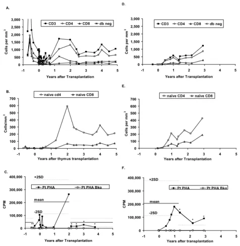
O estudo da cinética de estabelecimento do compartimento de células T após transplante de timo com um grau elevado de incompatibilidade HLA representa um contributo único para a compreensão da dinâmica de reconstituição do sistema imunitário. Observou-se uma reconstituição progressiva das células T naïve exibindo uma diversidade do repertório conservada apesar da incompatibilidade HLA do epitélio tímico. O estabelecimento da população Treg teve uma cinética semelhante às células CD4 mantendo níveis normais. Dados recentes sugerem que o desenvolvimento da população Treg no timo está dependente de um pequeno “nicho” que controla o número de células produzidas. É plausível especular que o FOXN1 desempenhe um papel nestes “nichos” e contribua para a regulação do número de Treg. Em contraste, observou-se a persistência da população DNαβ ao longo dos 5 anos de seguimento, sugerindo uma produção mantida eventualmente num possível rudimento tímico associado à mutação FOXN1.
A função do enxerto tímico alogénico foi ainda estimada pela quantificação do sj/βTREC, um rácio entre produtos precoces e tardios do rearranjo do TCR no timo, que representa uma medida indirecta do número de divisões intra-tímicas e que se correlaciona directamente com a produção tímica. O rácio sj/βTREC atingiu níveis comparáveis aos observados em crianças saudáveis. No entanto, 4 anos pós-transplante, documentou-se um declínio marcado do rácio sj/βTREC suportando uma diminuição da actividade do enxerto. Apesar disso, observou-se apenas uma pequena redução seguida de estabilização das células T naive, sugerindo que após reconstituição do sistema imunitário a homeostasia periférica consegue assegurar a manutenção da população T naive.
Assim, os nosso dados demonstram que é possível adquirir competência imunológica com transplante tímico independentemente da compatibilidade HLA, mesmo na ausência de sustentabilidade do enxerto sugerida pela diminuição do rácio sj/βTREC, o que tem implicações para o desenvolvimento de novas estratégias de reconstituição imunológica noutros contextos clínicos.
Estudos recentes sugerem um aumento compensatório da actividade tímica em resposta a uma diminuição do compartimento de linfócitos T. Tem sido atribuído um papel à citocina IL-7 neste processo para além dos seus efeitos na proliferação homeostática das células T na periferia. A infecção pelo HIV está associada a uma perda progressiva de células T CD4, sendo ainda controversa a capacidade do timo de compensar esta perda bem como de promover a reconstituição desta população após terapêutica antiretroviral (ART) em doentes infectados pelo HIV-1. Por outro lado, os efeitos periféricos da IL-7 podem minimizar a possível redução da actividade tímica quer devida à infecção directa dos timócitos pelo HIV quer mediada pela hiper-activação generalizada associada à infecção. De facto, a hiper-activação persistente do sistema imunitário, com a consequente anergia e aumento da susceptibilidade à apoptose, é considerada determinante na progressão para SIDA.
O segundo objectivo deste trabalho foi investigar a relação destas vias na infecção HIV/SIDA. A originalidade deste estudo residiu na comparação de doentes infectados pelo HIV-2, que mesmo na ausência de terapêutica antiretroviral têm virémia reduzida, com indivíduos infectados pelo HIV-1 com supressão terapêutica da virémia e diferentes graus de recuperação das células T CD4. A comparação com doentes infectados pelo HIV-1 não tratados e, portanto, com elevada virémia, permitiu concluir
que a linfopénia CD4 se associa a uma perda de células T naive, quer CD4 quer CD8, independentemente do tipo de infecção ou exposição à ART. Nos doentes HIV-1+ tratados com ausência de recuperação das contagens CD4 apesar de resposta virológica, várias estratégias parecem contribuir para suster o declínio das células T CD4 e proteger de infecções oportunistas, nomeadamente: menores níveis de activação imunológica e uma melhor utilização da IL-7 atestada pelo aumento dos níveis intracelulares de Bcl-2, uma molécula anti-apoptótica induzida pela IL-7.
O terceiro objectivo deste trabalho baseou-se no estudo de um doente com Doença Granulomatosa Crónica (CGD) e uma linfopénia CD4 persistente na ausência de infecção pelo HIV. A CGD constitui a deficiência fagocitária primária mais frequente (1:200000 nascimentos) e é causada por defeitos na capacidade oxidativa fagocitária. Este doente de 32 anos, com uma clínica de infecções tipicamente associadas à CGD, apresentou contagens de células T CD4 inferiores 200 células/l durante mais de 16 anos. O estudo efectuado revelou uma activação linfocitária generalizada, com diferenciação efectora terminal dos linfócitos T CD8 e uma redução da dimensão dos telómeros das subpopulações T, sugerindo um turnover celular aumentado devido a uma estimulação persistente do sistema imunitário. Além disso, observou-se uma depleção marcada das células T naive, com evidência de diminuição da timopoeise estimada pelo rácio sj/βTREC, apesar dos níveis elevados de IL-7 sérica. Este perfil imunológico tem sido associado a um risco elevado de infecções e sido considerado um factor preditivo de morte em indivíduos com idade avançada. O aparente esgotamento dos recursos imunológicos é particularmente relevante tendo em conta o aumento de esperança de vida dos doentes com CGD, devendo ser considerado na definição de indicações terapêuticas, incluindo transplante de células hematopoiéticas.
Em conclusão, este trabalho demonstra que diferentes vias podem ser exploradas para atingir competência imunológica mesmo na presença de profundas alterações linfocitárias. O seu estudo baseado em modelos humanos é fundamental para o desenvolvimento de novas estratégias de reconstituição imunológica.
Palavras chave: Células T, Timo, Imunodeficiência, FOXN1, HIV/SIDA, Doença
The thymus is essential for both the establishment of the peripheral T cell pool and the generation of the diverse T cell receptor (TCR) repertoire capable of dealing with new pathogens and controlling the escape of persistent infections. In humans, the thymus is almost fully developed at birth, with the rate of T cell production markedly decreasing after puberty. However, it is now clear that this central lymphoid organ plays an essential role in the lifetime “de novo” generation of T cells. The maintenance of naïve T cells is currently thought to depend upon a combination of peripheral T cell proliferation as well as to an age-dependent contribution of recent thymic emigrants.
The overall aim of this work was to investigate the relative roles of the thymus and the “periphery” in the maintenance/recovery of the human T cell compartment through the study of specific clinical models.
FOXN1 is a transcription factor, expressed by thymic epithelium, crucial for both the development of the thymus and prevention of its involution. Defects in FOXN1 in mice lead to athymia in association with total alopecia, due to its additional role in hair follicle differentiation (“nude-SCID mice”). Human FOXN1 deficiency was first reported by Pignata et al. in two sisters from Italy that, despite the evidence of athymia, exhibited a significant number of circulating T cells. We identified the same homozygous R255X mutation in a Portuguese child, who presented at 5 months of age with alopecia, respiratory failure due to Bacillus Calmette-Guérin (BCG) dissemination following routine neonatal BCG vaccination, and circulating T cells of non-maternal
origin at close to normal numbers. The first aim of this work was to investigate the T cell population generated in the presence of FOXN1 deficiency.
Circulating T cells were equally distributed between CD4, CD8 and, strikingly, an abnormally increased population of TCR αβ+ cells that expressed neither CD4 nor CD8 (double-negative, DNαβ), which usually represent less than 1% of the peripheral T cell pool. T cells were non-naïve, oligoclonal, activated, and unable to proliferate in vitro. The thymus is known to produce a regulatory CD4 T cell subset (Treg), with suppressive properties, fundamental for preventing autoimmunity. Currently they are best identified by expression of the forkhead box P3 transcription factor FoxP3. Notably, more than 40% of the CD4 subset expressed high levels of FoxP3 and had a clear regulatory-like T cell (Treg) phenotype. Thus, human FOXN1-deficiency due to R255X mutation was associated with significant numbers of oligoclonal T cells suggesting that, to a certain extent, T cell development still occured, albeit with altered positive/negative selection, as illustrated by the aberrant expansion of FoxP3+ and DN subsets.
As FOXN1 mutations impact on thymic epithelium rather than hematopoietic precursors, we predicted that thymic transplantation, although never performed before in this setting, could be a curative strategy. This was confirmed by the documentation of the clinical efficacy of HLA-mismatched thymic transplantation, as attested by the temporal association between the clearance of the ongoing BCG adenitis and the development of specific responses against mycobacterium antigens.
Our study of the kinetics of establishment of the T cell pool after HLA mismatched thymic transplantation provides unique data regarding the dynamics of replenishment of the immune system.
A progressive increase of naive (antigen non-experienced cells) T cells was also observed, resulting in the generation of a fully diverse CD4 T cell repertoire in spite of the HLA-mismatched thymic epithelia.
Reconstitution of the Treg pool occurred in parallel to that of the CD4 subset, leading to stable frequencies within the normal range. Thymic Treg development is currently thought to be dependent upon a small developmental niche that tightly controls Treg output. It is thus possible that FOXN1 plays a role in such niches, contributing to the thymic regulation of Treg numbers.
In contrast, a significant population of circulating DNαβ persisted, at relatively stable frequencies (17% of αβ T cells) and phenotype, throughout 5yrs of follow-up. Although the possibility that DNαβ T cells are long-lived cells generated pre-transplant cannot be excluded, it is also plausible that their persistence may reflect a continuous production of this population by a putative thymic rudiment.
The functionality of the allogeneic thymic graft was further estimated by sj/βTREC quantification; a ratio between early and late products of TCR rearrangements that was shown to represent an indirect measurement of thymocyte division-rate, and a direct correlate of thymic output. A progressive increase of the sj/βTREC ratio was observed, reaching levels comparable to those found in healthy children. Importantly, a sharp decline of sj/βTREC, accompanied by a decrease in the proportion of naïve cells was observed 4yrs post-transplant. Notably, these values plateaued thereafter, suggesting that steady-state equilibrium could be established after replenishment of the immune
system.
Importantly, our data showed that immunocompetence can be achieved through HLA-mismatched thymic transplantation, despite the lack of a sustained thymocyte-division rate (as evidenced by sj/βTREC). These novel data regarding the long-term sustainability of allogeneic thymic tissue and the immunological reconstitution achieved have implications for the design of immune-based therapeutic strategies to be used in other clinical settings.
Recent studies suggested the possibility of a thymic rebound as a compensatory feedback loop triggered by emptiness of the peripheral T cell pool, particularly in HIV infection. The cytokine 7 has been highlighted as a possible factor in this process. IL-7 has also a central role in peripheral T cell homeostasis. The IL-IL-7 driven peripheral T cell proliferation/survival is thought to be able to compensate any putative thymic impairment resulting either directly from HIV infection, or from the heightened state of immune-activation that characterizes HIV disease. In fact, the persistent immune stimulation, and the consequent T cell anergy and susceptibility to apoptosis, are considered key determinants of CD4 decline and AIDS progression.
The second specific aim of this work was to investigate the interplay of these pathways during HIV/AIDS. The originality of this study mainly resulted from the definition of the cohorts under investigation. HIV-1+ patients with discordant responses to ART (ART-Discordants, poor CD4 recovery despite suppression of viremia) were compared with HIV-2+ patients that exhibited a similar degree of CD4-depletion and reduced circulating virus in the absence of ART. Untreated HIV-1+
patients who are expected to have high viremia, as well as HIV-1+ patients under ART with successful virological and immunological responses were studied in parallel.
Low CD4-counts were associated with major naïve CD4 and CD8 depletion, irrespective of type of infection or ART-exposure. Several pathways were likely to contribute to the CD4-count stability and low rate of opportunistic infections documented in ART-Discordants in spite of their presumed thymic impairment, namely lower levels of T cell activation and a better ability to use IL-7 as indicated by the expression levels of the IL-7 induced, anti-apoptotic molecule, Bcl-2.
Our third specific aim was based on the study of an individual with chronic granulomatous disease (CGD), a primary defect in the phagocytic oxidative burst, who, despite being HIV negative, presented CD4 T cell depletion at levels similar to those found in advanced AIDS patients. CGD represents the most prevalent (1:200000 live births) primary phagocytic defect. This 32-year-old patient, presenting with typical CGD-associated infections, had a CD4 lymphopenia of less than 200 cells/l, for more than 16 years. Although there are previous reports of diminished T cell numbers in CGD patients, these studies did not include phenotypic and functional T cell analysis. We found a generalized immune activation, in conjunction with markers of increased cell-turnover, including a reduced telomere length of both the CD4 and CD8 subsets, and expansion of terminally-differentiated effector CD8 T cells. Additionally a marked
loss of naïve T cells was found, with evidence of impaired thymic production as assessed by sj/βTREC ratio, despite the increased IL-7 levels. This immunological profile has been considered a risk for infections, and was shown to be an independent predictor of death in aged subjects. Therefore, it is worth considering this putative exhaustion of immune resources in the evaluation of long-term therapeutic strategies,
including stem-cell transplantation, given the increasing life-expectancy of CGD patients.
Overall, our data provide evidence that, although immunodeficiency may be associated with profound lymphocyte disturbances, different pathways can be exploited to achieve immunological competence. The characterization of these pathways in human models is of importance for the definition and design of new strategies for immune reconstitution.
Keywords: T cells, Thymus, Immunodeficiency, FOXN1, HIV/AIDS, Chronic
Introduction
The main function of the immune system is to protect the individual from the continual exposure to potentially pathogenic microorganisms. Another fundamental property is to discriminate these foreign antigens from self-antigens and thus, to maintain immune tolerance.
T lymphocytes play a central role in this process, in two ways. Firstly, they act as orchestrators of the many cell types involved in both innate (non-specific) and adaptive (specific) immune responses. With regard to the latter, they provide the help necessary for B cell differentiation and antibody production. Secondly, they have key effector functions, as clearly illustrated by the contribution of cytotoxic T lymphocytes to the effective control of intracellular pathogens, particularly viruses. In order to carry out these diverse roles, it is necessary for T lymphocytes to possess a broadly reactive TCR repertoire able to react against foreign antigens whilst maintaining self-tolerance.
Potential strategies for T cell reconstitution have become a critical research area for clinical practice. The newly developed, more aggressive therapeutic interventions in the areas of oncology, auto-immune disease, and transplantation are frequently associated with a major side-effect: T cell depletion. It has been shown that the reconstitution of cellular immunity after hematopoietic stem cell transplantation is a critical determinant of its long-term success. On the other hand, in spite of the major
impact of antiretroviral therapy (ART) on the survival and morbidity of Human Immunodeficiency Virus (HIV)-1 infected patients, it is now clear that immune-based complementary therapies are needed to achieve the immunological recovery required to allow the discontinuation of the antiretroviral drugs. Moreover, although primary T cell immunodeficiencies are rare, they are frequently life-threatening conditions that require prompt diagnosis and therapeutic intervention.
The overall aim of this work is to investigate mechanisms involved in the preservation and recovery of the human T cell compartment. Therefore, the introduction has been divided into three main sections.
The first section comprises an overview of T cell physiology, focusing on the central role of the thymus in the establishment and maintenance of a diverse T cell population. Moreover, the main mechanisms thought to be involved in T cell homeostasis and in the regulation of T cell responses will be discussed.
The second part addresses the main causes of T cell deficiencies, with particular focus on the clinical settings that were explored in this work as a strategy to address the relative roles of the thymus and periphery in the maintenance/recovery of the T cell pool.
The final section provides a review of the most important, currently available, therapeutic strategies for achieving T cell recovery.
1.1 T cell physiology
1.1.1 T cell generation
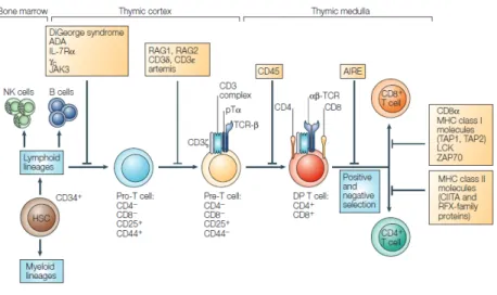
T lymphocytes are produced in the thymus. This primary lymphoid organ provides a unique environment that enables the differentiation of bone-marrow derived T cell precursors into mature T cells. The key feature of this process is the generation of a diverse repertoire of functional T cell receptors (TCR), with a reduced reactivity to self-antigens. As a primary site for T cell lymphopoiesis, the thymus is active, not only during the foetal stages of development, but also throughout postnatal life.
In the mouse, the thymic rudiment is first evident on day 11 of gestation, having evolved from the endoderm of the third pharyngeal pouch (1). The development of the thymus in humans closely follows that observed in the murine model of thymic development. The growth of the thymus primordium from the third pouch coincides with the onset of FOXN1 (Forkhead box protein N1) gene expression (2). The forkhead transcription factor FOXN1 is the best-known regulator of foetal thymus development (3). Its expression has been shown to be required for differentiation of thymic epithelial cells (4) and the induction of cortical and medullary differentiation in the thymus (5). Before undergoing the differentiation into the medullary and cortex compartmentalization, the thymus is colonized by hematopoietic precursors (1, 6, 7).
The thymic architecture consists of distinct anatomical compartments which include the subcapsular area, the cortex, the cortical medullary junction and the medulla (8). Thymic epithelial cells (TECs) constitute the major component of the stromal population. They can be subdivided according to their functional,
morphological, and antigen presentation capacity, into two main subpopulations, cortical (cTEC) and medullary (mTEC) epithelial cells. In addition to the TEC subsets described above, the thymus is composed of other cell types, including populations of haematopoietic (dendritic cells, macrophages, B cells) and nonhaematopoietic (fibroblasts, endothelial cells and others) origin. This stromal scaffold provides the specialized microenvironment necessary for the life-long attraction of haematopoietic precursor cells; the signalling infrastructure required to instruct early thymocyte differentiation; the factors to guide precursor cells to the different anatomical sub-compartments; the molecular constraints that are needed for the selection of immature T cells; and the molecules necessary for the functionally mature T cells to exit to the periphery. The hallmark of T cell development is the generation of T cells that express a functional TCR, whether it be TCR or TCR, able to recognize antigenic peptides bound to major histocompatibility complex (MHC) molecules on the surface of antigen presenting cells (APC).
The CD34+ precursor cells, which originate from bone-marrow stem cells, migrate to the thymus to undergo T cell development (9). These precursor cells enter the human thymus at the cortico-medullary junction. From this area they migrate towards the cortical region where proliferation and differentiation are initiated via interactions with the thymic stroma. The distinct stages of T cell development are defined by the sequential expression of cell-surface antigens (Figure 1) (10). Rearrangement of the TCR , and loci, as well as expression of the CD1a molecule mark the commitment to the T cell lineage. The definition of thymocyte differentiation steps is typically based on the cell-surface expression of CD4 and CD8, with undifferentiated populations
being classified as CD4-CD8- double negative (DN). In humans, three distinct DN stages can be recognized: a CD34+CD38-CD1a- stage that represents the most immature thymic subset, and the consecutive CD34+CD38+CD1a- and CD34+CD38+CD1a+ (Pre-T1) stages. Human DN thymocytes mature via a CD4 immature single positive (CD4ISP) stage (Pre-T2), which express CD4 in the absence of CD8. This population contains precursors for both the and T cell lineages. The CD4ISP stage is followed by an early double-positive (EDP) stage composed of cells expressing CD4 and the chain of CD8. EDP cells are the immediate precursors of double-positive (DP) cells, which are characterized by the co-expression of CD4 and the and chains of CD8. Following this DP stage, cells differentiate into CD4+ or CD8+ SP T cells that express a functional TCR and exit the thymus (8).
Figure 1:Early stages of T cell development in the thymus. From Spits et al (10).
The DN thymocytes mature in distinct areas within the cortex, as well as in the subcapsular region. Their proliferation, survival and differentiation are controlled by a combination of cell-autonomous factors and a number of stromal cell-derived signals
(including interleukin-7 (IL-7) and stem cell factor (SCF)) secreted by TEC; Wnt molecules, Hedgehog (an essential positive regulator of T cell progenitor differentiation); and Notch, amongst others (11-13).
TCR gene rearrangement plays a crucial role in thymocyte fate. Thymocytes can only survive to maturity if they successfully carry out combinations of gene rearrangements that will generate and , or and , chains, that can support the assembly of TCR/CD3, or TCR/CD3 complexes, respectively. The TCR diversity that can be generated within the thymocyte pool has been estimated to be as large as 1020 /-chain combinations.
The antigen-recognising, variable domains of TCR and chains are encoded by combinations of variable (V), diversity (D), and joining (J) gene segments (TCR chains), or V and J gene segments (TCR chains).
D-to-J recombination occurs first in the chain of the TCR. This process can involve either the joining of the D1 gene segment to one of six J1 segments, or the joining of the D2 gene segment to one of seven J2 segments. D-to-J recombination is followed by V-to-DJ rearrangements leading to the formation of the TCR C chain.
T cell commitment is thought to occur with the initiation of TCR gene rearrangement. -selection is the process by which precursor T cells with a productive rearrangement of the TCR locus are selected to undergo further differentiation, in the form of TCR rearrangements. The developmental stage at which -selection occurs in humans is still the subject of controversy (10). However, it was recently shown to occur as early as the CD34+CD38+CD1a+ stage (14). Thymocytes harbouring a rearranged
TCR locus generate a TCR molecule that pairs covalently with the invariant pre-TCR (pT), and noncovalently with CD3 signal-transducing molecules, resulting in formation of a pre-TCR complex at the cell surface.
The rearrangement of the chain of the TCR is followed by chain rearrangement. The subsequent assembly of the and chains results in formation of the -TCR that is expressed on a majority of T cells. As the TCR gene segments are embedded within the TCR locus, the V-to-J rearrangements lead to deletion of the locus from the chromosome (15).
After completion of TCR rearrangements, T cells are selected by a low-affinity interaction of the TCR heterodimer with self-peptides complexed with MHC antigens, a process termed positive selection. Thymocytes differentiate into single positive (SP) CD4 or SP CD8 thymocytes whose TCR recognises MHC class II or class I molecules, respectively. Death by neglect of thymocytes whose TCR cannot recognise antigen in the context of either MHC class I or II molecules ensures that only those thymocytes with appropriate TCR specificities survive and differentiate into functionally mature T cells (16). Thymocytes that express high-affinity receptors for self-peptide–MHC complexes expressed on thymic dendritc cell (DCs) are deleted through a process known as negative selection.
The usual end result of these combined selection mechanisms is the generation of a naïve T cell pool composed of cells with a stringently selected TCR repertoire, able to respond to foreign non-self antigens. They are exported to the periphery where the majority of them remain tolerant to the host’s tissues (self).
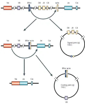
A variety of T cell receptor excision circles (TRECs) are formed from the excised DNA generated by the rearrangements of the TCR, and loci described above.
During TCR rearrangement, the end-to-end ligation of the recombination signal sequences flanking the -rec locus and the -J locus generates a single TREC containing a signal joint (sj) sequence (sjTREC), as shown in Figure 2. Coding-joint (cj)TRECs are produced during the TCR rearrangement of V to J gene segments.
Figure 2: T cell receptor excision circles. From Spits et al (10).
TRECs are not duplicated during mitosis, and are therefore diluted out with each cellular division (17, 18). Since recent thymic emigrants (RTE) are enriched in these molecules, TREC levels have been used to assess thymic function, through their quantification in peripheral blood using real time PCR technology.
The quantification of sjTREC as a marker of RTE, and measure of thymic output was first utilized by Douek et al (19). As expected, given the gradual loss of thymic function during aging, they showed a decreasing number of sjTRECs with age in
healthy individuals. Moreover, they also reported that sjTREC levels were decreased in HIV infected patients, but rapidly increased during antiretroviral therapy (19).
Since sjTRECs are not replicated during cell division, their levels can also be influenced by events occurring in the periphery, such as cell proliferation and differentiation, redistribution, or alterations in cell survival. All of these can lead to a dilution of TREC-bearing cells in the periphery. Thus, TREC quantification data requires cautious interpretation; particularly in those conditions, such as the process of immune reconstitution following bone marrow transplantation or during HIV infection, that are associated with the previously described confounding factors.
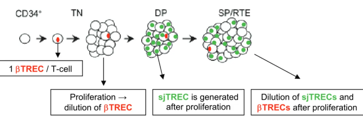
The level of thymic output has been shown to be primarily determined by the intrathymic proliferation of precursor T cells (20). Thus, a new assay was developed to estimate the relative changes in intrathymic proliferation occurring between the TN and early DP stages (21), as represented in Figure 3. This approach is based not only in the quantification of sjTRECs, generated at the DP stage, but also the DJTREC created during the previous DJ rearrangement and organization of the TCR locus. The proliferation occurring between these stages can then be estimated by the measurement of the ratio of TREC to sjTREC (sj/TREC ratio) (21). It was also shown that this parameter was not influenced at the periphery, given that both types of TREC were equally affected by the rates of proliferation and death of peripheral T lymphocytes (21).
Figure 3: Schematic representation of intrathymic proliferation occurring between late TN and early DP thymocyte differentiation stages and its effects on TREC and sjTREC levels.
TN: Triple Negative stage; DP: Double Positive stage; SP/RTE: Single Positive/ Recent Thymic Emigrant stage. Adapted from Dion, et al (21).
1.1.2 The naïve T cell compartment
RTEs emigrating from the thymus are incorporated into the pool of naïve T cells. The size of this pool remains relatively constant throughout adult life despite continuous antigenic stimulation and the reduction of naïve T cell production by the thymus. The maintenance of the naïve T cell pool is thought to mainly depend upon survival signals, such as those provided through TCR engagement of self-peptide-MHC complexes and by IL-7 (22-24). IL-7 has been shown to be a key cytokine involved in controlling the survival and homeostatic turnover of peripheral T cells (25-27). Its effects on T cells appear to be multifactorial and are regulated by the expression of its specific receptor, a heterodimer consisting of the IL-7 receptor chain (IL-7R) and common gamma chain (c). The binding of IL-7 to its receptor induces several signalling cascades, such as the JAK–STAT (Janus kinase–signal transducer and activator of transcription) and the PI3K (phosphoinositide 3-kinase) pathways, that promote lymphocyte survival (28, 29). Furthermore, IL-7 has been shown to inhibit
1 TREC / T-cell
Proliferation →
programmed cell death by up-regulating the expression of the anti-apoptotic molecule Bcl-2 (30).
Prior to contact with antigen, naive T cells continuously recirculate between blood and secondary lymphoid tissues (spleen, lymph nodes, Peyer’s patches and mucosal-associated lymphoid tissues) (31, 32). Entry into secondary lymphoid tissues is a highly regulated process. In the case of lymph nodes and Peyer's patches, specialized blood vessels called high endothelial venules (HEVs) serve as the entry point into the tissue. HEVs express a unique set of ligands that are recognized by homing receptors expressed on the naïve T cells’ surfaces. Amongst the most critical of these are the chemokine CCL21, that engages CCR7, and vascular addressins, that interact with CD62L (L selectin) (31). Thus, naïve T cells moving along HEV’s encounter IL-7, self-peptide–MHC complexes and CCR7 ligands, all of which cooperate to produce homeostatic survival signals (33). Continuous migration of T cells through the secondary lymphoid tissues is a key mechanism in providing antigenic surveillance. Within lymphoid tissues, these antigens are presented to T cells in the form of peptide fragments bound to MHC molecules expressed upon specialized APC, in particular DC. These cells are strategically positioned within a dense network in the T cell zones and are continuously surveyed by recirculating T cells for their expression of foreign peptide/MHC complexes.
Several surface molecules have been shown to be expressed by, and thus identify, naïve T cells. The co-expression of CD45RA and CD62L on CD4+ T cells is frequently used, and is currently thought to identify the majority of this population. With respect to CD8 T cells, the surface co-expression of CD45RA and CD27 is commonly used to
identify the naïve subset (34). However, the combination of these markers with other molecules, such as CCR7 or CD28, has enabled a more detailed definition of CD8 T cell differentiation states and/or functional capacity (35-37).
Some groups have shown that the CD31 (PECAM-1) molecule is expressed preferentially on the cell surface of naïve, TREC-rich T cells that have undergone a low number of cell divisions (38, 39). On this basis, Kimmig et al were able to distinguish two populations of naive T cells: “true” recent thymic emigrants (thymicnaive CD4 cells, that co-expressed CD45RA and CD31 and have high TREC content), and peripherally expanded naive CD4 cells (centralnaive CD4 cells) (38), as illustrated in Figure 4.
As previously discussed, aging is associated with decreased thymic activity that results in reduced numbers of thymicnaive CD4 cells (38). Triggering by self peptide/MHC complexes is thought to induce thymicnaive CD4 cells to proliferate and differentiate into centralnaive CD4 cells (38). This population featured lower TREC levels, expressed CD45RA and lacked CD31 expression. Of note, both thymicnaive and centralnaive CD4 cells are able to differentiate into CD31-CD45RA-CD45RO+ memory-effector cells upon antigen encounter (38).
Importantly, results from our lab suggest that IL-7 may play a role in the maintenance of CD31+ naïve CD4 T cells during adult life (40). In adults, this cytokine was shown to preferentially drive the proliferation of the CD31+ naive CD4 subset, and to increase or sustain the expression of CD31.
Figure 4: Post-thymic proliferation of human naive CD4 T cells. From Kohler et al (39).
Recently, protein tyrosine kinase 7 (PTK7) was described as a novel marker of human CD4 RTEs (41). A fraction of the naïve CD31+ CD4 subset was shown to express PTK7 and to contain higher levels of sjTREC as compared with the PTK7-CD31+ counterpart. Additionally, patients that underwent complete thymectomy were shown to have a more pronounced and persistent loss of PTK7+CD31+ than PTK7-CD31+ naive CD4+ T cells, suggesting that PTK7, unlike CD31, may identify those RTEs most recently produced by the thymus (41).
Upon antigen stimulation, naïve T cells differentiate into distinct cell types that play important functions during the development of an immune response.
1.1.3 The mounting of specific immune responses
Specific immune responses are mostly initiated in the T cell areas of secondary lymphoid organs where naïve T lymphocytes encounter antigen-loaded DC. T lymphocytes recognize antigen through the interaction of their TCR with foreign peptide-MHC complexes displayed on the surface of APCs, through which a tight synapse can form (34, 42). Activation of the T cell follows synapse formation and is associated with rapid clustering of TCR molecules, bound to peptide/MHC complexes upon the APC, and a consequent, local accumulation of intracellular signalling molecules. The intensity of signal that a T cell receives is dependent both on the number TCRs triggered by the peptide–MHC complexes, and the level of costimulatory signals that regulate the activation process (43). A large number of costimulatory/adhesion molecules expressed on T cells (CD28, LFA-1, CD40L, ICOS, OX40, CD2, CD27, and 41BB) (44, 45) bind to their receptors on the APC. Some costimulatory/adhesion molecules provide essential second signals for T cell activation, whilst others act by enhancing TCR triggering via stabilization of the synapse, and/or through the recruitment of intracellular signalling molecules (46, 47). A further level of co-stimulation is provided by the release of various cytokines that can act in an autocrine and/or paracrine fashion to enhance the process of T cell activation and subsequent downstream events.
The continuous TCR and cytokine stimulation induces T cell to divide and progressively differentiate into effector and memory subsets, during which they acquire the capacity to produce effector molecules, such as cytokines and cytolytic mediators (48, 49).
At later stages of the immune response, the clearance of pathogen from the site of infection, by effector cells, reduces the influx of antigen-loaded APC into the T cell zones.
After antigen withdrawal, the survival of activated T cells becomes dependent upon the expression of anti-apoptotic molecules such as Bcl-2 and the expression of receptors for homeostatic cytokines such as IL-7 and IL-15, as discussed below.
1.1.4 Memory-effector T cell subsets
During an immune response, antigen-specific T cells proliferate, generating a large number of effector cells that migrate to the distal site of infection to fight the invading pathogen. Some of these primed T cells develop into memory cells, which confer protection in peripheral tissues, through their ability to mount a more rapid and effective response to their cognate antigen. This process is known as a secondary immune response. Of note, memory T cells increase progressively with age as a consequence of T cell responses to diverse foreign and self-antigens.
Understanding the pathways of memory T cell differentiation in humans has been a central issue in immunology. Distinct memory T cell subsets have been defined on the basis of homing capacity and effector function (34, 50). These definitions, based on the expression of several differentiation markers, have failed to identify a clear phenotype for each putative subset. Nevertheless, both in humans and mice, memory cells have been shown to comprise of populations of “central” memory (TCM) and effector-memory (TEM) T cells. TCM and TEM cells were initially defined in the human immune system based on the absence or presence of immediate effector function, and
on the expression of homing receptors that allow cells to migrate to secondary lymphoid organs vs. nonlymphoid tissues (34, 50).
Human TCM are CD45RO+ memory cells that recirculate between blood and the secondary lymphoid organs, entering the latter via high expression of CCR7 and CD62L (51). These cells also feature high surface expression of CD127 and CD122, which allow them to readily respond to IL-7 and IL-15, promoting survival and homeostatic proliferation. Central memory T cells have little or no immediate effector function but readily proliferate and differentiate into effector cells in response to antigenic stimulation (51).
Effector memory T cells (TEM) migrate to inflamed peripheral tissues and display immediate effector function, as evidenced by their rapid production of effector cytokines, such as IFN-, and contain large amounts of cytolytic mediators, such as perforin. Human TEM are cells that have lost CCR7 expression, have a heterogeneous CD62L expression profile, and display characteristic sets of chemokine receptors and adhesion molecules that are necessary for homing to inflamed tissues (51).
Memory and effector CD8 T cells have been shown to play an important role in viral infections throught their cytotoxic activity and also because of their ability to produce various factors involved in suppression of viral replication, including cytokines and chemokines (51-53).
The surface expression of the costimulatory molecules CD27 and CD28 together with CD45RA have been used by several authors to discriminate between distinct stages of human CD8 T cell differentiation (36, 37, 54). A T cell differentiation pathway
has been proposed in which CD27+CD28+CD45RA+ naive cells progress through a CD27+/-CD28+/-CD45RA- to a CD27-CD28-CD45RA+ terminally differentiated effector phenotype with increased cytotoxic potential and reduced ability to proliferate (35, 51, 53).
Both IL-7 and IL-15 have been described as playing a role in the maintenance of memory T cells (55). In particular, IL-15 has been shown to have a crucial role in memory CD8 T cell generation and/or maintenance, as illustrated by the lack of memory CD8 T cells in IL-15R- and IL15-deficient mice (56, 57). Although IL-15 has been reported to have only a minimal role in the homeostasis of memory CD4 T cells (58), it has been recently published that antigen-specific CD4 memory cells are also dependent upon this cytokine for their basal homeostatic proliferation and long-term survival (59).
Effector CD4 T cells, also known as CD4 T helper (Th) cells, can be polarized into distinct subsets characterized by the acquisition of cytokine production and other specialized functions. Polarization of lymphocytes to T helper 1 (Th1) or Th2 cells is promoted by IL-12 and IL-4, respectively (60), as well as by the strength and duration of TCR stimulation (61). The differentiation processes involves upregulation of specific transcription factors (62, 63) and activation of STAT proteins (64).
As part of their differentiation programme, Th1 and Th2 cells down-regulate lymph-node homing receptors, and up-regulate the expression of those receptors necessary to enable their migration to inflamed non-lymphoid tissues, where they exert effector functions. Each lineage also expresses unique cytokine receptors, enabling them to respond to cytokines produced by accessory cells. Th1 cells are characterized
by their secretion of IFN- and are important activators of macrophages, NK cells, and CD8 T cells (65). They are thought to be involved in the defense against intracellular pathogens. Th2 cells secrete IL-4, IL-13, IL-15, IL-10 and IL-25 and are important for barrier defense at mucosal and epithelial surfaces, as well as in the control against parasites. These cells mobilize and activate eosinophils, basophils, mast cells and, alternatively, activated macrophages (66).
A third subset of IL-17-producing effector T helper cells, referred to as Th17 cells, has recently been characterized. The differentiation of Th17 cells is initiated by the activation of naive T cells in the presence of IL-6 plus transforming growth factor (TGF)-. This leads to the expression of the transcription factor ROR-t, and production of IL-17 (67). Th17 cells have been suggested to play a role in the induction of autoimmunity and inflammation. They act in concert with neutrophils and are important for defense against extracellular bacteria and fungi (68, 69).
Recently, IL-9 producing CD4 T cells have been described as a novel Th subset (TH9) (70, 71). Th9 differentiation has been shown, both in humans and mice, to require exposure to the cytokines TGF- and IL-4, and to be mediated through the expression of the transcription factor GATA-3. Of note, several inflammatory cytokines, such as IL-1, IL-6, IL-10, IL-21 and type I IFNs, enhance IL-9 production, suggesting a complex regulation of Th9 differentiation (72). The exact role of Th9 cells in the immune system has yet to be fully defined. Studies in mice have suggested this subset plays a role in tissue inflammation (70), immunity against helminth infections, as well as in allergy (71).
Follicular helper T cells, or ThF cells, are memory-effector CD4 T cells found in lymph nodes and identified by high expression of CXCR5 (73, 74). They are found at the periphery of B cell follicles and in germinal centers (GC). They are thought to mediate naïve B cell activation and GC formation, probably through the expression of CD40 ligand (CD40L) and secretion of IL-21 and IL-10.
1.1.5 Regulatory T cells
An important feature of the immune system is its capacity to discriminate between self and non-self, whilst establishing and maintaining a lack self-responsiveness. The maintenance of immunological self-tolerance is a tightly regulated process. As previously discussed, the deletion of self-reactive cells in the thymus plays a key role in this process. As this process is not perfect, autoreactive clones may occasionally escape into the periphery, hence the need for other mechanisms to maintain a state of tolerance in the periphery. So-called “peripheral tolerance” is maintained through a variety of mechanisms, including the presence of a population of regulatory T cells (Treg) that actively suppress autoreactive T cells (75-78).
Treg form a subset of CD4 T cells that either develop in the thymus (naturally occurring Treg; nTreg) or in the periphery (induced Treg; iTreg). The latter can be generated in a variety of circumstances; such as direct differentiation from naive T cells that have undergone TCR-stimulation in the presence of TGF-, or via the conversion of other pre-existing T cell subsets.
Treg constitute 5% to 10% of the peripheral CD4 T cell compartment (79). In both mice and humans, this population was first defined by their expression of the IL-2
receptor -chain (CD25), and their capacity to suppress the proliferation of other T cells. CD4+CD25+ Treg were first defined by Sakaguchi et al. (75) in the murine system. They showed that the transfer, into athymic nude mice, of lymphoid-cell populations depleted of CD4+ T cells expressing CD25 caused the spontaneous development of autoimmune disease. Additionally, reconstitution with CD4+CD25+ T cells prevented the development of autoimmunity.
In humans the CD4+ T cell subset expressing the highest levels of CD25 (termed CD25bright) was shown to be enriched in cells with regulatory properties (76).
The transcription factor FoxP3 has been shown to be specifically expressed by Treg, and is currently considered the most specific marker of these cells. This was further supported by the discovery that mutations in the FOXP3 gene in humans cause immune dysregulation, polyendocrinopathy, enteropathy, X-linked syndrome (IPEX). This syndrome is characterized by a high incidence of autoimmune disease, including type 1 diabetes and thyroiditis, inflammatory bowel disease, and allergic diseases (80, 81). The FOXP3 gene has been shown to be a key regulator of Treg development and functional activity (82-84). However, due to its nuclear expression, FoxP3 cannot be used to purify Tregs (83, 85).
Importantly, FoxP3 expression can be up-regulated as a result of T cell activation, at least in humans (86). The TCR stimulation of human naïve FoxP3-CD4+ cells was shown to induce FoxP3 expression without conferring suppressive activity on these cells (86). Thus, functional characterization of FoxP3+ cell is of major importance in order to definitively confirm their true identity, especially in the context of pathological conditions associated with hyperimmune T cell activation.
One of the current challenges in the field of regulatory T cell research remains the determination of the relative in vivo contributions of nTreg versus iTreg to the Treg pool as a whole. nTreg develop in the thymus and exit into the periphery.
In younger adults, around 10% of Foxp3+ Treg express CD45RA and are considered an unprimed, or naïve Treg population (87, 88). In addition, a large proportion of CD45RA+ Treg in adults express CD31, however its frequency rapidly declines with age (89). However, naïve CD45RA+ Treg have been shown to proliferate significantly less than their CD45RO+ counterparts (89, 90). Thus, the contribution of different Treg sub-populations to the maintenance of the total Treg pool in adults is yet not clear.
Regarding iTregs, it was recently suggested that human Tregs may be generated from rapidly dividing, differentiated memory CD4 T cells (91). Although these memory Tregs were highly proliferative, they were also highly susceptible to apoptosis and replicative senescence, suggesting they possessed a limited capacity for self-renewal (91).
In an attempt to clarify the dynamics of Treg cell differentiation, Sakaguchi et al. have recently reported that human FoxP3+CD4+ T cells can be separated into three functionally and phenotypically distinct subpopulations based on the expression of FoxP3 (90, 92). These included CD45RA+FoxP3low resting Treg cells (rTreg cells); CD45RA-FoxP3high activated Treg cells (aTreg cells); and cytokine-secreting CD45RA-FoxP3low non-Treg cells. Both rTeg and aTreg cells where shown to have in vitro suppressive capacity, whereas CD45RA-FoxP3low non-Treg cells did not. Importantly, they were able to distinguish the differentiation pathways of these subpopulations. FoxP3high aTreg cells were shown to originate from rTreg cells, although some FoxP3high Treg cells may also have arisen from FoxP3-CD4+ non-Treg cells (91). Moreover, a
large proportion of the FoxP3high aTreg cells proliferated in vivo and appeared to be recently activated, whilst nearly all rTreg cells did not express the cycling marker Ki-67. On stimulation, rTreg cells upregulated FoxP3 expression, and differentiated into aTreg cells, a process associated with a concomitant gain of proliferative capacity (90). These newly differentiated aTreg were also shown to be highly susceptibility to apoptosis. Thus, the identification of the inter-relationship between the variously described Treg subpopulations, and the unique and/or common functions they may serve, is fundamental to the understanding of their role in both disease states and the maintenance of peripheral tolerance.
Several other molecules, that may be potentially important for Treg function, have been shown to be expressed by this T cell subset. In particular, cytotoxic T lymphocyte associated protein 4 (CTLA-4) was shown to be constitutively expressed by the majority of Treg (93). It is not clear whether CTLA-4 expression is absolutely required for Treg generation, regulatory capacity or both. CTLA-4-deficient mice have been shown to be able to generate CD4+FoxP3+ Tregs that maintain their suppressive capacity in vitro and in vivo (94, 95), supporting the idea that CTLA-4 expression is not an absolute requirement for the development or suppressive function of Treg. Another molecule, CD39, has been shown to be expressed by immunosupressive Treg (96). However, in contrast to mice, where CD39 is ubiquitously expressed uponTreg cells, expression of this molecule in humans was shown to be confined toa subset of FoxP3+ cells of an activated effector/memory-like suppressor cell phenotype (96). Other groups have also shown that FoxP3 expression and suppressive capacity are enriched in CD4 T cells that express low levels of IL-7R (CD127), suggesting that the low CD127 expression can be used to isolate Treg (97).
The expression of the integrin 7 (CD103) has been shown to potentially distinguishes a unique functional subset of Foxp3+ T cells with suppressive properties in mice (98, 99). CD103 is expressed on CD4+ and CD8+ T cells in the intestine and other epithelial compartments. Treg cells expressing this marker were shown to display an effector/memory phenotype, possess more potent suppressor activity in vitro, and selectively home to inflamed tissue sites in vivo (98-100). However, in humans, this markers has been described to be expressed by only a minority of the Treg population (101, 102)
Several other surface markers have been put forward as candidates to identify Treg. Overall it is not known whether the apparent degeneracy of Treg marker expression reflects the existence of discrete in vivo Treg subsets, levels of Treg activation or even if it is potentially an artefact of experimental design.
1.1.6 Maintenance of T cell homeostasis
Claude Bernard was the first to realize that the body has control mechanisms able to maintain its internal equilibrium (in terms of body temperature and the levels of nutrients and waste products) in spite of changes in the environment (103). Later, this type of control was given the name homeostasis (104). The immune system is under homeostatic control, such that it can react to changes in the environment, enabling the maintenance of a relatively constant number of cells throughout the life of an individual (105, 106). Therefore, homeostasis of both naïve and memory T cell pool is a highly dynamic and tightly regulated process depending on a balance between generation, proliferation, differentiation, survival, and death.