Nuno Ricardo Lucas Soares
Licenciatura em BioquímicaNon - cell autonomous neuroprotective effect of
carbon monoxide: microglia-to-neuron communication
Dissertação para obtenção do Grau de Mestre em Bioquímica para a Saúde
Orientador: Cláudia Susana Fernandes Queiroga, PhD, CEDOC FCM/UNL
Co-orientador: Helena Luísa de Araújo Vieira, PhD, PI, CEDOC FCM/UNL
Setembro, 2015
Nuno Ricardo Lucas Soares
Licenciatura em BioquímicaNon - cell autonomous neuroprotective effect of
carbon monoxide: microglia-to-neuron communication
Dissertação para obtenção do Grau de Mestre em Bioquímica para a Saúde
Orientador: Cláudia Susana Fernandes Queiroga, PhD, CEDOC FCM/UNL
Co-orientador: Helena Luísa de Araújo Vieira, PhD, PI, CEDOC FCM/UNL
Júri: A definir
Nova Medical School – Faculdade de Ciências Médias da Universidade Nova de Lisboa
Acknowledgments
Gostava de expressar a maior gratitude a todas as pessoas que directa ou indirectamente me ajudaram a completar esta tese de mestrado.
Aos coordenadores e inteiro corpo docente do Mestrado de Bioquímica para a Saúde, por todo o auxílio e disponibilidade ao longo destes dois anos
Às minhas orientadoras, Doutora Helena Vieira e Doutora Cláudia Queiroga, pela oportunidade que me deram, mas principalmente por tudo o que me transmitiram ao longo deste ano. Pelos brainstorms, ensinamentos e conselhos cientificos, mas também pela confiança, encorajamento e pelo exemplo de liderança que sempre foram.
Às meninas do laboratório: Sofia, Pereira, Daniela e Sara, pela assistência, pela disponibilidade e pelos cafés, lanches e conversas.
Um agradecimento a todo o CEDOC, pelo sentido de entre-ajuda e comunidade que sempre senti. Um agradecimento especial à Marcia, amiga de outras bandas, pelos cafés e um obrigado também ao pessoal das futeboladas.
À Doutora Teresa Pais, agradeço pela gentileza e disponibilidade com a interpretação dos resultados.
À Doutora Cláudia Santos pela genorosidade que teve ao ajudar-nos com os ELISAs. Um obrigado também ao seu grupo, em particular ao Gonçalo, pela paciência e tempo que dispendeu para me acompanhar.
Aos meus ‘Amiguinhos e Amiguinhas’, por todos os momentos que partilhámos, desde os anos da FCUL e que nunca vou esquecer. Um obrigado especial aos rapazes, pelas noitadas de LoL, entre outras aventuras.
Por tanto terem contribuido para o que sou hoje, agradeço a toda a minha familia, em particular aos que me são mais próximos.
Aos meus tios, por todo o amor e apoio ao longo destes anos, por me ajudarem sempre a lutar contra as adversidades.
Ao meu irmão e aos meus primos, que considero também meus irmãos, agradeço pela companhia, pelos momentos de gozo e alegria, e por me fazerem sorrir.
Aos meus avós, em particular à minha avó ‘D’, pelo apoio e amor incondicional e principalmente pelas sopas e outras marmitas deliciosas que são diariamente o meu almoço.
À minha mãe agradeço por aquilo que não consigo colocar em palavras. Por me ajudar nesta batalha com a dedicação e amor de sempre. Por ser o pilar da nossa familia, mesmo nas piores alturas, e por dar tudo para que tenhamos as ferramentas certas para enfrentar o futuro.
À minha namorada, Joana, por ser a minha companheira e melhor amiga. Por me animar e acreditar sempre em mim nos dias em que nada corria bem e nas alturas de cansaço, e por todas as coisas, que por mais pequenas que sejam, me fazem sentir indiscritivelmente feliz todos os dias.
Obrigado a todos mais uma vez, esta tese de mestrado não teria sido possivel sem o vosso apoio.
Abstract
Brain ischemia is a widespread disease worldwide, being one of the main causes of mortality and permanent disability.
A portion of the damage that ensues following the ischemic event is caused by unrestrained inflammation, which is mainly orchestrated by exacerbated microglial activity. Hence, developing strategies for modulating microglial inflammation is a major concern nowadays.
The endogenous molecule carbon monoxide (CO) has been shown to possess anti-neuroinflammatory properties using in vitro and in vivo approaches. Thus, our objective was to study CO as modulator of microglial activity, in particular in what concerns their communication with neurons, by promoting neuronal viability and limiting inflammatory output of activated microglia.
A conditioned media strategy was established with BV2 microglia and SH-SY5Y neurons as cell models. CO-releasing molecule A1 (CORM-A1), a compound that releases CO spontaneously, was used as method of CO delivery to cells.
We found that CORM-A1 pre-treatment in BV2 cells yields neuroprotective results, as it limits cell death when SH-SY5Y neurons are challenged with conditioned media from LPS-activated microglia and the pro-oxidant t-BHP (tert-butyl-hydroperoxide). Thus, we assumed carbon monoxide promotes neuroprotection via inhibition of microglial inflammation, displaying a non-cell autonomous role.
CORM-A1 pre-treatment limited inflammation by inhibiting BV2 secretion of TNF-α and stimulating IL-10 production. These results reinforce that CO’s anti-inflammatory role confers neuroprotection, as the alterations in these cytokines occur concurrently with the increase in SH-SY5Y viability.
Finally, we showed for the first time that carbon monoxide promotes the expression of CD200R1, a microglial receptor involved in neuron-glia communication and modulation of microglia inflammation. Further studies are necessary to clarify this role.
Altogether, other than just highlighting CO as an anti-inflammatory and neuroprotective molecule, this work set the foundation for disclosing its involvement in cell-to-cell communication.
Resumo
A isquémia cerebral é uma das doenças mais predominantes a nivel mundial, sendo uma das principais causas de mortalidade e invalidez.
Parte da propagação de dano no cérebro é causado por inflamação descontrolada, causada principalmente por disfunção da microglia. Desta forma, existe a necessidade de tentar desenvolver estratégias para melhor compreender e modular as acções destas células.
O monóxido de carbono (CO), é uma molécula endógena com provas dadas como anti-neuroinflamatório em vários modelos. Assim, o principal objectivo do trabalho foi o estudo do CO como um modulador da acção da microglia, com principal foco dado à comunicação entre estas células e neurónios, tentando entender se existe um efeito neuroprotector por inibição da inflamação.
Um protocolo de meio condicionado foi estabelecido usando as linhas celulares BV2 e SH-SY5Y, de microglia e neurónio. A molécula CORM-A1, que liberta expontaniamente CO, foi usada como método de entrega da molécula às celulas.
Demonstrámos que o pre-tratamento de células BV2 com CORM-A1 gera neuroprotecção já que reduz a morte celular de neurónios SH-SY5Y quando são incubados com meio condicionado de microglia activada em conjunto com o pró-oxidante
t-BHP (tert-butil hidroperóxido). Assim, considerámos que o CO promove
neuroprotecção ao inibir as acções inflamatórias da microglia.
O papel anti-inflamatório da molécula CORM-A1 foi confirmado quando se verificou que pré-tratamento desta molécula em microglia BV2 limita a secreção de TNF-α mas estimula a secreção de IL-10.
Por último, a CORM-A1 induziu a expressão do receptor da microglia CD200R1, molécula que participa na comunicação neurónio-microglia e fundamental para a modulação das acções inflamatórias destas últimas.
Em suma, o nosso trabalho reforçou as propriedades anti-neuroinflamatórias do CO e uma capacidade de modular viabilidade neuronal através do seu efeito a nível de comunicação célula-célula.
Index
Acknowledgments ... v
Abstract ... ix
Resumo ... x
Figure Index ... xiii
Abbreviations ... xv
I. Introduction………..……….…..… 1
1.1 Brain cells – general introduction……….……. 3
1.2 Microglia, neuroinflammation and pathology……….…….. 6
1.3 Developing anti-inflammatory strategies and the importance of cellular cross-talk……….… 10
1.4 Carbon monoxide – Introduction and toxicity……….…… 11
1.5 CO as a therapeutic molecule? – Historical perspective………….….… 12
1.6 Carbon monoxide releasing molecules – A novel method for CO therapeutic delivery……….…… 14
1.7 CO and its cytoprotective properties – Molecular insight…………...…. 16
1.8 Final remarks and objectives………... 19
II. Materials and Methods 2.1 Cells and reagents……….... 23
2.2 LPS validation protocol………... 24
2.3 NO quantification……… 24
2.4 Optical microscopy………. 24
2.5 t-BHP validation protocol………... 25
2.6 Conditioned media/neuroinflammation protocol……… 25
2.7 Flow cytometry………...… 25
2.8 Supernatant collection protocol……….. 26
2.10 BV2 protein extract collection protocol………..…………. 26
2.11 Protein quantification………... 27
2.12 Western Blot………....………. 27
2.13 Statistical Analysis………... 28
III. Results 3.1 Experimental model establishment... 33
3.2 Microglia-neuron communication – neuron viability and carbon monoxide effect... 36
3.3 Characterization of microglia soluble factor secretion patterns in the presence of carbon monoxide... 41
3.4 Assessing expression levels of molecular players involved in neuron-to-glia communication... 44
IV. Discussion and Conclusion... 47
Figure Index
I. Introduction
Figure 1 – Illustration of the neuron’s morphology... 3 Figure 2 – Depiction of glial cells morphology and interaction with neurons... 6 Figure 3 – Confocal microscopy images with GFP-expressing microglia (green)
amongst YFP-expressing neurons (red)... 10
Figure 4 – MRI scans of 70 yeard old woman presenting stroke symptons, on
admission (A) and after an follow up scan after an ischemic event (B)... 11
Figure 5 – Reaction catalyzed by heme-oxygenase enzymes... 12 Figure 6 – Chemical structures of four of the most common CO releasing
molecules... 14
Figure 7 – Pivotal documented cytoprotective functions of HO-1 / CO and
respective cellular pathway involved... 16
Figure 8 – The most widely accepted model for CO driven ROS generation... 19 Figure 9 – Summary of the main approaches and questions to be answered during
this master thesis
II. Materials and Methods
Figure 10 – Simplified depiction of the experimental protocol developed……… 29
III. Results
Figure 11.1 – Effect of LPS treatment on BV2 nitric oxide production... 33 Figure 11.2 – Effects of an LPS 24 hours stimulus on the morphology of BV2
microglia cell line... 34
Figure 11.3 – Effect of t-BHP on SH-SY5Y cellular viability... 36 Figure 12.1 – Comparison of the effect of BV2 activated and resting conditioned
media with untreated neurons in overall SH-SY5Y viability, in the absence of CORM-A1 treatment for various concentrations of t-BHP... 37
Figure 12.2 – Effect of CORM-A1 pre-treatment on cell viability of SH-SY5Y
neurons treated with BV2 inactivated media and various concentrations of t-BHP... 39
Figure 12.3 – Effect of CORM-A1 pre-treatment on cell viability of SH-SY5Y
neurons treated with BV2 activated media and various concentrations of t-BHP... 40
Figure 13.1 – Effect of different treatments on BV2 pattern of nitric oxide
secretion... 41
Figure 13.2 –Effect of different treatments on BV2 pattern of TNF-α
secretion... 42
Figure 13.3 – Effect of different treatments on BV2 pattern of IL-10
secretion... 43
Figure 14.1 – Effect of different treatments on expression of CD200R1 in BV2
cells... 44
IV. Discussion and Conclusion
Figure 15 – CORM-A1 promotes neuroprotection through an inhibitory effect on
microglial inflammation (non-cell autonomous role), and potentially regulates neuron-microglia cross-talk by inducing the expression of CD200R1... 55
Abbreviations
ATP – Adenosine triphosphate; BBB – Blood-brain barrier;
BDNF – Brain derived growth factor; BSA – Bovine serum albumin;
CD200 – Cluster of differentiation 200;
CD200R1 – Cluster of differentiation 200 receptor 1; CNS – Central nervous system;
cGMP – cyclic guanosine monophosphate; CO – Carbon monoxide;
COHb – Carboxyhemoglobin;
CORM – Carbon monoxide releasing molecules; CX3CL1 – Fractalkine;
CX3CR1 – Fractalkine receptor;
DMEM/F12 – Dulbecco’s Modified Eagle Medium: Nutrient Mixture F-12; DMSO – Dimethyl sulfoxide;
EDTA – ethylenediamine tetraacetic acid; Egr-1 – Early growth response protein-1; ELISA – enzyme-linked immunosorbent assay; ERK – Extracellular-signal-regulated kinase; FBS – Fetal Bovine Serum;
GFP – Green fluorescent protein; HIF-1α – Hypoxia-inducible factor 1α;
HO – Heme oxygenase;
HRP – Horse radish peroxidase; IFN-γ – Interferon γ;
IL – Interleukin;
iNOS – Inducible Nitric oxide synthase; JNK – c-Jun N-terminal kinases;
LPS – Lipopolysaccharide;
MAPK – Mitogen activated protein kinase; MRI – Magnetic resonance imaging;
NADPH Oxidase – Nicotinamide adenine dinucleotide phosphate-oxidase; NGF – Nerve growth factor family;
NO – Nitric oxide;
Nrf-2 – Nuclear respiratory factor 2; NT-3 – Neurotrophin-3;
PACAP – Pituitary adenylate cyclase-activating peptide; PBS – Phosphate buffer saline;
PC – Pre-conditioning; PGE2 – Prostaglandin E2;
PI – Propidium iodide;
PPAR-γ – Peroxisome proliferator-activated receptor γ;
ROS – Reactive oxygen species;
RPMI 1640 – Roswell Park Memorial Institute 1640 sGC – solube guanylyl cyclase;
t-BHP – tert-butyl-hydroperoxide; TGF-β – Transforming growth factor β;
TLR-4 – Toll-like receptor 4; TNF-α – Tumor necrosis factor α;
_ I. Introduction
1.1 Brain cells – general introduction
Neurological function is of extreme importance for any kind of living being to perform fundamental biological activities. Any sort of disequilibrium is generally sufficient to undermine those functions, possibly resulting in dreadful consequences. The brain, which along with spinal cord belongs to the Central Nervous System (CNS), serves as the center of the nervous system for all of the vertebrate animals and it is responsible for controlling a large set of activities – motor function, respiration, blood pressure, food intake control, the ability to balance, sensory functions, memory, speech, senses, emotional responses and more (Chiel and Beer, 1997; Kandel, Schwartz and Jessell, 2000; Malenka and Bear, 2004).
Neurons are generally considered the most important cells in the brain (Kandel,
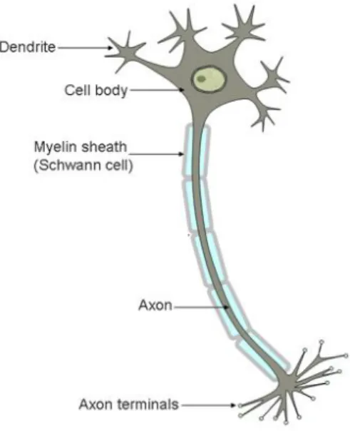
Schwartz and Jessell, 2000), highly specialized post-mitotic cell, characterized by a very peculiar morphology (Figure 1). Their main function is to transmit critical information to target cells, through neurotransmitters or electrochemical signaling (Kandel, Schwartz and Jessell, 2000). The neuron is typically comprised by the soma (cell body), its dendrites and the axon (with respective terminals) (Palay and Chan-Palay, 2011). This morphology favors the capacity of these cells to transmit information. The axon, protruding out of the soma, carries out an action potential that can be passed onto a different neuron, when it reaches the terminal. This is possible due to specialized communication between neurons – synapses (Kandel, Schwartz and Jessell, 2000; Palay and Chan-Palay, 2011). There are two types of synapses, chemical and electrical. In the first, the pre-synaptic neurons release neurotransmitters that bind to receptor molecules in the dendrites of the target neuron (post-synaptic cell), leading to the signal perpetuation (Kandel, Schwartz and Jessell, 2000; Palay and Chan-Palay, 2011). In electrical synapses, the signal propagation is carried through ions that flow onto the post-synaptic cell via specialized pores that physically connect the two cells (Kandel, Schwartz and Jessell, 2000; Palay and Chan-Palay, 2011). The electrical synapses conduct impulses in a faster manner, being therefore present in flight or fight responses or in the heart muscle, providing a smooth and uniform contraction. The chemical synapses, while slower, present other features, such as being more effective in signal amplification1. Synapses
involve a third part, glial cells (astrocytes), which communicate bidirectionally with the synaptic neuronal elements (Allen and Barres, 2009; Araque [et al.], 1999). These cells have a large input in modulating signal transmission(Allen and Barres, 2009; Araque [et
al.], 1999).
Figure 1 – Illustration of the neuron’s morphology. It can be described considering three parts: 1) cell body, where the majority of the organelles are located; 2) dendrites, that branch out of the cell body and that receive the stimuli from upstream cells; 3) axon, which arises from the cell body and functions as the conductive unit that transports the neurotransmitters or electrochemical signal onto the terminals. Axons are insulated by myelin sheaths. Adapted from http://scienceblogs.com/neurotopia/2006/07/19/stem-cells-for-spinal-cord-inj/
While highly important, neurons are also sensitive cells and neurogenesis (the process of stem cell differentiation into neurons) is limited during adulthood, occurring only in certain areas of the brain under tight regulation (Ming and Song, 2011). Therefore, neuronal cell death, particularly in the brain is a process capable of taking a heavy toll on the nervous system functioning.
The capacity of these cells to survive is highly reliant on glial cells. In addition to synaptic modulation, glial cells are responsible for an additional number of processes: (i) providing physical support to neurons, (ii) nourishing them with nutrients and oxygen, (iii) providing a first line of defense against pathogens, (iv) removing dead cells and toxic metabolites and (v) forming myelin sheets. In summary, glial cells enable the maintenance of a functional environment for neurons (Allen and Barres, 2009). Therefore, interaction between glia and neuron is of great importance.
There are three major types of glial cells – oligodendrocytes, astrocytes and
microglia (Figure 2).
Oligodendrocytes, the most abundant glial cells in the cortex (approximately 75%
of total glia) (Pelvig [et al.], 2008), are mainly responsible for producing myelin, which coats the axons, facilitating the propagation of the electrochemical signal (Baumann and Pham-Dinh, 2001). Their shape aids them in their function, by presenting long extensions originating from the cell body (represented in blue in figure 2).
Astrocytes make up for about 20% of glial cells in the cortex (Pelvig [et al.], 2008)
and display a ‘star-like’ shape, with several branches (represented in green in figure 2), even though different populations can exhibit heterogeneous morphology. Their range of actions comprises metabolic support, extracellular ion concentration regulation and synaptic transmission modulation (as previously stated). These cells also function as a repair system in the brain, acting on injured neurons and replacing cells that the CNS cells cannot regenerate. Astrocytes can modulate other glial cells actions, for instance promoting oligodendrocytes myelinating activity (Volterra and Meldolesi, 2005).
Microglia (depicted in brown in figure 2), the less common of the three (about 5 –
10 % of glial population in the cortex), are highly involved in critical processes for brain development, preservation of the neural environment, injury and repair. Microglia are simplistically referred as the ‘resident immune cells in the brain’ (Kettenmann [et al.], 2011; Kraft and Jean Harry, 2011; Weinstein and Möller, 2010). Belonging to a monocytic-macrophage lineage, they orchestrate a large number of immune responses (Kettenmann [et al.], 2011; Kraft and Jean Harry, 2011; Weinstein and Möller, 2010). Nevertheless, striking differences between microglia and common macrophages exist: microglia are under a greater level of regulation by the CNS environment and predominantly exist in a quiescent phenotype or resting state as opposed to an activated state (Kettenmann [et al.], 2011; Kraft and Jean Harry, 2011).
Figure 2 – Depiction of glial cells morphology and interaction with neurons. Astrocytes are represented in green, olygodendrocytes in blue, microglia in brown and neurons in orange. In addition, the cellular interactions are also simplistic representated. Adapted from http://imgkid.com/types-of-brain-cells.shtml
1.2 Microglia, neuroinflammation and pathology
Regardless of what nomenclature might suggest, microglia while on resting state, are not necessarily dormant or inactive. In contrast, they actively patrol several areas of the brain, acting more as a screening agent. It has been proven that microglia is capable of participating in the development, structuring and function of neuronal networks (Kettenmann [et al.], 2011), with distinct capability to express and secrete multiple neurotrophic factors (Elkabes, DiCicco-Bloom and Black, 1996; Morgan, Taylor and Pocock, 2004). This function is essential for promoting CNS tissue homeostasis during brain development (Napoli and Neumann, 2009; Neumann, Kotter and Franklin, 2009). In terms of morphology, in resting state, microglia presents a small cell body and a branched, ramified morphology, which facilitates its screening function (Figure 3A) (Kettenmann [et al.], 2011; Kraft and Jean Harry, 2011; Luo and Chen, 2012). Following activation, the soma tends to become larger, and the complexity of their shape is reduced by retraction of the branches, with a more round form being displayed, similar to that of regular macrophages (Figure 3B) (Kettenmann [et al.], 2011; Kraft and Jean Harry, 2011;
Luo and Chen, 2012). They also gain increased mobility and locomotion, which allows them to migrate to regions that are under greater threat.
Figure 3 – Confocal microscopy images with GFP-expressing microglia (green) amongst YFP-expressing neurons (red). Image A is relative to a 12 day mouse cortical slice and a resting state microglia phenotype can be seen, with highly ramified branches. Image B is relative to a 6 day mouse hippocampus slice. In this case, an activated population of microglia is present, with retracted branches and an enlarged cell body. Adapted from http://bioweb.biology.uiowa.edu/daileylab/projects.html
Phenotypic alterations occur when encountering any potential source of danger for the regular function of the CNS, namely pathological conditions (hypoxia, tumors, ischemia, infection) (Bosco, Steele and Vetter, 2011; Hur [et al.], 2010; Morigiwa [et al.], 2000), accumulation of proteins (amyloid-β, α-synuclein) (Jana, Palencia and Pahan, 2008; Lee [et al.], 2010), chemicals (LPS, alcohol, berberine) (Lu [et al.], 2010; McClain [et al.], 2011; Meng [et al.], 2008), cytokines (Hall [et al.], 1999; Iribarren [et al.], 2005; Krady [et al.], 2008; Tamakawa [et al.], 2004)and others. Microglia also undertakes major functional modifications – altered gene expression of surface molecules, intracellular enzymes and secreted mediators (Kettenmann [et al.], 2011).Expression and releasing of inflammatory factors, such as reactive oxygen species (ROS) intermediates (Colton and Gilbert, 1987; Infanger, Sharma and Davisson, 2006), Nitric oxide (NO) (Kraft and Jean Harry, 2011), proteases(Kraft and Jean Harry, 2011), arachidonic acid derivates (Kraft and Jean Harry, 2011), cytokines (like IL-1β, IL-6, TNF-α) (Hanisch, 2002; Lai and Todd, 2006) potentiate the phagocytic microglial activity, favoring the degradation of bacteria, viruses, debris, and induce cell death on compromised neurons.
The release of chemoattractive factors (Kraft and Jean Harry, 2011) leads to the recruitment of peripheral immune cells to the brain. In this last setting, microglia also function as antigen-presenting cells to various populations of recruited T cells, helping in advancing the adaptive immune response (Kraft and Jean Harry, 2011; Weinstein and Möller, 2010).
The state in which the CNS induces a short lived, beneficial protective immune action is called neuroinflammation. This response however, needs to be under a very tight control, paramount for a system with limited capacity for regeneration, since sustained and uncontrolled inflammation origins a detrimental environment, causing damage and decline of neuronal activity (Kettenmann [et al.], 2011; Kraft and Jean Harry, 2011; Weinstein and Möller, 2010). Namely, ROS can attack susceptible cellular constituents (Kraft and Jean Harry, 2011), TNF-α is an inducer of apoptosis (Hanisch, 2002) and certain proteases can disrupt the BBB and lead to overwhelming infiltration of circulating immune cells (Weinstein and Möller, 2010). This increase of peripheral immune cells can prolong and amplify the inflammatory response (Becker, 2001; Lai and Todd, 2006; Weinstein and Möller, 2010). Hence, the existence and severity of the brain insult is dependent on the duration of the inflammatory process, in addition to the production of sufficient anti-inflammatory mediators to balance out the response (Graeber and Streit, 2010; Kraft and Jean Harry, 2011; Weinstein and Möller, 2010).
Neuroinflammation has been associated with chronic neurodegenerative disorders like Alzheimer’s or Parkinson’s disease (Aschner [et al.], 1999; Glass [et al.], 2010; Kraft and Jean Harry, 2011) and inflammatory pathologies like Multiple Sclerosis (Glass [et
al.], 2010; Kraft and Jean Harry, 2011). The underlying processes that lead to microglia
dysfunction and ultimately the creation of a neurotoxic environment are yet to be well understood (McMahon [et al.], 2005).
An acute neurodegenerative disorder and a major cause of mortality in the western world (Go [et al.], 2013), brain ischemia, is another pathology associated with inflammation (Lai and Todd, 2006). In addition to its high levels of mortality, it is also a major cause of disability, potentially leading to other subsequent conditions like dementia or epilepsy (Raichle, 1983). In spite of the disease’s prevalence, therapies are largely inefficient, in particular due to short therapeutic time window, thus there is a great need for clinically relevant solutions (Raichle, 1983)
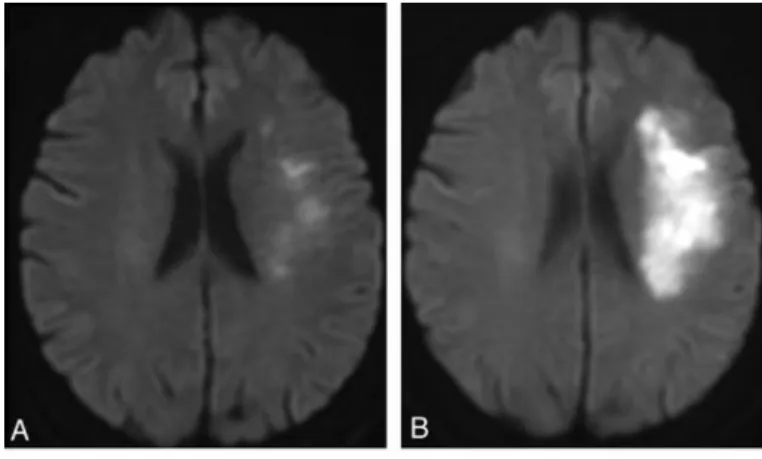
Brain ischemia is defined by the blockade of blood supply to tissue (through embolism, thrombosis, trauma, etc.), leading to extensive necrotic cell death in the area most affected (ischemic core). The surrounding area – ischemic penumbra, can have the same destiny and amplify the injury (Figure 4) through delayed apoptosis, or possibly remain viable; depending on the aftermath of the ischemic event, since that tissue is potentially salvageable. Restoration of blood circulation following the ischemic event can ensue and provoke further damage, mainly through production of ROS species and oxidative stress – reperfusion injury.
Figure 4 – MRI scans of 70 yeard old woman presenting stroke symptons, on admission (A) and after an follow up scan after an ischemic event (B). The propagation of damaged area from one picture to another is visible (bright white). Adapted from http://www.ajnr.org/content/33/3/545/F2.expansion.html
In the ischemic penumbra overwhelming inflammation can also cause a deleterious outcome. Up to two hours after injury onset, an inflammatory response is initiated by activation of microglia and astrocytes, which migrate to the site of injury, with the aim of clearing compromised tissue (Kraft and Jean Harry, 2011). However, overproduction of inflammatory compounds can render them cytotoxic. Recruitment of peripheral immune cells occurs shortly after insult and it is also a process that amplifies and prolongs the inflammatory response (Becker, 2001; Lai and Todd, 2006; Weinstein and Möller, 2010). With clinical purposes, protecting the penumbra is one of the main strategies for avoiding the propagation of damaged tissue and therefore ameliorating the outcome of pathology. Hence, many research approaches have been developed to understand how neuroinflammation can be modulated in the penumbra. In particular, the microglia –
neuron interaction is a subject of particular relevance for the better understanding of inflammatory processes and therefore for opening a possible therapeutic window.
1.3 Developing anti-inflammatory strategies and the
importance of cellular cross-talk
A quantity of compounds labeled anti-inflammatory have been used in order to modulate microglial pro-inflammatory activity – either naturally occurring, as spermidine (Choi and Park, 2012), pheophytin/clorophyll a (Park [et al.], 2014)and luteolin (Zhu [et
al.], 2014) or products of chemical synthesis like
4-[(Butylsulfinyl)methyl]-1.2-benzenediol (Jo [et al.], 2014) have all succeeded to some extent in reducing production and secretion of pro-inflammatory mediators like NO, IL-6, IL-1β, PGE2 and TNF-α in
in vitro models of LPS-stimulated microglial BV2 cell line. Luteolin in particular has
succeeded in inhibiting SH-SY5Y neuronal cell line apoptosis in a model of co-culture with LPS-stimulated BV2 cells (Zhu [et al.], 2014).
Even though these are obviously promising and encouraging results, many of the mentioned data result from simplistic studies (mono cultures) which, are far from in vivo reality in some endeavors. These models give no information regarding cell-to-cell communication between microglia and neurons, and how this cross-talk can ultimately affect microglial activity and the overall inflammatory activity in the CNS environment.
Recently, it has been speculated that interactions between microglia and astrocytes, and more importantly microglia and neurons are aspects that need to be factored when accounting for the outcome of microglia’s activity. Polazzi (Polazzi and Contestabile, 2002) has hypothesized that neurons, when under duress, can send on/off signals to microglia, with the loss of this specific communication being a potential reason for the neurotoxic function of microglia. Signaling mechanisms in which the two cell types physically interact, like CD200-CD200R1 and CX3CL1-CX3CR1 have been described and appear to be pathways by which neurons are able to modulate the inflammatory properties of microglia (Chitnis [et al.], 2007;Neumann, 2001; Sheridan and Murphy, 2013). Also, secreted neuropeptides like VIP and PACAP are molecules, which are up-regulated in injured neurons and can perform a similar role as modulators of microglial
activity (Fernandes, Miller-Fleming and Pais, 2014). These are just some examples of neuron-glia communication mechanisms involved in controlling inflammation (Fernandes, Miller-Fleming and Pais, 2014).
Thus, in order to study inflammation in the CNS and potential anti-inflammatory compounds, it is necessary to experimentally reproduce an in vivo like environment, in particular in what the communication between neurons and microglia is concerned, by using co-culture/conditioned media systems.
1.4 Carbon monoxide – Introduction and toxicity
Paracelsus has been quoted for once having said “All substances are poisons, there
is none which is not a poison. The right dose differentiates the poison”. This seems to be
the case regarding carbon monoxide (CO).
This gas has been famously referred as the silent killer since 1870 when Claude Bernard discovered its affinity for hemoglobin (yielding carboxyhemoglobin, COHb) (Prockop and Chichkova, 2007; Raub [et al.], 2000), which mainly accounts for its deadliness. The presence of COHb in the blood leads to a decrease in the capability to carry oxygen throughout the body, resulting in hypoxia (Prockop and Chichkova, 2007; Raub [et al.], 2000). It has also been claimed that systemic hypoxia facilitates CO binding to cytochrome c oxidase, inhibiting this enzyme activity and disturbing ATP synthesis and global mitochondrial functioning (Ryter, Alam and Choi, 2006). These properties stem from carbon monoxide’s inherent strong binding to transition metals in their respective reduced state, such as hemoglobin and others (myoglobin (Antonini and Brunori, 1971), soluble guanylyl cyclase (Antonini and Brunori, 1971), inducible nitric oxide synthase(Stevenson [et al.], 2001) or NADPH oxidase (Cross [et al.], 1982)).
1.5 CO as a therapeutic molecule? – Historical
perspective
Despite its associated toxicity, carbon monoxide is an endogenous product of the degradation of free heme, by heme-oxygenase (HO) (Tenhunen, Marver and Schmid, 1968). This enzyme has two known isoforms: HO-1, an inducible form which acts on a wide array of stress signals and HO-2, which is constitutively expressed (Cruse and Maines, 1988; Maines, Trakshel and Kutty, 1986; Ryter, Alam and Choi, 2006). HO is the only known enzyme capable of heme degradation, which when accumulated can become deleterious (Ryter, Alam and Choi, 2006).
This reaction yields not only CO, but additionally free iron (Fe2+) and biliverdin, which is subsequently converted to bilirubin by the action of biliverdin reductase (Ryter, Alam and Choi, 2006) (Figure 5).
Figure 5 – Reaction catalyzed by heme-oxygenase enzymes. O2 and NADPH function as cofactors and heme participates as the
prosthetic group, in addition to being the substrate. Adapted from (Ryter, Alam and Choi, 2006).
HO-1, the inducible isoenzyme and HMOX (Cruse and Maines, 1988), the gene that encodes for it, is considered one of the most stress - sensitive genes known to date, being stimulated by a wide array of agents and chemicals, like cytokines (IL-6, IL-10, IL-1α) (Lee and Chau, 2002; Mitani [et al.], 1992; Terry [et al.], 1999), LPS (Camhi [et al.], 1995; Kurata [et al.], 1996; Lutton [et al.], 1992), heavy metals (Keyse and Tyrrell, 1989;
Mitani [et al.], 1993) or reactive oxidative species (Keyse and Tyrrell, 1989). It is generally considered that any condition associated with an imbalance of the intracellular redox state stimulates its expression. This response appears to occur ubiquitously among most tissues of higher organisms (mammals, birds, fish) tested (Ryter, Alam and Choi, 2006). Nuclear factor like-2 (Nrf-2), a transcription factor involved in the regulation of expression of anti-oxidant proteins, appears to be a staple in the expression of HO-1 (Ryter, Alam and Choi, 2006).
HO-1 is known to have a number of beneficial cytoprotective effects, namely anti-inflammatory, anti-apoptotic and anti-proliferative actions, which have been well documented on numerous cell types (endothelial, epithelial, smooth muscle and others) and disease models (cardiac ischemia, ischemia/reperfusion injury, hypertension) (Brouard [et al.], 2000; Otterbein [et al.], 2003; Peyton [et al.], 2002). HO-1 deficient mice for example, tend to develop chronic inflammation (Poss and Tonegawa, 1997) and two humans were reported to have suffered HO-1 deficiency and died while still young due to mainly inflammatory related complications (Yachie [et al.], 1999;Radhakrishnan [et al.], 2011).
Some of the HO protective properties might be attributed to its products inherent cytoprotective properties. Biliverdin/bilirubin, are potent antioxidants that scavenge ROS (Stocker [et al.], 1987), and exogenous administration in rodents has provided beneficial effects in various models of disease (Fondevila [et al.], 2004; Sarady-Andrews [et al.], 2005; Yamashita [et al.], 2004). Iron (II), on the other hand, induces the expression of ferritin, a chelating protein, which limits the generation of free radicals by binding to this free metal (Balla [et al.], 1992).
CO, however, has long been considered as a catabolic waste product of the reaction. It was not until the 1990’s that CO started to be looked upon in a different light, when it was reported that this gaseous molecule could act as a neurotransmitter by inducing production of cGMP (Verma et al., 1993) in the brain, through its action on sGC. CO’s action as physiological regulator of cGMP was also associated with potent vasodilatory properties in various types of tissues (Morita [et al.], 1995, 1997; Hussain [et al.], 1997; McFaul and McGrath, 1987; Sylvester and McGowan, 1978)
This elicited interest from the scientific community led to numerous reports in the past two decades about the cytoprotective and anti-inflammatory effects of endogenous
CO and HO-1 activity (Ryter, Alam and Choi, 2006). With increasingly encouraging results, the focus is now more than ever on the CO/HO-1 axis and disclosing its properties and mechanisms of cytoprotection in order to translate this knowledge into therapeutics.
1.6 Carbon monoxide releasing molecules
–A novel
method for CO therapeutic delivery
CO not only has vasoactive properties but also has other cytoprotective characteristics that make the CO/HO-1 axis such an appealing target in various models of disease.
Administration of CO as gas presents several drawbacks: inhaled CO is not tissue specific and can increase carboxyhemoglobin levels, potentially provoking lethal hypoxia (Motterlini and Otterbein, 2010; Romão [et al.], 2012). Thus, treatment with inhaled CO is a delicate procedure that would require serious monitoring and medical technical devices, even though some therapeutic tests with inhaled CO in animal models of disease have shown moderate success (Romão [et al.], 2012).
One possible solution to overcome these limitations started to be developed in the early 2000’s by Motterlini and collaborators. Carbon monoxide-releasing molecules (CORMs) are pro-drugs, which are able to deliver CO in vivo and in vitro, in a more controlled manner (Motterlini [et al.], 2002).
CORMs are complex metal carbonyl compounds (M(CO)y) in which the carbonyl groups function as coordinated ligands of a transition metal (generally molybdenum, cobalt, iron, manganese or ruthenium) in the center (Motterlini [et al.], 2002). Nevertheless, the therapeutic usage of CORM molecules presents glaring difficulties, namely: transition metal compounds are highly toxic. Furthermore, solubility, stability, bioavailability and other pharmacological factors need to be modulated by tuning the nature of the ligands (Motterlini [et al.], 2002; Romão [et al.], 2012).
The emergence of CORMs however, can solve some of the issues that make inhaled CO unviable as a method of delivery. Potential therapeutic administration of CORM would not require specific equipment or same monitoring as inhaled CO (Motterlini and Otterbein, 2010; Romão [et al.], 2012) and could be consumed not only orally but
intramuscularly and intravenously. Other favorable feature of CORMs is the potential manageable tissue specificity. Controlled release for specific targeting is feasible by chemically altering CORMs to be more or less sensitive to environmental changes (pH, ROS concentrations) or coupling them with specific ligands, depending on the preferred targeted tissue/organ (Motterlini and Otterbein, 2010; Romão et al., 2012). Efficient tissue delivery means that lower doses of these compounds could be used, in contrast to what occurs with inhaled CO.
The first two commercially available CORMs, CORM-1 (Mn2CO10) and -2 (Ru(CO)3Cl2) (Figure 6 A and B), proved to release CO when stimulated and promoted vasodilation and hypotension in vivo (Motterlini [et al.], 2002). These two molecules were nevertheless only lipid-soluble. Motterlini et al. then succeeded in creating the first water soluble CORM – CORM-3 [Ru(CO)3Cl(glycinate)] (Figure 6 C), which displayed a very rapid release of CO when in contact with biological fluids (Clark [et al.], 2003). Simultaneously, a second water soluble, transition metal-less molecule emerged in the form of CORM-A1 [H3BCO2]Na2 (Figure 6D), a boranocarbonate whose carboxyl group is slowly converted and released as CO under a specific combination of controlled conditions of pH (slightly acidic) and temperature (around 37ºC) (Motterlini [et al.], 2005). The two main advantages of CORM-A1 are: the slower CO release rate (half life time of around 21 minutes) and it does not contain transition metal. While CORM technology progresses, molecules tend to become increasingly more physiological and effective for being used in both in vivo and in vitro settings.
Figure 6– Chemical structures of four of the most common CO releasing molecules – CORM-1 (A), 2(B), 3 (C) and A1 (D). Adapted from http://www.fasebj.org/content/19/2/284/F3.large.jpg
The described beneficial effects of CORMs range to various models of disease like: reduction of graft rejection in rodents (Clark [et al.], 2003; Musameh [et al.], 2006), attenuation of histamine release in human neutrophils and guinea pig mast cells (Vannacci
[et al.], 2007), anti-apoptotic effects in mice brain (Fiumana [et al.], 2003; Parfenova [et
al.], 2006), among others.
A particular set of studies by Motterlini’s group, has shown CO’s capacity to reduce neuroinflammation in in vitro models of LPS/IFN-γ (Bani-Hani [et al.], 2006a) and thrombin-activated BV2 microglia (Bani-Hani [et al.], 2006b) using CORM-3 as the delivery molecule. In addition, an in vivo study using rat models of hemorrhagic stroke has further succeeded in underlining CO as a potential anti-neuroinflammatory therapeutic molecule (Yabluchanskiy [et al.], 2012).By also using CORM-3 the authors were able to conclude that this molecule promoted a decrease in infarct area through inflammatory modulation.
In summary, this data supports the notion that CO can be used in therapy, particularly if an appropriate method of delivery is developed.
1.7 CO and its cytoprotective properties – Molecular
insight
CO seems to act as a signaling molecule – the induction of sGC activity and also its action on mitogen-activated protein kinases (MAPK) pathways, like p38 (Brouard [et
al.], 2000; Otterbein [et al.], 2000), ERK1/2 (Song [et al.], 2003) and JNK 1/2 (Morse [et al.], 2003) are well documented and it is involved in the generation of a protective
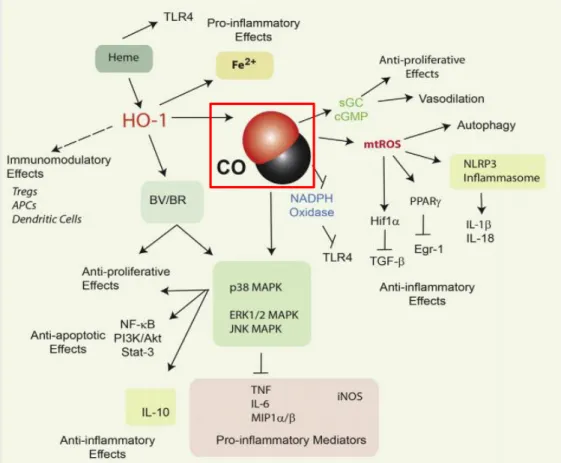
response. For inflammation in specific, altering the activity levels of these pathways, leads to decreased expression of inflammatory cytokines (TNF-α, IL-6, IL-1β) and increases in anti-inflammatory compounds like IL-10 (Bani-Hani [et al.], 2006b; Otterbein [et al.], 2000; Peyton [et al.], 2002). In addition to this, CO also promotes the expression of factors PPAR-γ and HIF-1α (Chin [et al.], 2007; Lancel [et al.], 2009) and modulates NADPH Oxidase activity (Nakahira [et al.], 2006), all of which inhibit signaling molecules involved in inflammatory signaling (Egr-1, TLR-4). Regarding brain ischemia in particular, recent studies have shown some involvement of CO in inhibiting the three mentioned MAPK pathways, after the onset of inflammation (Bani-Hani [et al.], 2006b).
Some of CO’s anti-inflammatory effects, among other documented cytoprotective mechanisms are illustrated in figure 7, presented below.
Figure 7 – Pivotal documented cytoprotective functions of HO-1 / CO and respective cellular pathway involved. Carbon monoxide displays anti-inflammatory properties, altering production of various pro and anti - inflammatory cytokines through inhibition/activation of various mediators involved in such processes. Examples of this are the hyperphosphorylation of the p38 MAPK pathway, inhibition of the activity of NADPH Oxidase and activation of PPAR-γ and HIF-1α transcription factors (Chin [et al.], 2007; Lancel [et al.], 2009; ,Nakahira [et al.], 2006). These last two cases are examples of molecular players activated by a burst in
mitochondrial ROS production, which is believed to be a direct consequence of the actions of CO. Also, CO displays anti- proliferative and anti - apoptotic capacities (Taillé [et al.], 2005, ,Vieira, Queiroga and Alves, 2008), acting on numerous other molecular signaling
pathways.These positive results stem from various kinds of models of disease, both in vitro and in vivo that have helped in unveiling carbon monoxide potential in therapy (Ryter, Alam and Choi, 2006; ,Motterlini and Otterbein, 2010). Adapted from (Ryter and Choi,
2015)
Helena L.A. Vieira team has shown that CO pre-treatment prevents neuronal apoptosis against different types of cell death inducers through pre-conditioning (PC) (Vieira, Queiroga and Alves, 2008). PC is a mechanism in which the exposure of cells to an early stimulus below the threshold of damage creates a resistance for later encounters, against similar or even different types of stronger stresses (Vieira, Queiroga and Alves,
2008). In this case, CO’s protection against apoptosis was attributed to stimulation of specific signaling pathways.
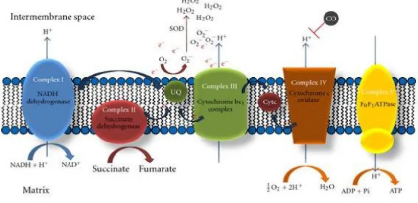
In fact, CO is capable of modeling a diverse number of molecular players, thus, its broad cytoprotective capacity (Figure 7). However, the specific and direct molecular targets of carbon monoxide are still under discussion. Because of CO cytotoxic studies, it is well accepted that it specifically binds to cytochrome c oxidase (complex IV), inhibiting its activity and mitochondrial respiration. Nevertheless, under low and physiological concentrations, CO also seems to bind to cytochrome c oxidase, leading to small amounts of ROS generation, mainly due to extensive electron accumulation at complex III, favoring generation of reactive species like anion superoxide (Nohl, Gille and Staniek, 2005) (Figure 8).
This shift into a pro-oxidant environment, with the presence of ROS as signaling molecules, triggers an array of cellular mechanisms (Bilban [et al.], 2008).
Figure 8 – The most widely accepted model for CO driven ROS generation. This gaseous molecule partially inhibits complex IV leading to electron accumulation at complex III level, facilitating radical formation. Adapted from (Queiroga, Almeida, and Vieira, 2012).
Regarding inflammation, there are some examples in which generation of oxygen species act as an intermediary for the carbon monoxide anti-inflammatory response. Some of those are: the upregulation of PPAR-γ and HIF-1α. and the activation of the NLRP3 inflamassome (Ryter and Choi, 2015), all of which dampen inflammation in some way, and are triggered by CO derived production of ROS (Bilban [et al.], 2006; Chin [et al.], 2007).
The effect of CO in cytochrome c oxidase however is still ambiguous and controversial – it has been speculated that CO’s capacity to promote ROS production
occurs by accelerating the process of mitochondrial respiration and therefore increasing the amount of O2 that is not totally reduced into water (Queiroga, Almeida and Vieira, 2012). Others claim that is not the case, stating that in human isolated mitochondria the enzyme’s activity is strongly hindered (Alonso [et al.], 2013).
In isolated mitochondria from rat brain (Almeida [et al.], 2012), a two-step response was detected in which the complex is inhibited in the first minutes, but after 30 minutes, its activity grows significantly, improving ATP production, ROS production and overall cellular metabolism.
Despite controversies regarding the exact mechanisms by which the ‘silent killer’ acts, it is clear that carbon monoxide possesses an innate ability to drive an overall cytoprotective mechanism.
1.8 Final remarks and objectives
Cerebral ischemia is the 2rd major cause of deaths in western society (Go [et al.], 2013). Despite this, therapeutic strategies are still, at large, inefficient (Go [et al.], 2013). Uncontrolled neuroinflammation is a major source of secondary damage on stroke, developing over a time period of hours to days after onset, in which microglia plays a central role by sustaining inflammatory signaling and mediating cytotoxic mechanisms. At the same time, this time window presents itself as an ideal opportunity for therapeutic intervention by targeting such cells.
Carbon monoxide is a promising therapeutic molecule with already proven anti-inflammatory potential. This has been established in various models of disease, both in
vitro and in vivo, limiting the pro-inflammatory output of macrophage and microglia cell
lines (Bani-Hani [et al.], 2006a, 2006b; Chin [et al.], 2007) in simplistic monolayer models and diminishing inflammation and tissue damage in a rat model of hemorrhagic stroke (Yabluchanskiy [et al.], 2012).
Thus, the final aim of this thesis is to study CO as a potential therapeutic agent – if and how can CO modulate activated microglial activity, promoting neuroprotection via suppression of inflammation. This thesis presents a novel approach, as the anti-neuroinflammatory role of CO is studied at the level of microglia and neuron
communication. This is of particular interest, since increasing evidences point out to neurons being able to influence the CNS environment, in particular by modulating microglia inflammatory mechanisms (Polazzi and Contestabile, 2002).
The majority of the experimental work will be done using a conditioned media system, which is appropriate for mimicking in vivo conditions since, even though it does not take physical interaction between cells into account, manages to mimic cellular communication through the release of soluble factors.
Figure 9 – Summary of the main approaches and questions to be answered during the master thesis.
After establishing the model (Task 1), specific questions (Tasks 2 and 3) will be targeted, as for instance: Does CO play a role in modulating neuron survival in the inflammatory context? If so, how does CO affect microglia inflammatory output? Does CO modulate molecular players involved in neuron-microglia cross-talk?
Answering these questions (Figure 9) will not only give a better depiction of the interplay between these cells and how it might be altered during pathology but also disclose new mechanisms by which carbon monoxide is capable of limiting inflammation. All this knowledge can be of particular value and a step forward for carbon monoxide as an eventual valuable therapeutic molecule in the near future.
2.1 Cells and reagents
For microglia, BV2 murine microglia cell line was used. Cells were cryopreserved at -80ºC in 90% (v/v) FBS and 10% (v/v) DMSO at passage 43. RPMI-1640 media (Sigma) was used as basal media, supplemented with 10% fetal bovine serum (Life Technologies), 4 mM L-glutamine (Life Technologies), penicillin (100 units/mL) and streptomycin (100 μg/mL) (Life Technologies). Cells were maintained at 75cm2 t-flasks and passages were performed three times a week in 1/4 dilution.
Regarding neurons, SH-SY5Y neuroblastoma cell line was used. Cells were cryopreserved at -80ºC in 90% (v/v) FBS and 10% (v/v) DMSO at passage 14. For cell maintenance, DMEM/F12 was used as basal media (Life Technologies) and supplemented with 10% fetal bovine serum (Life Technologies), penicillin (200 units/mL) and streptomycin (200 μg/mL) (Life Technologies). Cells were maintained at 75cm2 t-flasks and passages were performed once a week in 1/2 dilution. Both cell lines were maintained in a humified incubator at 37ºC and 5%CO2.
CORM-A1 was purchased from Sigma. Stock solutions of 5mM were prepared by diluting the reagent in MilliQ water. LPS (Sigma) was also diluted in MilliQ water to obtain a stock solution of 1 mg/mL. tert-butyl-hydroperoxide (t-BHP, Sigma) and Propidium Iodide (PI, Life Technologies) were acquired at a concentration of 7.8M and 1 mg/mL, respectively. For t-BHP, an intermediate solution at a final concentration of 3.9 mM was used by diluting the reagent in MilliQ water. Griess reagent (Sigma) was acquired and used at working concentration.
2.2 LPS validation protocol
BV2 cells were seeded onto 24-well plates (6 × 10⁴ cells/well). For validation of LPS as inflammatory stimulus (Figure 10), cells were treated with various concentrations (0; 100; 250; 500; 1000 and 2000 ng/mL) of this molecules 24 hours after seeding.
For analysis of the effect of CORM-A1 pre-treatment on NO secretion (Figure 10), BV2 cells were treated with this molecule (0; 12.5 µM) and 24 hours later with LPS (0; 500ng/mL).
2.3 NO quantification
24 hours after an LPS stimulus, 100 μL of supernatant from each well were collected and centrifuged (5 min, 10000 g). After this centrifugation, 50μL of the resulting supernatant were transferred onto a 96-well plate where the same volume of Griess reagent was added (Figure 10). After a 10 minute incubation step, the absorbance of the resulting solution was measured at 540 nm, using a Tecan Infinite F200 PRO microplate reader. NO concentration was calculated with reference to a standard curve constructed with known concentrations of sodium nitrite (Sigma).
2.4 Optical microscopy
The changes in BV2 morphology 24 hours following an LPS stimulus were analyzed using a light microscope Zeiss Axiovert 40 CFL (Figure 10). Zeiss microscope software ZEN was used for image acquisition and ImageJ software was used for image analysis.
2.5 t-BHP validation protocol
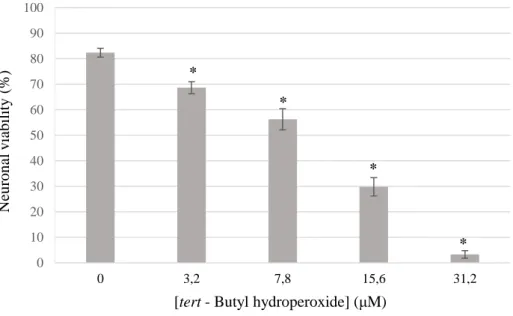
SH-SY5Y neurons were seeded onto 24-well plates (1 × 10⁵ cells/well). One day after seeding, cells were challenged with increasing concentrations of the cell death inducer t-BHP (0, 3.2; 7.8; 15.6; 31.2 µM) for 24 hours and cell viability was assessed by the means of flow cytometry (Figure 10).
2.6 Conditioned media/neuroinflammation protocol
BV2 cells were seeded onto 24-well plates (6 × 10⁴ cells/well). One day after, microglia was pre-treated or not, with various concentrations of CORM-A1 (0; 12.5; 25 µM), and then with LPS (0; 500 ng/mL), always with 24 hours in between (Figure 10).
SH-SY5Y grew on 24-well plates (1 × 10⁵ cells/well). One day after, the media where neurons were growing was removed and substituted with BV2’s, 24 hours after those had been stimulated or not with LPS, as described above. Simultaneously, the SH-SY5Y cells were also challenged with different values of t-BHP (0; 7.8; 15.6; 31.2 µM) (Figure 10). One day after, cell viability was assessed through flow cytometry.
2.7 Flow Cytometry
The supernatant and the adherent neurons from each well were collected (Figure 10). Cell detachment was performed using trypsin (Life Technologies). Subsequently, a centrifugation was performed (5 min, 350 g) with the resulting supernatant being discarded and the pellet resuspended in DMEM/F12 with 1 ng/mL of PI. The cells were thereafter incubated for 30 minutes at 37ºC and cell viability was evaluated using a flow cytometer (FACSCalibur, BD Biosciences).
2.8 Supernatant collection protocol
BV2 cells were plated at a density of 6 × 10⁴ cells/well in 24-well plates. 24 hours after, they were either pretreated with 12.5 µM of CORM-A1 or remained untreated, and 24 hours further, were similarly either stimulated with 500 ng/mL of LPS or continued untreated (Figure 10). One day after the stimulus, samples of conditioned media were collected and stored at -20ºC.
2.9 ELISA
Conditioned media (100 µL) from the supernatant collection protocol was used to measure TNF-α and IL-10 concentration levels. Two different ELISA kits were used: a Murine TNF-α Standard ABTS ELISA Development Kit (PeproTech) and a Murine IL-10 Mini ABTS ELISA Development Kit (PeproTech). In both cases, manufacturer’s instructions were followed throughout and in the end, absorbances at 405 and 650nm were inferred using a TecanInfinite F200 PRO microplate reader. Absorbance data was thereafter converted to concentration values using standard curves constructed by following the instructions presented on the kit.
2.10 BV2 protein extract collection protocol
BV2 microglial cells were plated at 6 × 10⁴ cells/well in 24-well plates. From here on, the cells were treated with CORM-A1 and LPS, with concentrations and time points equal to the ones used for the preparation of the supernatants in the ELISA protocol (Figure 10).
One day after the LPS stimulus the media was discarded from each well and a PBS (1.54mM NaCl, 20 mM KH2PO4, 34 mM Na2HPO4 pH 7.2) washing step was performed. Subsequently, a non-denaturating lysis buffer was used (20 mM Tris HCl pH 8, 137 mM
NaCl, 10% glycerol, 1% Igepal, 2mM EDTA and 1% protease inhibitor) and cells were scrapped and transferred to pre-cooled eppendorfs, where they were kept at constant agitation for 30 minutes at 4ºC. Afterwards, the samples were centrifuged for 12 minutes at 13400g with the supernatant being collected and kept at -20ºC.
2.11 Protein quantification
Cell extracts were diluted 10 times in MilliQ water and then transferred onto 96-well plates, at a final volume of 25μL per 96-well. 200μL of working reagent from Pierce BCA protein assay Kit (Thermo Scientific) were then added, followed by a 30 minute incubation step at 37ºC.The absorbance values from each wells were measured at 562 nm, using a Tecan Infinite F200 PRO microplate reader. Concentrations were calculated from absorbance values using a standard curve constructed using known concentrations of BSA.
2.12 Western Blot
Equal amounts of protein (25 µg for CD200R1) were separated by sodium dodecyl sulfate polyacrylamide gel electrophoresis (SDS-PAGE) on polyacrylamide gel (10%) for 1h15min, with the molecular markers used being BenchMark and MagicMark XP (both Life Technologies). The proteins were then transferred onto nitrocellulose membranes (1h15min), blocked with 7% BSA (Sigma) solution for 1h15min at RT and incubated overnight with primary antibodies (goat anti-CD200R1 dil. 1/200 in TTBS, Santa Cruz Biotechnology), at RT. Next, membranes were treated with secondary antibody (rabbit anti-goat HRP conjugated dil. 1/5000 in BSA, Abcam) for 1 hour at RT. Also, the membranes always undertook multiple washing steps with TTBS after blocking and incubation with both antibodies. Rouge Ponceau was used as internal control for checking total protein loading.
Immunoblots were exposed to electrochemiluminescence western detection reagent (Bio-Rad) 5 minutes and the reactive bands were detected after the membranes were exposed to X-ray film (Chemidoc, Bio-Rad). The resulting area and intensity of the bands were quantified using ImageLab software (Bio-Rad) and are presented as a percentage relative to the control (100%).
2.13 Statistical analysis
The data presented throughout is the mean ± standard deviation. Comparisons between different groups of conditions were analyzed using the one-way ANOVA test with p-values of less than 0.05 being considered statistically significant.
_ III. Results
3.1 Experimental model establishment
In order to create a proper experimental paradigm, some initial and more simplistic protocols were performed. For example, LPS, which is a molecule widely used in inflammation related studies as a trigger for microglia activation, was validated for promoting inflammatory responses in BV2 microglia cell line. Thus, different time points and concentration levels were tested in order to obtain optimal experimental conditions. NO production was the used readout for microglia activation, because it is a well-known robust inflammatory soluble factor (Kettenmann [et al.], 2011; Moncada, Palmer and Higgs, 1991), but also because the detection methods are fairly simple and swift.
It is important to mention that the protocol used measures nitrite (NO2-), a product of NO decomposition, but not authentic NO, as this compounds is highly reactive in the presence of oxidants like oxygen.
Also, we performed microcopy tests in order to further confirm the activation state of microglia, by analyzing morphological differences between resting and LPS-activated cells, one day after the stimulus.
0 50 100 150 200 250 300 350 400 0 100 250 500 1000 2000 [NO ]/[p rot ein ] (% Re lati ve to Co n tr o l) [Lipopolysaccharides] (ng/mL)
24 hours
*
*
*
*
*
AFigure 11.1 – Effect of LPS treatment on BV2 nitric oxide production.BV2 cells were treated for 24 hours (A) or 48 hours (B) with various indicated concentrations of LPS. Values were normalized with intracellular protein content. Differences between experimental conditions were analyzed by the one-way ANOVA test with the results being considered statistically significant when p-value < 0.05 (n = 7). The asterisk indicates statistical significance relative to the negative control.
Figure 11.2 - Effects of an LPS 24 hours stimulus on the morphology of BV2 microglia cell line. Representative micrographs showing BV2 cells, either untreated (A) or treated with 500 ng/mL of LPS for 24 hours (B). Phase contrast images were obtained from Zeiss microscope (400x magnification).
In both experiments, the presence of LPS (even at the lowest concentration) leads to a significant increase in NO secretion, pointing out to the fact that this molecule is inducing an inflammatory reaction in glial cells.
In figure 11.1A, the NO levels in the supernatant increase accordingly to the LPS concentration used, until reaching a ‘threshold’ at around 250 ng/mL, in which they stay
0 50 100 150 200 250 300 350 400 0 100 250 500 1000 2000 [N O ]/ [pr ot ei n] (% Re lativ e to Co n tro l) [Lipopolysaccharides] (ng/mL)
48 hours
*
*
*
B*
*
A Babout the same, regardless of the strength of the stimulus. On the other hand, in figure 11.1B, there is an evident LPS-concentration dependent response concerning the levels of NO compounds produced. When comparing the two graphs, it is also noticeable that the overall levels of nitric oxide in the supernatant are greater at 48 hours, which is easily attributable to the added 24 hours that these cells spent incubated with the pro-inflammatory reagent.
In figure 11.2, we were also able to see that LPS treatment promotes an alteration in cell morphology, as BV2 microglia present a more round-like shape, characteristic of inflammation activated cells.
In conclusion, LPS is a valid inflammatory trigger for the activation of BV2 microglia, and it will be used in the following protocols. The conditions adopted were 24 hours of LPS exposure at 500 ng/mL, based on both our obtained results and existing literature (Bani-Hani [et al.], 2006a, 2006b). Treatment for 24 hours is enough to activate microglial cells without longer periods of treatment.
As figure 11.1A illustrates, the differences on NO production at LPS concentrations between 250 – 2000 ng/mL were almost non-existing. Thus, choosing 500 ng/mL as the optimal concentration of LPS was mainly based on the existing microglia activation protocols on literature (Choi and Park, 2012; Jo [et al.], 2014; Kim [et al.], 2014).
The pro-oxidant tert-butyl-hydroperoxide (t-BHP) was validated as an inducer of cell death in SH-SY5Y neurons. t-BHP induces apoptosis via oxidative stress, which heavily contributes to neuronal damage in cerebral ischemia, in particular during the reperfusion phase. Thus, using this molecule mimics in vitro an existing mechanism in
Figure 11.3 – Effect of t-BHP on SH-SY5Y cellular viability.The neurons were treated for 24 hours with the cell death induce agent before being submitted to a flow cytometry protocol, with PI as a dye to determine cell survival by plasmatic membrane integrity. Differences between experimental conditions were analyzed by the one-way ANOVA test with the results being considered statistically significant when p-value < 0.05 (n = 6 - 12). The asterisk indicates statistical significance relative to the negative control.
As the figure 11.3 illustrates, t-BHP treatment of SH-SY5Y neurons promotes extensive cell death, in an evident concentration dependent manner. Therefore, t-BHP can be used as a robust inducer of cell death in a model of SH-SY5Y neuronal cell line, which will be used in the following experiments.
3.2 Microglia-neuron communication – neuron
viability and carbon monoxide effect
In order to validate that the medium from LPS-stimulated microglia creates a cytotoxic environment for neurons, acting synergistically with oxidative stress, a conditioned media based protocol was established.
Microglia is well capable of performing basal communication with neurons, but exacerbated microglia activation is a main reason for the development of a toxic environment in neuroinflammation (Kettenmann [et al.], 2011; Kraft and Jean Harry, 2011; Weinstein and Möller, 2010). This conditioned medium protocol mimics the in vivo
0 10 20 30 40 50 60 70 80 90 100 0 3,2 7,8 15,6 31,2 N eur onal v iabi li ty ( % )
[tert - Butyl hydroperoxide] (μM)
*
*
*