Leonor Nunes Rodrigues
Licenciada em Bioquímica
Unravelling the role of alpha 2,6 sialic acid on mouse
dendritic cells’ functions
Dissertação para obtenção do Grau de Mestre em Bioquímica para a Saúde
Orientador: Paula Alexandra Quintela Videira, Professora Auxiliar, Nova Medical School – Faculdade de Ciências Médicas e Faculdade de Ciências e
Tecnologia da Universidade Nova de Lisboa
Coorientador: Joseph T. Y. Lau, Investigador Principal, Roswell Park Cancer Institute, Buffalo, NY, USA
Leonor Nunes Rodrigues
Licenciada em Bioquímica
Unravelling the role of alpha 2,6 sialic acid on mouse
dendritic cells’ functions
Dissertação para obtenção do Grau de Mestre em Bioquímica para a Saúde
Orientador: Paula Alexandra Quintela Videira, Professora auxiliar, Nova Medical School – Faculdade de Ciências Médicas e Faculdade de Ciências e
Tecnologia da Universidade Nova de Lisboa
Co-Orientador: Joseph T. Y. Lau, Investigador Principal, Roswell Park Cancer Institute, Buffalo, NY, USA
Júri: A definir
Nova Medical School – Faculdade de Ciências Médicas da Universidade Nova de Lisboa
iii Acknowledgments
First of all, I would like to thank Dr. Paula Videira due to the excellent opportunity of working in her group and for being my mentor in this project. I am also very grateful due to her advice, teachings, availability and encouragement given over this year.
I am sincerely thankful to my mentor Dr. Joseph Lau, for accepting me in his group and for the given advice and knowledge. Moreover, I am very thankful to my mentor in the laboratory, Dr. Mehrab Nasirikenari due to all the support and scientific growth provided over this year.
I could not fail to thank Fulbright Commission of Portugal for the given opportunity to develop part of this project in USA.
To Neubauer family, I express the most sincere thank you for hosting me as family and for all the offered support.
I thank all my colleagues from both laboratories: Alex, Chris, Chuck, Joey, Melissa, Mindy, Val, Patrick (USA) and Graça, Guadalupe Inês Ferreira, Inês Iria, Liliana, Márcia, Mauro, Mylène Tiago and Zélia (Portugal), for the friendship, good times in the laboratory and help provided during my work.
I also would like to thank people from Buffalo for the good times during my stay, namely Melissa, Meriem, Camila and Karstin.
I would like to show my consideration to Cytometry Department of Roswell Park Cancer Institute, Buffalo, NY, USA, for all the teachings provided.
Moreover, I thank the animal facility and Microbiology Department of Nova Medical School- Faculdade de Ciências Médicas, Dr. José Ramalho, Dr. Diogo Casal and Isabel Silva for providing the needed resources to finish this project in Portugal.
I would like to thank my family and friends for all the help and encouragements. A special thanks to Ana, Inês, Jéssica, Jéssica Ramos, Katharina, Madalena, Margarida, Sílvia, Rita, Coelho, Fred, João Sousa, João Valadinha, Lucas, Nuno, Tomás Santos, Tomás Curveira and Ricardo for the friendship, presence and good times.
Lastly, but not least, I thank the people without whom the achievement of this project would have been impossible: my parents, who always believed in me, helped and instilled the necessary motivation and resources; my siblings Diogo and Beatriz for the needed distractions and help over this year and my boyfriend Guilherme, for supporting my choices, for the patient demonstrated over this year and for always being present.
v Abstract
Dendritic cells (DCs) are vital for immunomodulation and the initiation of adaptive immune responses, whereas sialic acids (Sias) are potential immunomodulators. These cells express high levels of sialyltransferase ST6Gal-1, responsible for transferring Sias to the terminal position of oligosaccharide chains. Indeed, DCs’ maturation is associated with decreased cell surface sialylation.
Although its biological significance is unknown, the soluble, extracellular form of ST6Gal-1 increases in cancers and inflammation. However, extracellular ST6Gal-1 was recently identified as modulator of hematopoiesis. Considering that DCs play a crucial role in the initiation of a productive anti-cancer immune response, a link between extrinsic sialylation by the extracellular ST6Gal-1 on DC function needs to be investigated.
We hypothesize that extrinsic α2,6 sialylation of DCs diminishes their maturation features upon lipopolysaccharide (LPS) stimulation. The main goal was to extrinsically α2,6 sialylate mice bone marrow derived DCs (BMDCs) and to evaluate their maturation and cytokine profiles upon LPS stimulation (by Flow Cytometry and ELISA, respectively). Unlike the hypothesis, we observed that BMDCs’ profile is not modulated, even using several approaches. In contrast, the consequence of lacking cell surface α2,6 Sias in DC maturation was assessed by analysing: 1) sialidase treated BMDCs, 2) BMDCs from mice lacking ST6Gal-1 and 3) DCs from mice airways, comparing wild type with ST6Gal-1 knockout mice. These results suggest that overall lack in α2,6 Sias is related with increased expression of major histocompatibility class II (MHC-II).
Although appearing to be controversial findings, other intracellular mechanisms might be occurring upon LPS-induced BMDC activation, probably reducing extracellular ST6Gal-1 effect. In opposite, the modification observed in DC profile of ST6Gal-1 knockout mice might be related to its predisposition to a more severe inflammatory status.
With this, the developed work opened future lines of investigation, namely exploring other factors involved in α2,6 (de)sialylation of DC, which might have influence in immunotherapy using DCs.
Key words: dendritic cells, sialidase, ST6Gal-1, extrinsic α2,6 sialylation, major histocompatibility class II
vii Resumo
As células dendríticas (CDs) são fundamentais na imunomodulação e iniciação de respostas imunes adaptativas, enquanto os ácidos siálicos (Sias) são potenciais imunomoduladores. Estas células expressam níveis elevados da sialiltransferase ST6Gal-1, que transfere Sias para a posição terminal de oligossacáridos. De facto, a maturação de CDs está associada a uma diminuição da sialilação na sua superfície celular.
Apesar de ter função biológica desconhecida, a forma solúvel, extracelular de ST6Gal-1 aumenta em cancros e inflamação. Ainda assim, esta foi recentemente identificada como moduladora da hematopoiese. Considerando o importante papel das CDs na iniciação de respostas anticancerígenas, uma ligação entre a sialilação extrínseca induzida por ST6Gal-1 extracelular e o seu papel na modulação de CDs deve ser identificada.
Neste trabalho hipotetizou-se que a sialilação α2,6 extrínseca de CDs diminui o seu perfil de maturação mediante ativação por lipopolissacarídeo (LPS). O objetivo principal foi sialilar extrinsecamente em α2,6 CDs da medula óssea de murganhos, avaliando os seus perfis de maturação e de libertação de citocinas, após estimulação com LPS (por Citometria de Fluxo e ELISA, respetivamente). Ao contrário da hipótese, o perfil celular não foi modulado, usando várias abordagens. Por outro lado, a consequência da falta de α2,6 Sias na maturação de CDs foi avaliada analisando: 1) CDs da medula óssea de murganhos tratadas com sialidase, 2) CDs da medula óssea e 3) CDs das vias aéreas, ambas de murganhos deficientes em ST6Gal-1, comparando com a estirpe selvagem. Estes resultados sugerem que a perta total de α2,6 Sias se relaciona com o aumento da expressão do complexo de histocompatibilidade principal de classe II.
Apesar de controverso, é provável existirem mecanismos inerentes à ativação por LPS, reduzindo a eficácia de ST6Gal-1 extracelular. Por outro lado, a modificação no perfil de CDs de murganhos deficientes em ST6Gal-1 poderá relacionar-se com uma predisposição para um estado inflamatório severo. Com isto, o trabalho desenvolvido abriu futuras linhas de investigação, nomeadamente explorar outros fatores envolvidos na (de)sialilação α2,6 de CDs, podendo ter impacto em imunoterapia com uso de CDs. Palavras-chave: células dendríticas (CDs), sialidase, ST6Gal-1, sialilação extrínseca α2,6, complexo de histocompatibilidade principal II
ix Table of contents
Chapter I ... 1
I.1. Immune system ... 2
I.1.1 Innate and adaptive immune system ... 2
I.2. Dendritic cells ... 2
I.2.1.1. General functions in the immune system ... 2
I.2.1.2. Subsets of DCs ... 4
I.2.1.3. Mice as powerful sources to study DCs ... 5
I.2.1.4. Therapeutic potential of DCs ... 6
I.3. Sialic acids ... 7
I.4. Sialyltransferases ... 9
I.4.1 ST6Gal-1: the membrane-anchored and the soluble forms ... 10
I.5. Sialidases ... 10
I.6. Lectins ... 11
I.7. Roles of Sias in the immune system ... 12
I.7.1 ST6Gal-1 in the modulation of immune functions ... 13
I.7.2 Extrinsic α2,6 sialylation: the new concept of distal immune regulation ... 14
I.7.1 ST6Gal-1 in the modulation of DCs functions ... 15
I.8. Context and aims of the work ... 18
Chapter II ... 21
II.1. Extraction of bone marrow cells from mice ... 22
II.1.1 Generation of bone marrow derived DCs ... 23
II.2. Assays to test the influence of extrinsic α2,6 sialylation in mice BMDCs’ activation profile ... 24
II.2.1 Assays to test the influence of extrinsic α2,6 sialylation during bone-marrow cells’ differentiation into DCs ... 25
II.2.2 Assays to test the influence of extrinsic α2,6 sialylation performed in a concentrated cellular volume ... 26
II.3. Evaluation of BMDCs’ surface markers by Flow Cytometry ... 26
II.4. Evaluation of cytokines by ELISA ... 28
II.5. Analysis of cells from bronchoalveolar lavage fluid of WT and ST6Gal-1 KO mice ... 30
x
II.5.1 Intra-tracheal injections ... 30
II.5.2 Bronchoalveolar lavage ... 30
II.5.3 Assess bronchoalveolar lavage fluid by Flow Cytometry ... 31
II.6. Analysis of data ... 31
Chapter III ... 33
III.1. General Introduction ... 34
III.2. Activation profile of mice BMDCs upon α2,6 extrinsic sialylation ... 35
III.2.1 Optimization of BMDCs’ generation ... 36
III.2.2 Influence of α2,6 extrinsic sialylation in BMDCs’ profile upon LPS stimulation ... 39
III.2.3 Influence of extrinsic α2,6 sialylation during bone-marrow cells’ differentiation into DCs in the modulation of their profile upon LPS stimulation .. 48
III.2.4 Influence of sequential addition of ST6Gal-1 followed by LPS in the role of extrinsic α2,6 sialylation modulating DCs’ profile upon LPS stimulation ... 51
III.2.5 Influence of a concentrated cell volume at ST6Gal-1 treatment in the role of extrinsic α2,6 sialylation modulating DCs’ profile upon LPS stimulation ... 55
III.2.6 ST6Gal-1 KO BMDCs’ profile upon extrinsic α2,6 sialylation upon LPS stimulation ... 61
III.3. Characterization of DCs from the airways of ST6Gal-1 KO and WT mice ... 66
Chapter IV ... 73
IV.1. General discussion of the Results ... 74
IV.2. Conclusions ... 83
References ... 85
xi Index of Figures and Tables
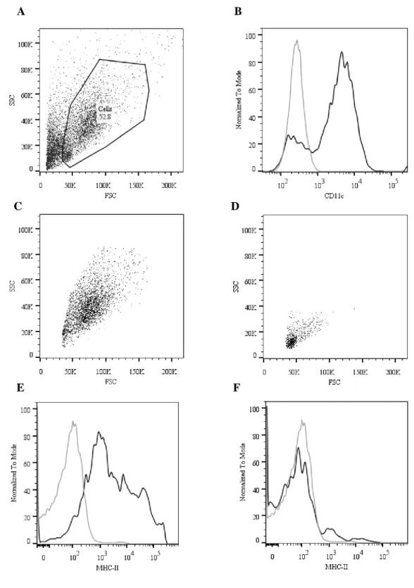
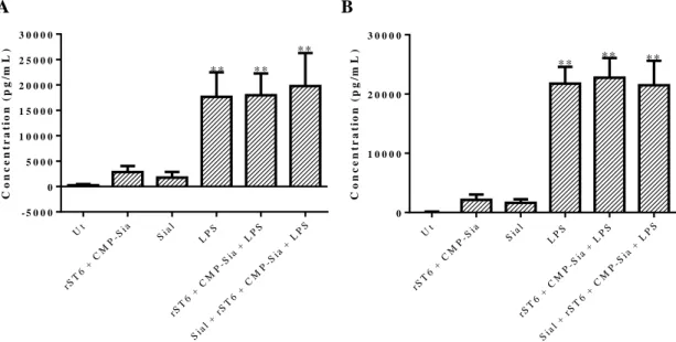
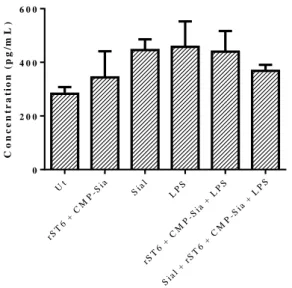
Figure I.1 - Representation for the chemical structure of Neu5Ac, a structure common to most of Sias.. ... 7 Figure I.2- Representation of Sias linked to glycosphingolipids or to O or N –glycans in glycoproteins, either in the cellular membrane as in secreted glycoproteins. ... 8 Figure III.1- Identification of BMDCs obtained within 8 days of differentiation, through Flow Cytometry analysis. ... 37 Figure III.2- Concentrations of TNF-α (A) and IL-6 (B) released by BMDCs, determined by ELISA. ... 39
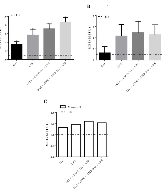
Figure III.3-Concentrations of IL-10 released by BMDCs, determined by ELISA. ... 40 Figure III.4- Percentage of cells within CD11c positive cells, which express high levels of CD86 (A), MHC-II (B) and are positive for CD80 (C), obtained by Flow Cytometry.. ... 42 Figure III.5- Fold increase of the MFIs for each condition comparatively to Ut, regarding the maturation markers CD86 (A), MHC-II (B) and CD80 (C), obtained by Flow Cytometry. ... 43
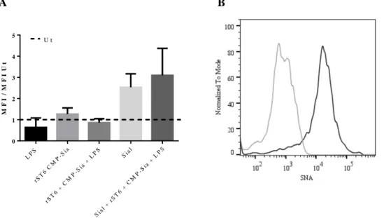
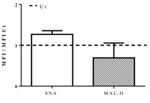
Figure III.6- A) Fold increase for the MFI of SNA to each condition comparatively to Ut. B) Histograms of unstained sample (grey solid line) and Ut sample (black solid line), representing a significant example of three independent experiments. ... 45 Figure III.7- Fold increase of Sial comparatively to Ut in the MFI of SNA and MAL-II, obtained within CD11c positive cells, by Flow Cytometry. ... 47 Figure III.8- A) Percentage of cells expressing CD86 (black solid bars) and MHC-II (black dashed bars), within CD11c positive cells, obtained by Flow Cytometry. B) MFI of CD86 (black solid bars) and MHC-II (black dashed bars), obtained within CD11c positive cells, by Flow Cytometry. ... 48 Figure III.9- Concentrations of TNF-α (black solid bars) and IL-6 (black dashed bars) released by BMDCs, determined by ELISA. ... 49
Figure III.10 – Fold increase of the MFI of SNA comparatively to Ut cells, obtained within CD11c positive cells, by Flow Cytometry. ... 50
Figure III.11- Concentrations of TNF-α (A) and IL-6 (B) released by BMDCs after 6 (black solids bars) and 24 hours (black dashed bars) of LPS stimulation, determined by ELISA. ... 52
xii
Figure III.12- Fold increase calculated for the MFIs of SNA about rST6 + CMP-Sia treatment performed during 4 hours and Sial treatment performed during 1 hour, comparatively to Ut and obtained by Flow Cytometry after 24 hours. ... 53
Figure III.13- Fold increase of the MFIs for each condition comparatively to Ut, regarding the maturation markers CD86 (A), MHC-II (B) and CD80 (C), obtained within CD11c positive cells, by Flow Cytometry.. ... 54
Figure III.14- Concentrations of TNF-α (A) and IL-6 (B) released by BMDCs within 6 (black solids bars), 24 (black dashed bars) and 48 hours (white solid bars, only performed for IL-6) after LPS stimulation, determined by ELISA.. ... 56 Figure III.15- Fold increase of the MFIs for each condition comparatively to Ut, regarding the maturation markers CD86 (A), MHC-II (B) and CD80 (C), obtained within CD11c positive cells, by Flow Cytometry. ... 58
Figure III.16- Fold increase of the MFIs for rST6 + CMP-Sia and Sial conditions comparatively to Ut, obtained for SNA and MAL-I after 1h of the respective treatments, by Flow Cytometry. ... 59 Figure III.17- Concentrations of TNF-α (A) and IL-6 (B), comparing WT with KO BMDCs, in the respective conditions, determined by ELISA.. ... 61 Figure III.18- A) Percentage of KO BMDCs expressing high levels of CD86 (black solid bars) and MHC-II (black dashed bars). B) MFI of CD86 (black solid bars) and MHC-II (black dashed bars), both within CD11c positive cells gate, obtained by Flow Cytometry. ... 62
Figure III.19- A) Histograms of SNA fluorescence in KO BMDCs for unstained (black line), Ut (grey line) and Sial + rST6 + CMP-Sia + LPS (blue line), obtained within CD11c positive cells, by Flow Cytometry. B) Fold increase in the MFI of SNA comparatively to Ut, from KO BMDCs in the respective conditions, also obtained within CD11c positive cells, by Flow Cytometry. ... 63
Figure III.20- Counter plots regarding CD86 vs SSC for Sial + rST6 + CMP-Sia + LPS conditions from KO BMDCs, obtained within CD11c positive cells gate, by Flow Cytometry.. ... 64 Figure III.21- FSC vs SSC density plots representing a significant example of BALF from ST6Gal-1 KO mice where PBS (A) and LPS (B) intra-tracheal injection were performed, assessed by Flow Cytometry after 24 hours.. ... 67
xiii Figure III.22- Counter plots of a significant example from 2 WT (A) and 3 KO mice (B), regarding Ly6G and CD11b markers, obtained by Flow Cytometry after 24 hours of LPS stimulation. ... 68
Figure III.23- Percentage of cells within Ly6G negative population expressing different levels of CD11c, for WT (black solid bars) and KO mice (black dashed bars) after 24 hours of LPS stimulation, obtained by Flow Cytometry. ... 69
Figure III.24- Evaluation of Ly6G negative, CD11c high population about the percentage of cells expressing CD11b, CD80, CD86 and MHC-II (A), with the respective MFIs (B), for WT (black solid bars) and KO (black dashed bars), after 24 hours of LPS stimulation, obtained by Flow Cytometry. ... 70
Figure III.25- Percentage of MHC-II + and MHC-II high populations within CD11c high, CD11b med population and CD11c high, CD11b – population (A), for WT mice (black solid bars) and KO mice (black dashed bars), whose respective MFIs are presented in B and were both obtained by Flow Cytometry after 24 hours of LPS stimulation. ... 71
Table II.1- Representation of the conditions to test in BMDCs’ activation assays and respective solutions prepared. ... 24
xv Abbreviations
AF Alexa Fluor
AP-1 Activation protein 1 APC Antigen presenting cells APC Allophycocyanin
APR Acute phase response
BACE Beta-site-amyloid precursor protein-cleaving enzyme 1 BAL Bronchoalveolar lavage
BALF Bronchoalveolar lavage fluid BCR B cell receptor
BMDC(s) Bone marrow derived dendritic cell(s) BV Brilliant violet
CCR7 C-C chemokine receptor type 7 cDC(s) Conventional dendritic cell(s)
CMP-Neu5Ac Cytidine-5’- monophosphate-N-acyl-neuraminic acid CMP-Sia Cytidine monophosphate sialic acid
Cy Cyanine
DC(s) Dendritic cell(s)
E. coli Escherichia coli
ELISA Enzyme linked immunosorbent assay FBS Fethal bovine serum
FITC Fluorescein isothiocyanate FMO Fluorescence minus one
FSC Forward scatter Gal Galactose
GalNAc N-acetyl-galactosamine
Galβ1,4GlcNAc Galactose β-1,4 N-acetyl-glucosamine
GM-CSF Granulocyte-macrophage colony stimulating factor HRP Horseradish peroxidase
IFN Interferon Ig Immunoglobulin IL Interleukin
IP Intraperitoneal
ITAM Immunoreceptor tyrosine-based activation motif ITIM Immunoreceptor tyrosine-based inhibitory motif
LPS Lipopolysaccharide
MAA Maackia amurensis agglutinin MAH Maackia amurensis hemagglutinin
MAL Maackia amurensis leukoaglutinnin MFI Median Fluorescence Intensity MHC Major histocompatibility complex mo-DC(s) Monocyte derived dendritic cell(s)
xvi
mRNA Messenger ribonucleic acid Neu Neuraminidase
Neu5Ac N-acetyl neuraminic acid NF Nuclear factor
OVA Ovalbumin P1 Promotor 1
PAMP(s) Pathogen associated molecular pattern(s) PBS Phosphate buffer saline
pDC(s) Plasmacytoid DC(s) PE Phycoerythrin
PerCP Peridinin chlorophyll
PRR(s) Pattern recognition receptor(s) rST6Gal-1 or rST6 Recombinant murine ST6Gal-1
SAMP Self-associated molecular patterns SEM Standard error of the mean
Sia(s) Sialic acid(s) Sial Sialidase
Siat1 KO Knockout mice for promotor 1 of ST6Gal-1 gene
Siglec Sialic-acid-recognizing immunoglobulin-like superfamily SNA Sacumbus Nigra Agglutinin
SSC Side Scatter
ST(s) Sialyltransferase(s)
ST3Gal β-galactoside α2,3 sialyltransferase ST6Gal β-galactoside α2,6 sialyltransferase
ST6Gal-1 KO β-galactoside α2,6 sialyltransferase I knockout ST6GalNAc N-acetyl-galactosamine α2,6 sialyltransferase
ST8Sia α2,8 sialyltransferase Th T helper
TCR T cell receptor TLR Toll like receptor(s)
TMB 3,3’,5,5’-tetramethylbenzidine TNF-α Tumour necrosis factor alpha
Ut Untreated WT wild type
Chapter I
Introduction
2
I.1. Immune system
I.1.1 Innate and adaptive immune system
The immune system includes all the cells and molecules that confer immunity, or defense from pathogens or infectious diseases. It includes the innate and the adaptive immune response. The former refers to the first line of defense, present in every multicellular organism, conferring a quick response to a potential pathogen. The innate response includes physical barriers such as epithelia, the action of phagocytic cells, serum proteins (as the complement) and cytokines, involved in inflammation. The adaptive immune response refers to a type of immunity with specific functions and memory. Indeed, the adaptive immune system is only present in vertebrates and is able to distinguish between similar pathogens, inducing a stronger response upon a second contact with the same antigen. In the adaptive immune system, the major players are the lymphocytes (T and B cells), which poses specific receptors, such as T cell receptors and antibodies, allowing the recognition of almost any antigen (Abbas, Lichtman and Pillai, 2012).
I.2. Dendritic cells
I.2.1.1. General functions in the immune system
Dendritic cells (DCs) were first described by Steinmann and colleagues in 1973, found in peripheral lymphoid organs of mice (Steinman and Cohn, 1973). DCs are the most important antigen presenting cells (APC) of the immune system, since they are the only cells that present antigens to naïve T cells (antigen inexperienced cells). Then, DCs are considered the bridge between the innate and the adaptive immune responses (Abbas, Lichtman and Pillai, 2012).
DCs own a set of features enabling them to play this role. First, they differentiate from immature to mature cells, upon a danger signal, adjusting their functions (Steinman and Cohn, 1973). They reside in different organs, such as the skin, the intestine, the lungs and secondary lymphoid organs (like the spleen and thymus), where they recognize and capture antigens, to display in their surface (Abbas, Lichtman and Pillai, 2012). Besides, DCs constantly endocyte self-antigens, contributing to tolerance, which is lost in autoimmune disorders, for example (Merad et al., 2013).
3 Otherwise, if an antigen is a potential dangerous, DCs phagocyte it and acquire a mature phenotype, migrating to the lymph nodes. There, they present antigens displayed through major histocompatibility complex (MHC) molecules to T cells. MHC can be class I or II. MHC-I is expressed in all nucleated cells, presents endogenous antigens (e.g. self or viral antigens) and may induce the activation of cytotoxic T cells, whereas MHC-II is expressed in every APC, presents phagocytosed antigens, mediating the activation of helper T cells . Other signals, such as the expression of co-stimulatory molecules, like CD80 and CD86, are required to activate T cells (Abbas, Lichtman and Pillai, 2012). Both belong to B7 family and bind to CD28 receptor in T cells, inducing their activation. CD86 is constitutively expressed in DCs at low levels, increasing quickly its expression after DCs’ activation. In the opposite, CD80 is only later expressed after DCs’ activation (Greenwald, Freeman and Sharpe, 2005). In addition, the release of pro-inflammatory cytokines, like interleukins (IL) 6, 12 and the tumour necrosis factor alpha (TNF-α), is needed to lead T cells towards a certain phenotype and immune response (Abbas, Lichtman and Pillai, 2012), (Merad et al., 2013). Furthermore, DCs also play a role in humoral immunity, by activation of B cells (Palucka and Banchereau, 2012).
Mature or activated DCs are characterized by reduction of the antigen uptake machinery (Granucci et al., 1999), modifications in their morphology (they acquire several dendrites) and the increased expression of receptors, such as C-C chemokine receptor type 7 (CCR7), allowing their migration to the lymph nodes. Mature DCs also have increased expression of MHC, co-stimulatory molecules and enhanced release of pro-inflammatory cytokines. (Banchereau and Steinman, 1998).
In order for DCs to capture antigens, they have a panoply of receptors that recognize pathogen associated molecular patterns (PAMPs). An example of pattern recognition receptors (PRR) is the family Toll like receptor (TLR). They can be found in the extracellular membrane of cells, or within the intracellular nucleus membrane (Takeda, Kaisho and Akira, 2003). Lipopolysaccharide (LPS) is a well-known PAMP, found in the outer membrane of Gram negative bacteria, being a useful tool to study the inflammatory response. Indeed, the engagement of LPS with TLR-4 induces a strong immune response from DCs (Dearman et al., 2009). The intracellular signalling activates transcriptor factors like nuclear factor (NF)-kB, activation protein 1 (AP-1) and interferon (IFN) regulating factors. Consequently, it occurs the expression and release of cytokines,
4
inducing the adaptive immune response (Takeda, Kaisho and Akira, 2003). (Zanoni and Granucci, 2010).
I.2.1.2. Subsets of DCs
DCs constitute a very heterogeneous population, where a single surface marker is not enough to distinguish them. There are also functional differences found within DCs’ subsets, besides the expression of surface markers, which are affected by the inflammatory status (Shortman and Liu, 2002), (Merad et al., 2013).. Nowadays, DCs obtained from mice are better characterized than human DCs, where several studies have the aim to establish homology between them. Because of this, is important to choose appropriate means to obtain DCs, depending on the goals of the investigation (Shortman and Liu, 2002).
Currently is known that myeloid or lymphoid precursors can originate the same subsets of DCs (Satpathy et al., 2012), (Sathe et al., 2013). Conventional DCs (cDCs) are the major subset within DC population and can be classified as migratory or resident (classification only applied in steady state) (Merad et al., 2013). The former includes, for example, dermal DCs and epidermal Langerhans cells, able to migrate to lymph nodes under inflammatory conditions (Shklovskaya, Roediger and Fazekas de St. Groth, 2008). Resident DCs can be found in secondary lymphoid organs, such as the thymus and the spleen (Vremec et al., 2000), constantly screening the blood and lymph (reviewd by Crespo, Lau and Videira, 2013). Conventional DCs express high levels of CD11c and MHC-II, but differentially express other markers, also depending on the tissue distribution and inflammatory status (Abbas, Lichtman and Pillai, 2012), (Merad et al., 2013). Taking this into account, cDCs are further divided in subclasses (Palucka and Banchereau, 2012).
Plasmacytoid DCs (pDCs) constitute a small subset of DCs, mainly found in the bloodstream or in lymphoid organs, whose DCs’ features arise upon inflammatory conditions (Abbas, Lichtman and Pillai, 2012). These cells look like plasma cells and express lower levels of CD11c and MHC-II, but express the B cell marker, B220. Their main function is the release of type 1 IFN, during viral infections (Merad et al., 2013).
5 Under inflammatory conditions, there are also DCs arising from monocytes in circulation (mo-DCs), emphasising the fact that the enormous diversity inherent to DCs’ subtypes is influenced by the surrounding microenvironment (Shortman and Liu, 2002).
I.2.1.3. Mice as powerful sources to study DCs
Since DCs are rarest cells, several methodologies were developed, in order to generate DCs in vitro. Indeed, the mouse has been a model of excellency to analyse DCs. (Inaba et al., 1992) demonstrated that mouse bone marrow CD34 positive precursors stimulated with granulocyte-macrophage colony stimulating factor (GM-CSF) generate large amounts of DCs. GM-CSF induces the maturation of granulocytes and monocytes (Syme and Glück, 2001). Later, GM-CSF and IL-4 were used to differentiate human monocytes into DCs, where IL-4 avoids the differentiation of monocytes into macrophages (Sallusto and Lanzavecchia, 1994), but also promotes the differentiation of monocytes into DCs lineage (Roy et al., 2004). The protocol developed to generate human DCs is nowadays used to generate mouse bone marrow derived DCs (Inaba et al., 2009). Immature DCs obtained through this protocol express high levels of CD11c, CD11b and medium levels of MHC-II, showing all the major functions related with DCs (Inaba et al., 1992).
Other techniques are useful to study DCs from specific tissues of mice, such as the lungs. DCs in the lungs have been studied in order to unravel the mechanisms underlying in pulmonary or allergic diseases, like allergic asthma (Kim and Lee, 2014). Albeit constituting only a minor population in the lungs, DCs have crucial roles screening inhaled air and migrating to mediastinal lymph nodes upon antigen contact, where they initiate the immune response (Jahnsen et al., 2006), (Hufford et al., 2012). Indeed, in a mouse model of asthma, CD11c positive DCs found in the airways induced the features of allergic asthma, which are abrogated upon their depletion (Julia et al., 2002). Different subsets of DCs are found in different compartments of mouse lungs, with specialized functions (Condon et al., 2011). In order to assess cells from the conducting airways and the alveolar space, bronchoalveolar lavage (BAL) of mouse lungs is performed (Heer, De
et al., 2005).This methodology is applied to patients with pulmonary chronic diseases for
diagnostic, but also in healthy patients for research purposes, despite only few studies were performed (Reynolds, 2000). In the alveolar compartment, in steady state, cDCs are
6
the major subset found within DC population, being CD11c high, CD11b positive or CD11c high, CD11b negative, CD103 (langerin) positive cells (Hufford et al., 2012). On the other hand, pDCs are rarest found in alveolar space, but increase under inflammatory conditions. Besides, DCs derived from circulating monocytes migrate to the alveolar compartment, when an inflammation occurs. They are characterized by the high expression of CD11c, expression of CD11b positive and monocyte lineage markers, like Ly6C (GeurtsvanKessel and Lambrecht, 2008).
I.2.1.4. Therapeutic potential of DCs
DCs are potential adjuvants to current therapies, improving the immune response against a certain antigen. Taking advantage of DCs’ plasticity, is possible to induce them towards a certain phenotype and different immune response (tolerogenic DCs or DCs activating cytotoxic T cells, for example), (Palucka and Banchereau, 2012).
Indeed, a major field in cancer immunotherapy relies in the administration of DCs obtained from the patient, which are loaded afterwards with cancer antigens (designated
ex vivo therapy). This protocol elicits tumour specific responses through the expansion of
T cells and has proven to be efficient in mice models of cancer, being implemented nowadays in clinical trials (Paczesny et al., 2004), (Steinman, 2008). Despite the vaccines have proven to be safe, several factors need to be improved to reach the success, namely avoiding the inactivation of DCs in the tumour microenvironment and finding the most appropriate antigens to elicit a strong and effective immune response (Palucka and Banchereau, 2012). An example of a promising vaccine relying on this principle is the PROSTVAC, used against prostate cancer, where a survival benefit was observed at Phase II Clinical trials (Kantoff et al., 2010).
7
I.3. Sialic acids
The concept of Glycobiology was first defined in 1980, to describe the study of the structure, biosynthesis, biology, and evolution of saccharides (sugars or glycans) distributed in nature, as well as proteins that recognize them (Varki et al., 2009). Over the years, the study of glycans in biological systems has gained its importance.
Glycosylation is an important posttranslational modification occurring mainly in the lumen of endoplasmatic reticulum and Golgi apparatus, where glycans are added to proteins and lipids. These modifications are performed by glycosyltransferases, distributed orderly, using a single nucleotide-sugar as a donor substrate. Then, these enzymes are responsible for glycan biosynthesis and structures found in glycoconjugates (Rabinovich and Croci, 2012).
There are many families of glycans, where the family of Sialic acids is one of them. Sialic acids (Sias) are expressed in the cells’ surface of all animals from deuterostome lineage (vertebrates and some higher invertebrates) and also in some pathogenic or symbiotic bacteria, which associate with vertebrates (Varki et al., 2009).
The number of members included in Sias family has increased over the years, where more than 50 structures are identified today in nature (Varki, 2010).
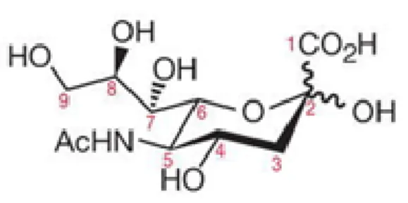
One of the most important structures of Sias, found in mammalian cells, is the N-acetyl neuraminic acid (Neu5Ac), which is a nine carbon backbone structure (Varki, 2007), represented in Figure I.1
Figure I.1 - Representation for the chemical structure of Neu5Ac, a structure common to most of Sias. The carbon numbers are represented, as well as the bonds of the anomeric centre, carbon 2 (C2). Adapted from (Varki et al., 2009).
8
N-glycans bind to asparagine residues in the sequence motif Asparagine-X-Serine-Threonine (Asn-X-Ser/Thr), whereas O-glycans bind to serine and threonine residues (Varki et al., 2009), as illustrated in Figure I.2. Sias are usually placed at terminal branches of N-glycans, O-glycans, and glycosphingolipids (gangliosides). Due to the type of linkages that Sias form with other molecules and the possible modifications in their primary structure, they increase the variety of structures found in glycoconjugates.
Figure I.2- Representation of Sias linked to glycosphingolipids or to O or N –glycans in glycoproteins, either in the cellular membrane as in secreted glycoproteins. The legend for the representation is shown in the upper left side of the chart. Adapted from (Varki, 2007).
Sias can remain in glycans attached to the membrane of cells or decorate secreted glycoproteins, as illustrated in Figure I.2. One of their particularly important features is the negative charge they confer to glycoconjugates, influencing the interactions of the cells with the microenvironment (Varki and Gagneux, 2012).
It is to notice that Sias are linked to other molecules through their anomeric carbon number 2 (C2) and can establish linkages to the carbons 3 or 6 of other glycans or to C8 of another Sia, designated respectively as α2,3, α2,6 and α2,8 Sias (Li and Chen, 2012).
9
I.4. Sialyltransferases
The attachment of Sias to glycoconjugates implies the existence of an enzyme able to transfer the substrate (donor) to an acceptor. The donor is the sugar cytidine-5’- monophosphate-N-acetyl-neuraminic acid (CMP-Neu5Ac) or CMP-Sia. The substrates can be galactose residues (Gal), N-acetyl-galactosamine (GalNAc) or Sias (Rao et al., 2009).
Sialyltransferases (STs) are enzymes responsible to transfer CMP-Neu5Ac to other glycans. Similarly with other glycosyltransferases, all animal STs are type II membrane proteins with signals for their Golgi localization (Varki et al., 2009). Most motifs of STs are highly conserved in vertebrates, where both mice and humans have 20 known STs. (Li and Chen, 2012). There are 4 classes of STs in mice and human, divided according with their substrate and acceptor specificity. ST3Gal (β-galactoside α2,3 sialyltransferase) family catalyses the transfer of CMP-Sia to terminal galactose through α2,3 linkages, where six subfamilies are identified; ST6Gal (β-galactoside α2,6 sialyltransferase) family has 2 subfamilies, catalysing the transfer of α2,6 Sias to galactose β-1,4 N-acetyl-glucosamine (Galβ1,4GlcNAc); the family ST6GalNAc (N-acetyl-galactosamine α2,6 sialyltransferase) owns six subclasses that transfer α2,6 Sias to N-acetyl-galactosamine (GalNAc); lastly, α2,8 sialyltransferase (ST8Sia) family induces the transfer of Sias in α2,8 linkage to terminal Sias, where 6 subfamilies are identified as well (Takashima, 2008), (Rao et al., 2009), (Harduin-Lepers, 2010).
ST genetic expression is modified during mammalian development and immune regulation, being also different within different cell types. Besides, ST families are differentially expressed (Rao et al., 2009). Indeed, in mammalian cells, α2,3 is the most common linkage of Sias formed by ST3Gal family, being α2,8 linkage, catalysed by ST8Sia family members, the rarest (Hennet et al., 1998).
In this dissertation we will focus in ST6Gal-1 subfamily (β-galactoside α2,6-sialyltransferase 1), responsible for most of α2,6 Sia linkages in Galβ1,4GlcNAc residues of N-glycans, with widespread tissue distribution, unlike ST6Gal-2 subfamily (Takashima, 2008).
10
I.4.1 ST6Gal-1: the membrane-anchored and the soluble forms
In humans, ST6Gal-1 gene is placed in chromosome 3, whereas in mice it is located in chromosome 16 (Dalziel et al., 1999). Either in humans as in mice, various and distal promotors control the genetic expression of ST6Gal-1 and have been a target of study (Hennet et al., 1998). Furthermore, ST6Gal-1 is highly expressed in hepatocytes and lymphocytes, where it catalyses α2,6 sialylation of serum glycoproteins and glycoproteins of antigen membrane receptors, respectively (Varki et al., 2009). ST6Gal-1 is usually at Golgi membrane, but can also be present in soluble form due to BACE (beta-site-amyloid precursor protein-cleaving enzyme 1) proteolytic action (Dalziel et al., 1999),(Jones et al., 2010). Liver is the major source of soluble ST6Gal-1, as shown by the creation of a Siat1ΔP1 mice (lacking the liver promotor P1 of ST6Gal-1 gene). These mice presented deficiency in serum levels of ST6Gal-1 under inflammatory status, suggesting that the liver promotor P1 is the most important regulating the levels of soluble ST6Gal-1 (Dalziel et al., 1999), (Appenheimer et al., 2003).
To compare the activity of both forms, the soluble form was deleted for the membrane anchor in different extends, where it retained the enzymatic fold and activity (Legaigneur
et al., 2001). Nevertheless, the specificity for glycan acceptor has diminished in the
soluble form, compared to the membrane anchored ST6Gal-1 (Legaigneur et al., 2001), (Kuhn et al., 2013).
I.5. Sialidases
In opposite to sialyltransferases, sialidases or neuraminidases are responsible for removing Sias from glycans. In mammals, there are four known neuraminidases, presenting homology between mouse and human: Neu1, 2, 3 and 4. Neu1 is expressed in the majority of cell types and is mainly located in the lysosome, but can be translocated to cell membrane. It regulates molecular adhesion and intracellular signalling, by desialylation of some receptors like TLR in immune responses (Stamatos et al, 2010), (Varki and Gagneux, 2012). Neu3 is a membrane associated protein also found in late endosomes, whose major function is to remove Sias from gangliosides. Neu2 is located in cytosol, playing a role in differentiation and malignancy. Lastly, Neu4 is found at lysosomes and mitochondria, but its functions are not well known (Stamatos et al., 2010).
11 Sialidases from vertebrates are unstable in extracellular fluids, unlike sialidases from bacteria, which are used to perform in vitro assays (Varki and Gagneux, 2012).
I.6. Lectins
Lectin is the general term referring to proteins which bind glycans. They are found in animals, plants and also in pathogens or toxins. In addition, these lectins can be intrinsic to an organism, or recognize glycans in other organism (Varki et al.,2009). Regarding the lectins that recognize Sias, there are three major families of lectins: Sialic-acid-recognizing Immunoglobulin-like superfamily (Siglec), complement factor H and selectins, which belong to C-type lectins family (Varki et al., 2009).
Siglecs are mostly found in cells of immune system and usually have an intracellular ITIM (immunoreceptor tyrosine-based inhibitory motif) or, not so frequently, ITAM (immunoreceptor tyrosine-based activation motif), regulating immune responses (Crocker, 2002), (Rabinovich and Croci, 2012). They are further divided in subclasses, wherein sialoadhesin, CD22 and CD33 are examples of Siglecs common to all mammals. Regarding CD33 related Siglecs, human and mice have different types expressed (Crocker, Paulson and Varki, 2007). Factor H is a down regulator of complement cascade, binding to Sias in cells’ surface and avoiding the constant complement activation. Selectins are present in the endothelia, leukocytes and platelets and induce leukocyte traffic (Varki and Gagneux, 2012).
Regarding lectins found in organisms which do not express Sias, such as plants, they are very useful to evaluate linkages to Sias, because they are specific, soluble and easy to isolate and obtain in higher amounts (Varki et al., 2009). For example, Sacumbus
Nigra Agglutinin (SNA) and Maackia amurensis Agglutinin (MAA) are widely used in
Glycobiology field. Both of them have hemagglutination abilities, i.e., they agglutinate red blood cells. SNA recognizes α2,6 Sias in terminal galactose residues (Shibuya et al., 1987), whereas MAA is divided in different classes. Maackia amurensis leukoaglutinnin I (MAL-I) preferentially binds Siaα2,3Galβ1-4GlcNAc residues and in a weaker extend Galβ1-4GlcNAc. In opposite, Maackia amurensis hemagglutinin (MAL-II) preferentially binds α2,3 Sias, despite some unspecific binding has been observed (Geisler and Jarvis, 2011). However, some controversial regarding MAL-I was reported, since some manufactures reported that MAL-I preferentially binds Galβ1-4GlcNAc, tolerating
12
instead the substitution of N-acetyl-lactosamine with α2,3 Sia binding to Gal (Vector Labs).
I.7. Roles of Sias in the immune system
Regarding the terminal position of Sias in cell membranes, they play important roles in interactions of cells with their microenvironment and in modulation of immune response (Varki et al., 2009), (Varki and Gagneux, 2012).
Sias can have a dual function, either masking important molecules, or being ligands of important receptors, as previously mentioned (Varki and Gagneux, 2012). By masking, Sias protect the host from pathogen interactions, such as proteases action. In addition, Sias prevent galectins binding, a lectin group that regulates several cellular events (Rabinovich and Croci, 2012).
As ligands of important receptors, Sias bind to factor H, recognizing self Sias (cis interaction) and avoiding autoimmune responses. As selectin ligands, Sias are involved in leukocyte traffic and chemokine derived migration of immune cells to lymph nodes (Varki and Gagneux, 2012). On the other hand, Sias are a recognition place to pathogens and can even be used as an energy resource, whereas in some cases as a tactic of molecular mimicry (Varki and Gagneux, 2012).
During stages of cellular differentiation and maturation, modifications occur in the genetic expression of STs, correlating with changes in Sias patterns (Crespo, Lau and Videira, 2013). The genetic expression of STs is also changed during embryogenesis, inflammation, some cancers and in autoimmune diseases, emphasizing the roles for Sias in several biological processes and disorders (Varki and Varki, 2007), (Rabinovich and Croci, 2012). In the following two sections, the importance of ST6Gal-1 in the modulation of immune functions will be detailed, namely its influence on DCs’ features and functions.
13 I.7.1 ST6Gal-1 in the modulation of immune functions
The functions of ST6Gal-1 in the immune system are often associated with its biological receptor, CD22 also known as Siglec-2, highly conserved between mice and human forms (Crocker, 2002).
To study the importance of α2,6 Sia in immune system, ST6Gal-1 knockout mice were generated (ST6Gal-1 KO) (Hennet et al., 1998), as this ST family is the major source of α2,6 Sia linkages, either in mice (Jones et al., 2010) as in humans (Varki, 2010). ST6Gal-1 KO mice showed deficient T cell-dependent antibody production, reduced B cell proliferation and antibody production (Varki et al., 2009), modified thymopoiesis and granulopoiesis and disorders on eosinophil and DCs’ profiles (Zhuo and Bellis, 2011).
Indeed, B cell functions in ST6Gal-1 KO mice are impaired due to the lack of α2,6 Sias in CD22 surface. In ST6Gal-1 KO mice, the threshold for activation of mature B cell receptor (BCR) increases due to absence of CD22-CD22 interactions, where α2,6 Sias are needed. In this case, CD22 co-localizes with BCR in cell membrane, recruiting phosphatases that will decrease BCR activation. Consequently, the threshold for BCR activation increases and the functions of B cells are supressed (Hennet et al., 1998), (Rabinovich and Croci, 2012).
Another interesting feature about α2,6 Sias is the different tissue distribution of Sias found within different species (Varki, 2010). Actually, the first role reported for Sias was being the receptors for Influenza virus. Interestingly, some Influenza virus preferentially bind to α2,3 Sias, abundant in birds’ airways, whereas humans’ airways have mostly α2,6 Sias in epithelial cells. This observation explained the initial resistance of humans to the virus, highlighting the protective role of α2,6 Sias, in this case (Varki and Varki, 2007).
Secreted glycoproteins usually have α2,6 Sias in their surface, modifying several of their features: they can have a different conformation, be retained in some cell membranes and clusters of receptors are changed as well. Therefore, events such as cellular migration and adhesion are influenced by α2,6 sialylation (Zhuo and Bellis, 2011). Actually, for immunoglobulin G (IgG) intravenous therapy, used in several autoimmune diseases, the α2,6 sialylation of N-glycans in Fc regions of IgG is required for anti-inflammatory effects (Kaneko, Nimmerjahn and Ravetch, 2006) (Jones et al., 2012).
14
Confirming the contribution of ST6Gal-1 in several disorders, its expression is up-regulated in some carcinomas as colon, breast, ovarian, gastric, cervix, choriocarcinomas, acute myeloid leukemia, and some brain tumours (Hedlund et al, 2008),,(Park and Lee, 2013). Besides, it promotes tumour cell growth and metastasis. In fact, in early studies, a bigger metastatic phenotype was observed in tumour mutant cell lines, correlating with increased sialylation and pronounced increase in the expression of ST6Gal-1 mRNA (Takano, Muchmore and Dennis, 1994). The presence of Sias in tumour cells’ surface also enables complement factor H binding, avoiding complement activation. In addition, α2,6 Sias bind inhibitory Siglecs of immune system cells, which probably dampens their functions helping to eliminate tumour cells (Varki et al., 2009).
I.7.2 Extrinsic α2,6 sialylation: the new concept of distal immune regulation For several years, ST6Gal-1 soluble was considered a product of metabolically inefficiency, with no biological significance, despite catalytic activity was noticed (Jamieson, McCaffrey and Harder, 1993).
Elevated ST6Gal-1 in the bloodstream of cancer patients is correlated with a bad prognosis. While these observations suggest an important role for extracellular ST6Gal-1 in cancer progression, its role in cancer remains to be elucidated, which will also determine if it can be used as a cancer biomarker (Park and Lee, 2013).
ST6Gal-1 is also released from liver into circulation, under inflammatory conditions, being part of the acute phase response (APR) (Kaplan et al., 1983). In addition, the release of ST6Gal-1 from liver was proved to be IL-6 dependent by using mice models (Dalziel
et al., 1999). It is tempting to think that soluble ST6Gal-1 could be a homeostatic
modulator, similar to other proteins released into bloodstream upon APR. Nevertheless, the role for ST6Gal-1 role in circulation, under APR, is unknown.
Extrinsic sialylation refers to the sialylation of glycans in distal cells or glycoproteins, occurring through the action of STs extrinsic to it, which could be present in bloodstream. This concept was previously dismissed, due to the absence of enough sugar substrates in circulation for STs to act distally (Zhu et al., 1998). Nevertheless, other roles were proposed for soluble STs, such as the possibility of acting like lectins, through linkages with carbohydrates, modulating then immune functions (Zhu et al., 1998). However, the concept of extrinsic sialylation is nowadays being revised, since appropriate sugar donors
15 are found in circulation, under certain conditions, like platelets (Wandall et al., 2012). In this case, circulatory STs could extrinsically add Sias in other cells’ surface, eventually contributing to the modulation of immune functions (Nasirikenari, Collins and Lau, 2011).
In asthma models of ST6Gal-1 KO, the number of neutrophils is increased comparing with WT mice, upon lung allergen stimulation. Supporting this, downregulation of circulatory ST6Gal-1 activity was noticed in WT mice, during lung allergen challenge (Nasirikenari et al., 2006). In addition, higher eosinophilia was noticed in ST6Gal-1 KO mice subjected to allergen stimulation, suggesting that lack in α2,6 Sia sensitizes these mice (Nasirikenari et al., 2010). These observations have been attributed to increased myelopoiesis (Nasirikenari et al., 2006). Corroborating a potential role for soluble ST6Gal-1, bone marrow myelopoiesis has decreased with in vitro treatment of marrow cells in myeloid colony forming assays, by adding physiological concentrations of ST6Gal-1 with appropriate sugar donors (Jones et al., 2010). Moreover, hematopoietic and stem cells numbers have decreased in vivo, upon increased levels of circulatory ST6Gal-1, released from the liver (Nasirikenari et al., 2014). The mechanisms underlying are still unknown and may be associated with CD22, since this receptor is related with decreased cellular proliferation, at least regarding B cell function (Crocker, Paulson and Varki, 2007).
I.7.1 ST6Gal-1 in the modulation of DCs functions
In the case of DCs, several changes in the glycosylation patterns are noticed during differentiation and maturation stages (Videira et al., 2008), (Bax et al., 2007).
Indeed, the genetic expression of ST6Gal-1 has increased during differentiation of human mo-DCs, correlating with an increased enzymatic activity and increased SNA binding to cells’ surface (Videira et al., 2008). In opposite to this observation, Neu1 and Neu3 genetic expression and activity increase upon differentiation of human monocytes into DCs, which appears to be controversial (Stamatos et al., 2010). Both observations suggest that the mechanisms regulating sialylation patterns of DCs during differentiation are complex, needing further elucidation.
The genetic expression of ST6Gal-1 is dramatically decreased upon DCs’ maturation either induced with LPS (activates TLR-4) or with modulators of other TLR or cytokines
16
(Bax et al., 2007), (Videira et al., 2008). However, upon DCs’ maturation, the binding of CD22 to α2,6Sia-Galβ14GlcNAc has increased comparing with immature DCs as well as the binding of SNA, unlike the genetic expression has suggested (Bax et al., 2007). Corroborating the former results, other study has proven that tolerogenic immature human mo-DCs have a higher content in α2,6 Sias. In this case, the genetic expression has also dramatically decreased upon DCs’ maturation with pro-inflammatory cytokines, but correlated well with a drastically decrease in SNA binding to mature mo-DCs. (Jenner et
al., 2006). Regarding the apparent controversial findings about SNA binding to human
mo-DCs upon maturation, is important to understand if these differences at protein level are related with the activation of a specific receptor or if they are dependent on another factor.
Taking into account the modifications in ST6Gal-1 expression upon DCs’ maturation, it is likely that removing Sias from DCs’ surface could influence related processes. Indeed, neuraminidase treatment of human mo-DCs decreased the number of particles endocytosed by DCs (Videira et al., 2008), where reduced expression of antigen uptake machinery is a common feature inherent to DCs’ maturation.
Nevertheless, later was proven that phagocytosis of Escherichia coli (E. coli) increases upon sialidase treatment. This suggests that this improvement is restricted to pathogens expressing sialylated structures, despite further elucidation is needed (Cabral
et al., 2013). Moreover, endogenous Neu1 activity improved the phagocytosis of DCs
from mice (Seyrantepe et al., 2010). In fact, DCs from Siat1-null KO mice (unable to produce α2,6 Sias in Gal residues) presented improved phagocytosis of E. coli, suggesting the implication of ST6Gal-1 in modulation of this function from DCs (Cabral et al., 2013).
In addition, sialidase treatment of human mo-DCs led to increased expression of MHC-II, MHC-I, CD80 and CD86, to improvement of T cells priming which increased their proliferation, and to increased mRNA expression of pro-inflammatory cytokines, such as IL-6, IL-12, IL-1β and TNF-α (Crespo et al., 2009). Corroborating these observations, endogenous expression of Neu1 and Neu3 increases in DCs stimulated with LPS, suggesting that it may have a role inducing maturation (Stamatos et al., 2010). In fact, in macrophages and DC cell lines, Neu1 activity for desialylation of TLR-4 upon LPS stimulation is required for further cellular activation (Amith et al., 2010). Besides,
17 DCs grown in the presence of endogenous Neu1 and Neu3 inhibitors decrease the amount of cytokines released from mo-DCs stimulated with LPS. This study also suggests that the hyper sialylation of specific moieties can be related with the specific decrease in maturation of DCs (Stamatos et al., 2010). This later observation highlights the importance of attributing roles for specific types of sialylation, and not only modifications in the general content of Sias by sialidase effect.
Regarding the role of ST6Gal-1 in DCs’ maturation, BMDCs from ST6Gal-1 KO mice presented a higher expression of MHC-II compared to WT, upon endocytosis. In addition, ST6Gal-1 KO mice showed a slight, but not impaired, decrease in endocytosis capacity. On the other hand, assessing DCs from blood, spleen and lymph nodes from ST6Gal-1 KO, their numbers and markers expression were similar to WT, whereas only pDCs expressed more MHC-II compared to WT pDCs (Crespo et al., 2009).
Furthermore, the quick changes found in DCs’ sialylation profile upon maturation were attributed to the activity of a ST in surface of human mo-DCs, through mechanisms still unknown (Cabral et al., 2010).
18
I.8. Context and aims of the work
DCs are vital for the initiation of adaptive immune responses and immunomodulation. The elucidation of mechanisms modifying DCs’ functions may have potential impact in immunotherapy, where improvements to DCs’ based vaccines efficacy need to be achieved (Palucka and Banchereau, 2012).
Previous work from our group demonstrated that sialidase treatment of human mo-DCs increase the expression of MHC-II, CD80, CD86, the genetic expression of released pro-inflammatory cytokines and improves the priming of T cells with their further activation. Moreover, ST6Gal-1 knockout mice presented more mature DCs, regarding MHC-II expression (Crespo et al., 2009). These findings suggest that ST6Gal-1-mediated sialylation might have an immunomodulatory role in DCs. Nonetheless, the mechanism underlying sialidase effect is unknown.
In our group, it was observed that soluble ST6Gal-1 decreases myelopoiesis in
vitro (Jones et al., 2010) and the numbers of hematopoietic and stem cells in WT mice
treated with soluble ST6Gal-1 (Nasirikenari et al., 2014). Moreover, in vitro treatment with ST6Gal-1 diminishes the release of pro-inflammatory cytokines in bone marrow derived macrophages (unpublished observations from Mehrab Nasirikenari, Joseph Lau). These findings suggest that extrinsic ST6Gal-1 has a key role in the modulation of immune functions. In addition, the implication of soluble ST6Gal-1 in several cancers (Swindall et al., 2013), (Park and Lee, 2013) emphasises the importance of unravelling its biological role. Considering the results from our groups all together, we hypothesize that DCs extrinsically treated with ST6Gal-1 diminish the expression of maturation markers and the release of pro-inflammatory cytokines.
In order to test this hypothesis, the main goal of this work was to assess the influence of in vitro extrinsic α2,6 sialylation in bone marrow derived DCs’ features, upon LPS stimulation. Bone marrow cells were chosen as the source of DCs, due to the high amounts of obtained cells, enabling the study of several combined conditions. Moreover, these cells present homology with human conventional DCs, suggesting that findings can be addressed to human DCs (Satpathy et al., 2012).
On the other hand, we intended to study sialidase effect in this type of DCs, establishing a homology with human mo-DCs. Lastly, we intended to address the specific role for ST6Gal-1 in DCs maturation, in vivo, namely in lung DCs, due to its importance
19 in several allergic and pulmonary diseases (Kim and Lee, 2014). To achieve this goal, we used ST6Gal-1 knockout mice stimulated with LPS and assessed, afterwards, the features of cells from their airways. Therefore, with this work we have addressed the effect of ST6Gal-1 in two important and representative DC subtypes.
The present work was first developed in Department of Molecular and Cellular Biology of Roswell Park Cancer Institute, Buffalo, NY, USA and finished in Glycoimmunology group of Chronic Diseases Research Center (CEDOC) from Nova Medical School – Faculdade de Ciências Médicas of Universidade Nova de Lisboa, Portugal.
Chapter II
Materials and Methods
22
II.1. Extraction of bone marrow cells from mice
All animal experiments of this work were approved by the Institute of Animal Care and Use Committee of Roswell Park Cancer Institute, Buffalo, NY, USA, and by animal facility from Nova Medical School – Faculdade de Ciências Médicas, Universidade Nova de Lisboa.
Wild type (WT) C57BL6 and ST6Gal-1 knockout (ST6Gal-1 KO) mice, 6 to 10 weeks of age, female or male were used. WT mice were purchased from Jackson Laboratories at Bar Harbor, Maine, USA and ST6Gal-1 KO were initially obtained from Dr. Jamey Marth laboratory from Molecular, Cellular, and Developmental Biology University of California, Santa Barbara, USA (Hennet et al., 1998) and successive backcrossed more than 10 generations into C57BL6 background (Roswell Park Cancer Institute, Buffalo, NY, USA). WT mice from animal facility of Nova Medical School – Faculdade de Ciências Médicas, Universidade Nova de Lisboa were kindly given by Dr. José Ramalho, from Molecular Biology group of CEDOC.
The mice were killed by CO2 asphyxiation. The femurs, tibiae and peroneum were
removed, and then, in sterile conditions, the bones were put in a petri dish with PBS 1X (from Corning). The bones were sniped in the ends with scissors. A 3 ml syringe was assembled with a 27 gauge needle and loaded with RPMI complete medium (Appendix I) in 1:10 dilution with PBS 1X. The bones were flushed till become white and then the cells were filtered with a 100 µm strainer. It was added Mouse red blood cell lysis buffer (Appendix I) to the cells in 1:2 proportion, in order to lyse erythrocytes (not needed to further differentiation of bone marrow precursor cells into DCs). Cells were centrifuged at 1400 rpm for 5 minutes. After that, is was discarded the supernatant and the pellet was ressuspended in RPMI complete medium. The cells were diluted in PBS 1X and counted using an automatic counter machine (from BioRad), or under the optic microscope with a Neubauer chamber. In the first case, the obtained number was multiplied by the dilution factor and considered the volume where the cells where ressuspended. In the former case, the total cells were counted, applying the following equation:
23 Where DF is the dilution factor and 104 refers to the volume of Neubauer
chamber. The number of cells per ml was obtained dividing the result of the equation by total volume where the cells were ressuspended. During this procedure, usually 25 to 30 million bone marrow cells were obtained, per mouse.
II.1.1 Generation of bone marrow derived DCs
The bone marrow cells from both WT and ST6Gal-1 KO mice, obtained as previously described, were cultured with 20 ng/ml (400 U) of recombinant murine GM-CSF (from Peprotech or Immunotools) and 10 ng/ml (50 U) of recombinant murine IL-4 (Biovision), in RPMI complete medium. Together, these cytokines induce the differentiation of bone marrow cells into DCs. Bone marrow cells were plated at 5 x 10⁵ cells per 3 ml of medium, in a 6 well plate, and incubated at 37 ºC in humid atmosphere with 5% CO2. In some assays, the bone marrow cells were cultured with 20 ng/ml of
GM-CSF (400 U), without IL-4.
Every two days, the culture medium was changed. Per well, it was discarded 1 ml of the old medium and replaced with 1 ml of fresh RPMI complete medium, supplemented with cytokines, in the same concentrations as mentioned before. By removing the old medium, other types of cells derived from bone marrow are eliminated, as well as metabolites produced by the cells (Inaba et al., 1992).
After 7 to 8 days of culture, the non-adherent cells were harvested, by gently pipetting, dislodging cellular aggregates, since DCs are non-adherent cells. In addition, using this method any eventual contamination of DC population with macrophages is prevented, since these are adherent cells (Inaba et al., 1992). The cells were centrifuged at 900 rpm for 10 minutes. This gentle way to harvest the cells was performed to avoid DCs’ activation.
After discarding the supernatant, the pellet was ressuspended in RPMI complete medium. The cells were counted in the same way as previously described and plated in 24 well plates, at 5 × 10⁵ cells per well, each one with 500µL of RPMI complete medium without cytokines. The number of cells obtained at this stage was usually between 5 to 10 million.
24
II.2. Assays to test the influence of extrinsic α2,6 sialylation in mice
BMDCs’ activation profile
With the aim of evaluating the influence of α2,6 extrinsic sialylation on bone marrow derived DCs (BMDCs)’ activation profile, different conditions upon lipopolysaccharide (LPS) stimulation were tested. These tests were run in the next day after BMDCs were obtained, as previously described in sections II.1 and II.1.1., and this protocol was applied either to WT as to ST6Gal-1 KO BMDCs.
To run the assays, five solutions were prepared in RPMI complete medium without fethal bovine serum (FBS) (Appendix I) supplemented with:
(1) 400 mU of sialidase/ml per 0.5 million cells, designated here as Sial (Clostridium
perfringens Neuraminidase from Roche);
(2) refers only to RPMI complete medium without FBS, designated as Untreated (Ut); (3) 100 ng/ml of lipopolysaccharide (LPS) (from Sigma);
(4) 10 µl/ml of recombinant murine ST6Gal-1 (further detailed in Appendix I and designed here as rST6Gal-1) + 100 µM of cytidine-5’- monophosphate-N-acyl-neuraminic acid (from Sigma, described here as CMP-Sia);
(5) 10 µl/ml of rST6Gal-1 + 100 µM of CMP-Sia + 100 ng/ml of LPS.
In sterile conditions, 400 µl of BMDCs’ culture medium was removed, per well, and added straightway 400 µl of the respective solutions. First, solution (1) was added, in the respective wells, incubating for 45 minutes, at 37 ºC, 5% CO2 .Then, the medium was
removed and the other solutions were added to the corresponding wells, as illustrated in Table II.1.
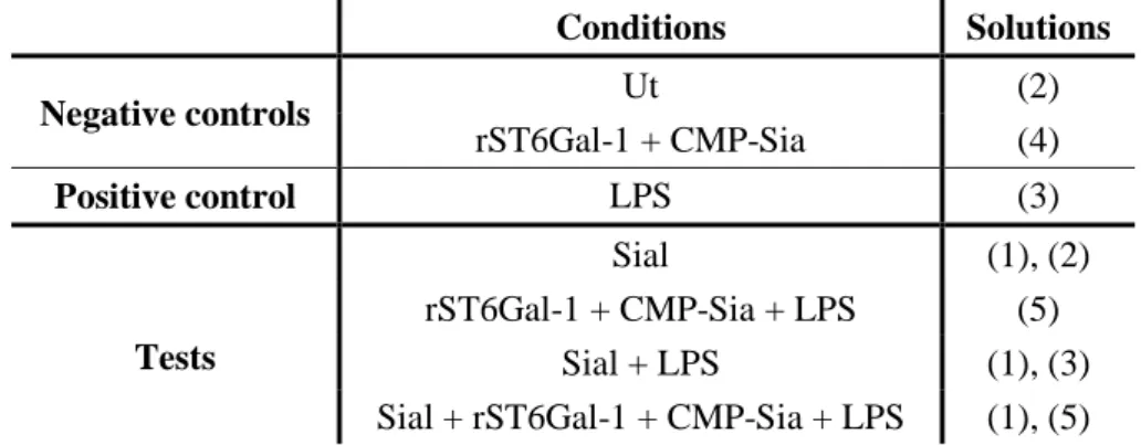
Table II.1- Representation of the conditions to test in BMDCs’ activation assays and respective solutions prepared. It is shown on the left column the conditions performed and the respective added solutions on the right column. Ut refers to Untreated, rST6Gal-1 to recombinant murine ST6Gal-1, CMP-Sia to cytidine-5’- monophosphate-N-acyl-neuraminic acid and Sial refers to sialidase.
Conditions Solutions Negative controls Ut (2) rST6Gal-1 + CMP-Sia (4) Positive control LPS (3) Tests Sial (1), (2) rST6Gal-1 + CMP-Sia + LPS (5) Sial + LPS (1), (3)
25 As mentioned in Table II.1, LPS condition was the positive control for BMDCs’ activation, whereas Ut and rST6Gal-1 + CMP-Sia conditions were the negative controls. The main goals to achieve with the performed tests will be further detailed in Chapter III, in section III.2. The cells were incubated in the respective conditions for 6 hours at 37 ºC, 5% CO2, whereas their supernatants were collected for ELISA after 6 or 24 hours. The
cells were also collected for Flow Cytometry analysis after 6 or 24 hours.
In some experiments, rST6Gal-1 + CMP-Sia and LPS were added to WT BMDCs in sequential steps, in order to test the influence of each component. In this case, incubation with rST6Gal-1 + CMP-Sia was performed during 4 hours, at 37 ºC, 5% CO2
followed by addition of RPMI complete medium without FBS with or without LPS. In this case, the supernatants were collected after 6 and 24 hours of BMDCs’ incubation at 37 ºC, 5% CO2 (for ELISA), whereas the cells were collected for Flow Cytometry analysis
after 24 hours.
In some experiments, BMDCs treated with Sialidase or ST6GalI were collected after 1 hour of the respective treatments, for Flow Cytometry assays with lectins.
Modifications to the protocol were applied to test different hypothesis, further detailed in the following sections.
II.2.1 Assays to test the influence of extrinsic α2,6 sialylation during bone-marrow cells’ differentiation into DCs
The influence of extrinsic α2,6 sialylation during WT bone-marrow cells’ differentiation into DCs was assessed. In this case, half of BMDCs were generated as described in II.1.1., whereas the other BMDCs were generated with RPMI complete medium supplemented with cytokines, but with 10 µl/ml of rST6Gal-1 + 100 µM of CMP-Sia, added every two days. After 8 days of differentiation, BMDCs were treated with RPMI complete medium without FBS with or without LPS (solutions 2 and 3, previously mentioned) and incubated for 6 hours, at 37 ºC, 5% CO2. Their supernatants
were collected after this time, similar to the cells, for ELISA and Flow Cytometry assays, respectively.