Renato Manuel Pereira
Alves
Efeitos do envelhecimento no proteoma mitocondrial
do músculo esquelético
Aging effects on the skeletal muscle mitochondrial
proteome
Renato Manuel Pereira
Alves
Efeitos do envelhecimento no proteoma mitocondrial
do músculo esquelético
Aging effects on the skeletal muscle mitochondrial
proteome
Dissertação apresentada à Universidade de Aveiro para cumprimento dos requisitos necessários à obtenção do grau de Mestre em Métodos
Biomoleculares, realizada sob a orientação científica do Doutor Francisco Manuel Lemos Amado, Professor Auxiliar do Departamento de Química da Universidade de Aveiro, e da Doutora Rita Maria Pinho Ferreira, Professora Coordenadora Convidada do Instituto Politécnico de Saúde - Norte
Apoio financeiro da FCT
(PTDC/DES/70757/2006) no âmbito do III Quadro Comunitário de Apoio.
o júri
presidente Prof. Dr. Artur Manuel Soares da Silva
professor catedrático do Departamento de Química da Universidade de Aveiro
Prof. Dr. José Alberto Ramos Duarte
professor catedrático da Faculdade de Desporto da Universidade do Porto
Prof. Dr. Francisco Manuel Lemos Amado
professor auxiliar do Departamento de Química da Universidade de Aveiro
Prof.ª Dr.ª Rita Maria Pinho Ferreira
agradecimentos Quero agradecer a todas as pessoas que de alguma forma contribuíram para realização deste trabalho.
Aos meus orientadores, Doutor Francisco Amado e Doutora Rita Ferreira, obrigado por me terem acolhido e por se dedicarem à difícil tarefa de pôr um “biólogo” a pensar nas “moléculas”.
Ao Doutor Rui Vitorino, obrigado pelos valiosos ensinamentos de espectrometria de massa. Sei que não foi fácil, mas agora sinto que valeu o esforço de ambos para que eu aprendesse um pouco que fosse.
À Sofia, obrigado por estares disponível para aquelas pequenas explicações que fazem a diferença.
A todos os elementos do grupo de espectrometria de massa, um “muito obrigado” por me terem acolhido e pelos bons momentos que passámos. Sei que a amizade não se vai esgotar por aqui e vamos continuar em contacto.
A todos os meus amigos que me apoiaram durante este período em que nem todos os momentos foram fáceis.
À minha família por terem permitido que chegasse a este ponto.
A todos, um sincero e sentido,
palavras-chave Mitocôndria, proteómica, stress oxidativo, Maldi-Tof/Tof, cadeia transportadora de electrões
resumo Este trabalho teve como objectivo determinar de que forma o envelhecimento afecta o proteoma mitocondrial do músculo-esquelético. Para tal, foram analisadas (i) alterações na composição (ii) variações da actividade dos complexos da cadeia respiratória, (iii) a oxidação de proteínas mitocondriais e (iv) os locais específicos de oxidação dos Complexo I da Cadeia de Transporte de Electrões (ETC). Simultaneamente, verificou-se de que forma o estilo de vida sedentário vs. não-sedentário pode condicionar cada um dos parâmetros observados. Para atingir estes objectivos ratinhos C57BL/6 machos foram sujeitos a um protocolo para simular o envelhecimento sedentário e não-sedentário. Os animais foram depois sacrificados e isoladas as mitocôndrias dos músculos dos membros traseiros. As proteínas mitocondriais foram depois separadas por 2D-PAGE e identificadas por espectrometria de massa. Após a comparação dos perfis de 2D-PAGE pode-se observar que a maioria das proteínas analisadas se encontra sobre-representadas no proteoma mitocondrial de ratinhos sedentários. Para avaliar se estas diferenças se correlacionavam com eventuais alterações na actividade dos complexos da cadeira respiratória, estes foram separados por BN-PAGE e corados com soluções específicas para determinar a sua actividade. Verificou-se uma diminuição na actividade dos complexos IV e V da cadeia respiratória que parece indicar que o aumento da quantidade de proteínas dos complexos da ETC no proteoma mitocondrial surge como um mecanismo de compensação para suprimir a diminuição da actividade da cadeia respiratória. Como parâmetro de oxidação proteica foi determinado o conteúdo em grupos carbonilos das amostras por slot blot. Verificou-se que há uma diminuição significativa do conteúdo em grupos carbonilos nas proteínas dos ratinhos que foram sujeitos a um estilo de vida não-sedentário. Para determinar os locais de oxidação do Complexo I da ETC, as sete subunidades deste complexo, identificadas previamente, foram sujeitas a espectrometria de massa tandem. Foram detectadas modificações em vários péptidos, sendo os resíduos de triptofano mais afectados pela oxidação. Contrariamente ao esperado, as subunidades membranares apresentaram-se mais modificadas que as subunidades mais próximas do centro catalítico. Os resultados permitem sugerir que as subunidades membranares são mais tolerantes aos efeitos da oxidação e que as subunidades do Complexo I podem ser substituídas individualmente quando são danificadas pelos efeitos da oxidação. Globalmente podemos concluir que a realização de actividade física moderada previne os efeitos do envelhecimento, nomeadamente a nível de danos oxidativos nas proteínas mitocondriais.
keywords Mitochondria, proteomics, oxidative stress, Maldi-Tof/Tof, electron transport chain
abstract This work aimed to assess how aging influences the skeletal muscle mitochondrial proteome. To achieve this, we have analyzed (i) alterations on its composition, (ii) variations on the activity of the respiratory chain complexes, (iii) the oxidation of mitochondrial proteins and (iv) the specific sites for oxidative modification on Complex I from the Electron Transport Chain (ETC). Simultaneously, we assessed how these parameters may be conditioned by a sedentary vs. a non-sedentary lifestyle. C57BL/6 male mice were subjected to a protocol to simulate sedentary and non-sedentary aging. The animals were then sacrificed and mitochondria from the hind limbs muscles were isolated. Mitochondrial proteins were resolved by 2D-PAGE and identified by mass spectrometry. Following a comparison of the 2DE profiles, we have observed that most of the analyzed proteins were up-regulated in sedentary mice mitochondrial proteome. To assess if these alterations were related to any functional variations, the respiratory chain complexes were resolved by BN-PAGE and differentially stained to determine the in-gel activity. We observed a reduction of the activity of Complexes IV and V in sedentary mice, which may suggest that the up-regulation of the respiratory chain complexes in sedentary mice may be a mechanism to overcome the loss of functionality. To assess the level of oxidative stress, we have determined the carbonyl content by slot blot and observed a significant decrease in the carbonyl content of non-sedentary mice. To locate the precise sites for oxidative damage in Complex I of the ETC, the seven subunits of this complex, previously isolated by 2D-PAGE, were subjected to tandem mass spectrometry. We have detected several modified peptides, with tryptophan being the most affected residue. Unexpectedly the membrane subunits presented more modification sites than the peripheral arm subunits. These results may suggest that membrane subunits are more tolerant to oxidative damage and Complex I subunits may be replaced by newly synthesized ones when oxidative damage becomes unbearable. In conclusion, we may assume that moderate physical activity attenuates the effects of aging, namely the oxidative damage of mitochondrial proteins.
TABLE OF CONTENTS
I. GENERAL INTRODUCTION ... 3
1. The aging process ... 3
1.1. The Reactive Species Theory of Aging ... 5
1.2. The mitochondrion and aging ... 6
2. The Electron Transport Chain ... 10
2.1. Composition of the Electron Transport Chain ... 10
2.2. The electron flow along the chain ... 12
2.3. Production of reactive species ... 14
2.4. Neutralization of reactive species - The antioxidant system .... 15
3. Protein oxidation in mitochondria ... 17
4. Skeletal muscle aging is influenced by physical activity ... 20
5. Methodological approaches on the study of mitochondrial aging ... 21
6. Aims of this thesis ... 24
II. SEDENTARY LIFESTYLE MODULATES THE AGING EFFECT ON MICE SKELETAL MUSCLE MITOCHONDRIAL PROTEOME ... 27
1. Introduction ... 27
2. Material and methods ... 28
2.1. Animals and Experimental Protocol ... 28
2.2. Skeletal Muscle Mitochondria isolation ... 29
2.3. Identification of the proteins present in the mitochondrial proteome ... 29
2.4. Analysis of the respiratory chain complexes by BN-PAGE and in-gel activity staining ... 30
2.5. Protein identification by Maldi-Tof/Tof mass spectrometry ... 31
2.6. Analysis of carbonylation in mitochondrial proteins. ... 31
3.1. 2D-PAGE and Maldi-Tof/Tof identification of proteins from
isolated mitochondria ... 32
3.2. Assessment of the carbonyl content ... 40
3.3. In-gel activity of respiratory chain complexes ... 42
4. Discussion ... 42
III. OXIDATIVE MODIFICATIONS ON COMPLEX I OF THE ELECTRON TRANSPORT CHAIN ... 49
1. Introduction ... 49
2. Material and Methods ... 50
2.1. Animals and Experimental Protocol... 50
2.2. Skeletal Muscle Mitochondria isolation ... 51
2.3. Two-dimensional polyacrylamide gel electrophoresis ... 51
2.4. Sample preparation for analysis by mass spectrometry ... 52
2.5. Protein identification by Maldi-Tof/Tof mass spectrometry ... 52
2.6. Identification and confirmation of oxidative modifications by Maldi-Tof/Tof mass spectrometry ... 53
3. Results ... 53
4. Discussion ... 66
IV. GENERAL DISCUSSION AND CONCLUSIONS ... 75
V. REFERENCES ... 79
VI. APPENDIX ... 93
1. Chemicals ... 93
2. Equipment ... 94
3. Reagents and solutions ... 95
3.1. 2D-Electrophoresis ... 95
3.2. BN-PAGE ... 96
3.3. Immunoblotting ... 97
3.4. Histochemical staining for in-gel activity ... 98
3.6. Maldi matrix ... 99
4. Procedures ... 100
4.1. 2D-PAGE ... 100
4.2. Staining with colloidal coomassie ... 101
4.3. In-gel tryptic digestion and acid extraction ... 101
4.4. BN-PAGE ... 102
4.5. Histochemical staining to determine the in-gel activity of the respiratory chain complexes ... 102
I. GENERAL INTRODUCTION
1. The aging process
“Aging is the progressive accumulation of changes with time that are associated with or responsible for the ever-increasing susceptibility to disease and death which accompanies advancing age” (Harman 1981). Aging refers to a multidimensional process of physical, psychological, and social change. During aging, organisms go through a series of alterations on every level, from molecules to the whole organism. The functional pathways involved in aging process may include responses to endogenous and exogenous changes, such like hormonal changes and damage accumulation; also, aging and longevity may be influenced by genes (Finch 1993; Finch and Tanzi 1997).
Biological, epidemiologic and demographic data have generated a number of theories that attempt to identify a cause or process to explain aging. These theories can be divided into four major categories, according to the biological level to which they apply, as evolutionary, molecular, cellular and systemic theories (Weinert and Timiras 2003). Some of the principal theories are listed in Table I.1 with a brief description.
Evolutionary theories argue that aging results from a decline in the force of natural selection. Longevity is a trait to be selected only if it is beneficial for fitness, as the natural selection acts primarily to maximize reproductive yield in an individual. The Mutation Accumulation Theory, for example, argues that some hereditary age-related diseases have not been selected against because the individuals bearing those genes reproduce long before the manifestation of the disease, thus being able to transmit their genetic information before natural selection could act (Medawar 1952).
Molecular theories argue that either altered gene expression on development genes or errors in the DNA replication, transcription or RNA translation accumulate and eventually lead to cell dead. While there is a basal rate or error in these events, if the cell looses the ability to repair errors, aberrant molecules accumulate and may induce cell death (Kanungo 1975; Dice 1993).
Cellular theories argue that is the cellular senescence the cause of aging. In this category falls: the Cellular Senescence-Telomere Theory, which
states that the increase in frequency of senescent cells, which may result from the telomere loss in chromosomes or cell stress, leads to aging of the organisms; the Hayflick Limit Theory established by Leonard Hayflick, which argues that cell are limited to a certain number of divisions and then die; and the Free Radical Theory, which will be described in detail later in this chapter (Dice 1993; Weinert and Timiras 2003).
The last category of theories is System theories, which argue that failures in physiological systems are the cause of aging. The Neuroendocrine Theory postulates that the neuroendocrine system eventually looses the ability to control the hormone production, and in fact the secretion of hormones and their effectiveness decline in advanced age. On the other hand, the Immunologic Theory argues that is the decline of the immune system that leads to aging and death (Weinert and Timiras 2003).
These categories, however, are often overlapping and the division is not always noticeable: age-related alterations on a molecular level, eventually lead to cellular alterations, which, in turn, lead to systemic failure with a strong reproductive and survival implications in evolution (Weinert and Timiras 2003). Despite all these theories, the search of a single cause for aging has been replaced by the view of aging as an extremely complex and multifactorial process (Kowald and Kirkwood 1996). A more comprehensive and realistic understanding of the aging process allow the division of aging theories in two major groups, genetic theories and stochastic theories (Weinert and Timiras 2003; Mota, Figueiredo et al. 2004). However, the division into two major categories is too general and less informative about the effects and their location.
Table I.1 – Classification and brief description of the main theories of aging (adapted from Weinert and Timiras 2003)
Biological level/Theory Brief description
Evolutionary
Mutation accumulation Mutations that affect health at older ages are not selected against.
Disposable soma Somatic cells are maintained only to ensure continued reproductive success; after reproduction, soma becomes disposable.
Antagonistic pleiotropy Genes beneficial at younger age become deleterious at older ages. Molecular
Gene regulation Aging is caused by changes in the expression of genes
regulating both development and aging.
Codon restriction Fidelity/accuracy of mRNA translation is impaired due to inability to decode codons in mRNA. Error catastrophe Decline in fidelity of gene expression with aging results in increased fraction of abnormal proteins. Somatic mutation Molecular damage accumulates, primarily to DNA/genetic material.
Dysdifferentiation Gradual accumulation of random molecular damage impairs
regulation of gene expression. Cellular
Cellular
Senescence-Telomere Theory
Phenotypes of aging are caused by an increase in frequency of senescent cells. Senescence may result from telomere loss (replicative senescence) or cell stress (cellular senescence).
Free radical and reactive species
Oxidative metabolism produces highly reactive free radicals that subsequently damage lipids, protein and DNA.
Wear-and-tear Accumulation of normal injury.
Apoptosis Programmed cell death from genetic events or genome crisis.
System
Neuroendocrine Alterations in neuroendocrine control of homeostasis results in aging-related physiological changes. Immunologic Decline of immune function with aging results in decreased incidence of infectious diseases but increased incidence of
autoimmunity.
Rate-of-living Assumes a fixed amount of metabolic potential for every living organism (live fast, die young).
1.1. The Reactive Species Theory of Aging
One of the most popular explanations for how aging occurs at the biochemical level was firstly proposed by Denham Harman in what he called “the free radical theory of aging” in 1956 (Harman 1956). This theory, however should be called “the reactive species theory of aging” as, it does not only explain aging with the production of free radicals, but also with other
non-radical reactive species, namely reactive oxygen species (ROS) and reactive nitrogen species (RNS). Apart from the naming issues, it is still one of the best known theories of aging, although controversial. All organisms live in an environment that contains ROS; mitochondrial respiration, the major source of energy for all eukaryotes, generates ROS by leaking electrons from the electron transport chain (ETC). The production of the superoxide radical on the ETC is counterbalanced by the action of superoxide dismutase, an enzyme that is found in all aerobic organisms and scavenges superoxide anions exclusively (Finkel and Holbrook 2000). The free radical theory of aging supposes that free radical reactivity is inherent in biology and results in cumulative damage to all the cellular components and ultimately to cellular senescence. In fact, the level of oxidatively damaged DNA and proteins is elevated in aged organisms (Stadtman, Starke-Reed et al. 1992; Hamilton, Van Remmen et al. 2001; Wanagat, Cao et al. 2001).
Over the past decade, the Free Radical Theory of Aging has gained wide acceptance because numerous studies have shown a strong correlation between increasing age and the accumulation of oxidative damage to cellular macromolecules (Warner 1994; Sohal and Weindruch 1996; Hamilton, Van Remmen et al. 2001; Wanagat, Cao et al. 2001).
1.2. The mitochondrion and aging
Mitochondria are recognized as an essential member of the eukaryotic cell cytoplasm, responsible for generation of most of the “energy currency” for the cell, ATP (Yaffe 1999; Koopman, Verkaart et al. 2005; Frazier, Kiu et al. 2006; Logan 2006). Apart from this, mitochondria also harbour essential parts of the urea cycle and are crucial for the breakdown of fatty acids, the generation of heat, and the biosynthesis of heme, pyrimidines, amino acids, phospholipids, and nucleotides (Koopman, Verkaart et al. 2005; Frazier, Kiu et al. 2006).
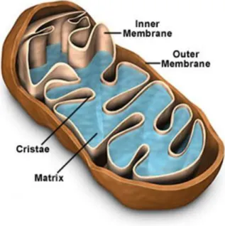
One important aspect of mitochondria is its dynamic morphology (Knowles, Guenza et al. 2002). The earliest electron microscopic observations of mitochondria defined their basic morphology as double-membrane organelles, typically rod-shaped, and about 1 micrometer in length (Scheffler
2001). The inner membrane is folded into "cristae" that project into the region inside the inner membrane called the "matrix." The area in between the inner and outer membrane is termed the "intermembrane space" (Figure I.1).
Figure I.1 - Schematic representation of a mitochondrion showing its internal configuration. (Adapted from http://www.cartage.org.lb)
Mitochondria display an amazing plasticity of form and distribution (Yaffe 1999; Collins, Berridge et al. 2002), which, along with its number, varies significantly between tissues and cell types and are further influenced by metabolic conditions and developmental stage (Rojo, Legros et al. 2002). The number of mitochondria in a cell is determined by the cell's specific function and energy needs. Cells such as muscle cells have many mitochondria while red blood cells have none. The distribution of mitochondria must strategically meet the cellular needs and signals from outside (Dimmer and Scorrano 2006). The mitochondrial plasticity may account for the discrepant observations of heterogeneity/homogeneity in mitochondrial form and function from different studies (Collins, Berridge et al. 2002). Furthermore, it emphasizes the conclusion that there is probably no single universal mitochondrial morphology and functional status and, even within an individual cell, we can have distinct populations with specific morphologies (Collins, Berridge et al. 2002).
The mammalian mitochondrion contains 4 to 5 copies of its own circular DNA molecule (mtDNA). This small molecule of 16 kb encodes for 37 genes, from which 13 are proteins, all belonging to the respiratory chain (Table I.2), 2 are rRNA (16S and 12S) and the remainder are mitochondrial tRNAs (Cogswell, Stevens et al. 1993; Taanman 1999; Wallace, Brown et al. 1999; Freyssenet, Irrcher et al. 2004; Hood, Irrcher et al. 2006; Cannino, Di Liegro et al. 2007). Although traditionally considered “naked” due to the lack of histones, a number of proteins interact with the mtDNA (Garesse and Vallejo 2001). The mtDNA has a significant contribute for mitochondrial function and integrity (Joseph, Rungi et al. 2004; Zeviani and Di Donato 2004). Defects in the synthesis of one of the 13 mtDNA- or nDNA-encoded respiratory subunits can lead to respiratory chain dysfunction and a wide range of pathogenic conditions, some of which affect skeletal muscle (Joseph, Rungi et al. 2004; Zeviani and Di Donato 2004).
Table I.1 – List of proteins encoded in the mitochondrial genome of Mus musculus obtained from the NCBI Genome database (http://www.ncbi.nlm.nih.gov/sites/entrez), accession number NC_005089, accessed on May 18th, 2008
Product Name Length Gi GeneID Locus
NADH dehydrogenase subunit 1 318 34538598 17716 ND1
NADH dehydrogenase subunit 2 345 34538599 17717 ND2
cytochrome c oxidase subunit I 514 34538600 17708 COX1
cytochrome c oxidase subunit II 227 34538601 17709 COX2
ATP synthase F0 subunit 8 67 34538602 17706 ATP8
ATP synthase F0 subunit 6 226 34538603 17705 ATP6
cytochrome c oxidase subunit III 261 34538604 17710 COX3
NADH dehydrogenase subunit 3 115 34538605 17718 ND3
NADH dehydrogenase subunit 4L 98 34538606 17720 ND4L
NADH dehydrogenase subunit 4 459 34538607 17719 ND4
NADH dehydrogenase subunit 5 607 34538608 17721 ND5
NADH dehydrogenase subunit 6 172 34538609 17722 ND6
Even though mitochondria contain their own genome and protein synthesizing machinery they are only semi-autonomous (Freyssenet, Irrcher et al. 2004; Logan 2006). Indeed, proteome analysis points to the existence of about 800 (yeast) to 1500 (human) different proteins in mitochondria (Wiedemann, Frazier et al. 2004), although more conservative estimations point out to 1000 mitochondrial proteins (Scheffler 1999). This indicates that the mitochondrial genome specifies only a few mitochondrial proteins (approximately 1%) (Poyton and McEwen 1996; Wiedemann, Frazier et al. 2004). So, the majority of mitochondrial proteins are encoded in the nuclear genome, synthesized in the cytosol and imported into the mitochondria post-transcriptionally (Butow and Avadhani 2004; Freyssenet, Irrcher et al. 2004; Logan 2006). Nonetheless, some of these nuclear-encoded mitochondrial proteins are co-assembled by RNA molecules encoded by the mitochondrial genome (Poyton and McEwen 1996; Wiedemann, Frazier et al. 2004).
The delayed onset and progressive course of mitochondrial diseases suggest that mitochondrial function may decline with age. This hypothesis is supported by multiple reports of age-related declines in mitochondrial gene expression and oxidative capacity (Wallace 1999; Barazzoni, Zanetti et al. 2005). Being one of the most mitochondria enriched tissues, skeletal muscle is no exception. For example, structural and functional changes in muscle during aging occur in a wide range of species, from C. elegans to humans (Nair 2005). Impaired mitochondrial enzyme activity and reduced mitochondrial protein synthesis rate has been linked to these structural changes in aging skeletal muscle (Yarovaya, Kramarova et al. 2002; Barazzoni, Zanetti et al. 2005), but the molecular level at which these alterations occur is not completely elucidated. A reduced mtDNA copy number may contribute to reduced mRNA abundance, which results in reduced mitochondrial protein synthesis and enzyme activity (Yarovaya, Kramarova et al. 2002; Barazzoni, Zanetti et al. 2005; Nair 2005). Muscle fibre composition and oxidative metabolism have also been shown to influence skeletal muscle mitochondrial gene expression in adult animal models (Williams, Salmons et al. 1986). The
overall effect is a reduced capacity for oxidative phosphorylation. The decreased availability of ATP may contribute to an overall reduction in the remodelling process that involves the synthesis and breakdown of proteins, both of which are energy consuming reactions in muscle (Nair 2005). However, this analysis is even complicated by the fact that skeletal muscle groups are highly heterogeneous with respect to oxidative metabolism and function (Barazzoni, Short et al. 2000).
2. The Electron Transport Chain
The oxidative phosphorylation (OXOPHOS) is possible due to an electrochemical gradient between the matrix and the intermembrane space. Within the inner membrane, a series of membrane-associated electron carriers constitute the respiratory chain, and are organized through a growing redox potential (Murray, Taylor et al. 2003). The electron transport chain (ETC) is a group of four protein complexes (Complexes I-IV) and two mobile carriers, ubiquinone and cytochrome c. Together with a fifth complex, the ATP synthase (Complex V), they constitute the respiratory chain which is responsible for the production of energy to the cell in the form of ATP (Figure I.2).
This set of complexes is located in the inner membrane of mitochondria and is organized by their redox potential, which allows the flow of electrons from the primary donor to the final acceptor through a series of redox reactions (Liu, Fiskum et al. 2002).
2.1. Composition of the Electron Transport Chain
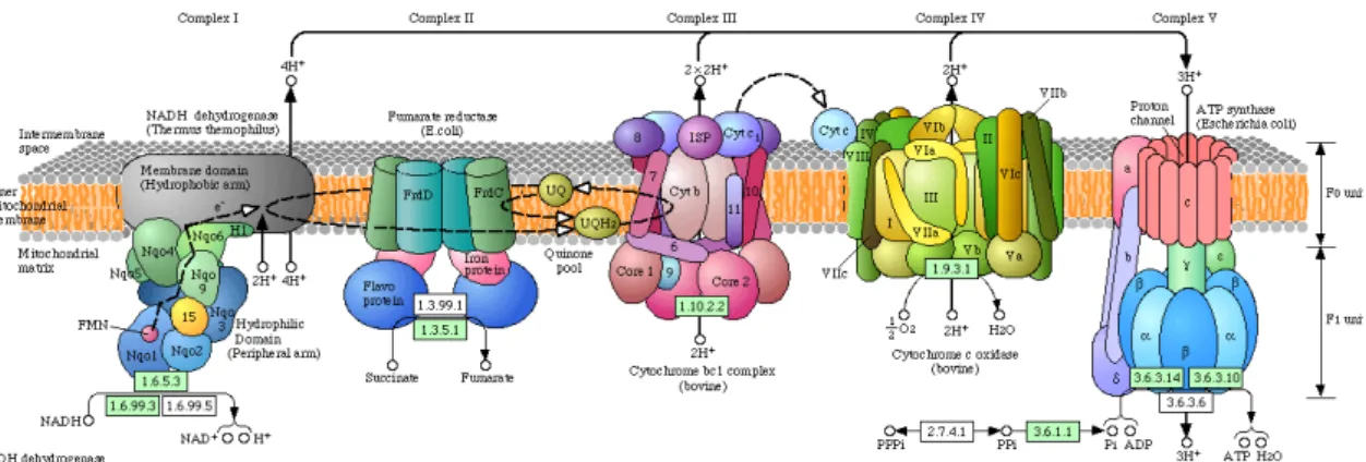
The four complexes that constitute the ETC are multisubunit protein complexes associated with prosthetic groups that allow the electrons to flow (Figure I.2).
Figure I.2 - Schematic representation of the respiratory chain complexes published in Kyoto Encyclopaedia of Genes and Genomes (KEGG) Pathway website (http://www.genome.jp/dbget-bin/get_pathway?org_name=mmu&mapno=00190). Due to the lack of complete tridimensional structures for each complex of Mus musculus, representations are based on the respective complexes of other organisms: Complex I is based on Termus
temophilus, Complex II and V on Escherichia coli and Complexes III and IV on bovine.
Complex I (C-I) is a NADH:ubiquinone oxidoreductase that catalyses the reduction of NADH to NAD+, transferring two electrons to ubiquinone. It has an
L-shaped form, with the long arm as a hydrophobic integral membrane protein and the short arm protruding to the matrix, which contains the FMN-containing flavoprotein and the iron-sulphur clusters (Navarro and Boveris 2007).
Complex II (C-II) is a succinate:ubiquinone oxidoreductase that reduces succinate to fumarate, transferring two electrons to ubiquinone. This complex is simultaneously part of the ETC and TCA cycle. It is an oligommeric complex and each monomer is composed of 4 subunits namely, a flavoprotein, with a FAD molecule covalently bound, an iron-sulphur protein and two hydrophobic smaller peptides that serve as membrane anchors. Complex II is the only complex in the respiratory chain that does not span the inner membrane and whose genes are only nuclear encoded (Cecchini 2003).
The mobile carrier between C-I/II and C-III is a lipid soluble benzoquinone with a long isoprenoid tail that may exist in three oxidation states: ubiquinone – fully oxidized, semiquinone – semi-oxidized, or ubiquinol – fully reduced. The most common form in mammals has 10 isoprene units and is referred as Coenzyme Q10 (Navarro and Boveris 2007).
Complex III (C-III) is an ubiquinol:cytochrome c oxidoreductase, hence it is responsible for the transfer of electrons to cytochrome c from ubiquinol. This complex has 9-10 polypeptides, three of which are associated with the redox centres b562, b566 and c1 hemes and an iron-sulphur cluster (Hatefi
1985). The membrane spanning region of each C-III monomer consists of 13 transmembrane helices, eight of which belong to cytochrome b (Xia, Yu et al. 1997).
Cytochrome c (cyt c) is a small peptide bound to a heme c group that is loosely associated to the mitochondrial inner membrane, facing the intermembrane space. It is reduced by the electrons transferred from C-III, transporting them to C-IV, where it is oxidized (Navarro and Boveris 2007).
Complex IV (C-IV) or cyt c oxidase (COX) is the final catalyst of the ETC. C-IV reduces O2 to H2O with four electrons from the reduced cytochrome
c, consuming four protons from the matrix (Navarro and Boveris 2007). The redox centres of C-IV are two heme a centres, heme a and heme a3, located in
two different environments, each one associated with a copper atom, respectively, Cua and Cua3 (Hatefi 1985).
2.2. The electron flow along the chain
The primary electron donors can be either NADH or succinate, creating two different pathways. When using NADH as the electron donor, the chain starts in Complex I; there, a pair of electrons is transferred to the flavine mononucleotide (FMN), leaving the NADH reduced to NAD+. The electrons then
flow through a series of eight iron-sulphur clusters reaching ubiquinone that is reduced to ubiquinol and heads out to Complex III. Contrastingly, when the electron donor is succinate, the chain starts in Complex II. Through this pathway, the succinate produced in Krebs cycle is reduced to fumarate by transferring two electrons to a flavine-adenine dinucleotide (FAD) in Complex II. Similarly to the NADH-pathway, the electrons flow from the FAD to ubiquinone through three iron-sulphur clusters. From this point on, both pathways follow the same steps. Complex III is responsible for the transfer of a pair of electrons from ubiquinol to two molecules cytochrome c. This latter will, in turn, transfer the electrons to Complex IV where the final reduction of
the chain occurs, and one molecule of O2 is reduced to two molecules of water
(Hatefi 1985).
One important feature that results from the flow of electrons along the chain is the transfer of protons from the matrix to the intracellular space. This transfer occurs against the electrochemical potential and is only possible because it is coupled with the redox reactions of the electron transport; otherwise it would require ATP to be achieved. The accumulation of protons on the intermembrane space creates an electrochemical potential against the matrix. This electrochemical potential is extremely important as protons return to the matrix through a specialized complex – the ATP synthase – which uses the energy released by the proton flow, back to the matrix, to phosphorylate ADP and thus produce ATP (Hatefi 1985).
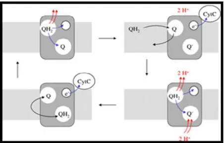
During the flow of electrons, one may consider an internal cycle that occurs in Complex III, which is called the ubiquinone cycle and was proposed by Mitchell (1976). Complex III has two binding sites for ubiquinone and during the cycle both binding sites are occupied. When ubiquinol is bound to the first site, and an oxidized ubiquinone is bound to the second site, the protons of ubiquinol are stripped off and expelled at the intermembrane space. One of its electrons is passed on first to a heme within complex III and then to cytochrome c; the other electron is passed on to the second molecule of ubiquinone, which is reduced to semiquinone. The ubiquinol molecule is now oxidized to ubiquinone and is replaced by a new molecule of ubiquinol that has been reduced in the preceding steps of the ETC. The protons and electrons of the new ubiquinol molecule are abstracted and split as for the first molecule and we now have a fully reduced ubiquinol in the second site. The molecules then switch places and thereby complete the cycle (Figure I.3) (Mitchell 1976). However, the electron flow through the ETC is not error proof.
Figure I.3 – Schematic representation of the ubiquinone cycle. Complex III has two binding sites for ubiquinone. Oxidation of one molecule of ubiquinone partially reduces the second one; oxidation of a new molecule of ubiquinone completes reduction of the second one and causes uptake of two protons from the cytosol. Protons released by oxidation are at all times released at
the cytosolic side. (obtained from http://watcut.uwaterloo.ca/webnotes/Metabolism/page-5.2.4.html)
2.3. Production of reactive species
During the electron flow along the chain, it may take place some electron leakage. By leaking from the normal flow, the electrons immediately react with O2 in the matrix to produce superoxide radical (O2•-). Current
estimations state that up to 5% of the oxygen consumed with ATP production on the respiratory chain, can be converted into O2•- during a normal
physiological state (Joenje 1989; Leeuwenburgh and Heinecke 2001; Wanagat, Cao et al. 2001). The biological toxicity of superoxide is, namely, due to its capacity to inactivate iron-sulphur cluster containing enzymes (which are critical in a wide variety of metabolic pathways), thereby liberating free iron in the cell, which can undergo Fenton chemistry and generate the highly reactive hydroxyl radical (•OH). In its hydroperoxyl radical form (•OOH),
superoxide can also initiate lipid peroxidation of polyunsaturated fatty acids. It also reacts with carbonyl compounds and halogenated carbons to create toxic peroxy (ROO•) or alkoxy (RO•) radicals. Moreover, superoxide can also react
with nitric oxide (•NO) to form peroxynitrite (ONOO−). All these radicals and
reactive species can induce damage in surrounding molecules such as proteins, nucleic acids, and lipids in a cascade, leading to the production of
more reactive species (Joenje 1989; Leeuwenburgh and Heinecke 2001; Wanagat, Cao et al. 2001; Stadtman and Levine 2003; Brookes 2005; Stadtman 2006).
2.4. Neutralization of reactive species - The antioxidant system
To counterpart the production of the reactive species, the cell developed mechanisms to neutralize them, such as enzymes or other molecules. The major mitochondrial enzymatic antioxidants are manganese superoxide dismutase, peroxiredoxin III, thioredoxin and thioredoxin reductase (Rabilloud, Heller et al. 2001).
Nearly all organisms living in the presence of oxygen contain isoforms of the superoxide scavenging enzyme, superoxide dismutase (SOD). SOD is a protein with metallic cofactors; eukaryotic cells have at least two forms of this enzyme, one is cofactored with copper and zinc and is located on cytosol, the other is cofactored with manganese and is located on mitochondria. Mn-SOD is an extremely efficient enzyme; it catalyzes the neutralization of superoxide nearly as fast as the two can diffuse together spontaneously in solution. The neutralization of superoxide is achieved by converting it into hydrogen peroxide (Warner 1994; Yamakura, Taka et al. 1998; Leeuwenburgh and Heinecke 2001).
Two systems have been proposed to neutralize hydrogen peroxide. The glutathione system includes glutathione, glutathione reductase, glutathione peroxidases and glutathione S-transferases, and was already shown to be present in mitochondria (Arai, Imai et al. 1996). Glutathione is a tripeptide (γ-glutamyl-Cys-Gly) that reduces any disulfide bonds formed within cytoplasmic proteins to cysteines by acting as an electron donor. Glutathione reductase recycles oxidized glutathione to its reduced form, using the electrons from NADPH. Glutathione peroxidase contains four selenium-cofactors that catalyze the breakdown of hydrogen peroxide and organic hydroperoxides. In addition, the glutathione S-transferases are another class of glutathione-dependent antioxidant enzymes that show high activity with lipid peroxides. These enzymes are at particularly high levels in the liver and also serve in detoxification metabolism. However, its presence in
mitochondria seems to be limited to the intermembrane space and in small amounts (Arai, Imai et al. 1999). The other proposed system includes peroxiredoxins and its regeneration system composed by thioredoxin and thioredoxin reductase (Rabilloud, Heller et al. 2001). Peroxiredoxins are peroxidases that catalyze the reduction of hydrogen peroxide, organic hydroperoxides, as well as peroxynitrite. They are divided into three classes: typical 2-cysteine peroxiredoxins; atypical 2-cysteine peroxiredoxins; and 1-cysteine peroxiredoxins. These enzymes share the same basic catalytic mechanism, in which a redox-active cysteine (the peroxidatic cysteine) in the active site is oxidized to a sulfenic acid by the peroxide substrate. The thioredoxin system contains the 12-kDa protein thioredoxin and the enzyme thioredoxin reductase. The active site of thioredoxin consists of two neighboring cysteines, as part of a highly-conserved CxxC motif, which can cycle between an active dithiol form (reduced) and an oxidized disulfide form. In its active state, thioredoxin acts as an efficient reducing agent, scavenging reactive oxygen species and maintaining other proteins in their reduced state. After being oxidized, the active thioredoxin is regenerated by the action of thioredoxin reductase (Lee, Yu et al. 1999; Lee, Kim et al. 2003).
Catalase is commonly localized in peroxisomes of most eukaryotic cells; however it was already demonstrated to be present in mitochondria (Nohl and Hegner 1978; Nohl and Jordan 1980; Radi, Turrens et al. 1991). This enzyme efficiently catalyses the conversion of hydrogen peroxide to water and oxygen, using either an iron or manganese cofactor. Lack of catalase in mitochondria of most cells can lead to the inefficient degradation of H2O2 in mitochondria,
especially under conditions of oxidative stress where the mitochondria generate more H2O2 than under normal conditions (Bai and Cederbaum 2001).
Another major group involved in the antioxidant response includes representatives of heat shock proteins (HSP), namely HSP27, HSP60 and the family of HSP70. The expression of these proteins is induced by heat shock and several other stressors, including oxidative stress (Fehrenbach and Niess 1999; Liu, Gampert et al. 2006). The family of HSP70 is represented by four members, the constitively expressed isoform heat shock cognate (HSC70), the inducible isoform HSP70 (HSP72) and two glucose regulated isoforms GRP75
and GRP78. Up-regulation of HSP72 has been shown to provide protection from ischemia–reperfusion-induced lipid peroxidation in rat myocardium (Demirel, Powers et al. 1998). Furthermore, overexpression of mouse HSP25 and human HSP27 exerts augmenting effects on cellular levels of GSH by increasing the activity of glutathione reductase and glucose-6-phosphatase dehydrogenase (Preville, Salvemini et al. 1999).
An imbalance between oxidants and antioxidants in favour of the oxidants, potentially leading to damage, is termed 'oxidative stress' (Sies 1991; Sies 1997). As stated before, the superoxide radical is the main source of reactive species because is starts a chain of events that lead to the production of more reactive species. Its main production site is the ETC, whenever electron leakage occurs. Two sites within the ETC have been identified as being the major sites for the electron leakage to take place, and hence be the sites for superoxide radical formation. These critical sites are the area near the redox centres of Complex I and the ubiquinone binding sites in Complex III. Thus, the reduction of NADH and the ubiquinone cycle seem to be critical events in the radical formation (Liu, Fiskum et al. 2002; Choksi, Nuss et al. 2007; Choksi and Papaconstantinou 2008).
3. Protein oxidation in mitochondria
The oxidative modification of proteins is a natural consequence of aerobic life and have also been discussed as regulatory mechanisms in oxidative stress signalling (Stadtman and Levine 2003; Viappiani and Schulz 2006; Chung, Miranda et al. 2008). Protein oxidation is also recognized to play a crucial role in the etiology and pathophysiology of some diseases (Stadtman and Levine 2003; Viappiani and Schulz 2006; Chung, Miranda et al. 2008).
The oxidative modifications of a protein can range from the simple oxidation of cysteine residues to changes caused by higher levels of oxidative stress, resulting in covalent crosslinking with other proteins, the formation of noncovalent aggregates or even formation of protein adducts with other lipid, carbohydrate, or nucleic acid radicals (Viappiani and Schulz 2006; Choksi and Papaconstantinou 2008). Studies on oxidatively modified proteins have
revealed an age-related increase in the level of protein carbonylation (Levine 2002; Chung, Miranda et al. 2008), oxidized methionine (Wells-Knecht, Lyons et al. 1997), cross-linked (Squier and Bigelow 2000) and glycated proteins (Baynes 2001) as well as catalytically less active enzymes (Rothstein 1985) that are more susceptible to protein degradation (Stadtman 2001).
One of the best-known markers of age-related protein oxidation is the carbonyl group content, which has been observed to increase with age (Levine 2002). The main carbonyl products of metal-catalyzed oxidation of proteins in
vitro have been shown to be glutamic and aminoadipic semialdehydes
(Requena, Levine et al. 2003; Pamplona, Dalfo et al. 2005; Soskic, Groebe et al. 2008). Lysine, arginine, proline, and threonine residues of proteins are particularly sensitive to metal-catalyzed oxidation, leading in each case to the formation of carbonyl derivatives. Peptide carbonyl derivatives are also obtained as fragmentation products of peptide bond cleavage reactions or can be formed by the interaction of protein amino acid side chains (cysteine sulfhydryl groups, histidine imidazole groups, and lysine amino groups) with lipid peroxidation products, including 4-hydroxy-2-nonenal, acrolein, and malondialdehyde. Glycation/glycoxidation reactions can also lead directly to carbonyl adducts and indirectly to the formation of N-carboxymethyl-lysine derivatives that, because of their strong chelating ability, are able to promote the generation of carbonyl groups by metal-catalyzed reactions (Stadtman and Levine 2003; Stadtman 2006).
In a recent study by Chung et al. (2008), several carbonyl-modified proteins in interfibrillar rat heart mitochondria were identified as putative targets of oxidation modifications. These authors observed age-associated changes of carbonyl levels for aconitate hydratase and the α- and β-polypeptide chains of the F1-ATP synthase. However, these studies did not
identify the precise location in the protein structure where these oxidative modifications occur.
Much of the knowledge about free radical–mediated post-translationally modified products in mitochondria is derived from in vitro studies (Stadtman and Levine 2003; Taylor, Fahy et al. 2003). Tyrosine residues may be oxidized by hypochlorite, peroxynitrite or by radicals formed in transition metal
ion-catalyzed Fenton and Haber-Weiss reactions (e.g. hydrogen peroxide/Fe2+). The resulting tyrosyl radicals may subsequently form intra- or
intermolecular Tyr–Tyr bonds (Kowald and Kirkwood 1996; Balasubramanian and Kanwar 2002; Soskic, Groebe et al. 2008). The exposure of mitochondria or proteins to simulated oxidative stress in vitro such as peroxynitrite-mediated nitration suggested that tyrosine from proteins like manganese superoxide dismutase is a susceptible residue (Yamakura, Taka et al. 1998; Taylor, Fahy et al. 2003).
Tryptophan is another residue commonly affected by oxidative modification. The double oxidation of tryptophan residues generates N-formylkynurenine, this can be generated enzymatically and non-enzymatically (Yamakura, Taka et al. 1998; Taylor, Fahy et al. 2003; Korlimbinis and Truscott 2006; Soskic, Groebe et al. 2008). In one of the few
in vivo studies about protein oxidation, Taylor et al. (2003) have examined the
occurrence of N-formylkynurenine throughout the mitochondrial proteome of normal human heart tissue and found evidence of selective oxidation in a subset of proteins associated predominantly with redox metabolism, including ETC proteins such as six Complex I subunits (75kDa, 30kDa, 23kDa, 20kDa, 18kDa and 13 kDa-B), two Complex III subunits (subunit IV and core protein 2), Complex IV subunit IV and ATP synthase γ and δ chain subunits.
Generally, these oxidative modifications to proteins can result either in reduction of normal function or in the gain of toxic function related with aging and age-associated diseases (Stadtman and Levine 2003). The unwanted accumulation of oxidized proteins in such conditions could reflect random DNA damage to one or more of the multitude of genes that are implicated in the synthesis of proteins that govern the generation of ROS, the antioxidant defence systems, and the proteolytic activities that degrade oxidized proteins, therefore being important reliable molecular markers of aging (Chao, Ma et al. 1997; Stadtman and Levine 2003).
While important to cellular function, post-translational modifications, particularly oxidation, are often difficult to detect using analytical techniques such as gel electrophoresis and mass spectrometry. These difficulties arise
from a number of reasons including the lower relative abundance of modified protein when compared to the unmodified protein (Distler, Kerner et al. 2007).
4. Skeletal muscle aging is influenced by physical
activity
Living an active lifestyle and at least maintaining mobility is essential for quality of life during old age (Ji 2002; Paterson, Jones et al. 2007). Skeletal muscle, a primary organ for locomotion, seems to be more prone to the deleterious effect of aging due to its post-mitotic nature (Sastre, Pallardo et al. 2003; Figueiredo, Ferreira et al. 2008). This age-associated deterioration, common in elderly humans and animals, is often referred to as sarcopenia. The term sarcopenia is defined as a loss of skeletal muscle mass and functionality that occurs with advancing age (Morley, Baumgartner et al. 2001; Carmeli, Coleman et al. 2002; Chabi, Ljubicic et al. 2008). This condition results in muscle weakness predicting several adverse outcomes, including disability, institutionalization and mortality (Carmeli, Reznick et al. 2000; Carmeli, Coleman et al. 2002; Short, Bigelow et al. 2005; Marzetti and Leeuwenburgh 2006). According to Carmeli and Reznick (1994), a variety of intrinsic and extrinsic factors appears to be involved in the aging skeletal muscle. Changes in intrinsic factors associated with aging include hormones, growth factors and energetic metabolism, whereas extrinsic factors include diet, exercise, injuries, and sedentary lifestyles. At a molecular level, it has been described altered gene expression, loss of cell division potential, increased protein degradation, tissue disorganization and increased vulnerability to stress (Yarovaya, Kramarova et al. 2002; Barazzoni, Zanetti et al. 2005; Nair 2005; Figueiredo, Ferreira et al. 2008; Figueiredo, Mota et al. 2008).
Mitochondria are intimately linked to the proper function of skeletal muscle, as these organelles constitute the main energy supply in contracting muscle (Chabi, Ljubicic et al. 2008). Dysfunctional mitochondria will be unable to meet cellular ATP demands compromising the cellular adaptability to physiological stress imposed to skeletal muscle across the entire lifespan, contributing to age-related muscle dysfunction and reduced aerobic capacity
(Short, Bigelow et al. 2005; Figueiredo, Ferreira et al. 2008). Several attempts to revert this situation have been made. In this sense, several studies discussed the effect of lifestyle on sarcopenia and on skeletal muscle mitochondrial plasticity. One of the classic responses to exercise is an increase in the oxidative capacity of skeletal muscle (Menshikova, Ritov et al. 2006). However, the merits of exercise for the well-being and maintenance of muscle tissue in old age are not always clear and depend on energy requirements, oxygen consumption and mechanical load predisposed by the type of exercise (Carmeli, Coleman et al. 2002). Moderate exercise was shown to increase muscular mass and strength, which counterparts for the loss of muscle mass associated with aging (Leeuwenburgh, Fiebig et al. 1994).
Moderate voluntary exercise has already been associated with an improvement in general health, increasing lifespan and improving neuromuscular coordination (Holloszy, Smith et al. 1985; Navarro, Gomez et al. 2004). This type of exercise also increases the activity of respiratory chain. In fact, it has been demonstrated that Complex IV activity suffered an increase in several tissues of mice subject to 24 weeks of chronic exercise, while the other Complexes showed no significant differences (Navarro, Gomez et al. 2004). On the other hand, Complexes I and IV showed a decrease in activity associated with aging (Navarro, Gomez et al. 2005), which was stopped by regular moderate exercise (Navarro, Gomez et al. 2004). Indeed, chronic moderate exercise was already shown to decrease the mitochondrial content in protein carbonyls and oxidation products (Navarro, Gomez et al. 2004).
5. Methodological approaches on the study of
mitochondrial aging
The pivotal role of mitochondria in the aging process is still controversial. Although cytochrome c oxidase deficient fibres are a real finding in skeletal muscle, the contribution of mitochondrial DNA mutations to human aging is not as clear (Brierley, Johnson et al. 1997). Indeed, other studies have failed to prove changes associated with age in mitochondria (Manzelmann and Harmon 1987; Bodenteich, Mitchell et al. 1991).
These controversies can be explained by that fact that most of these studies come from genetic analysis, namely polymerase chain reaction (PCR) analysis on mtDNA and nuclear DNA encoding for mitochondrial proteins (Cortopassi and Arnheim 1992). The genomic analysis is not sufficient to fully elucidate the mechanisms of aging. Expression levels of a protein depend not only on transcription rates of the gene, but also on additional control mechanisms, such as transcript stability, translational regulation and protein degradation. Moreover, both the activity and the function of proteins can be altered, mainly through post-translational modification (glycosylation, phosphorylation) or proteolytic cleavage (Boguski and Schuler 1995; Amson, Nemani et al. 1996; Harry, Wilkins et al. 2000). All these points could contribute to the presence of largely controversial observations using genetic analysis (Rustin, von Kleist-Retzow et al. 2000; Storm, Rath et al. 2002). Thus it is imperative to analyze other levels to disclose the aging mechanisms. Proteome analysis come as an obvious approach, as most of the alterations occurring during aging are due to post-translational events as previously stated (Stadtman and Levine 2003).
High throughput two-dimensional protein electrophoresis coupled with peptide mass fingerprinting analysis by mass spectrometry (MS) have become the most powerful techniques for modern proteome analysis (Gras, Muller et al. 1999). The mitochondrial proteome has been studied in tissues like the human placent cells or heart muscle (Rabilloud, Kieffer et al. 1998; Taylor, Warnock et al. 2002) and cell lines (Fountoulakis and Schlaeger 2003). As stated above, the number of mitochondrial proteins is estimated to be around 1000-1500. One of the largest proteomic studies of purified mitochondria was presented by Sickmann, Reinders et al. (2003) on yeast mitochondria, leading to the identification of 750 mitochondrial or mitochondria-associated proteins with a coverage of up to 90% of predicted yeast mitochondrial proteome. More recently, Zhang, Li et al. (2008) identified a total of 940 distinct proteins from murine cardiac mitochondria, among which, 480 proteins were not previously identified by major proteomic profiling studies.
The sensitivity and effectiveness of proteomic analysis has only recently risen through a whole new repertoire of high-throughput technical
developments. Especially, large-gel 2D electrophoresis analysis has now reached a technical state that offers the possibility to reveal the majority of the cellular proteins with reproducible results (Klose, Nock et al. 2002). Blue-native electrophoresis has greatly contributed to the mitochondrial protein complex investigations (Schagger and von Jagow 1991), while Western immunoblotting remains to be an effective proteomic strategy. New protein analytical methods such as mass spectrometry compatible for macromolecules, computational tools, and comprehensive databases for characterization of molecular structures of proteins led to large-scale strategies in protein identification.
Maldi-TOF/TOF made large-scale proteomics possible. By coupling a Maldi source with a TOF analyzer it is possible to analyse large biomolecules quickly and efficiently without ripping them apart (Constans 2005). To study proteins we need to isolate them and then digest with an enzyme, usually trypsin, that cleaves the protein in specific residues (after any Arg or Lys for trypsine). The number and size of peptides resulting from tryptic digestion of a protein is called the peptide mass fingerprint (PMF) and is specific for each protein, helping us to identify it. The number of peptides observed in a PMF and the accuracy to which they are measured determine the confidence of the protein identification (Westermeier and Naven 2002).
There are databases with the fragment pattern for almost every known protein. Mascot is one of the best known search engines that use the data produced by mass spectrometers to identify proteins. It incorporates code from Mowse algorithm, developed by Darryl Pappin (1993) and David Perkins (1999). It can quickly return the most probable protein to be in a given sample with a given set of mass peaks on a mass spectrum. Additionally, it allows us to introduce variable modifications in the search, such as oxidations, which produce mass shifts and alter the fragmentation pattern of a given protein.
Thus, a proteomic approach seems an attractive strategy of studying complex biology problems such as aging, in order to gain additional knowledge of protein localization, protein interaction and their influence on protein structure and function.
6. Aims of this thesis
Given the importance of mitochondria in the aging process we aim to characterize the alterations induced by aging in mice skeletal muscle mitochondrial proteome.
To achieve this we have:
- Identified how aging affects the constitution of the mitochondrial proteome;
- Identified which are the proteins most susceptible to oxidative damage;
- Determined how the activity of the respiratory chain complexes is affected by aging;
- Identified the location of oxidative modifications on Complex I subunits from the ETC, and relate these with the changes in the activity. Ultimately, by comparing the group of sedentary vs. non-sedentary mice, we have assessed the implications of a sedentary lifestyle on mitochondrial functionality and protein oxidative damage.
CHAPTER II – Sedentary lifestyle
modulates the aging effect on mice
skeletal muscle mitochondrial proteome
II. SEDENTARY LIFESTYLE MODULATES THE AGING
EFFECT ON MICE SKELETAL MUSCLE
MITOCHONDRIAL PROTEOME
1. Introduction
Skeletal muscle, a primary organ for locomotion, was shown to undergo age-associated deterioration in structure and function (Ji 2002; Johnston, De Lisio et al. 2008). Several strategies have been employed to revert this scenario enhancing endogenous antioxidant levels through dietary restriction, dietary antioxidant supplementation, and use of pharmaceutical antioxidant mimetics. However, none have been shown to successfully boost antioxidant defense in skeletal muscle (Ji 2002). Nonexhaustive exercise appear as a potent and effective countermeasure for skeletal muscle aging (Navarro and Boveris 2007; Paterson, Jones et al. 2007; Gomez-Cabrera, Domenech et al. 2008; Johnston, De Lisio et al. 2008). Indeed, moderate voluntary exercise was already shown to produce an increase in the lifespan of rodents (Holloszy, Smith et al. 1985), as well as improve neuromuscular coordination and exploratory activity (Navarro, Gomez et al. 2004). It was observed that some types of exercise induced an increase in muscle mass and strength, paralleled by an increase in mitochondrial content, antioxidant enzymes activity and in the synthesis of respiratory chain components (Navarro, Gomez et al. 2004; Menshikova, Ritov et al. 2006; Navarro and Boveris 2007; Paterson, Jones et al. 2007; Gomez-Cabrera, Domenech et al. 2008).
It is not surprising that mitochondria have been proposed to play a major role in aging since it is a major source of endogenously generated ROS (Boveris, Cadenas et al. 1976; Raha and Robinson 2000). Oxidative damage to the mitochondria can lead to an amplifying effect whereby damaged mitochondria release more ROS, further increasing mitochondrial oxidative damage (Harman 1972). With time, accumulation of damaged mitochondria is proposed to lead to a decrease in the capacity to produce ATP, the principal source of energy (Levine, Mosoni et al. 1996; Chang, Van Remmen et al. 2003). Thus, impaired mitochondria will contribute to age-related dysfunction
and namely a reduced aerobic capacity in muscle (Short, Bigelow et al. 2005; Figueiredo, Ferreira et al. 2008).
While other studies have been designed to evaluate the beneficial effect of moderate exercise in aging muscle, to the best of our knowledge, no work has evaluated the mitochondrial proteome alterations induced by voluntary moderate exercise in aged mice skeletal muscle and compare them with a state of absence of any physical activity. Hence, we proposed to study the effect of lifelong voluntary physical activity on mitochondrial functionality and proteome composition. To do so, we have evaluated alterations on protein regulation with 2D-PAGE, oxidative stress by assessing the protein carbonylation and functionality by determining the in-gel activity of the respiratory chain complexes. With this experimental approach we tested the hypothesis that age-related oxidative damage of proteins leads to a decline in organelle function that may be attenuated by moderate physical activity. In overall, our results provide evidence that moderate physical activity reverts some of the deleterious effects observed in sedentary aging.
2. Material and methods
2.1. Animals and Experimental Protocol
Male C57BL/6 strain mice aged two months were randomly divided into three groups (young-Y; old sedentary-S; old active-NS). After one week of quarantine, animals from Y group (n=5) were sacrificed whereas animals from S (n=5) and NS (n=5) groups were individually placed into standard cages 355x235x190 mm (Ref. 2150E, Tecniplast, Italy) and in cages equipped with running wheels (25 cm in diameter) with 364x258x350 mm (Ref. 1284L0106, Tecniplast, Italy), respectively, until sacrifice at 25 months old. NS mice performed an average of 7 Km per day during the experimental period. Animals from both groups were housed at constant temperature (21º–24°C) on a daily light schedule of 12 h of light vs. dark until sacrifice. All animals were provided with food and water ad libitum. Housing and experimental treatment of animals were in accordance with the Guide for the Care and Use of Laboratory Animals from the Institute for Laboratory Animal Research
(ILAR, 1996). The local Ethics Committee had approved the study and the experiments were complied with the current national laws.
2.2. Skeletal Muscle Mitochondria isolation
The animals were sacrificed by cervical dislocation, and hind limb muscles were extracted for preparation of isolated mitochondria, as previously described by Tonkonogi and Sahlin (1997). Briefly, muscles were immediately excised and minced in ice-cold isolation medium containing 100mM sucrose, 0.1mM EGTA, 50mM Tris-HCl, 100mM KCl, 1mM KH2PO4, and 0.2% BSA, pH
7.4. Minced blood-free tissue was rinsed and suspended in 10ml of fresh medium containing 0.2mg/ml bacterial proteinase (Nagarse E.C.3.4.21.62, type XXVII; Sigma) and stirred for 2 min. The sample was then carefully homogenized with a tightly fitted Potter-Elvehjen homogenizer and a Teflon pestle. After homogenization, three volumes of Nagarse-free isolation medium were added to the homogenate, which was then centrifuged at 700g for 10 min. The resulting supernatant suspension was centrifuged at 10,000g during 10 min. The supernatant was decanted, and the pellet was gently resuspended in isolation medium (1.3ml/100mg initial tissue) and centrifuged at 7,000g for 3 min. The final pellet, containing the mitochondrial fraction, was gently resuspended (0.4μl/mg initial tissue) in a medium containing 225mM mannitol, 75mM sucrose, 10mM Tris, and 0.1mM EDTA, pH 7.4. All mitochondrial isolation procedures were performed at 0–4°C. Mitochondrial protein concentration was spectrophotometrically estimated with the biuret method using bovine serum albumin as standard.
2.3. Identification of the proteins present in the mitochondrial proteome
Mitochondrial proteins (200μg) were solubilized in rehydration buffer and applied on an IPG strip Immobiline DryStrip pH 3-11 NL 13cm (GE Healthcare). After isoelectric focusing, the strips were prepared for the second dimension incubating in equilibration buffer with mild-agitation for 15 minutes. The second dimension was obtained on a 15% SDS-PAGE. After separation, the gels were fixed for 60 min with a 40% methanol/10% acetic acid solution
and stained overnight with colloidal coomassie. The gels were then destained with 25% methanol solution until background colour is cleared and scanned in a Bio-Rad G710 Densitometer. Digital images were analysed with PDQuest software (Bio-Rad) for matching and quantification. Protein spots were excised and tryptic in-gel digestion was performed. In brief, the gel pieces were washed twice with 25mM ammonium bicarbonate/50% acetonitrile, followed by a wash with 100% acetonitrile. They were then dried in vacuum and 25μl of 10μg/ml trypsin in 50mM ammonium bicarbonate was added to the dried residue. The samples were incubated overnight at 37°C with sequence-grade modified porcine trypsin. The tryptic peptides were extracted from the gel with formic acid and were then dried in vacuum and resuspended in 10μl of a 50% acetonitrile/0.1% formic acid solution.
2.4. Analysis of the respiratory chain complexes by BN-PAGE and in-gel activity staining
BN–PAGE was performed based on the method of Schagger and von Jagow (1991) with minor modifications. Briefly, pellet aliquots of mitochondria (400μg protein) were solubilized for 10 min on ice in 40μl of 50mM Imidazole, 1mM EDTA (pH 7.0), 50mM NaCl, 2mM 6-aminohexanoic acid, and 12μl digitonin (6.0g/g of protein) (Sigma). Proteins were separated on a 4 to 13% acrylamide–bisacrylamide BN–PAGE gel. For separation, cathode buffer (7.5mM Imidazole (pH 7.0) and 50mM tricine) containing 0.02% (w/v) Coomassie Blue G was used until the dye front had reached approximately one-third of the gel before switching for cathode buffer lacking Coomassie Blue G. Anode buffer contained 25mM Imidazole (pH 7.0). Native complexes were separated at 200V for 5 h at 4 ºC.
For identification of proteins resolved by BN-PAGE, the bands were excised and subjected to in-gel tryptic digestion as described above. For in-gel activity and histochemical staining assays of mitochondrial complexes the protocols of Zerbetto et al. (1997) were used. In brief, in-gel NADH:NTB reductase activity for complex I was determined by incubating the native gels in Complex I assay solution (2.5mg NTB and 10mL NADH (10mg/mL) were added to 1mL 5mM Tris/HCl, pH 7.4). Complex IV-specific heme stain in native
gels was determined using 10mL cytochrome c (5mM) and 5mg diaminobenzidine (DAB) dissolved in 1mL 50mM sodium-phosphate, pH 7.2. Complex V activity was analyzed by incubating the native gels with 35mM Tris, 270 mM glycine buffer, pH 8.3, that had been supplemented with 14mM MgSO4, 0.2% (w/v) Pb(NO3)2, and 8mM ATP. Lead phosphate precipitation
that is proportional to the enzymatic ATP hydrolysis activity, was stopped by 50% (v/v) methanol (30 min), and the gels were then transferred to water.
2.5. Protein identification by Maldi-Tof/Tof mass spectrometry
Mass spectra were obtained on a matrix-assisted laser desorption/ionization–time-of-flight MALDI-TOF/TOF mass spectrometer (4700 Proteomics Analyzer, Applied Biosystems, Foster City, CA, USA) in the positive ion reflector mode. A data-dependent acquisition method was created to select the five most intense peaks in each sample spot for subsequent tandem mass spectrometry (MS/MS) data acquisition, excluding those from the matrix, due to trypsin autolysis or acrylamide peaks. Trypsin autolysis peaks were used for internal calibration of the mass spectra, allowing a routine mass accuracy of more than 25ppm. Spectra were processed and analyzed by the Global Protein Server Workstation (Applied Biosystems), which uses internal Mascot software (Matrix Science, London, UK) on searching the peptide mass fingerprints and MS/MS data. Searches were performed against the Swissprot nonredundant protein database, allowing oxidation of methionine and acrylamide adducts (propionamide) of the cysteine residues as variable modifications. Positive identifications were accepted up to 95% of confidence level.
2.6. Analysis of carbonylation in mitochondrial proteins.
For protein carbonyl derivatives assay, a given volume (V) of sample containing 20μg of protein was derivatized with 2,4-dinitrophenylhydrazine (DNPH). Briefly, the sample was mixed with 1V of 12% SDS plus 2V of 20mM DNPH/10% TFA, followed by 30 minutes incubation in dark, after which 1,5V of 2M Tris-base/18.3% of β-mercaptoethanol was added for neutralization. After dilute the derivatised proteins in TBS to obtain a final concentration of 0.001μg/μL, a 100μL volume was slot-blotted into a nitrocellulose membrane.
Carbonylation was also assessed in 2-DE oxyblot as described by Conrad et al (2001). Briefly, the isoelectric focusing was carried out as described above. Then the IPG strips were derivatized with 10mM DNPH/2N TFA. After incubating for 20 minutes in dark, the reaction was stopped with 2M Tris-base/30% glycerol for 15 min. The strips were then prepared for the second dimension as described above. After separation by molecular weight, proteins were transferred for a nitrocellulose membrane.
Both slot blot and 2D-blot membranes were blocked with 5% (w/v) dry non-fat milk in TBS-T for 3 hours and then incubated overnight at 4ºC with a solution of anti-DNP antibody (Dako) in a dilution 1:2000 in blocking solution. The membranes were washed three times (10 minutes/each) with TBS-T and incubated for 2 hours with a solution of horseradish-conjugated anti-rabbit antibody (Amersham Pharmacia) in a dilution of 1:1000. Detection was carried out with enhanced chemiluminescence (Amersham Pharmacia). Quantitative analysis of slot blot was performed with Quantity One software (Bio-Rad). Mean and standard error of the mean were calculated and one-way ANOVA, followed by Tukey-Kramer multiple comparisons test were used to assess differences between groups. The level of significance was set at 5%.
3. Results
3.1. 2D-PAGE and Maldi-Tof/Tof identification of proteins from isolated mitochondria
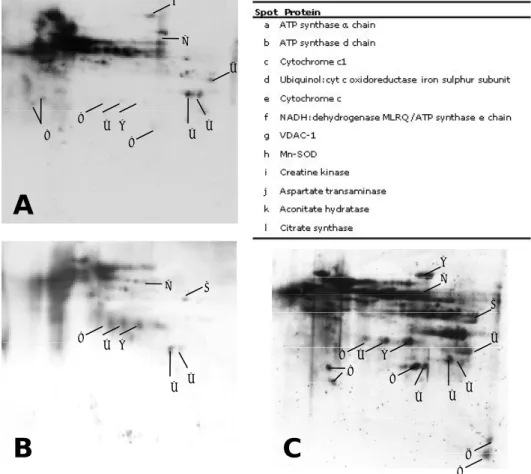
Mitochondrial proteins were resolved by 2D-PAGE (Figure II.1). Duplicates from all the experimental groups were subject to PDQuest analysis for matching the observed spots. This analysis identified an average per gel of 233 protein spots in the Y group, 205 spots in the NS group and 263 spots in the S group. A total of 496 spots were excised from the gels and, after Maldi-Tof/Tof analysis, 273 spots were positively identified containing 79 different proteins that are the most abundant in the mitochondrial proteome. The identified proteins were grouped in 4 major classes according to their physiological function: metabolic, membrane transport, stress and other proteins (Table II.1). Most of the identified proteins belong to oxidative phosphorylation system. We have found 75 spots corresponding to 33
different proteins of the respiratory chain, mainly subunits of Complex I. This figure corresponds to almost half the proteins identified, yet it is not surprising as the respiratory chain is the main pathway within mitochondria. Other well represented metabolic subclass was the TCA cyle, with 13 proteins identified in 79 distinct spots. Additionally, 8 proteins from β-oxidation and 2 proteins from the urea cycle were detected. Besides metabolic proteins, we have also identified proteins associated with membrane transport, namely ANT 1, VDAC-1, VDAC-2 and VDAC-3. Several proteins were detected in more than one spot; considering the theoretical values of molecular weight and pI, some of these spots appeared below the expected molecular weight, suggesting they are fragments. These was more evident for aconitate hydratase (34 spots), VDAC-1 (16 spots), citrate synthase, malate dehydrogenase and ATP synthase α chain (12 spots each).
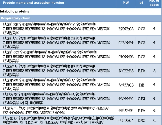
Table II.1 – List of all the identified proteins on the mitochondrial proteome, grouped in classes according to the physiological function.
Protein name and accession number MW pI No. of
spots Metabolic proteins
Respiratory chain
(Q91VD9) NADH-ubiquinone oxidoreductase 75 kDa subunit,
mitochondrial precursor (EC 1.6.5.3) (EC 1.6.99.3) (Complex I-75Kd) (CI-75Kd)
79697.53 5.51 2 (Q91YT0) NADH-ubiquinone oxidoreductase 51 kDa subunit,
mitochondrial precursor (EC 1.6.5.3) (EC 1.6.99.3) (Complex I-51KD) (CI-51KD)
50801.71 8.51 2 (Q91WD5) NADH-ubiquinone oxidoreductase 49 kDa subunit,
mitochondrial precursor (EC 1.6.5.3) (EC 1.6.99.3) (Complex I-49KD) (CI-49KD)
52591.64 6.52 2 (Q99LC3) NADH-ubiquinone oxidoreductase 42 kDa subunit,
mitochondrial precursor (EC 1.6.5.3) (EC 1.6.99.3) (Complex I-42KD) (CI-42KD)
40577.73 7.63 3 (Q9DCT2) NADH-ubiquinone oxidoreductase 30 kDa subunit,
mitochondrial precursor (EC 1.6.5.3) (EC 1.6.99.3) (Complex I-30KD) (CI-30KD)
30188.54 6.4 2 (Q8K3J1) NADH-ubiquinone oxidoreductase 23 kDa subunit,
mitochondrial precursor (EC 1.6.5.3) (EC 1.6.99.3) (Complex I-23KD) (CI-23KD) (TYKY subunit)
24022.75 5.89 1 (Q9CQJ8) NADH-ubiquinone oxidoreductase B22 subunit (EC 1.6.5.3)
(EC 1.6.99.3) (Complex I-B22) (CI-B22) 21838.68 7.83 1 (Q9D6J5) NADH-ubiquinone oxidoreductase ASHI subunit, mitochondrial