Complement C3c and C4c as predictors of death in heart failure
☆
Nuno Silva
a,b,c,⁎
, Sandra Martins
c,d, Patrícia Lourenço
a,e, Paulo Bettencourt
a,e, João Tiago Guimarães
b,c,da
Unidade I&D Cardiovascular do Porto, Faculdade de Medicina da Universidade do Porto, 4202-451 Porto, Portugal
b
Departamento de Bioquímica, Faculdade de Medicina da Universidade do Porto, 4202-451 Porto, Portugal
cServiço de Patologia Clínica, Centro Hospitalar São João, 4202-451 Porto, Portugal d
EPIUnit—Instituto de Saúde Pública da Universidade do Porto, 4050-600 Porto, Portugal
e
Serviço de Medicina Interna, Centro Hospitalar São João, 4202-451 Porto, Portugal
a b s t r a c t
a r t i c l e i n f o
Article history:
Received 20 October 2014
Received in revised form 29 December 2014 Accepted 21 February 2015
Available online 1 March 2015 Keywords:
Complement system C3c
C4c Heart failure
Background: Activation of the immune system is one of the several pathways suggested as involved in Heart Failure (HF). The complement system is a key component of innate immunity. We hypothesized that comple-ment proteins C3 and C4 can be an important predictor of death in patients with this condition.
Methods: 380 patients admitted with acute HF were recruited. They were followed up during 6 months. Serum C3c and C4c proteins were measured and groups were created and compared according to the 25th percentile cut-off value. A multivariate Cox-regression model was used to establish the prognostic value of both markers with the endpoints of HF and all-cause death.
Results: Median patients' age was 78 years and 49% of the patients were men. No major differences were observed in clinical characteristics of the groups. Patients with lower values of C3c and C4c had significantly higher values of BNP. During the 6 month period of follow up, 63 patients died, and 49 patients were due to HF. C4c showed univariate prognostic value, but not multivariate value. The multivariate-adjusted Hazard Ratios for the 6 month HF and all-cause death in patients with C3c values below 110.0 mg/dL were, respectively, 2.32 (95% CI: 1.25–4.28) and 2.52 (95% CI: 1.41–4.49).
Conclusion: Lower C3c levels are independently associated with higher risk of death. Our results reinforce the role of innate immunity in HF pathophysiology.
© 2015 The Authors. Published by Elsevier Ireland Ltd. This is an open access article under the CC BY-NC-ND license (http://creativecommons.org/licenses/by-nc-nd/4.0/).
1. Introduction
The knowledge on the pathophysiology of Heart Failure (HF) has evolved from a hemodynamic model to a more complex multisystem syndrome. Several pathways are pointed out as involved in the progres-sion of this condition. Recently, the activation of the immune system has received considerable interest[1,2]. It is becoming increasingly appar-ent that inflammatory mediators play a crucial role in the development and perpetuation of HF. In fact, the auto-perpetuation observed in HF
can be, in large part, due to an imbalance between inflammatory and
anti-inflammatory mediators, in both ischemic and nonischemic HF[3,
4]. These mediators are also known to be related to HF severity and as-sociated with prognosis[2,5]. One of the hallmarks of HF pathogenesis is the remodeling process. There is now evidence that innate immunity has a determinant role in this process. In fact, after myocardial injury
and damage of cardiac cells, innate immune system must be activated for a correct regeneration[6–8]. However, long-term chronic innate immune activation is detrimental as it leads to adverse left ventricular remodeling and HF worsening. This activation involves a variety of
inflammatory molecules and pathways, such as proinflammatory
cyto-kines, immune cells, autoantibodies formation and complement activa-tion[9,10]. The complement system is a key component of the innate immune system and comprises a cascade of more than 30 proteins. The complement system has a dual role, as a receptor, for example, for host infection and as an effector protein that can efficiently attract in-flammatory cells and also directly destroy cells by the membrane attack complex[11].
We hypothesized that in patients with an episode of acute HF, pro-teins from the complement system namely C3 and C4 can be involved in the pathophysiology of HF. We also hypothesized that besides their involvement, the magnitude of their activation can be related with HF outcome.
2. Methods
We conducted a prospective observational study between January 2009 and December 2010. During this period, patients admitted to the
☆ The authors take responsibility for all aspects of the reliability and freedom from bias of the data presented and their discussed interpretation.
⁎ Corresponding author at: Unidade I&D Cardiovascular do Porto, Faculdade de Medicina da Universidade do Porto, 4202-451 Porto, Portugal. Tel.: +351 912821468.
E-mail address:[email protected](N. Silva).
http://dx.doi.org/10.1016/j.ijcme.2015.02.001
2214-7624/© 2015 The Authors. Published by Elsevier Ireland Ltd. This is an open access article under the CC BY-NC-ND license (http://creativecommons.org/licenses/by-nc-nd/4.0/).
Contents lists available atScienceDirect
IJC Metabolic & Endocrine
Internal Medicine Department of a central Portuguese Hospital Center, with the primary diagnosis of HF, whether worsening or de novo HF, were eligible for study entry. Patients with acute coronary syndromes, with complaints attributable to causes other than HF, or with no echo-cardiographic structural or functional cardiac abnormalities were excluded.
Treatment decisions, timing of discharge, and discharge medication were at discretion of the attending physician and the physicians were aware of the ongoing registry.
An echocardiogram was performed within 72 h of admission to all eligible patients. Comprehensive echocardiographic assessment was performed using a multi-frequency matrix probe (Vivid6, GE Healthcare). The diagnosis of HF was made based on the European Society of Cardiology guidelines[12]. All HF etiologies were admitted. Patients with left ventricular systolic dysfunction (LVSD) and with HF with preserved ejection fraction were included in the registry. Normal systolic fraction was defined as a left ventricular ejection fraction (LVEF) above 50%. Treatment and time of discharge were decisions of the attending physician.
Fasting venous blood samples were collected from all patients be-tween 7 and 8 am on discharge day. Clinical and demographic data, as well as other relevant information were collected by interview upon the collection of the blood sample. B-type natriuretic peptide (BNP) was measured by a chemiluminescent immunoassay in the Architect i2000 automated analyzer (Abbott). Creatinine and C-reactive protein (CRP) were measured in the automated clinical chemistry Olympus AU5400 analyzer (Beckman-Coulter). C1i, C3c and C4c complement proteins were measured in the Dimension Vista 1500 nephelometer (Siemens). Hemoglobin was evaluated in an automated blood counter Sysmex XE-5000 (Emilio de Azevedo Campos).
Comorbidities were also recorded for each patient. Coronary heart disease was defined as either history of myocardial infarction, history or electrocardiographic evidence of ischemia, or coronary angiography confirmation. Diabetes mellitus was defined as either a history of diabe-tes or the current prescription of either an oral hypoglycemic agent or insulin. Anemia was considered when hemoglobin level was below 13 g/dL in men and 12 g/dL in women. Arterial hypertension was de-fined as the presence of previous diagnosis and record of antihyperten-sive pharmacological treatment. Renal dysfunction was considered when creatinine levels exceeded 1.5 mg/dL. Estimated glomerular filtra-tion rate (GFR) was calculated by the Cockcroft–Gault formula[13].
Patients were followed up during a 6 month period after their hospi-tal discharge, by consulting hospihospi-tal registries and/or by telephone
con-tact. The endpoints were defined as HF death, including worsening
congestion due to progressive pump failure and sudden cardiac death, and all-cause mortality.
All patients provided written informed consent to participate in the study. The study protocol conforms to the ethical guidelines of the dec-laration of Helsinki and was approved by the local ethics committee.
2.1. Statistical analysis
Continuous variables are presented as median (interquartile range) due to the skewed distribution. Normality of the variables was tested by the Shapiro–Wilk test. Categorical variables are presented as counts and proportions.
Patients were divided according to the cut-off value correspondent to the 25th percentile for C3c and C4c. The groups of patients created were compared. A Chi-Square test was used for the comparison of cate-gorical variables. Mann–Whitney test was used for comparing continu-ous variables once their distribution was skewed.
Kaplan–Meier test was used for estimating the survival function of patients in the 6 month follow-up with the outcome of HF death.
Spearman's coefficient was calculated to evaluate the correlation be-tween continuous variables.
A multivariate Cox regression analysis was used to assess the prog-nostic power of C3c and C4c. Variables with progprog-nostic impact in an uni-variate approach or known to influence HF prognosis in all groups were also included in the multivariate model built.
The p value considered for statistical significance was 0.05 for a con-fidence interval of 95%.
Data was stored and analyzed using SPSS software (SPSS Inc, Chicago, Illinois, 20.0).
3. Results
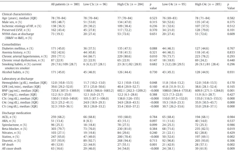
We included 380 patients discharged after hospitalization due to an acute episode of HF. Patients had a median age of 78 years old (interquartile (IQ) range: 70–84) and half of them were men (48.7%). Is-chemic HF etiology was considered in 36.3% of the patients, 56.6% had systolic HF and 19.3% were discharged with a New York Heart Associa-tion (NYHA) class of III or IV. Diabetes mellitus and anemia were comor-bidities in 45.0% and 42.6% of the patients, respectively. Arterial hypertension affected 77.1% of the patients, while patients suffering from renal dysfunction were 22.9%. Almost half of the patients revealed to have alcohol habits and 36.3% were current or ex-smokers. Patients' characteristics, comorbidities and laboratory assessments are shown inTable 1. When divided into groups according to the cut-off value of the 25th percentile of C3c and C4c, no major differences were noted in clinical characteristics. However, patients with lower C3c values showed a significantly smaller percentage of history of arterial hyper-tension. These patients were also less likely to be discharged on statins. They presented higher values of BNP (median 1188.8 pg/mL vs 602.1 pg/mL) and lower values of C4c and C1i. Patients with lower C4c values had an identical trend in BNP, C3c and C1i values. At dis-charge, they were also less likely to be discharged on statins or on beta-blockers, when compared with the ones with higher values of C4c (Table 1).
Patients were followed up for 6 months after discharge. During this period among the 49 patients that died due to HF, 22 had low C3c values and 21 low C4 values. Estimates revealed a significantly higher risk of death, by HF or other cause, in patients with lower values of C3c and C4c upon discharge.Fig. 1shows the survival curves estimator for the 6 month period according to discharge values of the 25th percentile for C3c (110.0 mg/dL) for HF death endpoint.
Table 2presents the predictors of 6 month HF and all-cause death in our group of patients in a univariate approach. These predictors for HF death were ischemic etiology of HF, NYHA class, history of arterial hy-pertension, higher BNP and lower hemoglobin, GFR, C3c and C4c values upon discharge. Besides the strong predictor for HF, BNP, had a hazard ratio (HR) of 1.02 for HF death (95% CI: 1.01–1.03) and all-cause death (95% CI: 1.01–1.02) per each 100 mg/dL of increase. Besides BNP, the markers C3c and C4c were also associated with shorter survival with a
HR of 0.98 (95% CI: 0.97–0.99) and 0.97 (95% CI: 0.95–0.99) for HF
and a HR of 0.98 for both in all-cause death, per each mg/dL of decrease.
Although C1i showed a significant difference between the groups
created, it did not reveal a significant HR concerning both endpoints in study. The prescription of angiotensin converting enzyme inhibitor
and beta-blockers showed to have a significant protective effect in
both endpoints.
In the multivariate adjusted models, in continuous and quartile anal-ysis, C3c association with 6 month endpoints was independent of the other prognostic predictors. The multivariate adjusted HR of 6 month HF and all-cause death, per mg/dL of C3c values was 0.99 (95% CI:
0.98–1.00). In patients with C3c values lower than 110.0 mg/dL, the
multivariate adjusted HR was 2.32 (95% CI: 1.25–4.28) for HF death
and 2.52 (95 %CI: 1.41–4.49) for all-cause death, comparing with pa-tients with higher C3c values. For papa-tients with C4c values lower than 25.15 mg/dL, the multivariate adjusted HR was not statistical significant, as well as in the continuous analysis. The multivariate adjusted models can be observed inTable 3.
Table 4presents the Spearman's correlations between the continu-ous variables in study. All complement proteins correlated with each other, with the stronger correlation between C3c and C4c. BNP had a negative correlation with C3c and C4c proteins. CRP only positively cor-related with C3c.
4. Discussion
We report that low values of C3c and C4c upon discharge of an acute HF episode are associated with unfavorable outcome in HF patients. The multivariate adjusted risk of HF or all-cause death at 6 months for pa-tients with C3c lower than 110.0 mg/dL was about 2.5 fold higher than for those with higher values at discharge. For C4c, although it showed prognostic value in a univariate approach, the same did not come through in a multivariate analysis.
The association between activation of the immune system and HF has been reported[14,15], with high levels of CRP as an indicative of se-verity of disease[16]. CRP mediates several protective processes, but may also have deleterious effects in HF, such as the up-regulation of
tumor necrosis factorα (TNFα) and interleukin (IL)-6 which are a
strong sign of this activation[3,4,17,18]. In fact, circulating levels of
pro-inflammatory cytokines are enhanced in the failing myocardium,
in both ischemic and nonischemic HF, and are long known to be related to disease severity and to predict poor survival[2,5]. Our data also corroborate this assumption. The levels of CRP, at discharge, had a
me-dian of 12.2 mg/L (IQR: 6.1–25.0) and showed a trend to predict HF
death (p value = 0.063) and a univariate statistical significant predic-tion value for all-cause death (p value = 0.013). Thus, once these are markers of immune activation and knowing that after an initial heart in-sult, the increased production of proinflammatory cytokines may
chal-lenge the surrounding tissue through propagation of the inflammatory
response and direct effects on the cardiac myocyte structure and func-tion, it is reasonable to assume that the complement proteins may also contribute to the destructive action of these factors and to the path-ogenetic sequelae of HF[14].
Table 1
Patients' demographics, clinical and laboratory characteristics and discharge medication; comparison between C3c and C4c groups. All patients (n = 380) Low C3c (n = 96) High C3c (n = 284) p
value
Low C4c (n = 95) High C4c (n = 285) p Value Clinical characteristics
Age (years), median (IQR) 78 (70–84) 78 (70–84) 77 (70–84) 0.523 76 (69–83) 78 (71–84) 0.582
Male sex, n (%) 185 (48.7) 51 (53.0) 134 (47.0) 0.315 50 (52.6) 135 (47.4) 0.375
Ischemic etiology of HF, n (%) 138 (36.3) 29 (30.2) 109 (38.4) 0.236 31 (32.6) 107 (37.5) 0.512
Preserved LVSF, n (%) 162 (43.4) 45 (27.8) 117 (72.2) 0.370 34 (21.0) 128 (79.0) 0.101
NYHA class at discharge (III&IV vs I&II), n (%)
73 (19.3) 20 (27.4) 53 (72.6) 0.651 20 (27.4) 53 (72.6) 0.609
Comorbidities
Diabetes mellitus, n (%) 171 (45.0) 36 (37.5) 135 (47.5) 0.088 44 (46.3) 127 (44.6) 0.767
Anemia history, n (%) 162 (42.6) 44 (45.8) 118 (41.5) 0.321 44 (46.3) 118 (41.4) 0.833
Chronic arterial hypertension, n (%) 293 (77.1) 64 (66.7) 229 (80.6) 0.010 70 (73.7) 223 (78.2) 0.293 Chronic renal dysfunction, n (%) 87 (22.9) 22 (22.9) 65 (22.9) 0.147 18 (18.9) 69 (24.2) 0.440 Smoking habits, n (%) current
smoker/ex smoker
29 (7.6)/109 (28.7) 6 (6.3)/27 (28.1) 23 (8.1)/82 (28.9) 0.682 3 (3.2)/28 (29.5) 26 (9.1)/81 (28.4) 0.296
Alcohol habits, n (%) 171 (45.0) 45 (46.9) 126 (44.4) 0.730 43 (45.3) 128 (44.9) 0.931
Laboratory at discharge
Hemoglobin (g/dL), median (IQR) 12.0 (10.8–13.5) 11.7 (10.2–13.0) 12.1 (10.8–13.6) 0.048 11.8 (10.4–13.2) 12.0 (10.8–13.5) 0.170 GFR (mL/min), median (IQR) 39.6 (29.2–52.0) 37.1 (25.8–50.6) 40.4 (29.9–52.7) 0.160 41.8 (31.9–51.3) 38.6 (28.1–52.4) 0.165 BNP (pg/mL), median (IQR) 725.8 (307.5–1369.9) 1188.8 (566.6–1683.2) 602.1 (262.3–1209.2) b0.000 1080.0 (384.4–1735.8) 609.9 (271.1–1284.0) 0.001 CRP (mg/L), median (IQR) 12.2 (6.1–25.0) 12.1 (6.0–21.7) 12.2 (6.1–28.6) 0.388 12.5 (7.3–22.0) 11.9 (6.1–28.7) 0.427 C3c (mg/dL), median (IQR) 130.0 (110.0–149.8) 101.5 (87.1–106.0) 138.0 (126–155) b0.000 110.0 (97.3–132.0) 135.0 (116.5–153.5) 0.000 C4c (mg/dL), median (IQR) 32.3 (25.2–41.1) 24.9 (18.9–29.3) 34.9 (28.8–43.5) b0.000 19.3 (16.0–23.3) 35.9 (30.5–43.7) 0.000 C1i (mg/dL), median (IQR) 32.3 (19.0–36.1) 30.3 (26.8–33.2) 33.4 (30.0–37.2) b0.000 30.7 (26.2–33.6) 33.0 (29.8–37.1) 0.000 Discharge medication ACEi, n (%) 259 (68.2) 66 (68.8) 193 (68.0) 0.784 65 (68.4) 194 (68.1) 0.984 ARA, n (%) 51 (13.4) 8 (8.3) 43 (15.1) 0.097 11 (11.6) 40 (14.0) 0.537 Spirolactone, n (%) 96 (25.3) 18 (18.8) 78 (27.5) 0.099 24 (25.3) 72 (25.3) 0.986 Beta-blocker, n (%) 303 (79.7) 73 (76.0) 230 (81.0) 0.384 68 (71.6) 235 (82.5) 0.019 Nitrates, n (%) 103 (27.1) 19 (19.8) 84 (29.6) 0.240 21 (22.1) 82 (28.8) 0.429 Statins, n (%) 247 (65.0) 47 (49.0) 200 (70.4) b0.000 50 (52.6) 197 (69.1) 0.002 Diuretic, n (%) 357 (93.9) 89 (92.7) 268 (94.4) 0.713 92 (96.8) 265 (93.0) 0.306 HF death 49 (12.9) 22 (44.9) 27 (55.1) 0.001 21 (42.9) 28 (57.1) 0.002
All cause death 63 (16.6) 29 (46.0) 34 (54.0) b0.000 24 (38.1) 39 (61.9) 0.009
IQR: interquartile range; HF: heart failure; LVSF: left ventricular systolic function; NYHA: New York Heart Association; GFR: estimated glomerularfiltration rate; BNP: B-type natriuretic peptide; CRP: C-reactive protein; C1i: C1 inhibitor; ACEi: angiotensin converting enzyme inhibitor; ARA: angiotensin II receptor antagonist.
Fig. 1. Kaplan–Meier survival curves according to C3c cut-off values of the 25th
percentile. Patients with lower values of C3c had higher 6 month risk of heart failure death after an acute heart failure episode.
Evidence also showed that extrahepatic tissues, such as the heart, produce complement proteins in ischemic and nonischemic
condi-tions[19,20]. As low complement levels are normally an indicator of
high tissue complement turnover[21,22], the low (peripheral) levels
in patients in our study may represent an increased (local) activation of complement in the heart. This activation and consequent lower values can be associated with the poor outcome of our patients, also supported with the evidence that these patients had significant higher BNP values, representing patients at a higher risk of death due to HF [23]. In fact, by the Spearman correlation C3c and C4c values signi ficant-ly correlate with BNP values in a negative way. In the heart, activated complement C3 was already shown to cause tachycardia, impairment
of atrioventricular conduction, left ventricular contractile failure, coro-nary vasoconstriction, and histamine release after injection into isolated guinea pig hearts[24]. Thus, besides being a good marker for overall complement activation, C3 seems to be of pathophysiological relevance in the cardiovascular system.
The consumption of serum C3 and C4 reflects the activation of the
classic and alternate pathways of the complement system[22]. CRP
values besides representing a state of chronic immune activation may also contribute to the understanding of complement activation. CRP ac-tivates the classical pathway of complement when bound to appropri-ate ligands. CRP efficiently activates the early proteins of the classical pathway (C1, C2, C4 and to a lesser extent C3) but does not generate consumption of the components of the membrane attack complex (C5–C9)[25]. Surface-bound CRP decreases the alternative complement pathway C3-convertase and C5-convertase activities, inhibits the
alter-native amplification loop and reduces deposition of C3b and lysis by
the lectin pathway[26]. In our patients the values of CRP did correlate positively with C3c values, but not with C4c. This positive relationship suggests that CRP can be related with the activation of C3c, by the
Table 2
Univariate Cox-regression between patients' characteristics and 6 month heart failure and all-cause death, after an acute heart failure episode.
HF death All-cause death
HR (95% CI) p Value HR (95% CI) p Value
Clinical characteristics, all patients (n = 380)
Age (per year) 1.02 (1.00–1.05) 0.111 1.03 (1.00–1.05) 0.041
Male sex 1.64 (0.92–2.93) 0.092 1.18 (0.72–1.94) 0.503
Ischemic etiology of HF 1.27 (1.05–1.54) 0.016 1.24 (1.03–1.50) 0.023
Preserved LVSF 1.56 (0.86–2.83) 0.146 1.01 (0.61–1.66) 0.983
NYHA class at discharge (III&IV vs I&II) 1.31 (1.08–1.60) 0.007 1.30 (1.09–1.55) 0.004 Comorbidities
Diabetes mellitus 0.87 (0.65–1.16) 0.332 0.83 (0.64–1.07) 0.144
Anemia history 1.00 (0.76–1.32) 0.986 1.12 (0.95–1.32) 0.194
Chronic arterial hypertension 0.47 (0.26–0.85) 0.012 0.50 (0.30–0.86) 0.011
Chronic renal dysfunction 1.11 (0.90–1.37) 0.338 1.23 (1.09–1.40) 0.001
Smoking habits 1.15 (0.95–1.39) 0.166 1.09 (0.90–1.32) 0.364 Alcohol habits 0.92 (0.52–1.63) 0.781 0.80 (0.48–1.33) 0.394 Laboratory at discharge Hemoglobin (per g/dL) 0.85 (0.73–0.98) 0.030 0.83 (0.73–0.95) 0.007 GFR (per mL/min) 0.98 (0.97–1.00) 0.045 0.98 (0.97–1.00) 0.010 BNP (per 100 pg/mL) 1.02 (1.01–1.03) b0.000 1.02 (1.01–1.02) b0.000 CRP (per 10 mg/L) 1.08 (1.00–1.16) 0.063 1.09 (1.02–1.16) 0.013 C3c (per mg/dL) 0.98 (0.97–0.99) b0.000 0.98 (0.98–0.99) b0.000 C4c (per mg/dL) 0.97 (0.95–0.99) 0.005 0.98 (0.96–0.99) 0.009 C1i (per mg/dL) 0.97 (0.92–1.02) 0.251 0.99 (0.95–1.03) 0.625 Discharge medication ACEi 0.49 (0.28–0.86) 0.013 0.54 (0.33–0.89) 0.016 ARA 0.56 (0.20–1.56) 0.266 0.66 (0.28–1.53) 0.328 Spirolactone 0.74 (0.37–1.49) 0.397 0.82 (0.45–1.48) 0.509 Beta-blocker 0.39 (0.22–0.69) 0.001 0.49 (0.29–0.83) 0.008 Nitrates 0.82 (0.48–1.42) 0.483 0.86 (0.55–1.35) 0.505 Statins 0.70 (0.40–1.25) 0.226 0.72 (0.43–1.20) 0.207 Diuretic 0.85 (0.41–1.75) 0.657 0.89 (0.51–1.56) 0.691
HR: hazard ratio; IQR: interquartile range; HF: heart failure; LVSF: left ventricular systolic function; LVSD: left ventricular systolic dysfunction; NYHA: New York Heart Association; GFR: estimated glomerularfiltration rate; BNP: B-type natriuretic peptide; CRP: C-reactive protein; C1i: C1 inhibitor; ACEi: angiotensin converting enzyme inhibitor; ARA: angiotensin II receptor antagonist.
Table 3
Multivariate Cox-regression model for C3c and C4c on 6 month HF death and all-cause death, after an acute HF episode.
HF deatha
All-cause deathb
HR (95% CI) p Value HR (95% CI) p Value C3c (per mg/dL) 0.99 (0.98–1.00) 0.030 0.99 (0.98–1.00) 0.027 C4c (per mg/dL) 0.98 (0.96–1.01) 0.149 0.98 (0.96–1.00) 0.097 C3c (1st Q vs others) 2.32 (1.25–4.28) 0.008 2.52 (1.41–4.49) 0.002 C4c (1st Q vs others) 1.84 (0.99–3.42) 0.055 1.66 (0.92–3.00) 0.091 HR: hazard ratio; HF: heart failure.
a
Model adjusted for: ischemic etiology of heart failure; New York Heart Association class at discharge (III&IV vs I&II); chronic arterial hypertension; hemoglobin (per g/dL); estimated glomerularfiltration rate; B-type natriuretic peptide (per 100 pg/mL); angioten-sin converting enzyme inhibitor; beta-blocker medication.
b Model adjusted for: age (per year); ischemic etiology of heart failure; New York Heart
Association class at discharge (III&IV vs I&II); chronic arterial hypertension; chronic renal dysfunction; hemoglobin (per g/dL); estimated glomerularfiltration rate; B-type natriuret-ic peptide (per 100 pg/mL); C-reactive protein (per 10 mg/L); angiotensin converting en-zyme inhibitor; beta-blocker medication.
Table 4
Spearman's correlations.
C1i C3c C4c CRP BNP
C1i Correlation coefficient – 0.369⁎ 0.295⁎ 0.305⁎ −0.070
p value b0.000 b0.000 b0.000 0.174
C3c Correlation coefficient 0.369⁎ – 0.515⁎ 0.106⁎ −0.308⁎
p value b0.000 b0.000 0.040 b0.000
C4c Correlation coefficient 0.295⁎ 0.515⁎ – 0.057 −0.217⁎
p value b0.000 b0.000 0.267 b0.000
C1i: C1 inhibitor; CRP: C-reactive protein; BNP: B-type natriuretic peptide. ⁎ Significant correlation.
alternative and lectin pathways. On the other hand, CRP correlated with C1i in a positive way. The main function of C1i is the inhibition of the classical pathway of complement to prevent spontaneous activation, with levels raising about 2 fold during inflammation[27,28]. In this study, the values of C3c and C4c positively correlated with C1i values. This correlation shows that the activation of the classical pathways due to a non-inactivation by C1i may also be present in HF.
To our knowledge, this study is thefirst one to strongly establish a
significant role for complement as a predictor marker of HF death in
an adjusted survival model. Previous studies have evaluated the pres-ence and prognostic role of complement system in HF. A study with a small number of patients (36 participants) with HF registered comple-ment activation in these patients[29]. In this same study, they have also shown an association of high SC5b9 levels with 6 month poor out-come, in an unadjusted model of survival. Another small study with a similar number of patients with HF (39 participants), also reported a systemic complement activation attenuated with intravenous immuno-globulin treatment[30]. In a larger study with 118 CHF patients, SC5b9 was also evaluated and correlated with CRP levels. Nevertheless, in these last two studies, the role as a predictor of death was not evaluated. Clearly, although these studies represent a necessary uncover of the subject potentiating other studies, the small number of participants and the absence of adjusted models are major weaknesses. A more re-cent study in 182 patients reported a significant association between complement C3a higher values and a combined endpoint of all-cause mortality or rehospitalization due to progression of HF[31]. In this study, the authors only focused in the analysis of the anaphylotoxin C3a, although also evaluating C3c and C4c levels. It is true that C3a is a complement activation product with powerful biological effects; never-theless this product is rather unstable and degradable upon freezing. In our study, we measured C3c and C4c, which are more stable comple-ment products[32]. In another recent study with 197 patients with sta-ble HF, the authors reported that patients with higher C3c levels exhibited a trend to better survival but were unable to statistically prove that prediction[33]. The major problem in that report was the di-vision of the patients according to the assay reference intervals, clearly not the proper way of studying the value of a biomarker, as was our in-tention in our cohort. In our study we divided the patients according to quartiles and in this way we could show an association between those that belonged to thefirst quartile, with the likelihood of dying due to complications of HF. Also, and differently from the two studies referred, as all patients were evaluated at discharge, we may assume with some level of certainty that they were in the same degree of immune activation.
5. Conclusion
Our results show that upon hospital discharge after an episode of acute HF, lower values of C3c are associated, BNP independently, with an approximately 2.5 higher risk of 6 month HF and all-cause death. Our results suggest that this marker can have a role in the identification of patients at increased risk of mortality.
Conflict of interests
The authors declare no conflict of interests, including specific finan-cial interests and relationships relevant to the subject of the paper.
Acknowledgments
This work was supported by the following grants: Portuguese Foundation for Science and Technology (SFRH/BD/79716/2011 and PIC/IC/82773/2007).
References
[1]Gong KZ, Song G, Spiers JP, Kelso EJ, Zhang ZG. Activation of immune and inflamma-tory systems in chronic heart failure: novel therapeutic approaches. Int J Clin Pract 2007;61(4):611–21.
[2]Apostolakis S, Lip GY, Shantsila E. Monocytes in heart failure: relationship to a dete-riorating immune overreaction or a desperate attempt for tissue repair? Cardiovasc Res 2010;85(4):649–60.
[3]Oikonomou E, Tousoulis D, Siasos G, Zaromitidou M, Papavassiliou AG, Stefanadis C. The role of inflammation in heart failure: new therapeutic approaches. Hell J Cardiol 2011;52:30–40.
[4]Heymans S, Hirsch E, Anker SD, et al. Inflammation as a therapeutic target in heart failure? A scientific statement from the Translational Research Committee of the Heart Failure Association of the European Society of Cardiology. Eur J Heart Fail 2009;11(2):119–29.
[5]Celis R, Torre-Martinez G, Torre-Amione G. Evidence for activation of immune sys-tem in heart failure: is there a role for anti-inflammatory therapy? Curr opin cardiol 2008;23(3):254–60.
[6]Engström G, Hedblad B, Janzon L, Lindgärde F. Complement C3 and C4 in plasma and incidence of myocardial infarction and stroke: a population-based cohort study. Eur J Cardiovasc Prev Rehabil 2007;14(3):392–7.
[7]Riedemann NC, Ward PA. Complement in ischemia reperfusion injury. Am J Pathol 2003;162(2):363–7.
[8]Walsh MC, Hart ML, Bourcier T, Bhole D, Takahashi M, Stahl GL. Role of complement in myocardial ischemia and infarction. In: Szebeni Janos, editor. The Complement System. US: Springer; 2004. p. 421–35.
[9]Hofmann U, Frantz S. How can we cure a heart“in flame”? A translational view on inflammation in heart failure. Basic Res Cardiol 2013;108(4):356.
[10]Flores-Arredondo JH, García-Rivas G, Torre-Amione G. Immune modulation in heart failure: past challenges and future hopes. Curr Heart Fail Rep 2011;8(1):28–37.
[11]Markiewski MM, Lambris JD. The role of complement in inflammatory diseases from behind the scenes into the spotlight. Am J Pathol 2007;171(3):715–27.
[12]Dickstein K, Cohen-Solal A, Filippatos G, et al. ESC guidelines for the diagnosis and treatment of acute and chronic heart failure 2008: the Task Force for the diagnosis and treatment of acute and chronic heart failure 2008 of the European Society of Cardiology. Developed in collaboration with the Heart Failure Association of the ESC (HFA) and endorsed by the European Society of Intensive Care Medicine (ESICM). Eur J Heart Fail 2008;10(10):933–89.
[13]Cockcroft DW, Gault MH. Prediction of creatinine clearance from serum creatinine. Nephron 1976;16(1):31–41.
[14]Gruson D, Ahn SA, Rousseau MF. Biomarkers of inflammation and cardiac remodel-ing: the quest of relevant companions for the risk stratification of heart failure pa-tients is still on going. Biochem Med 2011;21(3):254–63.
[15]Yndestad A, Damås JK, Oie E, Ueland T, Gullestad L, Aukrust P. Systemic inflamma-tion in heart failure—the whys and wherefores. Heart Fail Rev 2006;11(1):83–92.
[16]Elster SK, Braunwald E, Wood HF. A study of C-reactive protein in the serum of pa-tients with congestive heart failure. Am Heart J 1956;51(4):533–41.
[17]van Kimmenade RR, Januzzi JL. Emerging biomarkers in heart failure. Clin Chem 2012;58(1):127–38.
[18]Gullestad L, Ueland T, Vinge LE, Finsen A, Yndestad A, Aukrust P. Inflammatory cyto-kines in heart failure: mediators and markers. Cardiology 2012;122(1):23–35.
[19]Yasojima K, Schwab C, McGeer EG, McGeer PL. Human heart generates complement proteins that are upregulated and activated after myocardial infarction. Circ Res 1998;83(8):860–9.
[20]Oliveira GH, Brann CN, Becker K, et al. Dynamic expression of the membrane attack complex (MAC) of the complement system in failing human myocardium. Am J Cardiol 2006;97(11):1626–9.
[21]Rutkowski MJ, Sughrue ME, Kane AJ, Ahn BJ, Fang S, Parsa AT. The complement cas-cade as a mediator of tissue growth. Inflamm Res 2010;59:897–905.
[22]Chiu RC, Samson R. Complement (C3, C4) consumption in cardiopulmonary bypass, cardioplegia, and protamine administration. Ann Thorac Surg 1984;37(3):229–32.
[23]Maisel A. B-type natriuretic peptide levels: diagnostic and prognostic in congestive heart failure: what's next? Circulation 2002;105(20):2328–31.
[24]del Balzo UH, Levi R, Polley MJ. Cardiac dysfunction caused by purified human C3a anaphylatoxin. Proc Natl Acad Sci U S A 1985;82(3):886–90 [1985].
[25]Berman S, Gewurz H, Mold C. Binding of C-reactive protein to nucleated cells leads to complement activation without cytolysis. J Immunol 1986;136(4):1354–9.
[26]Bíró A, Rovó Z, Papp D, et al. Studies on the interactions between C-reactive protein and complement proteins. Immunology 2007;121:40–50.
[27]Davis 3rd AE. Biological effects of C1 inhibitor. Drug News Perspect 2004;17(7): 439–46.
[28]Cicardi M, Zingale L, Zanichelli A, Pappalardo E, Cicardi B. C1 inhibitor: molecular and clinical aspects. Springer Semin Immunopathol 2005;27(3):286–98.
[29]Clark DJ, Cleman MW, Pfau SE, et al. Serum complement activation in congestive heart failure. Am Heart J 2001;141(4):684–90.
[30]Aukrust P, Gullestad L, Lappegård K, et al. Complement activation in patients with congestive heart failure: effect of high-dose intravenous immunoglobulin treatment. Circulation 2001;104(13):1494–500.
[31]Gombos T, Förhécz Z, Pozsonyi Z, et al. Complement anaphylatoxin C3a as a novel independent prognostic marker in heart failure. Clin Res Cardiol 2012;101(8): 607–15.
[32]Okumura N, Nomura M, Tada T. Effects of sample storage on serum c3c assay by nephelometry. Clin Lab Sci 1990;3:54–7.
[33]Frey A, Ertl G, Angermann CE, Hofmann U, Störk S, Frantz S. Complement C3c as a biomarker in heart failure. Mediat Inflamm 2013;2013:716902.