DEPARTAMENTO DE BIOLOGIA ANIMAL
FORENSIC ENTOMOLOGY: DNA BARCODING
FOR COLEOPTERA IDENTIFICATION
Susana Fernandes Torres Lopes
Dissertação
Mestrado em Biologia Humana e Ambiente
2012
DEPARTAMENTO DE BIOLOGIA ANIMAL
FORENSIC ENTOMOLOGY: DNA BARCODING
FOR COLEOPTERA IDENTIFICATION
Susana Fernandes Torres Lopes
Mestrado em Biologia Humana e Ambiente
Dissertação orientada por:
Professora Doutora Deodália Dias
I
Nota prévia
Este estudo está inserido no projecto “Forensic Entomology: Morphometric and Molecular databank (mtDNA) to identify species (Diptera and Coleoptera) with forensic interest” (PTDC/SAU-ESA/101228/2008), financiado pela Fundação para a Ciência e Tecnologia (FCT).
A Dissertação de Mestrado foi redigida em Inglês e respeitou os critérios exigidos pela revista Medical and Veterinary Entomology, uma vez que consideramos que os resultados aqui apresentados, complementados com dados obtidos por outros membros da equipa, poderão ser submetidos a uma publicação nesta revista.
III
Agradecimentos
No decorrer deste ano tive o prazer de perceber que a elaboração de uma tese de mestrado é muito mais do que um trabalho individual, mas sim resultado do apoio e incentivo de um conjunto de pessoas. Desejo desta forma expressar os meus sinceros agradecimentos a cada uma delas.
Em primeiro lugar agradeço à minha orientadora, Professora Deodália Dias, pela confiança que depositou em mim desde o primeiro dia, pela orientação e palavras de incentivo quando as coisas corriam menos bem! Agradeço também pela disponibilidade que sempre demonstrou em ajudar-me, mais do que uma orientadora foi uma grande amiga!
Devo também os meus maiores agradecimentos à Rita Oliveira Dario e à Catarina Dourado a quem devo tudo aquilo que aprendi no laboratório durante este ano! Obrigada pela paciência, conselhos e ideias partilhadas. Saber que vos tinha como “apoio” ajudou-me muito nos momentos mais complicados. Obrigada ainda por todas as correcções e sugestões na tese, sem vocês este trabalho não seria o mesmo!
Agradeço também aos restantes membros do projecto de Entomologia Forense, Professora Teresa Rebelo e Ana Farinha, por terem estado sempre disponíveis a ajudar! Agradeço ainda à Susana Parra por me ter disponibilizado os Coleópteros que foram recolhidos para o seu estudo, à Viviana Padinha pelas identificações morfológicas e ao Carlos Aguiar pela preciosa ajuda!
Novamente à Catarina e à Rita, desta vez pela vossa amizade, por todas as conversas e momentos partilhados ao longo deste ano! Foi sem dúvida um enorme prazer conhecer-vos!
À Cátia, Nádia e Ana Calarrão que fizeram parte do meu trajecto académico, tornando-o único e inesquecível. Obrigada pela constante preocupação com o meu trabalho e pelo optimismo que sempre me transmitiram!
Agradeço também às melhores amigas de sempre, Melanie, Patrícia, Sara, Tânia e Telma! Obrigada por todos os momentos de boa disposição, pelas conversas sem sentido e pelas gargalhadas que demos juntas! Cada uma de vocês tem um lugar muito especial no meu coração!
IV
Ao Rúben, pela enorme paciência e compreensão que teve comigo durante este ano! Foste o meu grande apoio! Obrigada por ouvires todas as conversas sobre PCRs, purificações, sequenciações e contaminações! És uma pessoa muito especial!
A toda a minha família que sempre apoiou os meus sonhos! Obrigada por todo o apoio e carinho! Sei que hoje é também um dia de felicidade para todos vocês!
Por último não podia deixar de agradecer às duas pessoas sem as quais nada disto seria possível, Mãe e Pai! Este trabalho é para vocês que sempre acreditaram em mim, mesmo quando eu duvidava! Apesar de estarem no fim desta lista de agradecimentos, são sem dúvida nenhuma as pessoas mais importantes na minha vida! Não tenho palavras para vos agradecer por tudo!
Agradeço ainda a todas as pessoas que possam não ter sido directamente mencionadas mas que de alguma forma contribuíram para a realização deste trabalho.
V
Resumo
A Entomologia Forense é a ciência que recorre ao estudo de insectos e outros artrópodes e os aplica em investigações do foro legal. Para além de contribuírem para o processo de decomposição do corpo, os insectos podem ainda fornecer informações importantes em investigações criminais. A aplicação mais frequente da Entomologia Medicolegal é a determinação do tempo decorrido desde a morte do indivíduo (intervalo postmortem ou IPM), contudo, existem outras informações importantes como a ocorrência de deslocação do corpo, situações de negligência e associação do corpo com drogas ou toxinas.
Apesar da fauna associada ao cadáver incluir uma grande diversidade de artrópodes, Diptera e Coleoptera são as duas principais ordens de insectos que fornecem a maioria da informação em investigações forenses. As espécies pertencentes à ordem Diptera são as primeiras a colonizar o cadáver, sendo que a ordem Coleoptera tem o seu pico, em número e diversidade, durante os estádios mais avançados de decomposição. As espécies de insectos encontradas, assim como o seu padrão de colonização estão dependentes de inúmeros factores, sendo a região geográfica onde se encontram um dos mais importantes. Desta forma, para que seja possível uma aplicação sustentável da Entomologia Forense em investigações do foro legal, é extremamente importante a elaboração de bases de dados específicas para cada região geográfica. A identificação de espécies de insectos encontradas em cadáveres é a etapa mais crucial neste tipo de investigação. Este processo baseia-se na observação directa de caracteres morfológicos e uso de chaves dicotómicas. Contudo, trata-se de uma abordagem demorada e que muitas vezes não permite a identificação de espécimes em estádio imaturo. De modo a ultrapassar estas limitações foi proposta uma identificação molecular que se baseia na sequenciação de uma pequena região do genoma do espécime alvo, denominada por DNA barcode, e que posteriormente é comparada com sequências de referência que se encontram em bases de dados online, pelo que esta ferramenta é designada por “DNA barcoding”. O marcador molecular de eleição, usado no DNA barcoding é o gene mitocondrial Citocromo c oxidase subunidade I (COI).
VI
Contudo, a utilização de um único marcador para a identificação de espécies é controversa, pelo que se tem vindo a apostar cada vez mais numa abordagem que envolve outros genes. Uma outra região genómica que tem vindo a ser utilizada com sucesso, incluindo estudos com Coleópteros, é o Citocromo b (Cytb).
Desta forma, o presente estudo surge com o principal objectivo de proceder à identificação molecular de espécies com interesse forense, pertencentes à ordem Coleoptera, recolhidas na região de Aveiro, Portugal. Os indivíduos colectados foram identificados morfologicamente procedendo-se, de seguida, à extracção do DNA e posterior sequenciação dos marcadores moleculares COI e Cytb. Deste modo, pretendeu-se testar a eficácia do gene Cytb como marcador molecular e simultaneamente comparar a sua eficiência relativamente ao marcador COI.
Neste trabalho foram colectados 35 espécimes de Coleópteros, pertencentes às famílias Staphylinidae, Silphidae, Histeridae, Chrysomelidae, Curculionidae, Nitidulidae, Scarabaeidae e Carabidae. Destes 35 espécimes foram obtidas 30 sequências com 658 pb para o gene COI e 31 sequências com 433 pb para o gene Cytb. Embora a amplificação tenha ocorrido sem grandes complicações para ambos os genes, quando o gene Cytb foi analisado, foi possível verificar que 17 das 31 sequências eram idênticas, mesmo entre indivíduos pertencentes a famílias distintas. Este resultado veio demonstrar que esta região parece ser bastante conservada entre as diferentes espécies, impossibilitando a sua diferenciação. Assim sendo, esta região do Cytb mostrou não ser um bom marcador molecular para ser utilizado no DNA
barcoding destas espécies. Todas as sequências obtidas foram comparadas com
sequências de referência disponíveis nas bases de dados online GenBank e BOLD, de forma a corroborar a identificação morfológica. Porém, para a grande maioria dos indivíduos não foi possível a confirmação ao nível da espécie, uma vez que o DNA
barcoding é uma abordagem relativamente recente e consequentemente ainda existe
um grande número de espécies que não se encontra nestas bases de dados, impossibilitando uma comparação. As sequências obtidas neste trabalho irão contribuir para a implementação de uma Base de Dados Nacional, permitindo aumentar o conhecimento de espécies de Coleópteros com interesse forense, em Portugal, e que no futuro poderão ser utilizadas como evidências entomológicas em
VII
investigações do foro legal. As sequências serão também submetidas nas bases de dados GenBank e BOLD.
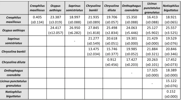
As variações intra e interespecíficas foram calculadas relativamente aos dados do COI, usando apenas sequências de indivíduos identificados até ao nível da espécie. Neste tipo de análise é importante ter em conta que o valor de variação intraespecífica deverá ser inferior a 3%, enquanto a variação interespecífica deverá ser superior a este limite, de forma a permitir a diferenciação das espécies. Outro critério está relacionado com a variação interespecífica, que deverá ser 10 vezes superior à média da variação intraespecífica. Contudo, ao analisar os resultados, foi possível observar que os valores de divergência intraespecífica para duas das espécies eram muito superiores ao esperado. Desta forma, foi necessário voltar a analisar a matriz inicial e tentar perceber quais os indivíduos que apresentavam um maior número de diferenças. Estas diferenças poderão estar relacionadas com o facto de os indivíduos terem sido morfologicamente mal identificados, podendo estar a considerar-se indivíduos de espécies diferentes como pertencentes à mesma espécie, obtendo-se assim valores errados. Outro critério usado na delimitação de espécies é a análise filogenética, onde cada espécie deverá surgir como um grupo monofilético. É importante ter em conta que geralmente a sequência barcode não apresenta um sinal filogenético suficientemente forte para determinar relações evolutivas, pelo que a análise destes dados deve ser realizada com precaução. A análise filogenética foi feita para ambos os genes através das metodologias Máxima Verosimilhança, Neighbour-Joining e Máxima Parcimónia. Relativamente à árvore obtida com os dados do COI, foi possível verificar que algumas espécies não agrupavam da forma esperada, o que nos leva a considerar, mais uma vez, que devido ao elevado grau de dificuldade, a identificação morfológica de alguns espécimes terá sido incorrecta. Em relação ao Cytb, visto que foram obtidas 17 sequências idênticas entre si, verifica-se que este gene não tem resolução para estimar relações filogenéticas nas espécies estudadas. Assim, este trabalho vem demonstrar que, na ordem Coleoptera, o COI é um marcador mais adequado do que o Cytb para ser usado no DNA barcoding.
IX
Abstract
Besides the contribution to the body decomposition process, insects are entomological evidences that can provide important information in criminal investigations. In order to study these events, it is essential to accomplish an accurate identification of species founded on corpses.
The main goal of this study was to perform a molecular identification of species with forensic interest, belonging to the order Coleoptera. With this purpose, two mitochondrial markers, COI and Cytb, were used. A total of 35 specimens belonging to the families Staphylinidae, Silphidae, Histeridae, Chrysomelidae, Curculionidae, Nitidulidae, Scarabaeidae and Carabidae were collected. Of the 35 Coleoptera specimens, there were obtained 30 sequences with 658 bp for COI gene and 31 sequences with 433 bp for Cytb gene. When Cytb sequences were analysed it was noticed that 17 of the 31 sequences were identical. This region appears to be highly conserved among different species, showing that this fragment is not a good molecular marker to be used for DNA barcoding of these Coleoptera species. Obtained sequences were compared with reference sequences in GenBank and BOLD databases. The majority of studied specimens did not have a sequence to be compared with, showing a lack of reference sequences for a great number of species.
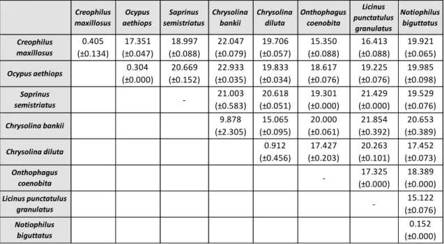
Intra and interspecific variation was determined for COI gene, using only specimens identified to species level. In this analysis it was realized that intraspecific divergence values for some species were much higher than expected (>3%). Therefore, the original dataset was reanalyzed to see which individuals presented greatest differences. Phylogenetic analysis was performed for both genes. Regarding the tree based on COI gene data, some species were not grouped as expected, leading us to consider that these specimens have been misidentified morphologically. About Cytb gene, since 17 obtained sequences were identical, phylogenetic analysis for this gene is not suitable. Comparing these two genes, results showed that COI gene is a most suitable marker to be used in DNA barcoding for the order Coleoptera.
XI
Index
Nota prévia ... I Agradecimentos ... II Resumo ... IV Abstract ... VIII List of Abbreviations ... XII Index of Figures ... XIV Index of Tables ... XVII. State of the Art ... 3
1.1. Forensic Entomology ... 3
1.1.1. Retrospective ... 3
1.1.2. Main Areas ... 5
1.2. Medical Entomology ... 6
1.2.1. Initial Postmortem Changes ... 6
1.2.2. Relationship between Insects and Corpses... 7
1.2.3. Insects with Forensic Importance ... 8
1.2.4. Role of Arthropods in Decomposition Process ... 10
1.3. Applications ... 11
1.3.1. Estimating the Postmortem Interval ... 11
1.3.2. Forensic Implications of Myiasis ... 14
1.3.3. Entomotoxicology: Insects as Toxicological Evidences ... 15
1.3.4. Gene Expression Analysis ... 15
1.3.5. Additional Applications ... 16
1.4. DNA Analysis ... 18
1.4.1. Species Identification ... 18
1.4.2. DNA Barcoding ... 19
1.4.3. Nuclear vs Mitochondrial DNA ... 20
1.4.4. Molecular Markers ... 22
1.4.4.1. Cytochrome c oxidase I ... 23
1.4.4.2. Cytochrome b ... 24
XII
II. Material and Methods ... 29
2.1. Sampling ... 29
2.2. DNA Extraction, Amplification and Sequencing ... 29
2.3. Data Analysis ... 31
III. Results and Discussion ... 35
3.2. Coleoptera Composition ... 37
3.3. Molecular Identification ... 39
3.4. Intra and Interspecific variation ... 42
3.5. Phylogenetic Analysis ... 46
3.5.1. Cytochrome c oxidase I ... 46
3.5.2. Cytochrome b ... 49
3.5.3. Concatenated data ... 51
IV. Final Considerations... 55
V. References ... 59
Appendices ... 73
Appendix I ... 75
Appendix II ... 77
XIII
List of Abbreviations
BLAST - Basic Local Alignment Search Tool
BLASTn - Nucleotide Basic Local Alignment Search Tool BOLD - Barcode of Life Data System
BOLD-IDS - Identification System of BOLD bp - Base Pair
BSA – Bovine serum albumine
CBOL - Consortium for the Barcode of Life cDNA - complementary DNA
COI - Cytochrome c Oxidase Subunit I Cytb - Cytochrome b
DNA - Deoxyribonucleic Acid
dNTP - Deoxyribonucleotide Triphosphate ML - Maximum Likelihood
MP - Maximum Parsimony mtDNA - Mitochondrial DNA
NCBI - National Center for Biotechnology Information NJ - Neighbor-Joining
NUMT - Nuclear Mitochondrial Pseudogene PCR - Polymerase Chain Reaction
PMI - Postmortem Interval RNA - Ribonucleic Acid rRNA - Ribosomal RNA
XV
Index of Figures
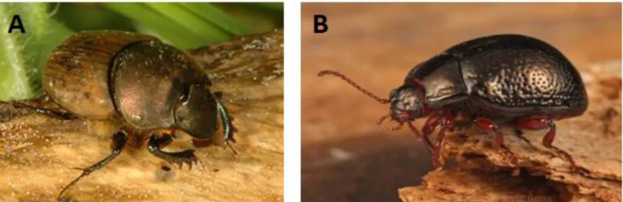
Fig. 1. Coleoptera species. A: Ontophagus coenobita; B: Chrysolina bankii ... 10
Fig. 2. Insect growth in relation to upper and lower thresholds (outlined area is the
area of growth) ... 13
Fig. 3. Gene map of Drosophila yakuba (Diptera) mtDNA molecule ... 21
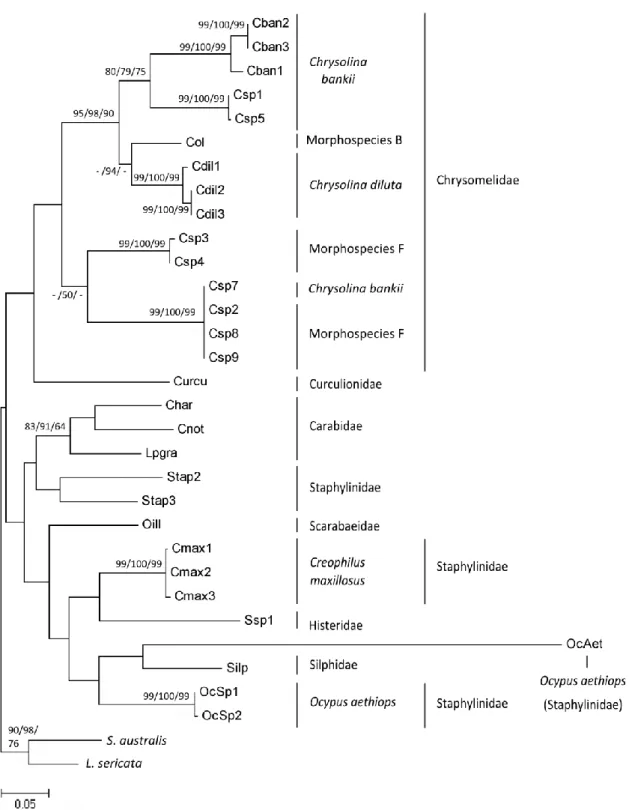
Fig. 4. Maximum likelihood phylogram (1,000 bootstrap replicates) based on 32
sequences of Cytochrome c oxidase I (COI) gene from 8 species, 7 morphospecies and 2 outgroups (Sarcophaga australis and Lucilia sericata - Diptera). Bootstrap values (above 50%) obtained for the analysis of ML, NJ and MP are shown next to the tree nodes ... 47
Fig. 5. Maximum likelihood phylogram (with 1,000 replicates) based on 33 sequences
of Cytochrome b (Cytb) gene from 8 species, 6 morphospecies and 2 outgroups (Sarcophaga australis and Lucilia sericata - Diptera). Bootstrap values (above 50%) obtained for the analysis of ML, NJ and MP are shown next to the tree nodes ... 50
Fig. 6. Maximum likelihood consensus phylogram (1,000 bootstrap replicates) based
on 29 sequences from 8 species, 6 morphospecies and 2 outgroups (Sarcophaga
australis and Lucilia sericata - Diptera). Bootstrap values (above 50%) obtained for the
analysis of ML, NJ and MP are shown next to the tree nodes ... 52
Fig. 7. Neighbour-joining consensus phylogram (1,000 bootstrap replicates) based on
29 sequences from 8 species, 6 morphospecies and 2 outgroups (Sarcophaga australis and Lucilia sericata - Diptera). Bootstrap values (above 50%) obtained for the analysis of ML, NJ and MP are shown next to the tree nodes ... 81
XVI
Fig. 8. Maximum Parsimony consensus phylogram (1,000 bootstrap replicates) based
on 29 sequences from 8 species, 6 morphospecies and 2 outgroups (Sarcophaga
australis and Lucilia sericata - Diptera). Bootstrap values (above 50%) obtained for the
XVII
Index of Tables
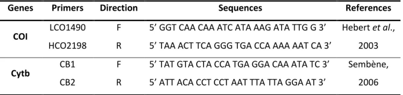
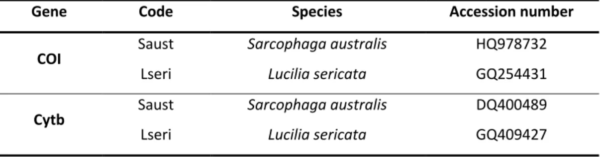
Table 1. Genes and primers used in this study ... 30 Table 2. Final concentration of reagents used in PCR amplification for COI and Cytb
genes ... 30
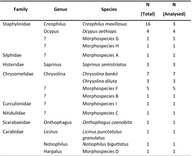
Table 3. Code and accession numbers of the specimens used as outgroup ... 32 Table 4. Specimens collected in this study: family, genus, species; number of
specimens of each species - N (Total); and number of analysed specimens – N (Analysed) ... 37
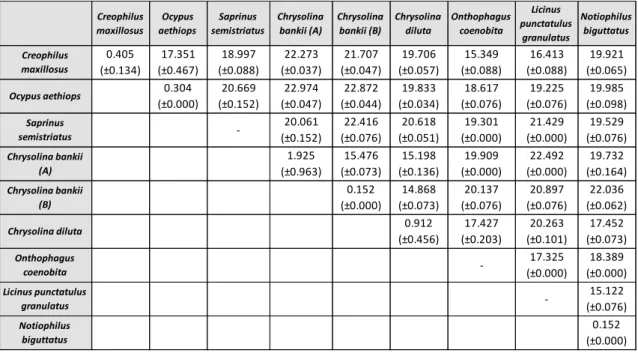
Table 5. Summary of intra and interspecific percentages of uncorrected p-distances
obtained for COI data of eight species. Standard deviation values are shown in brackets ... 43
Table 6. Summary of intra and interspecific percentages of uncorrected p-distances
obtained for eight species of the reanalyzed COI dataset. Standard deviation values are shown in brackets ... 44
Table 7. Summary of intra and interspecific percentages of uncorrected p-distances
obtained for eight species of the reanalyzed COI dataset. Standard deviation values are shown in brackets. A – Cban1, Cban2 and Cban3 specimens; B – Csp1 and Csp5 specimens ... 45
Table 8. Species identification, success of extraction and amplification for COI and Cytb
... 75
Table 9. Molecular Identification of insect specimens obtained for Cytb in GenBank
database ... 77
Table 10. Molecular Identification of insect specimens obtained for COI in BOLD and
3
I.
State of the Art
1.1. Forensic Entomology
“Forensic” can be defined as the use of scientific techniques to solve legal issues and “Entomology” as the study of insects. Thus, “Forensic Entomology” is a branch of forensic sciences which applies the study of insects and other arthropods with forensic interest in legal investigations (Jobling & Gill, 2004; Amendt et al., 2007).
1.1.1. Retrospective
Although Forensic Entomology has only begun to play an important role in criminal investigations over the past few decades, the potential of using insects to solve criminal cases has been known for centuries (Oliveira-Costa & Mello-Patiu, 2004).
The first documented case was written in 1235 by Sung Tz’u, a Chinese lawyer and criminalist, in a Legal Medicine book entitled “The Washing Away of Wrongs” (Benecke, 2001). In the 13th century, Sung Tz’u investigated a murder in a local village, in which a farmer had been stabbed in a rice field with a sharp weapon. The criminalist believed that a farming tool was the murder weapon, so he called all the farm workers and told them to place their working tools on the ground. He noticed that numerous blowflies were attracted by one of the sickles, probably due to the presence of invisible traces of blood. Confronted by this evidence, the stickle owner confessed to be the murderer (Amendt et al., 2004; Gupta & Setia, 2004).
During the Middle Ages, many sculptures, painters and poets have observed the decomposition process of human bodies, paying particular attention to the role of maggots. The realistic and detailed illustrations of corpses infested with maggots were usual (Benecke, 2001; Amendt et al., 2004). There are documents dating from 15th and 16th century that illustrate the importance of insects in decomposition, showing the pattern of body mass reduction and the skeletonization process by insect activity (Gupta & Setia, 2004).
4
In 1831, during the mass exhumations in France, the medical doctor Orfila noticed that insects and their larvae played an important role in body decomposition. That was an important step to understand the correlation between maggots and corpses decomposition (Benecke, 2001).
The first application in Europe is attributed to the French doctor Bergeret (in 1855), when the skeletonised remains of a child were found behind a chimney, by a workman, during a house redecoration. Bergeret studied the insect fauna discovered on these remains and made an estimation of the postmortem interval (PMI), i.e. time since death occurred, proving that the current occupants of the house could not have been the murderers since they had moved recently (Benecke, 2001; Gupta & Setia, 2004).
Later, in 1894, Mégnin published his most important book “La Faune des
Cadavres: Application de l’entomologie à la Medicine Légale”. In this book, he
describes the morphological features of different insect families that helped in their identification (Benecke, 2001; Gupta & Setia, 2004). He was the first to describe the relation between the chronological sequence of the decomposition process and insect colonization. He recognized eight invasion waves of arthropods related to the different stages of human decomposition on freely bodies, and two waves on buried bodies. This knowledge about insect succession became the basis for forensic entomologists to estimate the PMI (Campobasso et al., 2001; Gennard, 2007).
Despite Mégnin studies, Forensic Entomology was neglected for many years, not only due to the lack of specialized entomologists in cadaveric fauna, but mainly due to the distance between entomologists and medicolegal specialists (Pujol-Luz et
al., 2008).
Only in the second half of the 20th century the interest in Forensic Entomology emerged again. In Europe, the medical Doctor Marcel Leclercq and the Professor of biology Pekka Nuorteva were the first ones to employ Forensic Entomology for the determination of the PMI (Benecke, 2001; Amendt et al., 2004). Nowadays, Forensic Entomology is recognized in many countries as an important forensic tool (Amendt et
5
1.1.2. Main Areas
Forensic Entomology is the broad field where the study of insects and legal system interact. Insects can be used as evidence to solve crimes and many times are in the centre of legal disputes for causing damages (Pujol-Luz et al., 2008; Hall & Huntington, 2010).
Lord and Steveson (1986) divided this scientific field into three main areas: urban, stored products and medicolegal.
Urban Entomology deals with arthropods that affect humans and their environment, usually involving infestation of houses and other facilities by termites and cockroaches (Pujol-Luz et al., 2008; Hall & Huntington, 2010).
Stored products Entomology is related with cases regarding infestation of stored products or food contamination. Some examples are parts of insects in cereals, caterpillar in cans of vegetables and fly maggots in sandwiches from fast-food restaurants (Hall & Huntington, 2010). Arthropods can cause direct damage to food resources and can also be accidentally ingested. This can lead to toxic or allergic reactions and other health disorders. They also may defecate or regurgitate fluids, contaminating food with potential pathogens, which will be ingested by humans or other animals (Durden & Mullen, 2002).
Lastly, Medicolegal Entomology, also known as Medicocriminal Entomology, involves the study of necrophagous insects that colonize human corps to help to resolve crimes, most often crimes of violence like homicide and suicide (Pujol-Luz et
al., 2008).
Although less known, Wildlife Forensic Entomology is another area that should be taken into account. It is a more recent area proposed by Anderson (1999) that focuses on surveillance and protection of wild animals in captivity from neglect situations or physical abuse and on the resolution of questions about illegal hunting of protected species (Anderson, 1999; Hall & Huntington, 2010).
6
1.2. Medical Entomology
1.2.1. Initial Postmortem Changes
After death, a body undergoes different stages of decomposition. Decomposition process is responsible for returning the organic matter of a body to the ecosystem. This process consists on the metabolic transformation of organic matter into simple organic and inorganic compounds (Souza et al., 2008). During this process, the body undergoes several changes caused by cells autolysis (promoted by the chemical breakdown), tissue autolysis (due to the released enzymes) and external processes, where fungi, bacteria and arthropods are the main responsible (Campobasso et al., 2001; Amendt et al., 2004).
During the first 72 hours after death, the forensic pathologist has the function of analyse the interior and exterior of the cadaver to be able to estimate the time since death (PMI), as well as the manner and the cause of death, through the observation of body changes (Henβge & Madea, 2004; Schoenly et al., 2006). Several changes of the body may occur in the first 72 hours after dead.
One of the earliest changes of the body is the red discoloration of the skin(livor
mortis or lividity). Upon death, circulation stops and blood begins to settle to the
lowest parts of the body by the action of gravidity. Skin becomes red in those areas, but pale in the rest of the body (Goff, 2010). Body temperature also decreases (algor
mortis) because body ceases the regulation of its internal temperature and begins to
approximate ambient temperature. The size of the individual and the exposure to sunlight or heat are factors that can influence this stage. For a more accurate measurement, liver temperature is usually used (Amendt et al., 2004; Goff, 2010).
Another change is the stiffening of muscle fibbers due to the breakdown of glycogen and accumulation of lactic acid in muscles (rigor mortis) (Amendt et al., 2004). The duration of rigor mortis depends on the metabolic state of the body. If the individual has been involved in intense exercise immediately before death, rigor mortis will develop faster. It also depends on the body temperature: lower temperatures tend to accelerate the beginning of rigor mortis and to prolong its duration, while in warmer temperatures the opposite occurs (Henβge & Madea, 2004; Goff, 2010).
7
Skin slippage also occurs, resulting in the separation of epidermis from dermis, due to the production of hydrolytic enzymes. Consequently, skin can be easily removed as well as nails and hair. A large quantity of putrefaction gases causes a physical distortion of the cadaver. One of the formed gases is hydrogen sulphide (H2S),
which reacts with haemoglobin forming sulfhemoglobin, a green pigment that can be seen in blood vessels and in other areas of the body (Amendt et al., 2004; Goff, 2010).
All these changes occur within the first 72-96 hours after death. Beyond this period there is less medical information and no reliable estimation of PMI interval is possible (Amendt et al., 2004). Therefore, insects found on the corpse can provide an important source of information.
1.2.2. Relationship between Insects and Corpses
Arthropods are by far the largest and most important biological group in our planet (Benecke, 2001). Insects, the major group of arthropods, can travel considerable distances in search of food or a suitable place for laying their eggs (Castner, 2010). There are about one million species described, living in a wide variety of habitats, including crime scenes (vertebrate corpses provide an excellent food source) (Benecke, 2001; Amendt et al., 2004). However, not all insects found near a body are feeding on it or have oviposited there. Therefore, it is important to distinguish which insects can be used as forensic evidences (Benecke, 1998; Campobasso et al., 2001; Amendt et al., 2011).
Hundreds of arthropods species are attracted to corpses, mostly flies and beetles, but also mites, isopods, opiliones and nematodes. Some insects are attracted to a body shortly after death and soon begin their activities (Benecke, 2001; Goff, 2010). These species can feed, live and/or lay their eggs on the cadaver, depending on their biological preferences and on the state of decomposition of the body (Benecke, 2001).
Based on their food preferences and behaviour, carrion insects can be grouped into four ecological categories: necrophagous, predators or parasites, omnivorous, adventitious and accidental species.
8
Necrophagous species feed only on decomposing tissues of the body. They are the first to arrive and constitute the most important category to estimate the PMI (Wolff et al., 2001; Goff, 2010). Diptera species are dominant in this group, followed by Coleoptera (Campobasso et al., 2001).
Predators or parasites of necrophagous species feed on other insects (especially on Diptera larvae and pupae) or arthropods and are the second most important category. This group includes essentially Coleoptera and some schizophagous species that become predators during the last stages of their development (Amendt et al., 2004; Gennard, 2007; Amendt et al., 2011).
Omnivorous species such as wasps, ants and some beetles are not obligate necrophages and can feed both on the cadaver and associated fauna (Oliveira & Vasconcelos, 2010; Amendt et al., 2011). Large populations of these species may delay the rate of decomposition since they feed on necrophagous species (Campobasso et
al., 2001).
It is possible to find other species, such as adventitious species, which use the corpse as an extension of their own habitat or as a refuge, and accidental species, which have no real relationship with the corpse (Goff, 2010; Oliveira & Vasconcelos, 2010).
1.2.3. Insects with Forensic Importance
Although the fauna associated with a corpse includes a wide variety of arthropod taxa such as Hymenoptera, Lepidoptera and Acari, Diptera (flies) and Coleoptera (beetles) are the two main insect orders that provide the majority of information in forensic investigations, probably due to their activity and frequency on human remains (Campobasso et al., 2001; Castner, 2010; Schilthuizen et al., 2011; Horenstein et al., 2012).
Diptera species are the first to colonize the corpse. They are attracted in large numbers by the smell of inner viscera and can locate an odour source with high precision and deposit their eggs on the body only a few minutes after death.
9
Oviposition first occurs at wounds or natural orifices of the body, namely eyes, nasal openings, mouth and ears (Campobasso et al., 2001; Amendt et al., 2011; Zhuang
et al., 2011). Female insects do not oviposit in dehydrated tissues because their eggs
and pupae need protection, good source of food and humidity for their development (Schroeder et al., 2002). Later, the larvae will hatch from the eggs and feed on death tissues (Amendt et al., 2011). The families Calliphoridae, Sarcophagidae and Muscidae (Diptera) are the most important colonizers of corpses (Kulshrestha & Satpathy, 2001).
The majority of research has focused on Diptera species, being Coleoptera much less popular, probably due to the fact that Diptera are the firsts to colonize a body, and therefore gives a more accurate estimate of PMI (Cai et al., 2011; Schilthuizen et al., 2011; Zhuang et al., 2011).
Although the order Coleoptera (Fig. 1) is much more diverse than Diptera, both taxonomically and ecologically, little knowledge is available about this group (Kulshrestha & Satpathy, 2001; Schilthuizen et al., 2011). The order Coleoptera is the largest group within all insects and it is believed that over 6 000 000 species have been described (Kulshrestha & Satpathy, 2001). Coleoptera species have their peak in number and diversity during advanced and dry stages of decomposition (Wolff et al., 2001; Almeida & Mise, 2009). In cases where mummification occurs, beetles contribute to the decay of a corpse, since they are detritivores. Therefore, when badly decomposed or skeletonised corpses are found, they are the main entomological evidence for the investigation (Kulshrestha & Satpathy, 2001; Shroeder et al., 2002; Zhuang et al., 2011).
Some of the most important families with forensic interest are Staphylinidae, Silphidae, Histeridae Dermestidae, Nitidulidae, Cleridae, Carabidae and Scarabaeidae (Kulshrestha & Satpathy, 2001; Almeida & Mise, 2009; Anton et al., 2011). Silphids are the firsts to arrive, followed by staphylinids and histerids. As decomposition progresses, dermestids, nitidulids and clerids are attracted to the corpse (Ozdemir & Sert, 2009; Schilthuizen et al., 2011).
10
Fig. 1. Coleoptera species. A: Ontophagus coenobita (Photo: Angela Schwarz); B:
Chrysolina bankii (Photo: Joyce Gross).
1.2.4. Role of Arthropods in Decomposition Process
Although being a continuous process, corpse decomposition can be divided into five stages to facilitate their study and analysis. These stages are related to the eight waves or arthropod colonization proposed by Mégnin (1984), since each stage is attractive to a different group of insects, creating a succession pattern (Campobasso et
al., 2001; Gennard, 2007; Horenstein et al., 2012). The stages of postmortem changes
are known as fresh, bloated, active decay, post decay and skeletozination (Ozdemir & Sert, 2009).
Fresh stage starts in the moment of death and ends at the first signs of bloating.
There are no visible changes besides a slight muscular rigidity. Internal bacteria activity begins, producing odours that attract the first insects. Insect invasion begins on this stage, being Diptera species the first to arrive (Souza et al., 2008; Goff, 2010; Kreitlow, 2010).
Bloated stage is the easiest phase to distinguish. The abdominal region begins to
swell due to the gases generated through the metabolism of anaerobic bacteria and eventually the whole body becomes inflated. It is in this stage that oviposition can be well noted, particularly in natural openings of the body. The odour of gases may be responsible for the increasing of the number of species attracted by the corpse during this stage (Gennard, 2007; Souza et al., 2008).
Active Decay stage is recognized by the rupture of skin and rapid mass loss,
11
be easily removed from the body. In this stage, decomposition gases escape and strong odours of decomposition are present (Carter et al., 2007; Gennard, 2007; Goff, 2010). Diptera larvae are predominant, attracting predators as Coleoptera (Kreitlow, 2010).
In Post Decay stage most of flesh is removed from the corpse, remaining only skin, cartilage and bones. The biggest indicator of this stage is an increase of beetles (Coleoptera) and a reduction of flies (Diptera) on the body. Diptera larvae migrate to the soil to pupate (Carter et al., 2007; Gennard, 2007).
Skeletonization is reached when skin dries completely and only bones and hair
remain. There are no obvious groups of insects during this stage. This final phase is not always reached, because it needs specific conditions of temperature and humidity to arise (Arnaldos et al., 2004;Goff, 2010; Kreitlow, 2010).
The duration of each stage and the rate of decay are dependent of intrinsic factors such as age, constitution of the body and cause of death, but also extrinsic factors like geographical region, temperature, seasonality, wind, rainfall and humidity. Temperature is the most important factor, being directly proportional to decomposition rate (Ozdemir & Sert, 2009; Horenstein et al., 2012).
It is well noticed that some species are directly attracted by the corpse while others are attracted by the presence of different insects that they use as a food resource (Anderson, 2010). Insects succession and their times of arrival are also affected by geographic and seasonal variations, being temperature a key factor, affecting their growth and development rate (Arnaldos et al., 2004; Oliveira-Costa & Melo-Patiu, 2004; Souza et al., 2008; Horenstein et al., 2012).
1.3. Applications
1.3.1. Estimating the Postmortem Interval
As mentioned above, beyond 72 hours after death it is only possible to estimate PMI through the study of insect species found in the corpse (Amendt et al., 2004; Amendt et al., 2011). In fact, the major contribution of forensic entomology is PMI estimation (Catts, 1992; Amendt et al., 2007).
12
An accurate PMI estimation is essential in any homicide investigation to identify the victim, reconstruct events and death circumstances, establish the credibility of statements made by witnesses, narrow the field of suspects or even to indentify the real criminal (Amendt et al., 2007; Wells & Lamotte, 2010; Aments et al., 2011). Even when the cause of death is natural, it is important to estimate the time of death for legal matters such as inheritance and insurance (Wells & Lamotte, 2010).
Estimating PMI involves the setting of minimal and maximal probable time interval between death and discovery of the corpse (Campobasso & Introna, 2001). The exact time of death cannot be determined, but it is possible to do an estimative through the period of insect activity. The estimation of PMI based on entomological evidences is also referred as the period of insect activity or the time of colonization (Kreitlow, 2010). Hence, when insect specimens are found associated with a body, samples should be collected and processed as evidence, like any other biological sample (Campobasso & Introna, 2001; Amendt et al., 2007).
There are essentially two main approaches to determine PMI, briefly described afterwards.
During short periods, PMI determination is based on the life cycle of insects associated with the corpse or, more precisely, on a direct age estimation of immature insects (Amendt et al., 2007; Gennard, 2007). This approach infers a minimum PMI, which corresponds to the time when insects first colonized the body and not the actual time of death (Amendt et al., 2011). The maximum PMI cannot be estimated by this approach because even though flies may be attracted immediately to fresh remains, it is impossible to know when the females laid their eggs (Catts, 1992; Wells & Lamotte, 2010). For measure insect development it is required that immature insects are still present on the body (Higley & Haskell, 2010).
After an accurate identification of the species found on a corpse, the next step is to associate this data to the temperatures to which the larvae were exposed, since insects are poikilotherms (cold-blooded) and use the environmental temperature as a source of heat to their development (Oliveira-Costa & Melo-Patiu, 2004; Gennard, 2007; Amendt et al., 2011).
13
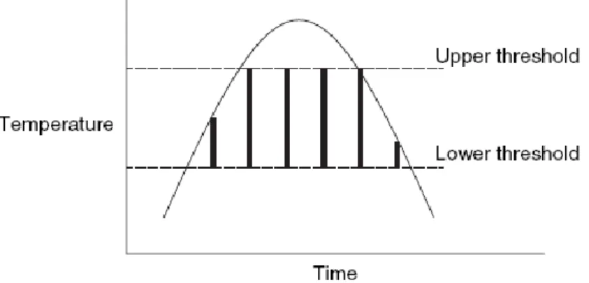
The most sophisticated technique used to estimate the rate of development of insects is named “accumulated degrees hours” or “accumulated degree days”, and it is based on the sum of temperature (in ºC) multiplied by time (Amendt et al., 2007). This model has a lower development threshold, i.e., the temperature value below which insect development stops (diapausa), and an upper development threshold, i.e., the temperature value above which the development slows down and can be lethal at some point. Between these two points there is a linear relationship between temperature and development rate (Fig. 2) (Amendt et al., 2007; Gennard, 2007).
It is required a specific number of accumulated degree hours to each stage complete their development (Amendt et al., 2011).
Fig. 2. Insect growth in relation to upper and lower thresholds (outlined area is the area
of growth) (Gennard, 2007).
The presence of a large number of larvae at the same location is an important factor that must be taken into account. The metabolic heat generated by a mass of maggots can be significantly higher than the ambient temperature, which is going to affect their development and consequently PMI estimation (Amendt et al., 2007; Wells & Lamotte, 2010).
Diptera specimens are good indicators of PMI over short periods of time due to their fast development. However, when they are no longer developing on carrion, Coleoptera species are also useful (Catts, 1992).
14
In cases of long postmortem periods, the first colonizers of the body have already left, so PMI estimation must be based on the succession pattern of arthropods found on a cadaver, although the two methods should be complementary (Amendt et
al., 2007; Kreitlow, 2010).
The succession pattern on corpses can be divided into different waves that correspond to different stages of decay (Amendt et al., 2004). Each of these stages is attractive to a different group of insects. This produces a faunal succession on corpses that can be used to determine PMI together with the knowledge of growth rates under specific environmental conditions (Benecke, 1998). This process can be used to estimate both minimum and maximum PMI. Succession data have been used to calculate a PMI up to 52 days and can be applied to a much longer time interval (Amendt et al., 2004; Wells & Lamotte, 2010).
1.3.2. Forensic Implications of Myiasis
The study of insects can also give information about cases of neglect or abuse (Benecke & Lessig, 2001). Some insects are attracted to ammonia odour, resulting from urine or faecal contamination. Patients in hospitals, elderly people or babies, who have not been assisted in their personal hygiene, occasionally suffer of an infection called myiasis (Hall & Huntington, 2010; Gennard, 2007).
Myiasis (from the Greek myia = fly) can be defined as the invasion of an organ or tissue of live humans or vertebrate animals by dipterous larvae, which feed on living or necrotic tissues, like wounds, ulcers or natural openings of the body (Catts & Mullen, 2002; Service, 2004). It can be classified by the level of dependency of the parasite to the host. There are three main categories: accidental, obligatory and facultative. Facultative parasites, such as blowflies (Calliphoridae) and fleshflies (Sarcophagidae) are the main group involved in this type of cases (Amendt et al., 2011). If a person was already infested when death occurred, insects present at that time could cause an overestimation of PMI (Wells & Lamotte, 2010). Entomological evidence recovered from the body can prove that negligence happened earlier than actual death (Benecke & Lessig, 2001).
15
1.3.3. Entomotoxicology: Insects as Toxicological Evidences
Entomotoxicology is a recent branch of Forensic Entomology that deals with the use of toxicological analysis of carrion feeding insects in order to identify drugs and toxins present in a death body (Introna et al., 2001; Carvalho, 2010). It can be applied in cases involving possible sudden death without obvious causes, criminal abuse by use of drugs, poisoning and suicide (Introna et al., 2001; Wolff et al., 2001; Gennard, 2007; Goff & Lord, 2010).
Insects, mainly larvae, which feed on a corpse, ingest and incorporate chemical metabolites of toxins into their own tissues (Wolff et al., 2001; Goff & Lord, 2010). It is possible to detect drugs and other chemical substances in insects and their chitinized remnants, recovered from badly decomposed bodies. These entomological techniques are important when the body is in an advanced stage of decomposition or when blood, urine or internal organs are not available or not suitable for analyses (Campobasso & Introna, 2001; Carvalho, 2010).
In recent years, some studies have been developed in order to understand the effects of drugs and toxins on carrion feeding insects (Introna et al., 2001; Goff & Lord, 2010). Studies showed that some drugs can delay insects colonization and also interfere with their rate of development and behaviour, which may affect an accurate PMI estimation. The decomposition process can be accelerated or decelerated depending on the substance and its concentration (Wells & Lamotte, 2010; Amendt et
al., 2011).
1.3.4. Gene Expression Analysis
Gene expression studies, a more recent application of Forensic Entomology, can help to determine the age of developing individuals (Tarone et al., 2007; Zehner et
al., 2009). There are development stages, such as eggs and pupae that exist over a
period of time without any morphological changes. Nevertheless, metamorphosis is characterized by several intrinsic processes that are regulated by several genes.
16
Therefore, a differential gene expression could be used to determine the age, since gene expression pattern is specific for each development stage.
For this type of analysis is necessary to determine mRNA quantification of specific genes through RNA extraction, followed by a reverse transcription into cDNA and quantification of this cDNA through real-time PCR (Tarone et al., 2007; Zehner et
al., 2009; Amendt et al., 2011). Although these studies are very expensive, they are
extremely important to achieve an accurate PMI estimation, since a more precise age of insects is given.
1.3.5. Additional Applications
Besides these applications, it is possible to obtain additional information through the study of insects found in a crime scene. For example, if a suspect have been bitten by an insect, it is possible to link him to the crime scene through DNA analysis from the blood ingested by the insect (DiZinno et al., 2002; Pujol-Luz et al., 2008). In some cases, it may be possible to prove that a body was moved after death from one area to another and it may provide information about the location of death itself, if insects, which live in a restricted geographical region, are found on a corpse in a different area (Wolff et al., 2001; Gennard, 2007). Besides that, the presence of live insects or their larvae in an empty house may indicate that the body has been removed after death (Benecke, 1998; Campobasso & Introna, 2001). Forensic Entomology can, as well, give information about the time of decapitation, dismemberment, postmortem artefacts and specific sites of trauma on the body because insects will lay their eggs in these places (Campobasso & Introna, 2001).
Thus, a good knowledge of general entomology is essential for the accurate interpretation of insect evidence.
Knowing the distribution, biology and behaviour of the insect species found in a place where a body has been discovered can be determinant for many types of forensic investigations by providing information on when, where and how the crime occurred (Amendt et al., 2007).
17
There are many crucial considerations and limitations in making such estimations (Higley & Haskell, 2010). Colonization pattern is different depending whether the body was buried (it will influence the time required for insects to reach the body and the species composition on the corpse), left on the soil surface or in the water (faunal succession will be different from that seen on soil surface) (Anderson, 2010; Amendt et al., 2004). Factors such as physical barriers (for example, presence of clothing), chemical barriers, climate factors, season, weather and location can influence the type and species of insects present, their development and consequently PMI estimation (Catts, 1992; Anderson, 2010; Goff, 2010).
Since there are so many parameters that can affect species colonization, it is extremely important to develop a geographical database of species with forensic interest to apply in criminal investigations (Hall & Huntington, 2010; Cainé et al., 2009). Due to ethical and logistical constrains of using human cadavers for this type of research studies, usually animal carcasses are used (Oliveira & Vasconcelos, 2010). The majority of these studies use domestic pig (Sus scrofa) as a model due to its high homology with human in internal anatomy, fat distribution, size of chest cavity, skin, intestinal flora due to omnivorous diet; consequently, the decomposition rate is similar of a human body with the same weight (Campobasso & Introna, 2001; Wolff et al., 2001; Schoenly et al., 2006; Anton et al., 2011; Horenstein et al., 2012). There are also other studies that test bait traps to capture species with forensic interest since they are a cheaper and easier alternative (Leccese, 2004; Hwang & Turner, 2005; Brundage
et al., 2011; Farinha et al., unpublished data).
In Portugal, forensic entomology studies concerning insect succession and species identification are also emerging (Gusmão, 2008; Marques, 2008; Prado e Castro & García, 2009, 2010; Prado e Castro et al., 2010a, 2010b, , 2010c, 2011, 2012) as well as studies employing DNA methodology to achieve species identification (Cainé
18
1.4. DNA Analysis
1.4.1. Species Identification
An accurate identification of insect species is the first and most crucial step in Forensic Entomology. Some species can seem similar but they may have considerably different growth rates, behaviours and habitat preferences (Zehner et al., 2004; Wells & Stevens, 2008). A misidentification may result in the application of an incorrect development data and consequently, the estimated PMI would be wrong (Benecke & Wells, 2001; Harvey et al., 2003).
Insects identification is typically based on morphological characters. This methodology often requires entomological experts, with specialized taxonomic knowledge and appropriate morphological keys, which are often incomplete or only effective for a particular life stage or gender, when species identification is mainly based on male genitalia (Harvey et al., 2003; Zehner et al., 2004; Valentini et al., 2008; Raupach et al., 2010). The proper use of morphological keys may also be affected by geographic variations or by the loss of some morphological characters, like colour patterns, due to preservation process (Wells & Stevens, 2008; Buhay, 2009). Moreover, for most immature stages, identification based on morphological characters is difficult and sometimes impossible due to similarities among species (Cainé et al., 2006; Raupach et al., 2010). For this reason, experts often prefer rearing the immature stages up to eclosion of the adult fly, and then apply identification keys to adult specimens. However, this approach is time consuming, delaying a criminal investigation, and is not always possible because the larval evidence can be already dead (Harvey et al., 2003; Amendt et al., 2011; Mazzanti et al., 2010). Besides that, the use of keys often demands such a high level of expertise that misdiagnosis are common (Hebert et al., 2003).
In order to overcome these difficulties, a molecular approach has been proposed. Molecular identification is based on species specific nucleotide sequences of certain genes (Cainé et al., 2006; Raupach et al., 2010; Amendt et al., 2011).
The development of Polymerase Chain Reaction (PCR) made a great contribution to the study of specimens at the molecular level, since through this
19
method it is possible to amplify in vitro a particular genomic sequence, allowing small amounts of DNA analysis (Gennard, 2007). This is a very fast and simple method when compared with morphological procedures. This approach also has the advantage of being applicable to any life stage of insects and even to damaged specimens, like puparium remains (Cainé et al., 2006; Mazzanti et al., 2010).
1.4.2. DNA Barcoding
Herbert et al. (2003) proposed a molecular identification system known as DNA barcoding, which provides a fast and accurate identification by using short standardized gene regions (DNA barcodes) of the target specimen (Hebert & Gregory, 2005; Hajibabaei et al., 2007). These sequences can be viewed as genetic labels that are present in every cell and each species will have a unique DNA “barcode” (Hebert et
al., 2003; Frézal & Lebois, 2008).
There is an international organization named Consortium for the Bacode Life (CBOL) that coordinates the DNA barcodes collection with the aim of building an international reference database for species molecular identification (Dawnay et al., 2007; Virgilio et al., 2010).
DNA barcoding has two main goals. The first is to use the barcode sequence to identify specimens (distinguish between known species) and the second one is to discover new species (species delimitation or description) (DeSalle et al., 2005; Frézal & Leblois, 2008).
After suitable laboratory procedures, obtained sequences are compared to reference sequences of known species, already deposited on an online database (Hebert & Gregory, 2005). For these purposes, two databases are commonly used: GenBank (www.ncbi.nlm.nih.gov/genbank/) and BOLD (www.barcodinglife.com/).
Hebert et al. (2003) proposed the use of a divergence threshold to delimit species, showing that the genetic distance between species almost always exceeds 3%. Another criterion is that the standard divergence threshold value should be ten times higher than the mean of intraspecific variation (Frézal & Leblois, 2008). However, the use of a divergence threshold to distinguish between intra and interspecific variations
20
should be done carefully, since these thresholds can suffer from statistical problems, compromising species identification (Valentini et al., 2008).
Phylogenetic reconstruction could also be used as a complementary analysis in species identification (Dawnay et al., 2007). Usually, DNA barcodes do not have sufficient phylogenetic signal to resolve evolutionary relationships, especially at deeper levels, so barcode-based trees should not be interpreted as phylogenetic trees (Hajibabaei et al., 2007). Nevertheless, they can be used to reveal the specimens closest relatives and narrow the choice of possible species (Benecke & Wells, 2001).
Since DNA barcoding is a relatively recent approach, there is a large number of undescribed species, which may constrain the representation of the overwhelming insect diversity on DNA barcode databases (Frézal & Leblois, 2008; Virgilio et al., 2010). Nevertheless, although DNA barcoding does not allow a complete taxonomic resolution, several studies demonstrate that the success in species identification exceeds 95% of the cases. Even when it fails it will reduce the option to a small number of congeneric taxa (Hebert & Gregory, 2005; Waugh, 2007).
Therefore, several studies showed that DNA barcoding is a reliable, cost effective and easy molecular identification tool for Diptera and Coleoptera species identification. This approach would be beneficial in the application of forensic insect evidences, since it allows an accurate identification in all life stages, which are often impossible to identify based on morphological characteristics (Nelson et al., 2007; Frézal & Leblois, 2008; Virgilio et al., 2010). DNA barcoding does not replace morphological taxonomists, but build alliances between molecular and morphological areas. In fact, DNA barcoding requires fully described voucher species to match the sequence of an unknown specimen to a reference sequence on a database (Hebert & Gregory, 2005; Dawnay et al., 2007; Waugh, 2007).
1.4.3. Nuclear vs Mitochondrial DNA
Molecular identification is based on the analysis of DNA sequences of the target specimen. Insects, as eukaryotic organisms, contain two distinct genomes, the nuclear and the mitochondrial genomes (Budowle et al., 2003).
21
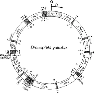
The nucleus of eukaryotic cells contains a double-stranded molecule of DNA, called nuclear DNA (nDNA) (Gennard, 2007; Lodish et al., 2008). Eukaryotic organisms also have DNA in mitochondria, named mitochondrial DNA (mtDNA), which is separated and distinct from the nuclear genome of the cell (Boore, 1999, Gennard, 2007). Insect’s mitochondria contain their own double-stranded circular genome, containing around 16,000 base pairs (bp) (Hu et al., 2009). Eukaryotic cells contain hundreds to thousands of mitochondria and each mitochondrion contains several copies of mtDNA. This property makes it easier to obtain mtDNA, especially in cases where the amount of DNA is very limited or degraded (Isenberg & Moore, 1999; Budowle et al., 2003). The number of mitochondria varies with the nature of the cell. The size of mtDNA also varies considerably between species. Despite this, the gene content of mtDNA is extremely conserved. With few exceptions, it encodes 37 genes, including 13 protein-coding subunits, 2 rRNAs of the mitochondrial ribosome and 22 tRNAs (Boore, 1999; Hwang & Kim, 1999).
Usually, the gene map of Drosophila yakuba mtDNA (Fig. 3) is used as a model, since gene arrangements of many insect species are identical to those (Boore, 1999).
Fig. 3. Gene map of Drosophila yakuba (Diptera) mtDNA molecule (Clary & Wolstenholme,
22
Mitochondrial genome contains a large non-coding region, which is involved in initiation and regulation of mitochondrial transcription and replication, known as control region or D-loop. This region is the main responsible for the differences in size of mtDNA between species (Isenberg & Moore, 1999; Hwang & Kim, 1999; Hu et al., 2009).
Mitochondrial DNA is maternally inherited, through cytoplasm (Lodish et al., 2008). Thus, the mtDNA of siblings and all maternal relatives is identical. The haploid and monoclonal nature of mtDNA simplifies the interpretation of DNA sequencing results because only one sequence can be detected (Benecke & Wells, 2001; Budowle
et al., 2003). On the other hand, does not allow the discrimination between individuals
of the same maternal lineage (Isenberg & Moore, 1999).
The mutation rate of mtDNA is several times higher than the nuclear genes probably due the low fidelity of mtDNA polymerase, inefficient repair mechanisms or the presence of free radicals formed during the phosphorylation process. The most frequently varying part of the mtDNA is the control region that appears to evolve at rates 5-10 times higher than nuclear genes (Hwang & Kim, 1999; Budowle et al., 2003; Witas & Zawicki, 2004).
The characteristics described above have made mtDNA a valuable tool in insect genomic studies. These includes a small genome with simple structure and organization, easy to isolate due their high copy number, lack of introns, haploidy, maternal inheritance, lack of recombination (which means that all differences result only from fixed changes), high mutational rates in different regions of the molecule and the availability of robust primers to amplify a target region. These features make it easy to obtain mtDNA sequences from unstudied insect species (Zhang & Hewitt, 1996; Branicki et al., 2003; Witas & Zawacki, 2004; Dawnay et al., 2007; Wells & Stevens, 2008).
1.4.4. Molecular Markers
Several molecular markers may be used for identification purposes, such as nuclear and mitochondrial DNA sequences.
23
To select a molecular marker it is important to take into account certain factors: the chosen segment of DNA must be relatively easy to isolate, easy to amplify, should have few insertions and deletions to facilitate sequence alignment and must have a mutation rate rapid enough to allow interspecific variation, allowing species discrimination, but sufficiently slow to minimize intraspecific variation (Blaxter, 2004; Hajibabaei et al., 2007; Waugh, 2007).
1.4.4.1. Cytochrome c oxidase I
Cytochrome c oxidase is a large transmembrane complex of proteins found in mitochondrion that acts as an oxidizing enzyme in the respiratory chain, which receives electrons from cytochrome complex and transfers them to oxygen molecules (Gennard, 2007; Waugh, 2007). This protein complex comprises four subunits encoded by the nuclear genome and three mitochondrially encoded, known as subunits I, II and III (Frati et al., 1997). Subunit I is the largest of the three mitochondrially encoded cytochrome oxidase subunits and was originally chosen to be used in genetic analysis due its great resolving-power for species discrimination, being the most conserved among the three subunits (Lunt et al., 1996; Hwang & Kim, 1999; Gennard, 2007; Frézal & Leblois, 2008).
DNA barcode itself consists of a 658 bp region of the cytochrome c oxidase I (COI), corresponding to nucleotide positions 1490 to 2198 of the Drosophila yakuba mitochondrial genome (Nelson et al., 2008). This mitochondrial gene has some characteristics which make it particularly suitable to be used as molecular marker. The universal primers for this gene are very robust, which enables the routine amplification of this specific segment (Hebert et al., 2003). Its size and structure are highly conserved across all aerobiotic organisms. On the other hand, the evolution of this gene is rapid enough to allow the discrimination of species and insertions/ deletions are rare (Lunt et al., 1996; Waugh, 2007).
Several studies demonstrate that COI gene has successfully been used for species identification of a large range of animal taxa (Harvey et al., 2003; Danway et
24
However, a lack of reference sequences in online databases prevents a match for a large number of species, limiting the use of COI. In addition, COI based identifications sometimes fail to distinguish closely related species, due to shared barcode sequence (Nelson et al., 2007). For example, this gene does not allow the distinction between sister species of the genus Lucilia (Raupach et al., 2010). In these cases, additional analysis of other COI segments, or even other gene, would be required to provide extra information in cases of uncertain species identification (Raupach et al., 2010; Oliveira et al., 2011). The idea of a multi-locus DNA barcoding approach has been increasingly applied in order to enlarge the power of sequence assignments (DeSalle et al., 2005; Frézal & Lebois, 2008).
The occurrence of non-funcional copies of mitochondrial sequences (nuclear mitochondrial pseudogenes – NUMTs) is an important factor that should be taken into account when COI gene is used as a molecular marker. NUMTs are copies of mitochondrial DNA sequences that have been translocated into the nuclear genome and could lead to some ambiguity in species identification since they could mimic mitochondrial copies of COI gene (Zhang & Hewitt, 1996; Frèzal & Leblois, 2008; Song
et al., 2008). When NUMTs have been alsoamplified, it is possible to see more than one band in PCR amplification or double peaks in chromatograms. Due to the random nature of nucleotide substitutions, NUMTs can also be detected when unexpected insertions/deletions, frameshifts or stop codons occur (Zhang & Hewitt, 1996; Grosso
et al., 2006). A fine phylogenetic analysis can also be useful in NUMTs recognition,
since their molecular evolution is much slower than the real mtDNA copy (Grosso et
al., 2006).
1.4.4.2. Cytochrome b
Another region of mtDNA that has been used as a molecular marker to species identification and to establish phylogenetic links is a fragment of the gene cytochrome b (Cytb) (Branicki et al., 2003; Prusak & Grzybowski, 2004; Hajibabaei et al., 2007).
25
Cytb is a transmembrane protein of the mitochondrial oxidative phosphorylation system responsible for the electron transport and thus the production of ATP, being only encoded by the mitochondrial genome. It is the central redox catalytic subunit of the ubiquinol cytochrome c reductase, an enzyme that is present in the respiratory chain of mitochondria. All eukaryotic organisms require this class of redox enzymes, and consequently Cytb, for energy conservation (Espositi et al., 1993; Prusak & Grzybowski, 2004).
This gene has been applied specially in studies with vertebrates, where the analysis proved to be a very sensitive, reliability and powerful technique, using primers given by Parson and his collaborators (Parson et al., 2000; Branicki et al., 2003). However, the use of Cytb gene as molecular marker has been applied to several species of animals, including Coleoptera (Barraclough et al., 1999; Kergoat et al., 2004; Balke et al., 2005; Pons, 2006; Sembène, 2006; Sembène et al., 2010; Ndiaye et al., 2011).
The typical variation on this region of mtDNA can be insufficient to enable unequivocal differentiation of species belonging to the same genus (Branicki et al., 2003). Although a few regions of Cytb sequences tend to be more conserved, other regions exhibit considerable variability and thus are valuable for determining the phylogenetic distance among species (Espositi et al., 1993).