Journal of Environmental
Analysis and Progress
Journal homepage: www.jeap.ufrpe.br/ 10.24221/JEAP.4.4.2019.2631.266-272 ISSN: 2525-815X
Anatomical characterization, histochemistry, and crystal analysis of the leaf
blade of Mangifera indica L.
Rafaela Damasceno Sáa, Girllene da Silva Cavalcantia, Deysielle Maria dos Santosa, Adolfo Santos da Silvaa, Rafael José Ribeiro Padilhab, Luiz Carlos Alvesb, Karina Perrelli Randaua
a Universidade Federal de Pernambuco-UFPE, Departamento de Ciências Farmacêuticas, Laboratório de
Farmacognosia. Av. Prof. Arthur de Sá, S/N, Cidade Universitária, Recife-PE, Brasil. CEP: 50740-521. E-mail: [email protected].
b UFPE, Laboratório de Imunopatologia Keizo Asami, Setor de Microscopia Eletrônica. Av. Prof. Moraes Rego, Cidade
Universitária, Recife -PE, Brasil. CEP: 50670-901.
A R T I C L E I N F O
Received 25 Jul 2019 Accepted 22 Out 2019 Published 29 Out 2019r
A B S T R A C T
Mangifera indica L., popularly known as mango, is an important fruitful widely used in Brazilian traditional medicine for the treatment of diabetes, anemia, diarrhea, hemorrhoids, indigestion, asthma, bronchitis, flu, hypertension, rheumatism, disorders of the liver and for tumors. In order to expand the pharmacobotanical information about the species, this study aimed to perform the anatomical characterization, histochemistry, and crystal analysis of the leaf blade of M. indica. For the anatomical characterization were used optical microscopy, polarization microscopy, and scanning electron microscopy. Histochemical tests were performed to evidence the presence of metabolites and microchemical analyzes by dispersive energy spectroscopy were done to determine the elemental chemical composition of the crystals. The microscopic analysis allowed the identification of characters useful in the identification of the species, such as the presence of glandular trichomes in the leaf blade. Through histochemistry was evidenced the presence of phenolic compounds, lipophilic substances, lignin, and starch in the leaf blade. The chemical nature of the crystals was determined to be calcium oxalate. The results contribute to the pharmacobotanical standardization of M. indica.
Keywords: Anacardiaceae, mangueira, quality control. Introduction
The Anacardiaceae family has
approximately 80 genera and 873 species, present mainly in tropical and subtropical regions around the world, extending to temperate regions (Pell et al., 2011). It is a family intensely studied for including species with fruits of economic importance, such as mango, cashew nut, pistachio, ambarella, yellow mombin, and red mombin (Lorenzi et al., 2015; Coelho et al., 2019).
Mangifera indica L., popularly known as mango, it is a perennial tree, 8-18 m in height, originating in India and Myanmar. It is one of the most planted fruitful in Brazil, with almost one hundred cultivars in the country (Lorenzi et al., 2015). Different parts of the plant, such as root, bark, leaves, flowers, fruits and seeds, are usually used for the treatment of diseases, such as diabetes, anemia, diarrhea, hemorrhoids, indigestion, asthma, bronchitis, influenza,
hypertension, rheumatism, liver disorders, and tumor (Shah et al., 2010; Santos, Nunes & Martins, 2012; Ribeiro et al., 2017).
The presence of bioactive compounds stimulate studies with this species, and there are several reviews pointing out the metabolites and proven pharmacological activities, such as antidiabetic, anticancer, analgesic, antipyretic, anti-inflammatory, anti-ulcer, and antibacterial (Shah et al., 2010; Ghuniyal, 2015; Jahurul et al., 2015; Parvez, 2016; Ediriweera, Tennekoon & Samarakoon, 2017; Lauricella et al., 2017).
Considering the wide distribution of this species, and the great variety of uses and interests, pharmacobotanical studies are also essential to support the correct identification of the species and to provide more information about the morphological characters. Therefore, this study aimed to carry out the anatomical and
histochemical characterization of the leaf blade of M. indica.
Material and Methods
The material was collected in the metropolitan region of Recife, Pernambuco, Brazil. The voucher specimen was deposited in the Herbarium Dárdano de Andrade Lima, of the Instituto Agronômico de Pernambuco (IPA), under registration number 91429.
Leaf-blades collected between the third and fifth nodes of three adult specimens were fixed in FAA 50 (formaldehyde, acetic acid, and ethyl alcohol) (Kraus & Arduin, 1997). For anatomical characterization, cross-sections and paradermal sections were obtained by hand, using an ordinary razor blade in the middle region of the leaf blades (Johansen, 1940; Oliveira & Akisue, 2009). Posteriorly, sections were clarified with sodium hypochlorite solution 50% (Kraus & Arduin, 1997). After washing in distilled water, the cross-sections were stained according to the technique described by Bukatsch (1972) using safranin and astra blue. Paradermal sections were stained with methylene blue 1% (Krauter, 1985). Semipermanent histological slides were prepared to contain the sections following usual plant anatomy procedures (Johansen, 1940; Sass, 1951). Images of semipermanent histological slides were used with the software LAS EZ (Leica), obtained by a digital camera (Leica ICC50 W) coupled to an optical and polarized microscope (Leica DM750M).
Considering the anatomical
characterization under Scanning Electron Microscopy (SEM), samples of fresh leaf blades were fixed in 2.5% glutaraldehyde and post-fixed using 2% osmium tetroxide solution. The material was submitted to dehydration in ethanol series until critical point drying (Bal-Tec CPD 030), mounted onto SEM stubs, using the double-sided adhesive tape and sputter-coated with gold (Leica EM SCD 500) (Haddad et al., 1998). The samples were analyzed with a scanning electron microscope (Quanta 200 FEG) in the Centro de Tecnologias Estratégicas do Nordeste (CETENE).
Histochemical tests were done on cross-sections of fresh leaf blades obtained by hand, using an ordinary razor blade (Johansen, 1940), using the reagents: potassium dichromate 10% for phenolic compounds (Gabe, 1968), vanillin chloridric for tannins (Mace & Howell, 1974), Sudan III for lipophilic substances (Sass, 1951), antimony trichloride for triterpenes and steroids (Mace, Bell & Stipanovic, 1974), Dragendorff’s reagent for alkaloids (Yoder & Mahlberg, 1976), Lugol's iodine reagent for starch (Johansen, 1940), phloroglucinol for lignin (Johansen, 1940), and hydrochloric acid 10% aiming to establish the nature of the crystals (Jensen, 1962). Controls were conducted at the same time with the tests (Johansen, 1940; Sass, 1951). Semipermanent histological slides with the cross-sections were analyzed under an optical microscope (Alltion).
The elemental composition of crystals was analyzed in cross-sections of leaf blades processed following the same methodology described for the analysis in SEM. The chemical microanalyses were done by Energy Dispersive Spectroscopy (EDS) with an X-ray detector attached to the Zeiss-EVO-LS15 scanning electron microscope.
Results and Discussion
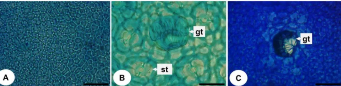
In the frontal view, the epidermal cells show slightly sinuous and thick walls on both sides of the leaf (Figure 1AB). The leaf blade is hypoestomatic, with anomocytic stomata (Figure 1B). According to Metcalfe & Chalk (1950), family Anacardiaceae is characterized by hypoestomatic or amphistomatic leaf blades. Multicellular glandular trichomes were found on both faces of the leaf (Figure 1BC). Rocha et al. (2015) did not mention the presence of trichomes in a study with the leaf of M. indica; the authors found non-glandular trichomes in the leaf of Spondias purpurea. The presence of non-glandular trichomes has also been described in the leaf of S. mombin (Vasconcelos, Vasconcelos & Randau, 2016).
Figure 1. Frontal view of the leaf blade of Mangifera indica L. under optical microscopy. A, C: adaxial face; B: abaxial face. Abbreviations: gt = glandular trichome; st = stomata. Bars: A, B = 50 µm; C = 20 µm. Fonte: Sá (2019).
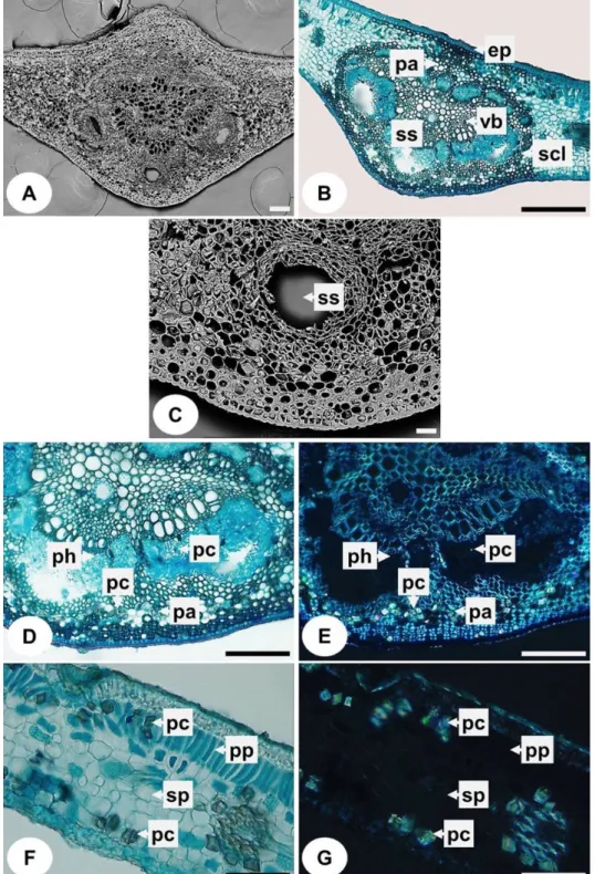
The midrib, in cross-section, is biconvex (Figure 2A). The similar shape of the midrib was also found by Rocha et al. (2015). The epidermis is uniseriate, covered by thick cuticle (Figure 2B). The vascular bundle has a biconvex shape and is collateral, surrounded by sclerenchyma (Figure 2B), corroborating some studies (Santhan, 2014; Rocha et al., 2015). In the phloem are secretory structures (Figure 2BC). The presence of
secretory structures is the main characteristic of the family Anacardiaceae (Metcalfe & Chalk, 1950). In vegetative organs, these structures are found mainly in the phloem and pith. There are studies in the literature reporting the composition of the substance of these structures and, also, the process of development (Lacchia & Carmello-Guerreiro, 2009). Prismatic crystals are visualized in the phloem and the parenchyma (Figure 2DE).
Figure 2. Cross-sections of the leaf blade of Mangifera indica L. A, C: scanning electron microscopy; B, D, F: optical microscopy; E, G: polarization microscopy. A-E: midrib; F, G: mesophyll. Abbreviations: ep = epidermis; pa = parenchyma; pc = prismatic crystal; ph = phloem; pp = palisade parenchyma; scl = sclerenchyma; sp = spongy parenchyma; ss = secretory structure; vb = vascular bundle. Bars: A, D, E = 100 µm; B = 200 µm; F, G: 50 µm; C = 50 µm. Fonte: Sá (2019).
The mesophyll presents an organization of the dorsiventral type, with one-two layers of palisade parenchyma and around six-eight layers of spongy parenchyma (Figure 2F). Santhan (2014) observed two-three layers of palisade parenchyma in the species. Prismatic crystals are visualized in the palisade parenchyma and the spongy parenchyma (Figure 2FG).
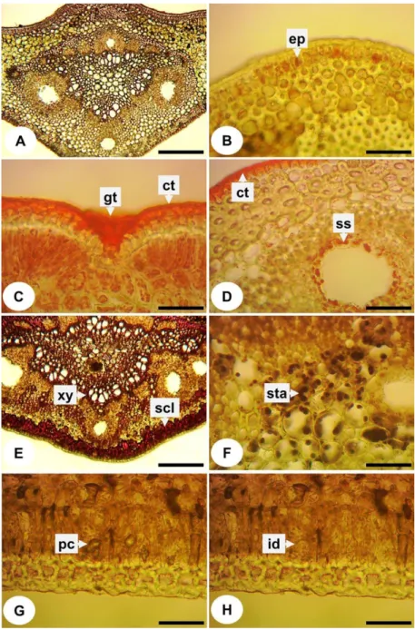
Figure 3A shows the cross-section of the leaf blade without the addition of reagents. Phenolic compounds were evidenced in the epidermis (Figure 3B) and lipophilic compounds in the cuticle (Figure 3CD), glandular trichomes (Figure 3D), and secretory structures (Figure 3D).
Lignin was observed in xylem,
sclerenchyma (Figure 3E), and starch was found in the parenchyma of the midrib (Figure 3F). The hydrochloric acid 10% showed that the crystals are of calcium oxalate, after dissolution thereof (Figure 3GH). The tests for alkaloids, tannins, triterpenes, and steroids were negative, differing from the literature (Okwu & Ezenagu, 2008; Helen et al., 2013; Somkuwar & Kamble, 2013; Nwankwo & Osaro-Mathew, 2014; Dhital, 2017; Diso et al., 2017; Divyalashmi & Sharmili, 2017). However, in these studies, phytochemical tests were performed, and the samples were collected in different periods and countries, which explains this divergence of results.
Figure 3. Histochemistry of the leaf blade of Mangifera indica L. A: control; B: potassium dichromate 10%; C, D: Sudan III; E: phloroglucinol; F: Lugol's iodine reagent; G, H: hydrochloric acid 10%. Abbreviations: ct = cuticle; ep = epidermis; gt = glandular trichome; id = idioblast; pc = prismatic crystal; scl = sclerenchyma; ss = secretory structure; sta = starch; xy = xylem. Bars: A, E = 200 µm; B, C, D, F, G, H = 50 µm. Fonte: Sá (2019).
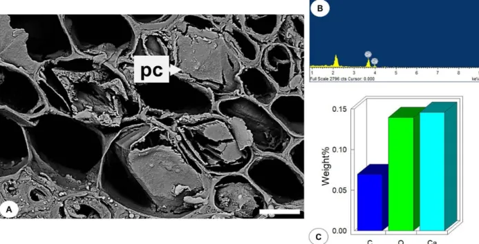
Through SEM-EDS analysis, it was possible to confirm that the chemical composition of the crystals present in the leaf of M. indica is of calcium oxalate (Figure 4ABC). Oxalic acid is an organic acid widely distributed in various organisms and a natural component of a large number of plants. It is a product where calcium ions are obtained in the environment, and oxalate ions are derived from plant metabolism (Ilarslan, 1997).
The morphology of the crystals produced may be of a single type throughout the plant, or various types, each being specific for a given organ or even several types within the same organ, but in different tissues. Despite this wide variety of distribution among species, the morphology of the crystal produced, and its distribution is constant within a species, and this constancy of crystal shape and distribution indicates the strong genetic regulation of crystal deposition (Franceschi & Nakata, 2005).
Figure 4. Scanning electron micrograph and elemental composition of the crystals of the leaf blade of Mangifera indica L. A: prismatic crystal; B: analysis of the elemental composition of the crystal; C: percentage of the chemical constituents of the crystal. Abbreviations: pc = prismatic crystal. Bar: A = 6 µm. Fonte: Sá (2019).
Conclusion
Although there are already some studies about this species, the present study provided new anatomical information useful for the correct identification of M. indica, such as the presence of glandular trichomes in the leaf. The histochemical tests allowed the determination of the sites of accumulation and synthesis of the metabolites, beyond the characterization of the chemical nature of the crystals, which was also confirmed by SEM-EDS analysis. The results contribute to the pharmacobotanical standardization of this species.
Acknowledgements
The authors are grateful to Coordenação de Aperfeiçoamento de Pessoal de Nível Superior (CAPES) for financial support through fellowship
awards and to Conselho Nacional de
Desenvolvimento Científico e Tecnológico (CNPq) for research funding (409452/016-5).
References
Bukatsch, F. 1972. Bemerkungen zur
doppelfärbung Astrablau-Safranin.
Mikrokosmos, 61, (8), 255.
Coelho, B. E. S.; Nascimento, M. M.; Carvalho, I. R. B.; Sousa, K. D. S. M.; Machado, N. S.; Neto, A. F. 2019. Curva de secagem solar e avaliação físico-química da manga" Espada". Journal of Environmental Analysis
and Progress, 4, (3), 187-194.
https://doi.org/10.24221/jeap.4.3.2019.2548. 187-194
Dhital, K. S. 2017. Phytochemical screening and antioxidant activities of Mangifera indica leaves grown in temperate region of the Nepal. Journal of Pharmacognosy and Phytochemistry, 6, (3), 205-209.
Diso, S.; Ali, M.; Mukhtar, S.; Garba, M. 2017. Antibacterial activity and phytochemical screening of Mangifera indica (Mango) stem and leaf extracts on clinical isolates of
methicillin resistant Staphylococcus aureus. Journal of Advances in Medical and Pharmaceutical Sciences, 13, (1), 1-6. https://doi.org/10.9734/JAMPS/2017/31127
Divyalashmi, L.; Sharmili, A. 2017.
Phytochemical analysis and antibacterial activity of Mangifera indica L and Piper betle. International Journal of Pharma and
Bio Sciences, 8, (2), 84-91. http://dx.doi.org/10.22376/ijpbs.2017.8.2.p8 4-91 Ediriweera, M. K.; Tennekoon, K. H.; Samarakoon, S. R. 2017. A review on ethnopharmacological applications,
pharmacological activities, and bioactive compounds of Mangifera indica (mango). A
review on ethnopharmacological
applications, pharmacological activities, and bioactive compounds of Mangifera indica (mango). Evid. Based Complementary
Altern. Med, 2017.
https://doi.org/10.1155/2017/6949835 Franceschi, V. R.; Nakata, P. A. 2005. Calcium
oxalate in plants: formation and function. Annual Review of Plant Biology, 56, (1), 41-71.
https://doi.org/10.1146/annurev.arplant.56.0 32604.144106
Gabe, M. 1968. Techniques histologiques. Paris: Masson & Cie.
Ghuniyal, J. 2015. Ethanomedical, chemical, pharmacological, toxicological properties of Mangifera indica: a review. International Journal of Pharma Research & Review, 4, (10), 51-64.
Haddad, A.; Sesso, A.; Attias, M.; Farina, M.; Meirelles, M. N.; Silveira, M.; Benchimol, M.; Soares, M. J.; Barth, O. M.; Machado, R. D.; Souto-Padrón, T.; Souza, W. 1998. Técnicas básicas de microscopia eletrônica aplicadas às Ciências Biológicas. Rio de Janeiro: Sociedade Brasileira de Microscopia Eletrônica.
Helen, P. A. M.; Aswathy, M. R.; Deepthi, K. G.; Mol, R. R.; Joseph, J. J.; Sree, S. J. 2013. Phytochemical analysis and anticancer activity of leaf extract of Mangifera indica (kottukonam varika). International Journal of Pharmaceutical Sciences and Research, 4, (2), 823-828.
Ilarslan, H.; Palmer, R. G.; Imsande, J.; Horner, H. T. 1997. Quantitative determination of calcium oxalate and oxalate in developing seeds of soybean (Leguminosae). American Journal of Botany, 84, 10422-1046. https://doi.org/10.2307/2446147
Jahurul, M. H. A.; Zaidul, I. S.; Ghafoor, K.; Al-Juhaimi, F. Y.; Nyam, K. L.; Norulaini, N. A.; Sahena, F.; Mohd Omar, A. K. 2015. Mango (Mangifera indica L.) by-products and their valuable components: A review.
Food Chem., 183, 173-180.
https://doi.org/10.1016/j.foodchem.2015.03. 046
Jensen, W. A. 1962. Botanical histochemistry, principles and practice. San Francisco: W. H. Freeman.
Johansen, D. A. 1940. Plant microtechnique. New York: McGraw-Hill Book Co. Inc.
Kraus, J. E.; Arduin, M. 1997. Manual básico em métodos de morfologia vegetal. Rio de Janeiro: EDUR.
Krauter, D. 1985. Erfahrungen mit Etzolds FSA-Färbung für pflanzenschnitte. Mikrokosmos, 74, 231-233.
Lacchia, A. P. S.; Carmello-Guerreiro, S. M. 2009. Aspectos ultra estruturais dos canais secretores em órgãos vegetativos e reprodutivos de Anacardiaceae. Acta Botanica Brasilica, 23, (2), 376-378.
http://dx.doi.org/10.1590/S0102-33062009000200009
Lauricella, M.; Emanuele, S.; Calvaruso, G.;
Giuliano, M.; D'Anneo, A. 2017.
Multifaceted Health Benefits of Mangifera indica L. (mango): the Inestimable Value of Orchards Recently Planted in Sicilian Rural
Areas. Nutrients, 9, (5), E525.
https://doi.org/10.3390/nu9050525.
Lorenzi, H.; Lacerda, M. T. C.; Bacher, L. B. 2015. Frutas no Brasil: nativas e exóticas (de consumo in natura). São Paulo: Instituto Plantarum de Estudos da Flora.
Mace, M. E.; Bell, A. A.; Stipanovic, R. D. 1974. Histochemistry and isolation of gossypol and related terpenoids in root of cotton seedlings. Phytopathology 64, 1297-1302.
Mace, M. Z.; Howell, C. R. 1974. Histochemistry and identification of condensed tannin precursors in roots of cotton seedlings. Can.
J. Bot. 52, (11), 2423-2426.
https://doi.org/10.1139/b74-314
Metcalfe, C. R.; Chalk, K. L. 1950. Anatomy of the dicotyledons: leaves, stem, and wood in relation to taxonomy with notes on economic uses. Oxford: Clarendon.
Nwankwo, I. U.; Osaro-Mathew, R. C. 2014.
Assessment of the phytochemical
components of Mangifera indica (leaf) and Musa paradisiaca (roots) extracts and their antibacterial activity against some common pathogenic bacteria. Journal of Pharmacy and Biological Sciences, 9, (1), 8-11.
Okwu, D. E.; Ezenagu, V. 2008. Evaluation of the
phytochemical composition of mango
(Mangifera indica Linn) stem bark and leaves. International Journal of Chemical Sciences, 6, (2), 705-716.
Oliveira, F.; Akisue, G. 2009. Fundamentos de farmacobotânica e de morfologia vegetal. 3ª ed. São Paulo: Atheneu.
Parvez, G. M. M. 2016. Pharmacological activities of mango (Mangifera indica): a review. Journal of Pharmacognosy and Phytochemistry, 5, (3), 1-7.
Pell, S. K.; Mitchell, J. D.; Miller, A. J.; Lobova, T. A. Anacardiaceae. In: Kubitzki, K. 2011.
Eudicots: Sapindales, Cucurbitales,
Myrtaceae. India: Springer-Verlag Berlin Heidelberg, p. 7-50.
Ribeiro, R. V.; Bieski, I. G. C.; Balogun, S. O.; Martins, D. T. O. 2017. Ethnobotanical study of medicinal plants used by
Ribeirinhos in the North Araguaia
microregion, Mato Grosso, Brazil. Journal of
Ethnopharmacology, 205, 69-102.
https://doi.org/10.1016/j.jep.2017.04.023 Rocha, L. A.; Rocha, A. M.; Pacheco, A. C. L.;
Abreu, M. C. 2015. Diferenças foliares morfoanatômicas de quatro espécies da família Anacardiaceae. Caderno de Pesquisa,
série Biologia, 27, (2), 35-48.
http://dx.doi.org/10.17058/cp.v27i2.6798 Santhan, P. 2014. Leaf structural characteristics of
important medicinal plants. International Journal of Research in Ayurveda and
Pharmacy, 5, 6, 673-679.
http://dx.doi.org/10.7897/2277-4343.056137 Santos, M. M.; Nunes, M. G. S.; Martins, R. D.
2012. Uso empírico de plantas medicinais para tratamento de diabetes. Revista Brasileira de Plantas Medicinais, 14, (2), 327-334. http://dx.doi.org/10.1590/S1516-05722012000200012
Sass, J. E. 1951. Botanical microtechnique. 2nd. ed. Ames: The Iowa State College Press. Shah, K. A.; Patel, M. B.; Patel, R. J.; Parmar, P.
K. 2010. Mangifera indica (Mango). Pharmacognosy Review, 4, (7), 42-48. http://dx.doi.org/10.4103/0973-7847.65325 Somkuwar, D. O.; Kamble, V. A. 2013.
Phytochemical screening of ethanolic extracts of stem, leaves, flower and seed kernel of Mangifera indica L. International Journal of Pharma and Bio Sciences, 4, (2), 383-389.
Vasconcelos, A. L.; Vasconcelos, A. L.; Randau,
K. P. 2016. Pharmacognostic
characterization of Spondias mombin L. (Anacardiaceae). Pharmacognosy Journal, 8,
(6), 513-519.
http://dx.doi.org/10.5530/pj.2016.6.1
Yoder, L. R.; Mahlberg, P. G. 1976. Reactions of alkaloid and histochemical indicators in laticifers and specialized parenchyma cells of Catharanthus roseus (Apocynaceae). American Journal of Botany, 63, (9),
1167-1173.