UNIVERSIDADE DE LISBOA
FACULDADE DE CIÊNCIAS DA UNIVERSIDADE DE LISBOA
DEPARTAMENTO DE BIOLOGIA VEGETAL
DISSERTAÇÃO
Alpha-synuclein phosphorylation role on
Parkinson’s disease
Filipa Magalhães Silva
Mestrado em Biologia Molecular e Genética
UNIVERSIDADE DE LISBOA
FACULDADE DE CIÊNCIAS DA UNIVERSIDADE DE LISBOA
DEPARTAMENTO DE BIOLOGIA VEGETAL
Alpha-synuclein phosphorylation role on
Parkinson’s disease
Filipa Magalhães Silva
Mestrado em Biologia Molecular e Genética
Dissertação orientada por:
Professor Doutor Tiago Fleming Outeiro
Cell and Molecular Neuroscience Unit – Instituto de Medicina Molecular (IMM)
Professora Doutora Maria Margarida Blasques Telhada
Faculdade de Ciências da Universidade de Lisboa
Todas as afirmações efetuadas no presente documento são de exclusiva responsabilidade do seu autor, não cabendo qualquer responsabilidade à Faculdade de Ciências da Universidade de Lisboa pelos conteúdos nele apresentados.
i Resumo
A doença de Parkinson (DP) é uma doença neurodegenerativa caracterizada pela presença de inclusões proteicas designadas por corpos de Lewy (LB). Os LB são agregados intracelulares que se formam nos neurónios dopaminérgicos, localizados na substantia nigra, mas que podem ocorrer em outras áreas do cérebro à medida que a doença progride. Os LB são constituídos principalmente pela proteína alfa-sinucleína (aSyn), sendo esta a principal característica desta doença. A aSyn é uma proteína envolvida tanto nos casos de doença idiopática, como familiar da DP. Vários resíduos de aSyn são fosforilados quer na forma solúvel quer na forma agregada da proteína. Sendo que, aproximadamente 90% da aSyn presente nos agregados é fosforilada no resíduo serina 129 (S129), em contraste com apenas 4% de fosforilação neste resíduo em cérebros saudáveis. Apesar da fosforilação no resíduo S129 ser a mais bem estudada, pouco se compreende sobre o seu papel na doença. Por outro lado, estudos recentes realizados em culturas de células, em Drosophila e em cerébros humanos demonstraram a existência de fosforilação no resíduo tirosina 125 (Y125). Sabe-se que a fosforilação deste resíduo suprime a oligomerização. Para além disso, durante o envelhecimento, a fosforilação Y125 é naturalmente reduzida, sendo essa redução mais notável nos pacientes com DP. A falta de informação sobre a fosforilação no resíduo Y125 está relacionada com a dificuldade de detectar aSyn fosforilada neste resíduo em cérebros de humanos. Para além disso, é difícil estabelecer a ligação entre a progressão da doença e os níveis de aSyn fosforilados no resíduo de Y125. Devido a todas estas razões torna-se importante estudar o papel da fosforilação da aSyn nos resíduos S129 e Y125 na patogénese da DP.
A levedura Saccharomyces cerevisiae (S. cerevisiae) é um organismo eucariota e unicelular versátil que apresenta uma organização intracelular complexa e cujas vias metabólicas são conservadas. Deste modo, este modelo é amplamente utilizado para compreender processos biológicos complexos encontrados em eucariotas superiores. S. cerevisiae é um modelo de estudo validado para a investigação da DP, sendo que já foi demonstrado que aSyn humana expressa em células de levedura induz toxicidade e formação de inclusões citoplasmáticas à semelhança do que foi observado noutros modelos biológicos, nomeadamente em modelos celulares de mamífero e em modelos animais de PD. Para além disso, existem várias vantagens para a utilização da levedura, como um organismo modelo, tais como a facilidade de manipulação em laboratório; o fato do seu genoma estar totalmente sequenciado; de apresentar um tempo de geração curto e também devido ao fato de ser possível manipular geneticamente através da aplicação simples de métodos clássicos de genética molecular.
ii O principal objetivo do presente trabalho foi estudar o papel da fosforilação de aSyn na etiologia da DP, no que diz respeito ao efeito a nível de toxicidade e propensão para formar inclusões, utilizando como modelos S. cerevisiae e células humanas. Para isso, foram utilizadas três formas de aSyn: a forma selvagem (WT) e as formas não fosforiladas nos resíduos Y125 (Y125F) e S129 (S129A ou S129G), respetivamente. Observou-se que o bloqueio da fosforilação no resíduo Y125 (Y125F aSyn) diminui a percentagem de células com inclusões, enquanto, que o bloqueio da fosforilação no resíduo S129 (S129A aSyn ou S129G aSyn) aumenta a percentagem de células com inclusões. Em paralelo, observou-se que a mutação Y125F não altera a toxicidade da aSyn expressa na levedura S. cerevisiae enquanto, que a mutação S129G aumenta a toxicidade desta.
Para validar os resultados em levedura, foi utilizado um modelo de agregação de aSyn, previamente descrito, numa linha celular humana de neuroglioma (H4). Neste modelo, a co-transfecção de uma forma modificada de aSyn (fundida a uma forma truncada de proteína verde fluorescente (GFP), e designada por SynT) juntamente com sinfilina-1, uma proteína neuronal também identificada nos LBs, resulta na formação de inclusões citoplasmáticas, as quais foram observadas através do método de imunocitoquimica. Utilizou-se este modelo estabelecido de agregação de aSyn para investigar o papel das mutações Y125F SynT e S129G SynT na formação de inclusões, em comparação com a situação controlo, WT SynT. Observou-se que as mutações Y125F SynT e S129G SynT resultaram no aumento do número de células com inclusões, o que indica que o bloqueio da fosforilação nos resíduos Y125 e S129 aumenta a propensão da aSyn para agregar no modelo de agregação H4. Além disso, observou-se que o padrão de formação de inclusões na mutação Y125F SynT era diferente do observado na WT SynT e S129G, apresentando agregados mais pequenos.
Neste estudo mostrou-se que existem evidências de uma correlação entre fosforilação, agregação e toxicidade celular de aSyn usando o modelo simples, mas poderoso da levedura S. cerevisiae, e um modelo de células neurogliais humanas. Os resultados obtidos também contribuíram para a compreensão da base molecular da doença de Parkinson, tal como de outros distúrbios neurodegenerativos (isto é, outras sinucleionopatias, como, multiple system atrophy (MSA), pure autonomic failure (PAF), Doença de Lewy bodies). É importante referir que a forma não fosforiladado do resíduo Y125, neste caso, a mutação Y125F, deve ser considerada para estudos posteriores, pois obtivemos resultados diferentes quando se bloqueou a sua fosforilação quer em células de levedura quer em células neurogliais humanas. Isto pode significar que não há uma conservação de mecanismos entre espécies ou então que os resultados obtidos dependem do facto da sinfilina-1 não estar presente em levedura; será algo que terá interesse aprofundar. Também se pode explicar a discrepância de resultados com o facto de o modelo
iii H4 se basear na co-expressão de sinfilina-1, uma proteína que não tem homologo em levedura mas quando expressa em levedura se sabe aumentar a propensão da aSyn para agregar. A fosforilação de Y125 aSyn, apesar de pouco conhecida e estudada considera-se muito importante, pois muitos estudos sugerem que uma alteração neste resíduo pode estar relacionada com a causa da doença neurodegenerativa e que pode ter uma influência negativa sobre a oligomerização, e consequentemente na agregação de aSyn.
Por outro lado, a mutação S129G aSyn conduz ao aumento da formação de inclusões em levedura e a formação de inclusões nas células H4. Assim, concluímos que S129 é um resíduo que tem efeitos semelhantes nos dois modelos usados, sendo que provavelmente a sua fosforilação é um mecanismo conservado entre espécies. Seria de extremo interesse de futuro, estudar em mais detalhe as cinases e as fosfatases envolvidas na regulação da fosforilação deste resíduo. Em conjunto, estes novos resultados demonstram que diferentes resíduos fosforilados na aSyn resultam em diferentes níveis de agregação e toxicidade, sendo que os efeitos da fosforilação de S129 se mostraram indubitavelmente consistentes entre espécies. Por outro lado, a fosforilação de Y125 não apresentou essa mesma consistência. Uma explicação possível pode estar relacionada com o fato das cinases/fosfatases que regulam tirosinas em aSyn nas células humanas poderem não estar tão conservadas em células de levedura como as vias que fosforilam/desfosforilam serinas. Deste modo, torna-se interessante de futuro estudar os dois resíduos em simultâneo e verificar o que acontece quando os dois locais estão disponíveis ou não para serem fosforilados.
O presente trabalho contribuiu para esclarecer o papel de uma modificação pós-traducional, a fosforilação, no processo de agregação de aSyn, no contexto da DP. Conhecer melhor estes mecanismos moleculares pode vir a permitir o desenvolvimento de novas estratégias para intervenção na DP, tal como noutras sinucleinopatias.
iv Abstract
Alpha-synuclein (aSyn) is a protein involved in both idiopathic and familial Parkinson´s disease (PD) cases, being the major component of Lewy body inclusions (LBs), the typical pathological hallmark of this disorder. It is estimated that ~90% of the aggregated aSyn in LBs is phosphorylated in S129 residue, in contrast with only 4% of phosphorylation in the same residue in normal brain.
Saccharomyces cerevisiae is a versatile eukaryotic organism that has being extensively used to understand the complex biological processes found in higher eukaryotes and it is a validated model in PD. Indeed, it has already been shown that aSyn induces toxicity and inclusion formation in a similar manner to that observed in other cell and animal model systems.
The objective of this work was to study the role of phosphorylation of aSyn in the etiology of PD. For this, we used three forms of aSyn: wild shape (WT) and non-phosphorylated forms in the residues Y125 (Y125F) and S129 (S129A or S129G), respectively. We observed that blocking the phosphorylation at residue Y125 reduced the percentage of cells with foci while blocking the phosphorylation at residue S129 increased the percentage of cells with foci. In parallel, we observed that the Y125F mutation did not change the toxicity of aSyn expressed while the S129G mutation increased its toxicity. Subsequently, the aggregation resulting from the expression of Y125F aSyn and S129G aSyn was analyzed in the mammalian cell aggregation model based on co expression SynT and synphilin-1. Both mutations increased the formation of aSyn inclusions in those cells.
Taken together, these new results provide an important insight into the role of phosphorylation in the aggregation process. A better understanding of these molecular mechanisms may enable the development of new strategies for intervention in PD and other synucleinopathies.
v List of Abbreviations
aa amino acid
AD Alzheimer’s disease
aSyn alpha-synuclein
BCA bicinchoninic acid
bp base pairs
BSA bovine serum albumin
C Celsius
CO2 carbon dioxide
CYC1 cytochrome-c oxidase
DLB dementia with LBs
DNA desoxyribonucleic acid
EDTA Ethylenediaminetetraacetic acid
E.coli Escherichia coli
FBS Fetal Bovine Serum
GAL Galactose
GAPDH glyceraldehyde-3-phosphate dehydrogenase protein
GFP green fluorescent protein
HD Huntington’s disease
H4 neuroglioma cells
kDa kilo Dalton
LB Luria-Bertani Broth
LBs Lewy bodies
LB/Amp Luria-Bertani Broth/ampicilin
LB/Agar Luria-Bertani Broth/agar
LiAc lithium acetate
mM Millimolar
MCS multiple cloning site
mL Milliliter
MSA multiple system atrophy
NAC non-amyloid componente
Ng nanogram
NGS normal goat serum
OD 600 nm optical density read at 600 nm
vi
PAGE Polyacrylamide gel electrophoresis
PBS phosphate buffered saline
PCR polymerase chain reaction
PD Parkinson’s disease
PEG polyethyleneglicol 4000
PFA Paraformaldehyde
pS129 aSyn aSyn phosphorylated at S129 pY125 aSyn aSyn phosphorylated at Y125
Raf rafinose
rpm revolutions per minute
RT room temperature
SC synthetic complete
SDS Sodium dodecyl sulfate
SN substantianigra pars compacta
SynT aSynuclein fused with a non-fluorescent GFP fragment
S129 serine 129
S129A 129 replaced by alanine
S129G 129 replaced by glycine
TAE Tris acetate EDTA
TCA trichloroacetic acid
TE Tris-EDTA
WT wild type
YEP Yeast Extract Peptone
YPD Yeast extract Peptone Dextose
Y125 tyrosine 125
Y125F 125 replaced by phenylalanine
vii Acknowledgements
I would like to thank Prof. Dr. Tiago F. Outeiro for the opportunity and support throughout this thesis.
I am very grateful to Dr. Sandra Tenreiro for her direct supervision, for the dedication, and great incentive for the development of this project at every stage of the dissertation process. I would also like to thank her for believing in me, for keeping me focused and for her friendship.
Thank you to the UNCM team for all the dedicated time, help and transmission of knowledge scientific and for the personal and scientific growth each one provided me. Special thanks to Pedro Antas for all the support and the help during this project; to Leonor Fleming for the constructive critiques and scientific discussions; to Elissa Basso for all the help in the experiments and the good times and Susana Gonçalves for constant encouragement and for her sympathy. I thank them all for their friendship.
I also want to thank to Prof. Dra. Maria Telhada to accept my co-supervisor.
I would like to thank Donata Wawrzycka (University of Wroclaw, Poland) for kindly providing haploid strains with double insertions of the appropriate aSyn.
I would also like to thank António Temudo and José Rino for all the technical support with microscopy.
Last but not least, I would like to give a special “thank you” to my family for always believing in me. To my mother Isabel Magalhães, to my father Agostinho Silva and my brother André Magalhães for being my support and strength and for their words of advice. A special thanks to Manuel António for constant encouragement, support and friendship. It is a real pleasure being with you. I would like to thank my friends Marilia, Catarina and João for their friendship and for the good and not so good times and especially to Rita for helping me in the process of writing this thesis.
viii Index Resumo... i Abstract... iv List of Abbreviations... v Acknowledgements ... vii Index………..………. viii List of Figures... xi
List of Tables... xiii
1.Introduction... 1
1.1. Parkinson’s Disease and Synucleinopathies... 1
1.2. Alpha-synuclein... 2
1.3. Alpha-synuclein phosphorylation... 3
1.4. Yeast as a model to study neurodegenerative disease... 4
1.4.1. Saccharomyces cerevisiae Life Cycle... 5
1.5. Mammalian cells lines model of aSyn aggregation... 6
1.5.1. SynT and Synphilin-1... 6
2. Aims... 7
3. Materials and Methods... 8
3.1 Molecular biology techniques used in Escherichia coli... 8
3.1.1. Plasmids used in this study………...………..…………. 8
3.1.2. Growth Media………..………..…….…… 8
3.1.3. Transformation of E.coli Supercompetent cells ……… 8
3.1.4. Extraction and purification of plasmids... 8
3.1.5. Restriction Digestion……….……….……… 9
3.1.6. DNA extraction from agarose gel ………..………. 9 3.1.7. Generation of Y125F and S129G phosphor-mutants in yeast and m mammalian vectors………..………... 9
ix
3.1.8. Cloning strategy of yeast integrative vectors ………...….. 10
3.2. Molecular biology techniques used in S.cerevisiae... 11
3.2.1. Yeast strains ………...………. 11
3.2.2. Growth media………..……….……... 11
3.2.3. Yeast transformation………... 11
3.2.4. Construction of haploid yeast strains with single genome insertions of the aSyn forms………... 12
3.2.5. Growth conditions and spot assays………..…… 13
3.3. Molecular biology techniques used in Human neuroglioma cells………...…... 13
3.3.1. Construction of psi Y125F and S129G aSyn mammalian express vectors………..……….. 13
3.3.2. Cell culture and cell transfection………..………. 13
3.3.3. Immunocytochemistry…………...………..……… 14
3.4. Fluorescence microscopy and image analysis………...…….. 14
3.5. Protein extraction………...………..………….. 15
3.6. Western blot analysis... 15
3.7 Data and statistical analysis………...….……….. 15
4. Results... 16
4.1. Construction of yeast strains with double insertions of the Y125F, S129G o or WT SNCA humangene... 1 16 4.1.1. Construction of yeast integrative expression vectors……….………… 16
4.1.2..Construction of yeast strains with single insertions of the SNCA gene……….. 17
4.1.3..Construction of yeast strains with double insertions of the SNCA gene……….. 19
4.2. Using yeast as a model to study aSyn phosphorylation………... 20
4.2.1. Effect of Y125F on aSyn toxicity and inclusions formation…... 20
4.2.2..Effect of S129G on aSyn toxicity and inclusion formation in comparison to S129A aSyn………. 21
x
4.3. Using H4 aggregation cell model to study aSyn phosphorylation……… 23
4.3.1. The effects of aSyn phosphorylation on inclusions formation... 23
5. Discussion and Conclusions ... 26
6. References... 29
7. Annexes... 33
Annex 7.1………...………..……… 33
Annex 7.2………..……….……….…….……… 34
xi List of Figures
Figure 1. Schematic representation of aSyn and the positions of genetic
mutations……….………...………. 2
Figure 2. aSyn phosphorylation sites……....………. 4
Figure 3. The life cycle of S. cerevisiae……….………...………. 5
Figure 4. Schematic representation of SynT………..……..………. 6
Figure 5. Maps of the yeast expression vectors (A) pRS304-GAL and (B) pRS306-GAL………... 11
Figure 6. Maps of the yeast expression vectors of S129G aSyn in (A) pRS304-GAL and (B) pRS306- GAL plasmids ………. 16
Figure 7. Schematic representation of aSyn nucleotide and amino acid sequence………...………..………... 17
Figure 8. Construction of yeast strains with single genome insertion of the indicated phosphor-mutants…………..………... 18
Figure 9. Representative electrophoresis to confirm genome insertions with plasmids containing different auxotrophies.………. 18
Figure 10. Haploid strains obtained from dissected spores from independent tetrads and independent matings…..………..………. 19
Figure 11. Effect of Y125F on aSyn toxicity in yeast cells carrying single or double insertions of the encoding gene..……… 20
Figure 12. Effect of Y125F on aSyn foci formation in yeast cells carrying single or double insertions of the encoding gene ………. 21
Figure 13. Effect of S129G on aSyn toxicity in yeast cells carrying single or double insertions of the encoding gene………... 21
Figure 14. S129G aSyn shows a similar phenotype to S129A aSyn….……… 22
Figure 15. S129G aSyn shows similar foci formation to S129A aSyn in yeast cells...……… 22
Figure 16. S129 phosphorylation levels of WT aSyn, Y125F aSyn, S129G aSyn and S129A aSyn expressed in yeast cells, determined by immunoblotting analysis..……….. 23
Figure 17. Representative images of the phenotypes observed in the H4 cells co-transfected with WT, Y125F or S129A aSyn and synphilin-1……….. 24
Figure 18. Effect of S129G and Y125F mutations on SynT inclusions formation in H4 cells………..
xii Figure 19. Immunoblotting analysis of aSyn Y125F and S219G mutants co-expressed
xiii List of Tables
Table 1. Primers used for site-directed mutagenesis………. 10 Table 2. Primers used for confirmation of the insertion of the integrative vectors in the
expected genome locus………...…...………... 12
Table 3. Plasmids used in this study……….. 33
Table 4. S. cerevisiae strains used in this study……….. 34 Table 5. Growth media used for S. cerevisiae in this study………...………… 36
1 1. Introduction
1.1. Parkinson’s Disease and other synucleinopathies
Parkinson’s disease (PD), originally described by James Parkinson in 1817, is the second most common late-onset neurodegenerative disorder after Alzheimer’s disease [1]. The neuropathological hallmark of the disease is represented by the deposition of alpha-synuclein (aSyn) in the Lewy Bodies (LB), which are intracellular aggregates characteristically found in the dopaminergic neurons affected by the disorder [2]. PD is known to affect around 1% of the population over the age of 65 [3] and 4-5% of the population by the age of 85 [2]. It is clinically characterized by tremor, rigidity, akinesia (reduction in movement) and bradykinesia (slowed movement) correlated to the degeneration of dopaminergic innervation involved in the movement [4]. In fact, the initial defects in the motor system result from the progressive loss of the dopaminergic neurons in the substantia nigra pars compacta (SNpc), which consequently cause a reduction in the dopamine levels. In the late stage of the disease the neurodegeneration affects also the other brain regions, with patients experiencing non-motor symptoms such as autonomic dysfunction, sleep disturbances and neuropsychiatric symptoms [5, 6].
The majority of the PD cases are sporadic (about 95%) with unknown aetiology, demonstrating that the disease may result from a combination of environmental, epigenetic and genetic factors [6, 7]. Indeed, environmental factors such as pesticides, have been implicated as risk factors for PD pathogenesis [8, 9]. The genetic factors may then represent susceptibility to disease that, either acting alone or in combination with other environmental factors, might predispose an individual to PD [10, 11] . Only 5% of the cases are familiar and are associated to specific gene mutations [12]. In the last years several mutations responsible for the disease were discovered [10]. These studies suggest the presence of common mechanisms underlying both familial and sporadic forms, like the presence of specific protein within the LBs. It is believed that these bodies may present a protective mechanism of the disease, where dysfunctional and misfolded proteins are sequestered, although there are increasing evidences suggesting a toxic role for aSyn intermediate oligomers species formation [13, 14].
In this scenario, synucleinopathies constitute a distinct group of neurodegenerative disorders that share a common pathologic hallmark, the presence of aSyn aggregates in surviving neurons. These disorders are characterized by the deposition of intracellular inclusions composed largely of fibrillar aSyn, although the distribution of aSyn aggregates is significantly different according to the disease [14-16]. This group of neurodegenerative disorders includes Parkinson’s disease (PD), dementia with Lewy bodies (DLB), Lewy body
2 variant of Alzheimer’s disease, multiple system atrophy (MSA) and pure autonomic failure (PAF) [17-19].
These disorders are characterized by different molecular and cellular pathological mechanisms, however, they point out to a key role for aSyn misfolding that may allow us to understand if aSyn intracellular inclusions are a cause or a consequence of neuronal death, and help us to design new therapeutic strategies [20].
1.2. Alpha-synuclein
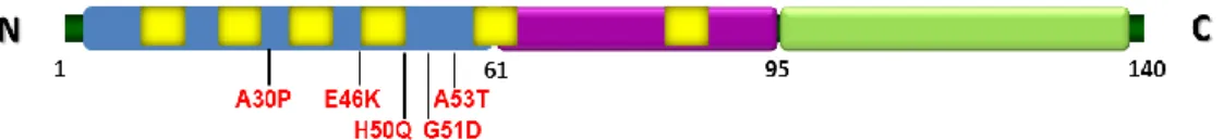
aSyn was initially associated with PD pathogenesis by being characterized as a primary component of the LB, and by the subsequent discovery of early onset familial PD cases caused by three different autosomal dominant missense mutations (A30P, E46K and A53T) present in the gene encoding aSyn [2, 21, 22]. Recently, new missense mutations, H50Q and G51D, were discovered, but few is known about it [23, 24]. Also duplications and triplications of its locus were associated with the early onset of PD pathogenesis [25, 26].
aSyn is a 14.5 kDa protein of 140 amino acids, with three major domains [27]. The N-terminal is an amphipatic lysine rich domain that is characterized by containinng a highly conserved region of seven 11-residue imperfect repeats with a hexameric motif (KTKEGV) [28, 29]. The hydrophobic central region, comprising residues 61-95, also known as NAC domain, with two additional KTKEGV repeats [30, 31]. The carboxy terminal (C-terminal) domain enriched in acidic residues is negatively charged, comprises the majority of post-translational modification sites and has been involved in the majority of aSyn proteins interactions [32-34] (Fig.1).
The function of aSyn is not yet truly understood, but several studies suggest that this protein might have a role in synaptic development, function and plasticity [35, 36]. aSyn found only in vertebrates, is expressed throughout the central nervous system and particularly enriched in neural pre-synaptic terminals, where may function as a chaperone-like protein or may be involved in synaptic vesicular regulation [37]. Numerous factors have been reported to affect the aggregation process of aSyn, however, few proteins and compounds have been identified as able to inhibit its aggregation propensity [38]. In addition aSyn is known to interact with several proteins, such as synphilin-1, in synaptic vesicles [39].
Figure 1. Schematic representation of aSyn and the positions of genetic mutations. The N-terminal
3
repeats are represented in yellow. In purple the NAC domain. In green the C-terminal domain. (adapted from Tenreiro S and Outeiro, unpublished).
1.3. Alpha-synuclein phosphorylation
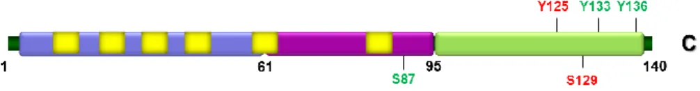
Several aSyn post-translational modifications, such as nitrosylation, ubiquitinylation, oxidation, truncation and phosphorylation have been identified and significant advances have been made towards the identification of new ones. Among these, phosphorylation is strongly linked to the pathogenesis of PD. In fact, among the aSyn post-translational modifications present in the LB, phosphorylation in Serine 129 (S129) is the most frequent [17, 40-42].
Phosphorylation regulates the structural and functional properties of proteins in health and disease conditions. The role of phosphorylation (a reversible post-translational modification) in the aggregation and toxicity of aSyn is currently the subject of intense investigation [41] .
Phosphorylation of aSyn at one or multiples sites may play an important role in the regulation of LB formation and neurotoxicity in vivo, but also on its physiological function [43, 44]. Several studies showed that aSyn is predominantly phosphorylated on serine residues. Residues S87 and S129 were identified as possible phosphorylation sites. S129 is the more phosphorylation site in the C-terminal region of aSyn [40-42, 45] . Phosphorylation of aSyn is known to occur also on tyrosine residues 125, 133 and 136 (Y125, Y133, and Y136), but very little is known about its biological significance [43, 46].
aSyn phosphorylation status influences the affinity of aSyn for other proteins and thus alters the biological processes regulated by these interactions. One way to study aSyn toxicity or aggregation related with phosphorylation, is by mimicking this modification by mutating specific serine or tyrosine residues to another amino acid (aa). Depending on the animal model used, the effect of phosphorylation of aSyn using the phospho-mimicking approach has yielded different results. Drosophila and several rat models have also been used to determine which form of aSyn confers toxicity [43, 46] (Fig.2).
While there are reports suggesting that pS129 phosphorylation promotes aSyn inclusion formation [47, 48], others suggest that phosphorylation prevents or does not affect inclusion formation [49]. Studies in Drosophila and transgenic mouse models of PD showed that phosphorylation S129 (pS129) aSyn mediates aSyn neurotoxicity and inclusion formation [46, 50]. These results suggested that modulation of pS129 aSyn had a role in inducing neuronal dysfunction [51, 52]. However studies in rats and in C. elegans failed to reveal any toxicity associated with the accumulation of phosphorylated aSyn [52, 53] .
4 Cell culture model systems have also shown that three adjacent tyrosine residues (Y125, Y133, and Y136) in the aSyn C-terminal (phosphorylation of these residues suppresses aSyn oligomerization) can be phosphorylated as well as in human and Drosophila brain [54].
Phosphorylation of wild type human aSyn at Y125 in Drosophila was shown to be protective against aSyn neurotoxicity. Interestingly, during the normal aging process both in humans and flies Y125 phosphorylation decreases. In addition, cortical tissue from patients with dementia with LB showed less phosphorylation at Y125. aSyn neurotoxicity in Parkinson disease and others synucleinopathies may result from the loss of the neuroprotective action of Y125 phosphorylation, which might inhibit toxic oligomer formation [54]. The lack of further studies on Y125 is related to the difficulty of detecting pY125 aSyn in human brains and the correlation between the progression of the disease and the levels of aSyn tyrosine phosphorylation [55].
Figure 2. aSyn phosphorylation sites. Schematic representation illustrating the various aSyn residues
that can be phosphorylated in vitro (represented in green) and the ones detected in LBs inclusions (shown in red) (adapted from Tenreiro and Outeiro, unpublished).
1.4 Yeast as a model to study neurodegenerative disease
The yeast model S. cerevisiae has been extensively used as a tool in the study of neurodegenerative diseases, such as Huntington’s disease (HD), Alzheimer’s disease (AD) and PD, since it recapitulates key cellular pathways involved in neurodegeneration [56]. S. cerevisiae is an extremely powerful model for molecular biologists and extensive knowledge has been produced on its fundamental cellular mechanisms [57]. Simple yeast cells have made possible to gain insight into neurodegenerative diseases and to uncover and establish basic aspects of both normal and abnormal aSyn biology [58, 59].
Despite the absence of an aSyn ortholog in S. cerevisiae, it is still possible to study the human gene via its heterologous expression in yeast, generating a humanized model system [58, 60]. Heterologous expression of aSyn in yeast induces toxicity in a concentration-dependent manner and is associated with the formation of cytoplasmic protein inclusions as observed in other cellular model systems and human PD brain [58]. Key cellular processes between yeast and higher eukaryotes are highly conserved. This conserved cellular network between yeast and higher eukaryotes has been benefitial to identify and study genes known to be involved in human diseases [61, 62]. Other features
5 obtained through the use of multicellular model systems have also been successfully recapitulated in yeast, namely the involvement of protein quality control system, production of reactive oxygen species and apoptosis [56]. Furthermore, in yeast, several proteins with very diverse functions were identified as genetic modifiers of aSyn toxicity and alterations of these pathways, together with the accumulation of aSyn, contribute to the same cellular dysfunction observed in PD pathology [61, 62].
1.4.1 The life cycle of Saccharomyces cerevisiae
Yeast cells propagate as haploid a, haploid α or diploid a/α cell types. The haploid cells undergo a simple life cycle of mitosis and growth. Under conditions of high stress they will, in general, die. On the other hand, during its vegetative cycle, the diploid a/α cells (the preferential 'form' of yeast) undergo meiosis and sporulation to produce an ascus that contains four haploid spores, which can proceed on to mate. After germination, two of these spores become cell type a and the other two become cell type α; these are divided to produce a mother and a daughter cell that have the same mating type as the original spore (Fig.3). As the second mitotic division occurs, the daughter duplicates its DNA, undergoes budding and division without switching mating type, whereas the mother switches the mating type before S phase. At the end of two mitotic divisions of the original spore, four cells are generated, being two a and other two α mating types. Four cells are then generated after two mitotic divisions of the original spore (two with a mating type and two with α) [63, 64].
Figure 3. The life cycle of S. cerevisiae. The diagram shows yeast life cycle. Mating of haploids gives
6
contains the four haploid products (spores) that results from the meiosis of the diploid cell. To analyze the individual spores, the ascus wall is digested with degradation enzymes, and the individual spores are separated from each other by micromanipulation. The spores begin to grow on nutrient media and form colonies that can be tested for their mating type and for other markers (Adapted from http://www.uta.edu/search/?q=saccharomyces).
1.5. Mammalian cells model of aSyn aggregation
1.5.1. SynT and synphilin-1
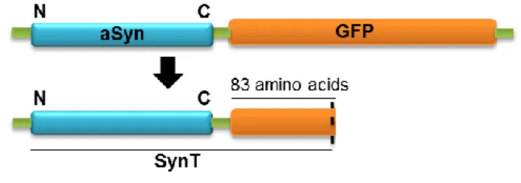
The SynT aggregation model used in this study was first described by McLean and colleagues and has been extensively used since then. This model is based on the expression of aSyn tagged with a truncated, non-fluorescent fragment of GFP (SynT) (Fig.4). In this model, overexpression of SynT together with synphilin-1 in a neuroglioma cell line (H4) promotes the formation of aSyn inclusions [65].
Figure 4. Schematic representation of SynT. This construction allows us to study aSyn aggregation in vitro in different mammalian cell lines.
Synphilin-1 is a presynaptic protein that was first identified by yeast-two-hybrid screening as a protein that interacts with aSyn and it is composed of 919 amino acid residues. aSyn interacts in vivo with synphilin-1 via its N-terminal and co-localizes with aSyn in LBs of PD patients. Similarly to aSyn, synphilin-1 is present in the majority of LB of patients with PD being present in almost 90% of LB from substantia nigra and other regions of the brain, suggesting its importance to the pathogenesis of PD [66, 67]. Moreover, synphilin-1 seems also to be required for inclusion formation [68].
7 2. Aims of the study
In order to study the effect of aSyn serine and tyrosine phosphorylation on its toxicity and aggregation we set two major aims:
- To determine the role of phosphorylation of specific residues Y125 and S129 on aSyn toxicity and inclusions formation. For this purpose, an established yeast S. cerevisiae model, a versatile eukaryotic organism, was has been already extensively used to understand the complex biological processes found in higher eukaryotes, involving aSyn post-translational modification in PD.
- To validate the main achievements obtained in yeast in a higher eukaryotic model of PD, using a human neuroglioma cell line (H4). For that aim, we generated the constructs Y125F and S129G to express mutant forms of aSyn and to investigate the effects of the different mutations on aSyn inclusions in cultured cells.
8 3. Materials and Methods
3.1. Molecular biology techniques used in Escherichia coli
3.1.1. Plasmids used in this study
The plasmids used in this study are listed in Annex 7.1.
Bacterial cultures of E. coli transformed with those plasmids were routinely preserved in glycerol stocks prepared in 2 mL vials of cryopreservation. For that, 500 µL of bacterial culture were mixed with 500 µL of glycerol (50% v/v) and stored at -80°C for future use.
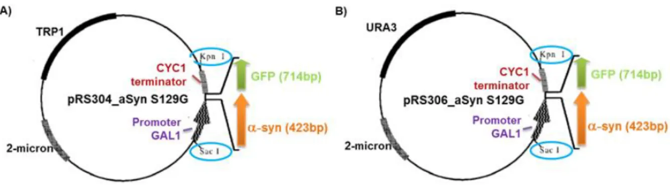
The vectors chosen for the heterologous expression of aSyn in S.cerevisiae were pRS304 and pRS306 and were used to express human aSyn under a galactose-inducible promoter and fused with GFP (green fluorescent protein) which enables the evaluation of protein expression and subcellular localization by fluorescence microscopy. They have a yeast TRP1 and URA3 selectable marker, respectively, and E.coli ampicillin resistance marker.
3.1.2. Growth media
The E. coli were grown at 37ºC overnight in specific medium (Luria-Bertani Broth - LB). The transformed bacteria were streaked onto plates supplemented with 2% agar containing 100 µg/mL of ampicillin (LB/Amp) as selection antibiotic and incubated overnight at 37ºC. After visible growth, the plates were kept at 4ºC.
3.1.3. Transformation of E.coli supercompetent cells
E. coli supercompetent cells were thawed on ice. 10 µL of transforming DNA were mixed with 100 µL thawed supercompetent bacteria cells (Nzytec). After that, mixed transformation was kept in ice for 30 min. Heat-shock of bacteria cells was performed by placing the tubes at 42ºC for 45 sec and the cells were placed on ice for 2 min. The resulting solutions added to 900 µL of LB medium without antibiotics and incubated for 1 hour at 37ºC. The transformed bacteria were spread on LB/Agar plates containing 100 µg/mL of ampicillin as selection antibiotic and incubated overnight at 37ºC.
3.1.4. Extraction and purification of plasmids
Random colonies were picked from the LB/Agar plates. These selected colonies were used to inoculate in 3 mL liquid LB/Amp medium, grown overnight with agitation (200 rpm) at 37ºC. The bacterial cultures were harvest by centrifugation and the plasmid DNA purified using NZYMiniPrep Kit from Nzytech. DNA extraction was performed following the manufactured recommendations. The plasmid DNA was eluted in 30 µL of preheated (42ºC)
9 sterile water. DNA concentration was determined at Nanodrop® (Thermo Scientific) or stored at -20ºC for further use.
3.1.5. Restriction digestion
To prepare the DNA for cloning procedures or to analyse the resulting clones multiple restriction digestion was performed. These restriction reactions were preformed both to confirm the presence of a gene in the expression vector and also to clone genes into expression vectors.
For cloning proposes, vectors and inserts were subject to enzymatic restriction. About 2-3 µg of DNA was digested in a final volume of 40 µL. The appropriated restriction enzymes were added using 1U for each 500 ng/µL of DNA and 4 µL of the corresponding enzyme buffer. For restriction map analysis of the clones obtained from the cloning procedures, a similar enzymatic restriction reaction was performed, but then 1 µL of the miniprep DNA was digested in a final volume of 10 µL.
The reactions were incubated at 37ºC for 2-3 hours or overnight and the resulting DNA fragments were visualized in agarose gel.
3.1.6. DNA extraction from agarose gel
The agarose gel was prepared in TAE 1x buffer and ethidium bromide was added before gel solidification. For visual tracking of DNA fragments migration during electrophoresis, it was applied the Orange G loading buffer (1/3 of the total volume of the samples to the DNA samples). The resolving agarose gel allowed separation of DNA fragments at a constant voltage of 80V.
For further cloning, the bands corresponding to the DNA fragments of interest were cut from the gel and DNA was purified using a specific kit.
3.1.7. Generation of Y125F and S129G phospho-mutants in yeast and mammalian vectors
Using the aSyn WT or SynT (aSynEGF deletion mutant (WTSynGFPΔ155) [65] and synphilin-1 [69]) constructs as a template, the mutations Y125F and S129G phospho-mutants were introduced by site-directed mutagenesis (QuickChange Site-Directed Mutagenesis Kit, Agilent Technologies).Briefly, a PCR was carried out using the PfuTurbo® DNA Polymerase (Stratagene, La Jolla, CA, USA) (1.25 U), the plasmid DNA template (10 ng) and the mutagenesis primers (62.5 ng, each primer) (Table 1).
10 Table 1. Primers used for site-directed mutagenesis.
The following conditions were used to perform the PCR reaction: 1 min at 95ºC, 18 cycles x [50 s at 95ºC, 50 s at 60ºC, 8 min at 68ºC] and 10 min at 68ºC. The resulting products were incubated with DpnI at 37ºC, overnight. The DpnI endonuclease specifically digests methylated and hemimethylated DNA allowing the selective digestion of the DNA template (methylated) and the selection of the mutated constructs synthesized de novo (not methylated). Accordingly, the digested DNA solutions contained only the mutant plasmids. Then, mutant plasmids were transformed in supercompetent E. coli cells as described in 3.1.3.
Two independent clones of each mutagenesis reactionwere sequenced by means of external DNA sequencing services to confirm the mutations introduced. The results of sequencing were analysed using the Basic Local Alignment Search Tool.
E. coli transformants of the positive clones were preserved in glycerol stocks as described above.
3.1.8 Cloning strategy of yeast integrative vectors
The vectors chosen for the heterologous expression of aSyn in S. cerevisiae were pRS304-GAL and pRS306-GAL. These were a shuttle integrative yeast expression vectors that have an inducible GAL (galactose) promoter, carrie a yeast TRP1 and URA3 selectable marker, respectively, and an E. coli ampicillin resistance marker (Fig.5).
The Y125F aSyn mutant forms were obtained by site-directed mutagenesis as shown in 3.1.7. While S129G aSyn were obtained by sub-cloning Gal1-aSyn S129G-CYCterminator from p426 yeast plasmid to either prs304 or prs306 integrative plasmids, using SacI/KpnI. The tyrosine (Y) residue at position 125 of the aSyn polypeptide chain was replaced by the amino acid phenylalanine (F) and the serine (S) residue at position 129 of the aSyn polypeptide chain was replaced by the amino acid guanine (G).
The different aSyn genes are fused to a GFP tag (green fluorescent protein) which enables the evaluation of protein subcellular localization by fluorescence microscopy.
Primer Phospho-mutants Sequence 5’ to 3’
Y125F-S Y125F mutagenesis 5' CCTGACAATGAGGCTTTTGAAATGCCTTCTGAG 3'
Y125F-AS Y125F mutagenesis 5' CTCAGAAGGCATTTCAAAAGCCTCATTGTCAGG 3'
S129G-S S129G mutagenesis 5' GGCTTATGAAATGCCTGGTGAGGAAGGGTATCAAG 3'
11 Figure 5. Maps of the yeast expression vectors (A) pRS304-GAL and (B) pRS306-GAL.These
expression vectors carry a TRP1 and URA3 selection marker, respectively. The GAL promoter controls the expression of generated fusion proteins. Here are shown the restriction sites of the multiple cloning site (MCS).
3.2. Molecular biology techniques used in S. cerevisiae
3.2.1. Yeast strains
The yeast strains used in this study are listed in Annex 7.2. Yeast strains were stored in glycerol stocks prepared by mixing 500 µL of yeast culture added to 500 µL of glycerol (50 % v/v) and stored at -80°C for future use. For further manipulations, strains were defrost and cultivated in solid medium. These plates were incubated at 30ºC until visible cell growth. The cultures were then stored at 4ºC.
3.2.2. Growth media
For routine work, yeast strains were grown at 30ºC in YPD medium and when necessary in synthetic complete (SC) medium lacking specific amino acids in Annex 7.3.
3.2.3. Yeast transformation
Yeast transformation protocol is based in the permeabilization of the yeast cellular membrane by lithium acetate (LiAc), its destabilization by TE and added PEG 40%. These compounds allowed the entry of DNA molecules in the cell.
Yeast cell were prepared prior to manipulation: in the first day, a single colony of each was picked into 5 mL of YPD and incubated at 30ºC with 250 rpm until late afternoon. The optical densities (ODs) were measured at 600 nm and yeast pre-cultures were diluted in 10 mL per transformation to obtain an OD between 0.5-0.7 (generation time of yeast cells in YPD were 90 min). In the second day, cells were harvest by centrifugation, washed with 30 mL of sterile water and resuspended in 1mL of sterile water. Then, pellet was washed in 0.5 mL of a solution of the LiAc 10x (1M LiAc pH 7.5) mixed with TE buffer 10x (0.1 M Tris pH 7.5; 0.01 M EDTA pH 7.5). For each transformation, 50 µL of yeast cells were added to the corresponding eppendorf along with 5 µL of previously boiled denatured salmon sperm. Ten
12 µL of transforming DNA were added and 300 µL of a solution containing PEG 50 %, TE 10x and AcLi 10x. The mixture was homogenized and incubated at 30ºC with orbital agitation for 30 min. The eppendorf was transferred to a 42ºC bath for 20 min. Cells were washed and were resuspended in 100 µL of sterile water, and plated onto appropriate selective medium.
The plates were incubated at 30ºC for 2-3 days and the transformants obtained were streaked onto fresh plates.
3.2.4. Construction of haploid yeast strains with single genome insertions of the aSyn forms
The integrative plasmids already available and the ones here constructed (point 3.1.8) were used to construct new yeast strains either in the W303.1A or the W303.1B background, carrying in its genome single integrations of WT aSyn, S129A aSyn, or Y125F aSyn, with or without a N-terminal fusion with GFP.
Namely, 3 µg of the integrative vectors of interest either in the pRS304 or in the pRS306 backbone were linearized with EcoRV as described in point 3.1.5. Then DNA was purified using a specific Kit (Wizard SV Gel and PCR Clean-Up System). The obtained DNA was them used to transform either W303.1A or W303.1B as described in point 3.2.3.
After strains were obtained, the correct insertion of the integrative vectors was verified by PCR using a specific pairs of primers (table 2).
Table 2. Primers used for confirmation of the insertion of the integrative vectors in the expected genome locus. Primer Sequence 5’ to 3’ Y125F-S 5' CCTGACAATGAGGCTTTTGAAATGCCTTCTGAG 3' Y125F-AS 5' CTCAGAAGGCATTTCAAAAGCCTCATTGTCAGG 3' S129G-S 5' GGCTTATGAAATGCCTGGTGAGGAAGGGTATCAAG 3’ S129G-AS 5' CTTGATACCCTTCCTCACCAGGCATTTCATAAGCC 3' S129A-S 5' CTTATGAAATGCCTGCTGAGGAAGGGTATC 3' S129A-AS 5' GATACCCTTCCTCAGCAGGCATTTCATAAG 3'
The following conditions were used to perform the PCR reaction: 5 min at 95ºC, 30 cycles x [30 s at 95ºC, 30 s at 55ºC, 2 min at 72ºC] and 10 min at 72ºC. After the PCR reaction was performed to confirm the PCR by agarose gel (1% agarose gel TAE 1x was run 60 min at 90V). Transformants were positive for vectors integrations were preserved at 4°C or used.
These haploid strains were then used to generate diploid strains by mating. Briefly, yeast cells from each haploid strain were mixed on a YPD agar plate and incubated at 30ºC for 2 days. Then, cells were picked, ressuspended in sterile watter and diploids were selected in minimal medium by URA and TRP auxotrophy.
13 These diploid strains were used to obtain haploid strains with double insertions of the appropriate aSyn by our collaborator Donata Wawrzycka. Briefly, diploids were by sporulation and tetrad dissection using a micromanipulator. The haploid strains obtained from each spore was analysed regarding auxotrophies and mating type.
The phenotypic characterization of these haploid strains was performed in several haploids obtained from dissected spores from independent tetrads and independent matings.
3.2.5. Growth conditions and spot assays
In the afternoon of the first day a pre-inoculum of the yeast strains of interest was prepared from fresh cultures in 5 mL YEP-Raffinose liquid media and incubated at 30ºC with orbital agitation (200 rpm) overnight. Optical density at 600 nm (OD600 nm) was read in the next afternoon and yeast cells were diluted into 5 mL of the same medium and incubated at 30ºC, with orbital agitation (200 rpm) until the standardized culture OD600 nm=0.5 was reached the next morning. The volume of yeast culture needed to inoculate a new culture with an initial standardized OD600 nm=0.2 was centrifugated and the cells were resuspended in YEP-Galactose liquid media and incubated at 30ºC, with orbital agitation (200 rpm), for 6 hours.
Serial dilutions (1:3) of the cell suspensions were prepared and spotted directly onto the agar plates that were then incubated at 30ºC and growth of yeast strains was monitored during 3 days.
3.3. Molecular biology techniques used in human neuroglioma cells
3.3.1. Construction of psi Y125F and S129G aSyn mammalian expression vectors
The SynT aggregation model used in this study was a starting point for the production of the Y125F and S129G mutant constructs. We generated two different phospho-mutant constructs using the SynT as a template. These mutations were introduced by site-directed mutagenesis (as described in 3.1.7).
3.3.2. Cell culture and cell transfection
The experiments were carried out in Human neuroglioma cells (H4) (ATCC HTB-148, LGC Standards, Barcelona, Spain) which were maintained in OPTI-MEM® I (Gibco, Invitrogen, Barcelona, Spain) supplemented with 10% Fetal Bovine Serum (FBS). The cells were grown at 37°C in an atmosphere of 5% CO2, under controlled conditions. For selected method cells were counted and seeded at a specific density of cells and on different types of plates. The density was maintained among the different sizes of plates in order to obtain
14 comparable results with different techniques. For microscopy experiment, cells were counted and seeded at a density of 120.000 cells/cm2 on glass- bottom 35 mm dishes (10 mm glass surface diameter, MatTek Corporation, Ashland, MA, USA) and for protein extraction cells experiment, cells were counted and seeded at a density of 175.000 cells/cm2 in 100 mm dishes (Techno Plastic Cultures AG, Switzerland). After 24 hours prior to transfection, H4 cells were transfected with the different combinations of plasmids. So, DNA transfection (Roche diagnostics, Mannheim, Germany) was used to transiently transfect cells (the cells are 60-80% confluent upon transfection) with the different combinations of plasmids, according to the manufacturer’s instructions.
3.3.3. Immunocytochemistry
The H4 cells were washed three times with PBS 1x, after 24 hours of incubation in the conditions described below. Cells were then fixed in 4% paraformaldehyde (PFA) at room temperature (RT) for 10 min. Cells were washed with PBS three times and thenwere permeabilized in 0.5% Triton X at RT for 20 min. The Triton X was removed but not washed. Cells were blocking in 1.5% normal goat serum (NGS) at RT for 1 hour. After blocking cells were incubated with primary antibody (mouse anti-aSyn, 1:1000) overnight at 4ºC. The primary antibody was removed and washed with PBS for three times. The secondary antibody was added (anti-mouse IgG-Alexa488, 1:1000, Invitrogen) and incubated at RT for 2 hours. Cells were washed more three times with PBS. Cells were maintained in PBS at 4ºC for further analyse fluorescent microscopy.
3.4. Fluorescence microscopy and image analysis
Fluorescence microscopy was performed using a Zeiss Axiovert 200 M Widefield fluorescence microscope equipped with a digital Axiocam from Zeiss (objective 40x and 63x, EC Plan-NeoFluar, Dry, NA (0.75)).
The yeast cultures grown as described in 3.2.4 were centrifuged at 3000 rpm, at 30ºC for 5 min and visualized under the microscope. The percentage of cells with aSyn inclusions was then determined by counting at least 500 cells per strain using ImageJ software.
Transfected H4 cells were fixed with 4 % paraformaldehyde described in 3.3.2. Slides were subjected to fluorescence microscopy. The proportion of cells with aSyn inclusions was then determined by counting at least 60-100 cells per culture using ImageJ software.
15 3.5. Protein extraction
For protein extraction, equal amounts of yeast cells were harvest and incubated with Trichloroacetic acid (TCA) (10%) at -20ºC for 20 min. Cells were than lysed using MRB buffer [50 mM sodium phosphate, 25 mM MES, pH 7.01 1% SDS, 3 M urea, 0.5% B-mercaptoethanol, 1mM sodium azide and proteases and phosphatases inhibitors (Roche diagnostics, Mannheim, Germany)] and disrupted with glass beads (3 cycles of 30 sec in the beadbeater and 5 min on ice). The samples were incubated at 70ºC for 10 min, and unlysed cells and membranes were removed by centrifugation at 10.000 g for 1 min at 4ºC and the supernatant containing the proteins was collected.
For H4 cells, the medium was removed. Cells were washed three times with PBS and harvest with Lysis buffer NP4O (with proteases inhibitor cocktail tablet).The plates were scraped and the cells were collected to an eppendorf and frozen at -80ºC for 15 min to disrupt membranes. Cells were always kept on ice during the extraction procedure, to avoid protein degradation. Cells were then sonicated 10 sec at 5 mA putted in 2 min in the ice twice (Soniprep 150 sonicator (Albra, Milano, Italy). After, cells were centrifuged at 0.7 rcf for 10 min at 4ºC. Proteins that were in the supernatant were collected.
The protein concentration was determined by means of the BCA Protein Assay Reagent Kit (Thermo Fisher Scientific Inc., Rockford, IL, USA), following manufacturer’s instructions.
3.6. Western blot analysis
Protein sample buffer (200 mM Tris-HCl pH 6.8, 6% 2-mercaptoethanol, 8% SDS, 40% glycerol, 0.4% bromophenol blue) was added to each protein sample that was then heated for 10 min at 100ºC. Protein samples were run in SDS-PAGE. After, resolved, proteins were transferred to a nitrocellulose membrane using a Trans-Blot Turbo transfer system (Bio-Rad). Immunoblotting was performed following standard procedures with the listed antibodies: aSyn (BD Transduction Laboratories, San Jose, CA, USA), pS129 aSyn (Wako Chemicals USA, Inc., Richmond VA, USA) and Y125F Abcam ab10789, GAPDH (Ambion, Cambridgeshire, UK) antibody served as loading control for yeast.
3.7. Data and statistical analysis
Results are shown as the averages of at least three independent experiments and are represented as the means ± SD. Differences amongst treatments were detected by analysis of variance with the Tukey HSD (honest significant difference) Multiple Comparison Test using SigmaStat 3.10 (Systat). Values of p<0.05 were considered significant.
16 4. Results
4.1 Construction of yeast strains with double insertions of the Y125F, S129G or WT SNCA human gene
4.1.1. Construction of yeast integrative expression vectors
In this work it was decided to use integrative expression vectors in order to guarantee stable expression levels of aSyn. Accordingly, the first step consisted to construct the plasmids that were not available yet and to use them in parallel with other already available and characterized in our laboratory (UNCM/IMM).
The strain W303.1A was chosen for this study, as several strains constructed in this background were, as mentioned above, were already available in the laboratory. This strain was also used and characterized by Outeiro and Lindquist, 2003. This study showed that when the gene encoding aSyn, SNCA is cloned into a 2 µ vector (pRS426GAL) and is expressed in this strain (W303.1A) results toxic, decreases yeast growth and augments the number of cells presenting foci. Since 2 µ expression of aSyn results quite toxic, cells have tendency to reduce the plasmid copy number and protein expression might be considerable variable in a yeast cell population; therefore in our study it was preferred to use the aSyn integrated strains.
The aSyn mutants forms Y125F and S129G, where the phosphorylation in this resides is blocked were obtained either by site-directed mutagenesis as described in point 3.1.7. or by subcloning as described in 3.1.8. (Fig.6).
Figure 6. Maps of the yeast expression vectors of S129G aSyn in (A) pRS304-GAL and (B) pRS306- GAL plasmids. These expression vectors contain a Gal promoter which controls the expression of the
fusion proteins S129G aSyn; a MCS with multiple restriction sites and a TRP1 or URA3 selection marker, respectively.
17 In Figure 7 there is a schematic representation of the nucleotides and aminoacid sequences of aSyn where the site-directed mutagenesis was performed.
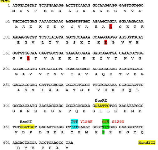
Figure 7. Schematic representation of the aSyn nucleotide and amino acid sequences. Upper line
represents the nucleotides sequence and the below one represents the protein sequence. Codons modified by site-directed mutagenesis for Y125F are shown in blue, while the ones modified by S129G are shown in green.
In more detail, Y125F mutation occurred through the replacement of tyrosine for phenylalanine, which blocks phosphorylation and allows studying the effect of phosphorylation on this residue. Indeed, S129G mutation occurred through the replacement of serine for a glycine residue. This mutation mimics aSyn unphosphorylated form and represents a novel mutant as alternative to the widely characterized S129A, where the serine was replaced by an alanine.
4.1.2. Construction of yeast strains with single insertions of the SNCA gene The plasmids obtained in 4.1.1. were used to construct haploid yeast strains with single insertions of Y125F aSyn in the genome (see 3.2.3). Integrative plasmids containing the mutation Y125F and bearing URA3 or TRP1 as auxotrophic markers, were cloned. Using
18 this construction and S129G; aSyn without GFP, S129A without GFP (already available in the laboratory) were made single integrations in the genome of W303.1A and W303.1B. Integrative vectors were designed in order to address a specific auxotrophic marker to integrate aSyn gene by homologous recombination (Fig.8).
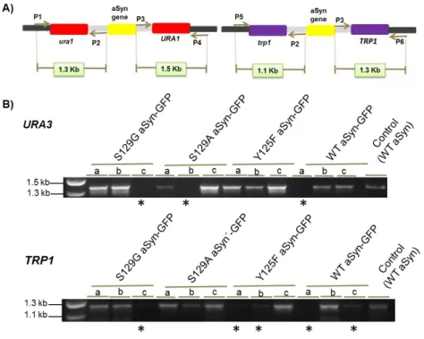
Different insertions in genome were performed with different mating types. The plasmids used were pRS306 and pRS304, which present different auxotrophies, URA and TRP respectively. The insertion of the integrative vectors in the expected genome locus was confirmed by PCR using specific pairs of primer. Figure 9 shows the PCR primers used and their respective products.
Figure 8. Construction of yeast strains with single genome insertion of the indicated phospho-mutants. Integrative yeast vector with different selective markers have been used to clone each aSyn
mutant and integrated in genome.
Figure 9. Representative electrophoresis to confirm genome insertions with plasmids containing different auxotrophies. (A) PCR primers used and their respective products. (B) The insertion of the
19
of primers. S129G aSyn-GFP; S129A aSyn-GFP and Y125F aSyn-GFP represents the unphosphorylated forms and WT aSyn is the control (VSY72). a, b, c represents different of colonies. * indicates negative candidates while all the remaining strains were positive for the correct genome insertion of the integrative vectors.
4.1.3. Construction of yeast strains with double insertions of the SNCA gene A normal cell division of yeast leads to their genetically identical progeny. However, in this study we crossed different strains of yeast in order to produce new strains with unique genetic combinations.
For this purpose yeast strains of each mating type were mated. The next step was to induce sporulation of the cells mentioned above by removing nutrients from their growth media. During this sporulation cells undergo two rounds of cell division, resulting in four spores in which the chromosomes have been distributed. This process generates four haploid yeast cells that are contained in an ascus and a single unit can be grown as an haploid cells.
W303 diploid strains were used to obtain haploid strains with double insertions of the appropriate aSyn by our collaborator Donata Wawrzycka (University of Wroclaw, Poland). Briefly, haploid cells were induced by sporulation and tetrads were dissected using a micromanipulator. The haploid strains obtained from each spore was characterized by auxotrophic marker and mating type.
The phenotypic characterization of these haploid strains was performed in several haploids obtained from dissected spores from independent tetrads and independent matings (Fig.10).
Figure 10. Haploid strains obtained from dissected spores from independent tetrads and independent matings. The picture represents the stains obtained by budding (as described in 3.2.4).
Throughout the plate are observed haploid strains resulting from de germination of different tetrads obtained from the mating of, in this case the strains SC208/220 (see Annex 8.2).
The VSY strains which contain a double genome insertion of WT aSyn where used as control strains. These strains were previously described and characterized by Sancenon et al., 2010.
Then we performed the characterization of the tetrads in order to study the toxicity and formation of foci in the Y125F and S129G residues.
20 4.2. Using yeast as a model to study aSyn phosphorylation
4.2.1. Effect of Y125F on aSyn toxicity and foci formation
We next investigated the role of blocking phosphorylation on residue Y125 on aSyn aggregation and toxicity using the phospho-mutant Y125F aSyn. Expression of this mutant was under the regulation of a galactose-inducible promoter which was turned on upon transfer of cells from raffinose and to galactose media.
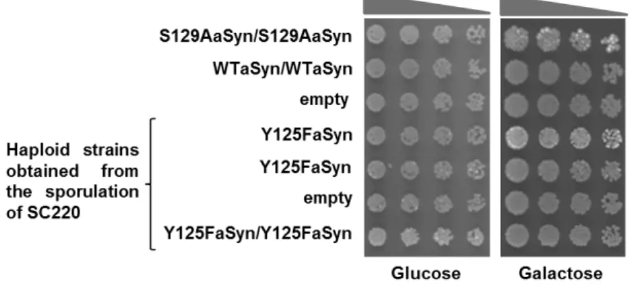
To evaluate the effect of blocking phosphorylation on Y125 on aSyn toxicity, the strains selected previously expressing one or two copies of Y125F aSyn (haploid strains obtained in 4.1.3), which resulted from the same tetrad were analysed. A replica of one of these set of strains was compared to strains expressing two copies of WT aSyn (VSY72) or S129A aSyn mutant form (VSY73) (that mimics the unphosphorylated form of aSyn) by spotting assay (Fig. 11). The control strain (VSY71) contained an empty vector.
Expression of two copies of WT aSyn was not toxic for yeast as previously described [58], whereas two copies of S129A aSyn (VSY73) caused mild toxicity. Our results showed that either one or two copies of Y125F aSyn did not cause cellular toxicity, as it presented identical growth as for the WT strains (Fig.11). Therefore, no significant phenotype was visible when phosphorylation on Y125 was blocked.
Figure 11. Effect of Y125F on aSyn toxicity in yeast cells carrying single or double insertions of the encoding gene. Spotting assay of serial dilutions of cell suspensions, initially adjusted to OD600 nm=0.05+-0.005, that where applied as spots (4 µl) onto the surface of the solid medium either with glucose (aSyn expression repressed) or galactose (aSyn expression induced). Results shown are from one representative experiment from at least three independent experiments.
Furthermore, we analysed cells expressing WT and Y125F aSyn-GFP by fluorescence microscopy. After 6 hours of induction of aSyn expression, cells expressing WT aSyn already showed the presence of aSyn foci (Fig.12). Surprisingly, we did not observe formation of foci in cells expressing either one or two copies of Y125F aSyn.
21 Figure 12. Effect of Y125F on aSyn foci formation in yeast cells carrying single or double insertions of the encoding gene. Fluorescence microscopic visualization and intracellular localization of the WT and
phospho-mutant Y125F aSyn fused to GFP 6 hours post induction of aSyn expression. Scale bar is 10 µm.
4.2.2. Effect of S129G on aSyn toxicity and foci formation
In these experiments, we compared the effect of two mutations (S129A and S129G) that prevent the phosphorylation of S129 residue on aSyn toxicity. Expression of these mutants was under the regulation of a galactose-inducible promoter (as described in 4.3.1.).We previously showed that aSyn toxicity and foci formation was enhanced (Tenreiro et al., unpublished results) by S129A mutation that blocks aSyn phosphorylation. However, whether this was simply due to structural effects caused by the S129A substitution in aSyn or due to blocking phosphorylation was not completely clear. To clarify this, we replaced the serine 129 by glycine, which also blocks phosphorylation, following the same procedure as in 4.2.1. (haploid strains obtained in 4.1.3). The spotting assays were performed with haploid strains that resulted from the same tetrad and were compared to strains expressing two copies of WT aSyn (VSY72) or S129A aSyn mutant form (VSY73) (that mimics the unphosphorylated form of aSyn). The control strain (VSY71) contained an empty vector. We observed that the toxicity caused by expression of S129A and S129G aSyn is identical (Fig.13).
Figure 13. Effect of S129G on aSyn toxicity in yeast cells carrying single or double insertions of the encoding gene. Spotting assay of serial dilutions of cell suspensions, initially adjusted to OD600
nm=0.05+-22 0.005, that where applied as spots (4 µl) onto the surface of the solid medium either with glucose (aSyn expression repressed) or galactose (aSyn expression induced). Results shown are from one representative experiment from at least three independent experiments.
In particular, yeast strains carrying two copies of human SNCA integrated in the genome, encoding either WT, S129A and S129G mutant aSyn C-terminally tagged with GFP were used to to be compared side by side and pursuit the work. Confirming the results apresented in fig. 13, the expression of S129A and S129G aSyn is more toxic for yeast cells than expression of WT aSyn, as evaluated by spotting assay (Fig.14).
Figure 14. S129G aSyn shows a similar phenotype to S129A aSyn. Cell viability of yeast cells
expressing either WT aSyn, S129A aSyn or S129G aSyn, after 6 hours of aSyn expression induction, assessed by spot assay. Cell suspensions were adjusted to OD600 nm=0.05±0.005 and used to prepare 1/3
serial dilutions that were applied as spots (4 µl) onto the surface of the YPD rich medium and incubated at 30ºC for 2 days. Results shown are from one representative experiment from at least three independent experiments.
Furthermore, after 6 hours of induction of aSyn expression, aSyn foci formation in cells expressing the WT, S129A or S129G was quantified by fluorescence microscopy. The majority of cells expressing either S129A or S129G aSyn displayed foci (90%). In contrast, only 70 % of cells expressing WT aSyn presented foci (Fig.15).
Figure 15. S129G aSyn shows similar foci formation to S129A aSyn in yeast cells. Intracellular
localization of the WT, S129A or S129G aSyn-GFP (left panel) and percentage of yeast cells containing aSyn foci, after 6 hours of aSyn expression induction, assessed by fluorescence microscopy (**p < 0.01;