Human polyomaviruses and cancer: an overview
Jose´ Carlos Mann Prado, Telma Alves Monezi, Aline Teixeira Amorim, Vanesca Lino, Andressa Paladino,
Enrique Boccardo*
Departamento de Microbiologia, Instituto de Ciencias Biomedicas, Universidade de Sao Paulo, Sao Paulo, SP, BR.
Prado JC, Monezi TA, Amorim AT, Lino V, Paladino A, Boccardo E. Human polyomaviruses and cancer: an overview. Clinics. 2018;73(suppl 1):e558s
*Corresponding author. E-mail: [email protected]
The name of the family
Polyomaviridae
, derives from the early observation that cells infected with murine
polyomavirus induced multiple (
poly
) tumors (
omas
) in immunocompromised mice. Subsequent studies showed
that many members of this family exhibit the capacity of mediating cell transformation and tumorigenesis
in different experimental models. The transformation process mediated by these viruses is driven by viral
pleiotropic regulatory proteins called T (tumor) antigens. Similar to other viral oncoproteins T antigens target
cellular regulatory factors to favor cell proliferation, immune evasion and downregulation of apoptosis. The
first two human polyomaviruses were isolated over 45 years ago. However, recent advances in the DNA
sequencing technologies led to the rapid identification of additional twelve new polyomaviruses in different
human samples. Many of these viruses establish chronic infections and have been associated with conditions in
immunosuppressed individuals, particularly in organ transplant recipients. This has been associated to viral
reactivation due to the immunosuppressant therapy applied to these patients. Four polyomaviruses namely,
Merkel cell polyomavirus (MCPyV), Trichodysplasia spinulosa polyomavirus (TSPyV), John Cunningham
Polyoma-virus (JCPyV) and BK polyomaPolyoma-virus (BKPyV) have been associated with the development of specific malignant
tumors. However, present evidence only supports the role of MCPyV as a carcinogen to humans. In the present
review we present a summarized discussion on the current knowledge concerning the role of MCPyV, TSPyV,
JCPyV and BKPyV in human cancers.
KEYWORDS:
MCPyV; BKPyV; JCPyV; TSPyV; Human cancer.
’
INTRODUCTION
Polyomaviruses (PyVs) are icosahedral, nonenveloped viruses
that are approximately 45 nm in size. The capsid is composed
of 72 pentameric capsomers and surrounds a circular,
double-stranded viral DNA genome that is approximately 5,5 kbp.
Inside the virion, the DNA is associated with the cellular
histones H2A, H2B, H3 and H4, forming the viral
mini-chromosome. In addition, inside the cell, the minichromosome
is found to be associated with histone H1 (1). These viruses are
relatively resistant to treatment with formalin and are not
affected by organic solvents (2).
Currently, the International Committee on Taxonomy of
Viruses (ICTV) has divided the
Polyomaviridae
family into
5 genera:
Alphapolyomavirus,
with 37 species that infect animals
and humans, including human PyVs (HPyVs) 5, 8, 9, 12 and
13;
Betapolyomavirus
, with 29 species that infect animals and
humans, including HPyVs 1 and 4;
Deltapolyomavirus
with
only HPyVs 6, 7, 10 and 11;
Gammapolyomavirus
, with seven
animal virus species, including PyVs that infect birds; and,
finally, an unassigned genus that contain three species of
animal PyVs (3,4).
Biology of polyomaviruses
The genomes of PyVs can be divided into three regions:
the control region; the early region, which encodes early
proteins; and the late region, which encodes late proteins
with structural function. The control region contains the
origin of replication and the promoters that regulate the
expression of early and late genes. However, this region does
not encode any protein or functional RNA. This region is
involved in the regulation of the viral life cycle by
modu-lating replication and transcription. Both strands of the PyV
DNA code for proteins. Early genes are expressed from one
strand immediately after infection. On the other hand, late
genes are expressed from the opposite strand after viral
genome replication. The mRNA transcribed from both
early and late regions exist in at least two isoforms due to
alternative processing. In addition, the expression of a viral
microRNA has been observed in the John Cunningham
polyomavirus (JCPyV), BK polyomavirus (BKPyV) and Merkel
cell polyomavirus (MCPyV) (5,6). This microRNA is encoded
by the DNA strand complementary to the large T antigen
(see below) and, at least in MCPyV, this RNA seems to
regu-late the expression of this viral protein (5).
The proteins encoded by the early genes are involved in
the regulation of viral transcription and genome replication.
These proteins are known as
‘‘
T antigens
’’
and received this
name because they were recognized by antibodies from
DOI:10.6061/clinics/2018/e558s
Copyright&2018CLINICS–This is an Open Access article distributed under the terms of the Creative Commons License (http://creativecommons.org/licenses/by/ 4.0/) which permits unrestricted use, distribution, and reproduction in any medium or format, provided the original work is properly cited.
No potential conflict of interest was reported.
Received for publication onJanuary 4, 2018.Accepted for publication onMay 15, 2018
Commemorative Edition: 10 years of ICESP
1
rodents bearing tumors (7). Different T antigen isoforms exist
and are named depending on the viral species from which
they originate. The transforming potential of the viral group
is directly related to the expression of T antigens and was
initially established in studies using simian virus 40 (SV40) (8).
The large T (LT) antigen is a nuclear protein that is
approxi-mately 700 amino acids. However, alterations in its
phosphor-ylation pattern may change the location of this protein within
the cell (9,10). The LT antigen regulates both viral transcription
and genome replication (11). In addition, the LT antigen as
well as the other T antigen isoforms expressed in human and
animal PyVs are pleiotropic proteins that affect the function
and expression of several cellular proteins involved in the
regulation of cell proliferation (see below). The neutralization
of the functions of these proteins is critical for the induction of
the entry of the host cell into the cell cycle, making the DNA
replication machinery available and allowing viral genome
replication (12-15).
The late region harbors the genes that encode structural
proteins found in the viral capsid, such as VP1 and VP2.
Some species also express the structural proteins VP3 and
VP4. VP2 and VP3 are important for viral entry into the host
cell. However, the role of VP4 during the viral life cycle
remains a matter of debate. Previous studies have suggested
that VP4 acts as a viroporin to disrupt the nuclear envelope
and mediate viral release (16-19). However, in a recent study,
Henriksen et al. showed that human renal proximal tubule
epithelial cells transfected with BKPyV genomes carrying
start codon substitutions in VP4, predicted to abolish the
production of this protein, released comparable amounts of
viral particles in the supernatant as cells transfected with WT
genomes (20). It is clear that additional studies are needed to
determine the role of VP4 in PyV biology. In addition, some
PyVs, including JCPyV, BKPyV and SV40, present an open
reading frame (ORF) that encodes a regulatory cytoplasmic
protein named agnoprotein (21). The involvement of
agno-protein in viral release has been suggested in species that
express this protein, including SV40, JCPyV and BKPyV (22).
Studies conducted during the past two decades have
shown that the expression of VP1 in different systems leads
to the production of structures called virus-like particles
(VLPs), which are similar to the viral capsid (23-27). After
being expressed, the structural proteins accumulate in the
cellular nucleus, contributing to the mounting of the virion.
These proteins are found in different amounts in viral capsids,
with VP1 being the major protein in the formation of
pent-amers. The carboxy end of VP1 extends outside the pentamer
and interacts with surrounding pentamers. These interactions,
mediated by VP1 together with Ca
2+and disulfide bonds,
contribute to the stabilization of the capsomers and capsid
structure (2).
The mechanism of viral entry into the host cell seems to
depend on the virus and cell type under study. For instance,
cell internalization via caveolin- or clathrin-dependent
endo-cytosis, as well as entry via other mechanisms, has been
previously described (28,29). Once in the cytoplasm, the viral
capsid suffers alterations that expose the nuclear localization
signals present in proteins VP2 and VP3 and mediate nuclear
import. After reaching the nucleus, the capsid is completely
dismounted, and the viral genome is exposed and maintained
in the episomal form. Next, the early region is transcribed to
produce an mRNA molecule that, after processing, will
gene-rate the large T antigen and its other isoforms. The LT antigen
protein will first mediate viral genome replication. This event
may also be regulated by epigenetic mechanisms due to the
association of viral DNA with cellular histones (30). Only
after genome amplification will the LT antigen promote the
transcription of the late region to express the structural proteins.
Infective virions are mounted in the nucleus, and the release of
these virions may depend on the occurrence of cell lysis (2,31).
The majority of PyV infections are asymptomatic;
how-ever, these infections may cause alterations in cell cultures
and induce tumors in immunocompromised laboratory animals,
including newborn mice. The transforming potential of PyVs
has been demonstrated
in vitro
using cells from different
organisms (32). Early studies conducted in
immunocompro-mised animals injected with murine polyomavirus
(MuPyV)-infected cells showed the formation of multiple (
poly
) tumors
(
omas
) and served to name the group (7).
Human polyomaviruses
The discovery and characterization of HPyVs occurred in
different technological contexts. In 1971, two isolates were
described, and for decades, these species remained the only
members of the family that infected humans (33,34). It was
not until 2007, and only after major advances in DNA
sequencing technologies, that other twelve PyVs were
identi-fied in human samples, increasing the total number of HPyVs
to fourteen (14,35-44). The timeline of HPyV discovery is
sum-marized in Figure 1.
Serological studies have shown that exposure to most
HPyVs occurs early in life and that infection prevalence in
adults may be high (45). Interestingly,
immunocompro-mised individuals exhibit higher viral loads of these agents
suggesting that immunocompromised individuals are more
susceptible to reactivation of these viruses (14,46-49). Among
the PyVs that affect humans and are associated with
impor-tant diseases in immunocompromised individuals, the most
relevant are JCPyV, BKPyV, MCPyV and Trichodysplasia
spinulosa PyV (TSPyV). These PyVs are ubiquitous and are
highly prevalent in the normal human population. In general,
infection by these agents is asymptomatic in healthy
indi-viduals, with prevalence values as high as 80% in adults,
as determined by serological studies (27,50). On the other
hand, in immunocompromised persons, infection with BKPyV
is associated with the development of nephropathies and
hemo-rrhagic cystitis, while infection with JCPyV is associated with
progressive multifocal leukoencephalopathy (PML). In
addi-tion, MCPyV is associated with the development of Merkel
cell carcinoma (MCC) and is considered a group 2A
carcino-gen (probably carcinocarcino-genic to humans) by the International
Agency for Research on Cancer (51).
It is accepted that PyV-associated tumors develop after
the interruption of the viral life cycle. This may be caused
by accidental viral integration into de cellular genome. In the
case of MCPyV and BKPyV, viral cycle interruption may
be caused by rupture of the VP1 gene (52,53) or by loss of
the carboxy-terminal domain of the LT antigen due to
non-sense mutations. This domain of the LT antigen is needed for
the normal cycle to promote viral genome replication (52,37).
Therefore, viral replication is interrupted while truncated
versions of the LT antigen and its isoforms are expressed (52).
The presence of the LT antigen is a universal characteristic
of the members of the
Polyomaviridae
family. LT antigens
share high identity between the members of the PyV genus,
which allows recognition by cross-hybridization in
west-ern blots performed with a few specific antibodies (54).
2 Human polyomaviruses and cancer
The N-terminal region of this protein contains a DnaJ domain,
which contributes to viral replication and mediates the
bind-ing of the cellular chaperone HSc70. This protein also contains
an LXCXE motif that binds the members of the
retinoblas-toma protein family pRb, p107 and p130. Together DnaJ
and LXCXE disturb the pRb/E2F complexes, promoting the
progression of the cell cycle (11). The C-terminal domain of
the LT antigen harbors a conserved threonine residue that,
when phosphorylated, competes with cyclin E1 and Myc to
bind to FBXW7. The protein FBXW7 (F-box) is part of the
ubiquitin ligase complex formed by Skp1/culin/F-box (SCF).
As such, LT prevents the degradation of cyclin E1 and Myc
and contributes to cell growth and proliferation (31).
The LT antigen also presents a nuclear localization sequence
(NLS), an origin-binding domain (OBD) and a helicase domain.
The OBD and the helicase domain are critical for viral genome
replication (55). Finally, LT antigens from many, but not all,
HPyVs exhibit a p53-binding domain (56,57). The
colocaliza-tion of p53 and the LT antigen has been demonstrated in the
cytoplasm of cultured cells of BKPyV-positive neuroblastoma
and prostate cancer (58,59). It is believed that this interaction
may block the expression of p53-regulated genes in response
to DNA damage (57). Another cellular target of early PyV proteins
is the phosphatase PP2A (60). This protein is inactivated by
T antigens from SV40, JCPyV and MCPyV (59). PP2A is a
criti-cal regulator of the mitogen-activated protein kinase (MAPK)
signaling cascade and has many functions. This protein
contri-butes to the control of cellular metabolism by regulating the
activities of different enzymes involved in glycolysis, lipid
metabolism and catecholamine synthesis (61). In addition, this
protein regulates various critical processes, such as cell cycle
progression, DNA replication and transcription, protein
trans-lation, signal transduction, cytoskeletal dynamics, cell
mobi-lity and apoptosis. As such, PP2A plays an important role in
cell transformation and cancer (62-65).
Most HPyVs have the potential to cause nonneoplastic
diseases in the context of immunosuppression (54). However,
this association has only been confirmed for a few of these viruses.
Among the 4 PyVs associated with diseases in
immuno-suppressed humans, two are associated with proliferative
diseases of the skin. MCPyV is associated with MCC, while
TSPyV is the etiological factor for Trichodysplasia spinulosa
(TS). The two other viruses are JCPyV, which is associated
with PML, and BKPyV, which is a leading cause of chronic
dysfunction in renal transplant patients, urethral stenosis
and nephropathy. The main characteristics of these viruses
will be presented in the next sections. Moreover, the cell
transforming potential exhibited by different HPyV proteins
in vitro
raises the intriguing possibility that some of these
agents, in addition to MCPyV, may be associated with specific
human cancers. Therefore, several studies conducted by
dif-ferent groups around the world have addressed the presence
of all HPyVs in different human tumors. Table 1 presents a
comprehensive summary of the main results obtained.
Merkel cell polyomavirus and Merkel cell carcinoma
In 2008, the identification of the fifth HPyV, detected in
samples of MCC, was reported, and this virus was named
MCPyV. The authors performed digital transcriptome
sub-traction (DTS) in MCC samples and identified one sequence
that exhibited no similarity with human transcripts. In-depth
analysis of the transcript using nucleotide databases showed
that this sequence was related to the T antigen sequence
from simian lymphotropic polyomavirus (LPyV) and BKPyV.
The viral genome, which was detected in 80% of the MCC
samples, was amplified by
primer walking
and sequenced.
The study also showed that in most of the MCPyV-positive
tumors, the viral DNA exhibited a clonal integration pattern
within the cell genome. The same integration pattern detected
Figure 1 -Timeline of the discovery of human polyomaviruses. The timeline shows the name of the virus, the year of discovery and the type of sample from which the virus was isolated. For complete references, see the text. BKV or BKPyV, human polyomavirus BK or human polyomavirus 1; JCV or JCPyV, John Cunningham or JC polyomavirus or human polyomavirus 2; KIPyV, Karolinska Institute polyomavirus or human polyomavirus 3; WUPyV, Washington University polyomavirus or human polyomavirus 4; MCPyV, Merkel cell polyomavirus or human polyomavirus 5; HPyV6, human polyomavirus 6; HPyV7, human polyomavirus 7; TSPyV, Trichodysplasia spinulosa polyomavirus or human polyomavirus 8; HPyV9, human polyomavirus 9; MWPyV, Malawi polyomavirus or human polyomavirus 10; STLPyV Saint Louis polyomavirus or human polyomavirus 11; HPyV12, human polyomavirus 12; NJPyV, New Jersey polyomavirus or human polyomavirus 13; LIPyV, Lyon IARC polyomavirus or human polyomavirus 14.
3
CLINICS 2018;73(suppl 1):e558s Human polyomaviruses and cancer
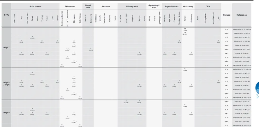
Table 1
-
Summary of studies addressing the presence of human polyomaviruses in tumor samples.PyVs
Solid tumors Skin cancer Blood cells Urinary tract Gynecologic
tract Digestive tract Oral cavity CNS
Method Reference
Solid tumor
Lung
Head and n
e
ck
Breast Uveal Thym
us
Kidney Liver Thyroid
Keratoacanthomas SCC,BC
C,m
e
lanom
a
Skin cancer Spitz naevus Leukemia Lymp
hom
a
Soft tissue Ewing's
Osteo
s
ar
com
a
Sarcoma Kaposi Prostate
Urin
ary tract
Uroteli
a
l
Bladder Urete
r
Ov
ary
Cervix
Gynecologic tract
Stomach
Large bowel Colo
rectal
Digestive System
Tonsil
s
Oral cavity
Brain
Meningomas Neurobl
asto
ma
CNS
HPyV1 (BKPyV)
0
(0/75) PCR Arthur
et al., 1994 (207)
45,8 (11/24)h
SB Dörries
et al., 1987 (208)
100 (5/5)
50
(6/12) 86 (50/58)g
PCR De Mattei
et al., 1995 (209) 0-100
k
0-6 k
0-44 k
0-100k 0.5-85k
64-76k 0-89k
PCR/IHC/SB Ambed et al.,2009 (210)
100
(18/18)i PCR Flaegstad et al., 1999 (58) 25,5
(13/51)a (15/51) 29,5 (14/48) 29,2 qPCR Loutfy et al., 2017 (211) 60
(10/15) 58 (15/26)
58 (15/26)
50
(2/4) PCR/SB Monini et al., 1995 (212) (22/26) 85 PCR Russo et al., 2008 (213) (0/109) 0 (0/68) 0 qPCR Csoma et al., 2016 (214) (0/38) 0 PCR Herberhold et al., 2017 (205) (0/123) 0 PCR Corbex et al., 2014 (215) 0
(0/19) 0 (0/1)
0 (0/4)
0
(0/3) 0
(0/7) 0
(0/9) PCR Schmitt et al., 2011 (216)
(7/32) 22 PCR Fioriti et al., 2003 (217) (0/236) 0 (0/85) 0 (0/65) 0 (0/50) 0 (0/137) 0 (0/36) 0 (0/94) 0 (0/65) 0 IHC Toptan et al., 2016 (54) 44,5
(65/146) PCR Drop et al., 2017 (191) (104/646) 16 PCR Rogers et al., 2017 (218) (0/5) 0 (0/2) 0 (0/15) 0 (0/10) 0 PCR Volter et al., 1997 (219) (0/37) 0 PCR Ramqvist et al., 2014 (220) (0/86) 0 PCR Gheit et al., 2012 (221) 0
(0/20) PCR Gellrich et al., 2005 (222) (2/22) 9 (3/25) 12 PCR Haeggblom et al., 2017 (223)
6,2
(7/113) PCR Polesel
et al., 2012 (171)
0
(0/94) PCR Campello
et al., 2010 (170)
HPyV2 (JCPyV)
11,8
(6/51)a 29,5 (15/51)
25
(12/48) qPCR Loutfy et al., 2017 (211) (0/21)0 h
SB Dörries et al., 1987 (208)
(0/75) 0
PCR Arthur
et al., 1994 (207) (0/18)0 i PCR Flaegstad et al., 1999 (58)
(0/38) 0 PCR Herberhold et al., 2017 (205) (0/19) 0 (0/1) 0 (0/4) 0 (0/3) 0 (0/7) 0 (0/9) 0 PCR Schmitt et al., 2011 (216) (0/236) 0 (0/85) 0 (0/65) 0 (0/50) 0 (0/137) 0 (0/36) 0 (0/94) 0 (0/65) 0 IHC Toptan et al., 2016 (54) (9/56) 16 PCR Vilkin et al., 2012 (161)
9
(3/32) PCR Fioriti et al., 2003 (217) (0/37) 0 PCR Ramqvist et al., 2014 (220) (0/86) 0 PCR Gheit et al., 2012 (221) 0
(0/20) PCR Gellrich et al., 2005 (222) (0/22) 0 (0/25) 0 PCR Haeggblom et al., 2017 (223)
65,5
(74/113) PCR
Polesel et al., 2012 (171)
0
(0/94) PCR Campello
et al., 2010 (170)
Sarcoma
4
Human
polyomavir
uses
and
cancer
Prado
JCM
et
al.
CLINICS
2018;73
(suppl
Table 1 -
Continued.PyVs
Solid tumors Blood
cells Sarcoma Urinary tract
Gynecologic
tract Digestive tract Oral cavity CNS
Method Reference
Solid tumor
Lung
Head and n
e
ck
Breast Uveal Thym
us
Kidney Liver Thyroid
Keratoacanthomas SCC,BC
C,m
elanom
a
Skin cancer Spitz naevus Leukemia Lymp
hom
a
Soft tissue Ewing's
Osteo
s
ar
com
a
Sarcoma Kaposi Prostate
Urin
ary tract
Uroteli
a
l
Bladder Urete
r
Ov
ary
Cervix
Gynecologic tract
Stomach
Large bowel Colo
rectal
Digestiv
e
System
Tonsil
s
Oral cavity
Brain
Meningomas Neurobl
asto
ma
CNS
HPyV3 (KIPyV)
(0/30) 0 qPCR Teramoto et al., 2011 (224) (0/183) 0 Serological Colombara et al., 2016 (225) (0/123) 0 PCR Corbex et al., 2014 (215) (0/56) 0 PCR Giraud et al., 2009 (226) (0/100) 0 qPCR Csoma et al., 2018 (227) (0/109) 0 (0/68) 0 qPCR Csoma et al., 2016 (214)
(0/50) 0 qPCR Gustafsson et al., 2012 (228) (0/38) 0 PCR Herberhold et al., 2017 (205) 0
(0/37) PCR Ramqvist et al., 2014 (220) 0
(0/19) 0 (0/1)
0 (0/4)
0
(0/3) 0
(0/7) 0
(0/9) PCR Schmitt et al., 2011 (216) 0
(0/236) 0 (0/85)
0
(0/65) 0
(0/50) 0
(0/137) 0 (0/36)
0 (0/94)
0
(0/65) IHC Toptan et al., 2016 (54) 0
(0/86) PCR Gheit et al., 2012 (221) 0
(0/32) 0
(0/2) 0
(0/2) 0
(0/1) 0
(0/1) 0
(0/20) 0
(0/16) PCR Duncavage et al., 2009 (229) (0/22) 0 (4/25) 16 PCR Haeggblom et al., 2017 (223)
0
(0/113) PCR Polesel
et al., 2012 (171) (0/56) 0 PCR Giraud et al., 2009 (226) (0/100) 0 qPCR Csoma et al., 2018 (227)
(0/38) 0 PCR Herberhold et al., 2017 (205) (0/109) 0 (0/68) 0 qPCR Csoma et al., 2016 (214)
(0/50) 0 qPCR Gustafsson et al., 2012 (228) (0/30) 0 qPCR Teramoto et al., 2011 (224) (0/183) 0 Serological Colombara et al., 2016 (225) (0/19) 0 (0/1) 0 (0/4) 0 (0/3) 0 (0/7) 0 (0/9) 0 PCR Schmitt et al., 2011 (216) (0/236) 0 (0/85) 0 (0/65) 0 (0/50) 0 (0/137) 0 (0/36) 0 (0/94) 0 (0/65) 0 IHC Toptan et al., 2016 (54) (0/37) 0 PCR Ramqvist et al., 2014 (220) (0/86) 0 PCR Gheit et al., 2012 (221) (0/32) 0 (0/2) 0 (0/2) 0 (0/1) 0 (0/1) 0 (0/20) 0 (0/16) 0 PCR Duncavage et al., 2009 (229)
(0/22) 0 (0/25) 0 PCR Haeggblom et al., 2017 (223)
0
(0/113) PCR
Polesel et al., 2012 (171)
HPyV4 (WUPyV)
Skin cancer
5
CLINICS
2018;73(
suppl
1):e558s
Human
polyoma
viruses
and
cancer
Prado
JCM
et
Table 1 -
Continued.PyVs
Solid tumors Blood cells Sarcoma Urinary tract Gynecologic tract Digestive tract Oral cavity CNS
Method Reference
Solid tumor
Lung
Head and n
e
ck
Breast Uveal Thym
us
Kidney Liver Thyroid
Keratoacanthomas SCC,BC
C,m
e
lanom
a
Skin cancer Spitz naevus Leukemia Lymp
hom
a
Soft tissue Ewing's
Osteo
s
ar
com
a
Sarcoma Kaposi Prostate
Urin
ary tract
Uroteli
a
l
Bladder Urete
r
Ov
ary
Cervix
Gynecologic tract
Stomach
Large bowel Colo
rectal
Digestiv
e
System
Tonsil
s
Oral cavity
Brain
Meningomas Neurobl
asto
ma
CNS
HPyV5 (MCPyV)
(0/56) 0 PCR Giraud et al., 2009 (226) (8/38) 21,1 PCR Herberhold et al., 2017 (205) (40/112) 35,7 qPCR Salakova et al., 2016 (47) (0/50) 0 qPCR Gustafsson et al., 2012 (228)
(0/183) 0 Serological Colombara et al., 2016 (225)
(1/123) 0,8 PCR Corbex et al., 2014 (215) (0/19) 0 (0/1) 0 (0/4) 0 (2/3) 67 (0/7) 0 (0/9) 0 PCR Schmitt et al., 2011 (216) (0/236) 0 (0/85) 0 (0/65) 0 (0/50) 0 (0/137) 0 (0/36) 0 (0/94) 0 (0/65) 0 IHC Toptan et al., 2016 (54) 0
(0/37) PCR Ramqvist et al., 2014 (220) 25,9
(50/193)d qPCR Scola et al., 2012 (46) 36
(20/56) PCR Dworkin et al., 2009 (230) 34,4
(10/29)e PCR Cohen et al., 2015 (231) 23,3
(7/30) PCR Joh et al., 2010 (232) (20/112) 17,9 PCR Hashida et al., 2013 (233) (30/167) 18 PCR Kim et al., 2017 (234) (4/86) 4,7 PCR Gheit et al., 2012 (221) (0/687) 0 (0/45) 0 (0/124) 0 (0/28)0 f (0/108) 0 (0/172) 0 (0/44) 0 (0/39) 0 PCR Sastre-Garau (235) et al., 2009 (0/32) 0 (0/2) 0 (0/2) 0 (0/1) 0 (0/1) 0 (0/20) 0 (0/16) 0 PCR Duncavage et al., 2009 (229)
(8/22) 36 (3/25) 12 PCR Haeggblom et al., 2017 (223)
2,7
(3/113) PCR
Polesel et al., 2012 (171)
HPyV6
(2/38) 5,3 PCR Herberhold et al., 2017 (205) (6/112) 5,4 qPCR Salakova et al., 2016 (47)
(0/123) 0 PCR Corbex et al., 2014 (215) (0/19) 0 (0/1) 0 (0/4) 0 (0/3) 0 0(0/7) (0/9) 0 PCR Schmitt et al., 2011 (216) (12/79)15,1 b (3/25) 12 qPCR Schrama et al., 2012 (203)
(13/91) 14,3 qPCR Fava et al., 2016 (236) (66/299)22 c PCR Beckervordersandforth et al., 2016 (204) (0/236) 0 (0/85) 0 (0/65) 0 (0/50) 0 (0/137) 0 (0/36) 0 (0/94) 0 (0/65) 0 IHC Toptan et al., 2016 (54) (0/37) 0 PCR Ramqvist et al., 2014 (220) (9/193)4,7 d qPCR Scola et al., 2012 (46)
(0/22) 0 (0/25) 0 PCR Haeggblom et al., 2017 (223)
Skin cancer
6
Human
polyomavir
uses
and
cancer
Prado
JCM
et
al.
CLINICS
2018;73
(suppl
Table 1 -
Continued.PyVs
Solid tumors Skin cancer Blood
cells Sarcoma Urinary tract
Gynecologic
tract Digestive tract Oral cavity CNS
Method Reference
Solid tumor
Lung
Head and n
e
ck
Breast Uveal Thym
us
Kidney Liver Thyroid
Keratoacanthomas SCC,BC
C,m
elanom
a
Skin cancer Spitz naevus Leukemia Lymp
hom
a
Soft tissue Ewing's
Osteo
s
ar
com
a
Sarcoma Kaposi Prostate
Urin
ary tract
Uroteli
a
l
Bladder Urete
r
Ov
ary
Cervix
Gynecologic tract
Stomach
Large bowel Colo
rectal
Digestiv
e
System
Tonsil
s
Oral cavity
Brain
Meningomas Neurobl
asto
ma
CNS
HPyV7
(1/38) 2,6 PCR Herberhold et al., 2017 (205) (2/112) 1,8 qPCR Salakova et al., 2016 (47)
(0/123) 0 PCR Corbex et al., 2014 (215) (0/19) 0 (0/1) 0 (0/4) 0 (0/3) 0 (0/7) 0 (0/9) 0 PCR Schmitt et al., 2011 (216) (0/91) 0 qPCR Fava et al., 2016 (236)
2,5
(2/79)b 0
(0/25) qPCR Schrama et al., 2012 (203) 0
(0/236) 0 (0/85)
0
(0/65) 0
(0/50) 0
(0/137) 0 (0/36)
0 (0/94)
0
(0/65) IHC Toptan et al., 2016 (54) 0
(0/37) PCR Ramqvist et al., 2014 (220) 0
(0/193)d qPCR Scola et al., 2012 (46) 0
(0/22) 0
(0/25) PCR Haeggblom et al., 2017 (223)
HPyV8 (TSPyV)
(0/38) 0 PCR Herberhold et al., 2017 (205) (0/123) 0 PCR Corbex et al., 2014 (215) (0/91) 0 qPCR Fava et al., 2016 (236)
(0/19) 0 (0/1) 0 (0/4) 0 (0/3) 0 (0/7) 0 (0/9) 0 PCR Schmitt et al., 2011 (216) (0/236) 0 (0/85) 0 (0/65) 0 (0/50) 0 (0/137) 0 (0/36) 0 (0/94) 0 (0/65) 0 IHC Toptan et al., 2016 (54) (0/37) 0 PCR Ramqvist et al., 2014 (220) (0/193)0 d qPCR Scola et al., 2012 (46)
(0/22) 0 (0/25) 0 PCR Haeggblom et al., 2017 (223) (0/109) 0 (0/68) 0 qPCR Csoma et al., 2016 (214)
(0/38) 0 PCR Herberhold et al., 2017 (205) 0
(0/123) PCR Corbex et al., 2014 (215) 0
(0/236) 0 (0/85)
0
(0/65) 0
(0/50) 0
(0/137) 0 (0/36)
0 (0/94)
0
(0/65) IHC Toptan et al., 2016 (54) 0
(0/37) PCR Ramqvist et al., 2014 (220) 0
(0/193)d qPCR Scola et al., 2012 (46) (0/22) 0 (0/25) 0 PCR Haeggblom et al., 2017 (223)
HPyV9
7
CLINICS
2018;73(
suppl
1):e558s
Human
polyoma
viruses
and
cancer
Prado
JCM
et
Table 1 -
Continued.PyVs
Solid tumors Skin cancer Blood cells Sarcoma Urinary tract Gynecologic tract Digestive tract Oral cavity CNS
Method Reference
Solid tumor
Lung
Head and n
e
ck
Breast Uveal Thym
us
Kidney Liver Thyroid
Keratoacanthomas SCC,BC
C,m
elanom
a
Skin cancer Spitz naevus Leukemia Lymp
hom
a
Soft tissue Ewing's
Osteo
s
ar
com
a
Sarcoma Kaposi Prostate
Urin
ary tract
Uroteli
a
l
Bladder Urete
r
Ov
ary
Cervix
Gynecologic tract
Stomach
Large bowel Colo
rectal
Digestiv
e
System
Tonsil
s
Oral cavity
Brain
Meningomas Neurobl
asto
ma
CNS
HPyV10 (MWPyV)
(0/100) 0 qPCR Csoma et al., 2018 (227)
(7/38) 18 PCR Herberhold et al., 2017 (205) (0/236) 0 (0/85) 0 (0/65) 0 (0/50) 0 (0/137) 0 (0/36) 0 (0/94) 0 (0/65) 0 IHC Toptan et al., 2016 (54) (0/37) 0 PCR Ramqvist et al., 2014 (220)
HPyV11 (STLPyV)
(0/100) 0 qPCR Csoma et al., 2018 (227)
(0/38) 0 PCR Herberhold et al., 2017 (205) (0/236) 0 (0/85) 0 (0/65) 0 (0/50) 0 (0/137) 0 (0/36) 0 (0/94) 0 (0/65) 0 IHC Toptan et al., 2016 (54)
HPyV12
(0/38) 0 PCR Herberhold et al., 2017 (205) (0/236) 0 (0/85) 0 (0/65) 0 (0/50) 0 (0/137) 0 (0/36) 0 (0/94) 0 (0/65) 0 IHC Toptan et al., 2016 (54)
HPyV13 (NJPyV)
(0/38) 0 PCR Herberhold et al., 2017 (205) 0
(0/236) 0 (0/85)
0
(0/65) 0
(0/50) 0
(0/137) 0 (0/36)
0 (0/94)
0
(0/65) IHC Toptan et al., 2016 (54)
PCR, polymerase chain reaction; qPCR, quantitative polymerase chain reaction; SB, Southern blot hybridization; IHC, immunohistochemistry; IFA, immunofluorescence; ISH,in situhybridization; DDrk, DNA– DNA reassociation kinetics
Sample description and number (when available):
a) (18) Breast; (8) rectal; (7) liver; (3) brain; (3) ovarian; (2) cervical carcinoma; (2) plural mesothelioma; (2) testicular carcinoma; (1) laryngeal carcinoma; (1) bladder; (1) non-small cell lung carcinoma (NSCLC); (1) penis sarcoma; and (2) synovial sarcoma.
b) (21) Squamous cell carcinoma (SCC); (18) basal cell carcinoma (BCC); (20) melanoma; and (20) MCV-neg MCC. c) (86) SCC; (109) BCC; (45) tricoblastoma; (59) kerathoacanthoma.
d) BCC; SCC; keratoacanthoma; microcystic adnexal carcinoma; atypical fibroxanthoma; facultative SCC precursor lesions; actinic keratosis (AK); and SCCin situ(SCCis). e) Only melanoma tissues from patients treated with serine/threonine-protein kinase B-raf (BRAF) inhibitors.
f) Skin no MCC - BCC, melanoma and other. g) Tumors from 8 histological types.
h) Tumors from 11 histological types (total n = 24 and 21 for BKPyV and JCPyV, respectively). i) (13) Primary tumors, (4) post treatment and (1) liver metastasis.
k) Range of prevalence from different studies using serological and molecular assays.
8
Human
polyomavir
uses
and
cancer
Prado
JCM
et
al.
CLINICS
2018;73
(suppl
in primary tumors was observed in the derived metastases,
supporting the notion that viral integration preceded the
clonal expansion of tumor (37).
MCC, a rare and aggressive neoplasia, was described in
1972 by Toker as a trabecular carcinoma of the skin (66). Data
from the Netherlands show an incidence rate of 0,35 cases
per 100.000 per year (0,35/100.000), while the incidence in
the USA is 0,24/100.000 (67,68). On the other hand, in
Queensland (Australia), where most habitants are Caucasian,
the incidence increases to 1,6/100.000 (69). This tumor is
more prevalent in men (61% of the cases) than in women,
particularly in white individuals older than 65 years (68-70).
This pathology is well described by the acronym AEIOU:
‘‘
Asymptomatic/lack of tenderness, Expanding rapidly,
Immune suppression, Older than age 50, and UV-exposed
site on a person with fair skin
’’
(71). MCC occurs as
fast-growing reddish-blue nodules located mainly over soft
tissues, sometimes with telangiectasia, in areas exposed to
intense solar radiation. The occurrence of MCC varies from
41% to 50% in the head and neck, 32% to 38% in the limbs
and 12% to 14% in the trunk of the body (72). In addition,
MCC can be detected in anatomic areas with low UV
expo-sure, such as genitalia and mucosa (71). Interestingly, a
retro-spective study conducted in the USA showed that MCC
cases in black people occur mainly at the extremities of the
inferior limbs (73). The ultimate diagnosis of MCC is given
by histopathological analysis of biopsies. This tumor presents
small ovoid cells with hyperchromatic nuclei, which are
char-acteristic of neuroendocrine tumors. The tumor
architec-ture may be trabecular, nodular or diffuse (74). The best
immunohistochemical markers for this pathology are
neuro-filaments and cytokeratin 20 (75).
Merkel cell polyomavirus biology and epidemiology
.
The MCPyV genome comprises 5387 bp (isolate MCC350,
EU375803) and exhibits the characteristic organization of the
family (Figure 2). The early region of this virus expresses the
large T (LT), small (sT), and 57kT antigens (76). In addition,
in 2013, the existence of a fourth ORF, expressed from an
alternative transcription initiation site located in the second
exon of the gene coding for the LT antigen was described.
The protein coded by this gene is called alternate frame of the
large T ORF (ALTO); this protein is expressed in replicating
infected cells and remains in the cytosol. The role of this
protein in the viral cycle and associated diseases has not been
determined. Intriguingly, the ORF that encodes this protein
has been found to be mutated in tumor tissues, suggesting
that this protein may play a role in viral pathogenesis (77).
The importance of the LT antigen in the pathology mediated
by this virus is highlighted by the fact that silencing of this
Figure 2 -Schematic representation of the genomes of BKPyV, JCK, MCPyV and TSPvV. The early and late regions (gray) are transcribed from opposite strands of the genome. The early region is transcribed in the counterclockwise direction and harbors the genes coding for the different T antigen isoforms as indicated. The late region expresses the structural genes (VPs) and the agnoprotein ORF (when present). BKPyV, JCV and MCPyV express a microRNA from the opposite strand of the early region. The noncoding control region (NCCR) contains the origin of genome replication and the promoters for the regulation of transcription. For details, see text.
9
CLINICS 2018;73(suppl 1):e558s Human polyomaviruses and cancer
antigen in MCPyV-positive MCC-derived cells inhibits cell
growth and induces senescence (78). Several studies have
demonstrated that MCPyV genomes present in tumors
exhibit mutations in the 3
’
region of the gene encoding the
LT antigen, mainly at the region upstream of the helicase
domain and downstream of the gene encoding the sT antigen
(53,79-81). The accumulation of mutations in this portion of
the LT antigen is important in the process of carcinogenesis
since these mutations downregulate viral replication and
viral load, allowing immune evasion while retaining the ability
to promote unscheduled cell proliferation. This phenomenon
is possible because the mutated form of the LT antigen always
preserves the domains that are involved in interactions with
cellular factors, including the domain that targets pRb (82).
The sT antigen, a 186-amino-acid protein, harbors the site for
PP2A binding. This site is conserved among PyVs and plays a
role in cellular transformation (60). In addition, the sT antigen
promotes LT-antigen-dependent MCV genome replication by
sequestering the F-box and WD repeat domain containing
7 (FBXW7) component of the Skp, Cullin, F-box
(SCF)-containing ubiquitin ligase responsible for LT antigen
degra-dation by the proteasome (76,83). The amino acids at position
91 to 95 of the sT antigen are required for this function and
define the large T stabilization domain (LSD). Mutation of
the LSD in the sT antigen leads to downregulation of the
LT antigen and prevents viral genome replication. Mutations
in this domain also prevent rodent cell transformation and
induction of cellular oncoproteins, including c-Myc and cyclin E,
by the sT antigen (84). Moreover,
in vitro
and
in vivo
obser-vations indicate that LSD integrity is required for sT mediated
induction of supernumerary centrosomes, appearance of
aneuploid cells, accumulation of chromosomal breaks and
micronuclei (85). Importantly, sustained expression of the sT
antigen in MCPyV is required for tumor cell proliferation,
which has been linked to sT-antigen-mediated stabilization of
the eukaryotic translation initiation factor 4E-binding protein 1
(4E-BP1), which leads to increased cap-dependent translation
in infected cells (86). Moreover, a recent study conducted
using an
in vivo
model of MCC showed that expression of the
sT antigen with an intact LSD domain was critical for tumor
initiation. On the other hand, coexpression of LT did not affect
the frequency of tumor establishment (87). Finally, the MCPyV
sT antigen is more frequently detected in human MCC tumors
than the LT antigen. These observations suggest a critical role
for the sT antigen in MCPyV-mediated carcinogenesis. As
previously mentioned, MCPyV integration into the genome of
the host cell, which interrupts normal viral cycle regulation,
is a critical step in MCC development (76). To date, there
has been no report of MCCs harboring MCPyV DNA in the
episomal state. Importantly, a truncated form of the LT
anti-gen or its complete smaller isoforms continue to be expressed,
altering cell homeostasis (88-91).
Finally, the MCPyV early region expresses a microRNA
with no identified cellular target but complementary to the
3
’
portion of the LT antigen, suggesting the involvement of
this microRNA in the regulation of the expression of the viral
protein (5).
The late region harbors the genes that encode the
struc-tural proteins VP1, VP2 and VP3. Interestingly, VP3 is not
detected in MCPyV-infected cells or in MCPyV virions.
More-over, alteration of the initiation codon of the VP3 ORF does
not alter the infectivity of MCPyV in cell culture. These
observations indicate that VP3 may be expressed under only
certain conditions (92).
The study of MCPyV prevalence in the human
popula-tion suggests that this virus is part the skin microbiota (38).
Exposure to this agent occurs early in life, as demonstrated by
serological surveys, which showed that 20% to 40% of
chil-dren less than five years old test positive for antibodies against
this virus. In addition, positivity increases to 80% in
indivi-duals more than 50 years old (27,50,93-97). Transmission
may occur via direct contact with the skin or saliva (98). In
addition, airborne as well as fecal-oral routes of transmission
have been proposed (99-101). A prospective study conducted
with bisexual and homosexual adults who were controlled at
six month intervals showed that primary infection is
asympto-matic in most of the cases. Analysis of clinical variables such as
fever, presence of sprouts, diarrhea or loss of weight, as well
as cytological tests involving the counting of erythrocytes
and lymphocytes (including CD4 and CD8 populations) were
unable to differentiate control individuals from those that had
seroconverted (97). As described above, MCPyV is considered
to be a part of the skin microbiota. However, detection of
viral DNA is very frequent in patients with MCC, even at sites
that are distant from the lesion (80,102). Viral DNA has been
detected in blood, eyebrows, nasal swabs and aspirates,
and adrenal glands (80,99,101-104). The presence of this virus
has been analyzed in other tissues and was not detected in
samples from the central nervous system (105). However,
analysis of viral presence in lymphoid tissues led to
uncon-clusive results (106-108).
Pathogenesis of Merkel cell polyomavirus
.
Merkel
cells are located at the basal layer of the epithelia of the
skin and oral mucosa and are in direct contact with the tactile
neural discs, to which these cells transmit mechanical
infor-mation. Merkel cells are associated with afferent
demyeli-nated neurons of the dermis (109). In the skin, these cells are
part of the somatic sensorial system and are classified as
exteroreceptors. However, in the epithelium of the mouth,
the format and function of these cells increase in
com-plexity (110). Although MCC is diagnosed by the detection of
specific markers, namely, cytokeratin 20 and CD56, the origin
of Merkel cells remains a matter of debate (111). Considering
the analysis of the expression of different cellular markers,
several hypotheses have been proposed, including epithelial
stem cells and pre-pro-B lymphocytes being the precursors
of Merkel cells (112-116). However, independent of the
anatomic locations of these cells, when observed by electron
microscopy, Merkel cell tumors exhibit neuroendocrine
granules, suggesting the neuroendocrine origin of these
malignancies (117). Results from a recent study suggest that
dermal fibroblasts may be the primary cells infected by
MCPyV (118). Interestingly, analysis of MCPyV expression
and replication in dermal fibroblasts from different species
demostrated that only human and chimpanzee cells were
permissive for the production of infectious MCPyV (119).
However, no viral DNA or protein has been detected in the
dermis adjacent to MCPyV-positive MCCs (120).
Immunosuppression is another important factor in MCC
development and progression. In fact, immunocompromised
individuals exhibit a higher (16-fold) relative risk for MCC
than the normal population (70). Immunosuppression allows
the establishment of persistent infections, even in the
pre-sence of immunogenic viral antigens (121). In addition, similar
to other tumor viruses, including human papillomaviruses,
Kaposi
’
s sarcoma herpesvirus and human T lymphotropic
10 Human polyomaviruses and cancer
virus type-I, MCPyV exhibits a series of immune evasion
mechanisms. For instance, it has been observed that the LT
antigen inhibits the activity of the transcription factor C/EBP
b
,
which downregulates the expression of Toll-like receptor 9 (122).
This fact makes the cell unable to detect unmethylated
double-stranded DNA in its cytoplasm (123). Moreover, the sT antigen
binds NF-
k
B (NEMO/IKK-
g
), altering an important pathway
involved in innate immunity (124). In addition, MCPyV-positive
MCCs present lower levels of MHC I than MCPyV-negative
tumors. Usually, cells lacking MHC I expression are eliminated
by natural killers (NK) cells. Importantly, MCPyV reduces the
expression of the NK-activating receptor group 2, member D
(NKG2D), allowing the survival of tumor cells expressing
low levels of MHC I (125). However, MHC I expression may
be induced in MCPyV-positive cells in response to IFN-
g
,
which may prove relevant for MCC treatment (126).
Studies conducted in different populations have shown
that patients diagnosed with MCC are at higher risk of
developing second neoplasias (66,127-129). Among these
neoplasias, malignant skin tumors are the most frequent,
highlighting the effect of UV radiation in the genesis of
these different types of tumors (127,130). In addition,
lym-phoid leukemia is also common in MCC patients (131).
Importantly, in many cases, the second neoplasia is an
independent primary MCC (132-138).
Finally, the presence of MCPyV has been analyzed in
different human tumors. The results from several studies
addressing this issue are summarized in Table 1.
John Cunningham polyomavirus
The human polyomavirus JCPyV is genetically related to
BKPyV and SV40. The first report of this virus was made by
Zurhein and Chou more than fifty years ago (139). Using
electron microscopy, these authors observed the presence of
particles similar to papovavirus in oligodendrocytes present
in demyelinated areas of the brains of patients with PML (139).
The virus was then isolated after the inoculation of brain
extracts from a patient with PML (patient John Cunningham)
in primary human fetal glial cells (33).
Different studies have shown that 40-60% of all adults
exhibit IgG antibodies against the JCPyV VP1 protein (31,51).
Initial subclinical infections occur during childhood, and the
virus establishes lifelong infections in specific sites, such
as the proximal kidney tubule. On rare occasions, the virus
may be reactivated. This phenomenon is more frequent in
immunosuppressed individuals, such as patients with AIDS
or recipients of organ transplants, than in
non-immunocom-promised individuals (31). Viral reactivation may lead to the
development of PML, a fatal demyelinating disorder of the
central nervous system caused by the destruction of
oligo-dendrocytes as a consequence of the lytic viral cycle (11,140).
In addition, this virus has been associated with renal diseases
in immunosuppressed individuals and organ transplant
recipients (141-144).
Viral biology and epidemiology
.
The 5130-bp genome
of JCPyV (J02226) presents the same general characteristics
that are typical of PyVs, as described above (1) (Figure 2).
This virus expresses three structural proteins, namely, VP1,
VP2 and VP3, from its late region, with VP1 being the major
capsid protein (145). In addition, this virus expresses three
regulatory proteins. The LT and sT antigens are expressed
from the early region, while the gene coding for agnoprotein
is located in the late region. Three splicing variants have been
reported for the LT antigen, namely, T
0135
, T
0136, and T
0165,
which are expressed in infected cells during the lytic cycle
(146,147). In addition, JCPyV expresses a microRNA that
may be involved in regulation of the LT antigen, as reported
for MCPyV (5).
The JCPyV LT antigen shares structural and functional
homology with LT antigens from other PyVs. Similar to
LT antigens from other PyVs the JCPyV LT antigen and
its splicing variants are multifunctional proteins that
inter-act with viral and host DNA and proteins affecting their
functions. As previously mentioned, LT antigens are
impor-tant for the induction of DNA replication in infected cells,
allowing the virus to usurp the DNA replication
machin-ery to amplify its genome (10,11). This occurrence fosters
viral multiplication in permissive cells and viral
transmis-sion. However, JCPyV infection of nonpermissive cells may
lead to cellular transformation (57). Expression of the LT
anti-gen may be regulated by the expression of a viral microRNA
complementary to the 3
’
region of the early mRNA. In
addi-tion, this microRNA targets the cellular mRNA that expresses
UL16-binding protein 3 (ULBP3) (6), probably leading to
inhibition of the antiviral response of NK cells (148).
Other viral proteins are involved in the control of the cell
cycle and viral replication. JCPyV also expresses an sT antigen.
As described above for TSPyV and JCPyV, the sT antigen
binds the phosphatase PP2A, promoting cell proliferation
(149). In addition, sT targets members of the pRb family,
further affecting cell cycle control (150). The JCPyV
agnopro-tein has been described as a multifunctional factor (151).
Functional elimination of this protein by deletion or mutation
leads to a dramatic downregulation of viral genome
replica-tion and transcripreplica-tion (22). However, the effect of this
pro-tein on host cell homeostasis is not clearly understood. The
agnoprotein of JCPyV may bind several cellular factors,
including p53, YB-1, Ku70, FEZ1, HP1
a
, PP2A, AP-3, PCNA,
and
a
-SNAP. In addition, this protein can bind LT, sT and VP1
and regulate the viral cycle (22,152). JCPyV variants carrying
deletions in the gene coding for the agnoprotein have been
linked to the development of severe encephalopathy, which is
of clinical relevance (153).
JCPyV inoculation in animal models, including rodents
and nonhuman primates, that are not permissive to the
replication of this virus resulted in the formation of tumors.
Since then, the tumorigenic potential of this virus in humans
and the association of this virus with the development of
some human malignancies has been a matter of debate (154)
(Table 1). To date, no conclusive prospective studies
support-ing a causal association between JCPyV infection and cancer
development in humans have been conducted. Several
case-control studies, sometimes nested within cohort studies, have
been conducted to establish the association between JCPyV
seropositivity and specific human tumor types, including
colo-rectal cancer (155-161), lymphoma (162,163), central nervous
system tumors (164-166), esophageal carcinoma (167),
carci-noma of the bladder (168), and prostate cancer (169). These
results should be interpreted with caution since anti-JCPyV
antibodies may persist for decades, indicating previous
expo-sure to the agent but not viral reactivation. On the other hand,
detection of JCPyV by qPCR in the urine indicates active
replication and has been applied for the detection of the virus
in cases of colorectal and bladder carcinomas (170,171).
To date, the oncogenic potential of this virus has not been
clearly established. Moreover, although JCPyV DNA has been
11
CLINICS 2018;73(suppl 1):e558s Human polyomaviruses and cancer
detected in a varying percentage of gastrointestinal tumors,
the IARC classifies this virus as a group 2B carcinogen,
indicat-ing that JCPV is possibly carcinogenic to humans (51,161).
It is well established that immunosuppressed
indivi-duals exhibit an increased risk of cancer. Few studies have
addressed the prevalence of JCPyV in tumors of
immuno-suppressed patients. In a recent study, Bolting et al. observed
a higher prevalence of JCPyV DNA in the normal mucosa of
the gastrointestinal tracts of patients who received
immuno-suppressant therapy than in immunocompetent control
indivi-duals (23,7% vs. 6,3%; p = 0,02) (172). Importantly, organ
transplant recipients exhibited a relative risk of 10.4
(preva-lence 35,3%) for carrying viral DNA. Altogether, these results
suggest that persistent viral infection in immunosuppressed
individuals may be a risk factor for tumor development.
Further studies are needed to confirm this hypothesis. Another
study compared the prevalence of JCPyV between the
normal colonic epithelium and adenomatous polyps from
liver transplant recipients (LTRs) and normal and adenoma
tissue samples from control patients (173). The authors
observed that LTRs exhibited higher prevalence of JCPyV
DNA in the normal colonic mucosa than the control patients
(67% vs. 24%, p = 0.025). In addition, the JCPyV LT antigen
protein was detected at a higher proportion in adenomas from
LTRs than in those from immunocompetent patients (50% vs.
5%, p
o
0.001) (173). These results suggest that JCPyV may be
reactivated under immunosuppressive conditions.
Altogether, the data discussed above underscore the need
for further molecular and epidemiological studies to gain
insights into the mechanisms of JCPyV pathogenesis.
Mole-cular studies conducted to better characterize the impact of
viral proteins in cellular processes will be needed to determine
the mechanisms of JCPyV-mediated cell transformation. In
addition, epidemiological, prospective and multicentric
stu-dies will be necessary to determine the existence of causality
between JCPyV and specific human cancers.
BK polyomavirus
Ninety percent of adults worldwide have been exposed
to BKPyV. As is the case for the other PyVs discussed in this
review, initial BKPyV infection occurs during childhood (174).
The virus seems to be transmitted via multiple routes,
includ-ing respiratory, urine-oral, feco-oral, and transplacental and
via transplantation of infected organs (174-179).
Four BKPyV genotypes, namely, I, II, III and IV have been
described based on variations in the nucleotide sequence
of the gene coding the structural protein VP1 and constitute
different serotypes (49,180) (Figure 2). BKPyV expresses
two minor structural proteins, namely, VP2 and VP3, which
participate in nuclear entry upon infection and virion
mounting (181). In addition, BKPyV expresses a microRNA
with similar functional characteristics as those described for
JCPyV (5,6,148). Finally, the BKPyV late region expresses an
agnoprotein important for the productive life cycle of the
virus (182,183). Agnoprotein has been shown to interact
with the N-ethylmaleimide-sensitive factor attachment
pro-tein alpha (
a
-SNAP), affecting the secretion of fusion reporter
proteins and supporting a role for this viral protein in the
regulation of exocytosis (184). In addition, agnoprotein also
targets the proliferating cell nuclear antigen (PCNA),
down-regulating DNA synthesis and cell proliferation
in vitro
(185).
This observation suggests that agnoprotein may inhibit viral
DNA synthesis at late stages of the viral cycle to allow virion
mounting. The early region of the BKPyV genome expresses
LT and sT antigens. Recently, the expression of a truncated
form of the T antigen (trT) has been reported (186).
After primary infection, the virus may establish persistent
infection in uroepithelial cells, oligodendrocytes and
mono-nuclear cells from the blood (49,174). In a great majority of
the cases, the infection is asymptomatic, and the virus can
be detected in the urine of 0,5% to 20% of the healthy
population (178,187). However, BKPyV can be reactivated in
organ transplant recipients due to the immunosuppressant
therapy used in these patients. Viral reactivation is associated
with increased viral load and destruction of infected tissues (49).
In fact, BKPyV is one of the leading causes of kidney
plant failure, and the prevalence of this virus in kidney
trans-plant recipients is high (10% to 60%). In addition, this virus
is associated with ureteric stenosis and nephropathy (178,179).
Viral reactivation has also been associated with old age,
pre-gnancy, diabetes mellitus, congenic immunodeficiency and
AIDS (179). Finally, BKPyV has also been detected in
HIV-associated salivary gland disease (HIVSGD), suggesting that
this virus may also exhibit tropism for this organ (188).
As observed for other PyVs, the complete BKPyV genome
or fragments containing the early genes are able to transform
several cell types from different animals in cell culture systems.
However, transformation of human cells by BKPyV is
ineffi-cient (189). The LT antigen seems to be the main
transforma-tion-associated protein in BKPyV. This protein transforms
rodent cells and immortalizes human cells in the presence
of activated oncogenes such
ras
and
myc
(189,190).
In vitro
studies have shown that the LT antigen from BKPyV binds
p53 as well as members of the pRb family (183).
BKPyV DNA has been detected in several human cancers,
including carcinomas of the lung, pancreas, liver, urogenital
tract, head and neck (191,192). In addition, this virus has
been detected in rhabdomyosarcoma, Kaposi
’
s sarcoma and
brain tumors (192). The results from these and other studies
are presented in Table 1. In addition, a recent study showed
that patients with bladder cancer exhibit higher median
antibody titers against this virus than matched controls.
Moreover, it was observed that the risk of bladder cancer was
significantly increased in individuals exhibiting high
anti-body titers against BKPyV and MCPyV (168).
As in the case of JCPyV, BKPyV is considered a possible
human carcinogen (2B) by the IARC. Therefore,
comprehen-sive prospective epidemiological studies are needed in order
to prove or refute the role of BKPyV in human cancers.
Concluding remarks
The proven cell transforming ability of different members
of the
Polyomaviridae
family has promoted to studies to
deter-mine the existence of an association between different HPyVs
and cancer development. The first HPyVs were described more
than 45 years ago. However, recent technological advances have
led to the swift identification of several PyVs in human samples.
Epidemiological and molecular data has established MCPyV as a
bona fide
tumor virus. Moreover, BKPyV and JCPyV are possibly
involved in the etiology of specific human malignancies.
In fact, there is a possibility that other HPyVs may be
related with different malignancies in humans. Multiple
studies conducted during the last decade have addressed the
presence of almost all the known HPyVs in human tumors
(Table 1). For instance, TS lesions associated with TSPyV
infec-tion that affect individuals who receive immunosuppressing
12 Human polyomaviruses and cancer