Mini-Mental State Examination and
proton spectroscopy of the posterior
cingulate in Alzheimer disease
Hae Won Lee
1, Paulo Caramelli
2, Maria Concepcion Garcia Otaduy
1,
Ricardo Nitrini
3, Claudia da Costa Leite
1Abstract – To compare metabolite ratios in the posterior cingulate with the Mini-Mental State Examination (MMSE) test scores in patients with mild or moderate Alzheimer disease and in controls. Methods: We evalu-ated 29 patients with mild or moderate Alzheimer disease and 15 controls by proton spectroscopy with the voxel located in the posterior cingulate. The MMSE was applied to all patients and controls. The metabolic ratios: N-acetyl-aspartate/creatine (Naa/Cr), mio-inositol/creatine (mI/Cr) and mio-inositol/N-acetyl-aspartate (mI/Naa) were obtained and then post-processed using the MRUI software (magnetic resonance user interface). Results: Correlation between Naa/Cr and mI/Naa ratios in the posterior cingulate with the MMSE was observed, and a positive correlation with Naa/Cr and negative correlation with mI/Naa were seen. The mI/r ratio presented no correlation with MMSE scores. Conclusion: The positive correlation with Naa/Cr, and negative correlation with mI/Naa may corroborate that neuronal density/viability is associated to a higher MMSE score.
Key words: proton spectroscopy, posterior cingulate, single voxel, MMSE test, Alzheimer disease.
Mini-exame do estado mental e espectroscopia de prótons do giro do cíngulo posterior na doença de Alzheimer Resumo – Comparar as razões dos metabólitos obtidas no giro do cíngulo posterior e os escores do Mini-Exame do Estado Mental (MEEM) em pacientes com doença de Alzheimer leve e moderada em controles cognitivamente normais. Métodos: Nós avaliamos 29 pacientes com doença de Alzheimer leve e moderada e 15 controles utilizan-do espectroscopia de prótons com volume de interesse único localizautilizan-do no cíngulo posterior. Os escores utilizan-do teste MEEM foram aplicados em todos os pacientes e controles. As razões dos metabólitos N-acetil-aspartato/creatina (Naa/Cr), mio-inositol/creatina (mI/Cr) e mio-inositol/-acetil-aspartato (mI/Naa) foram obtidas e pós-proces-sadas usando o software MRUI (magnetic resonance user interface). Resultados: Foram encontradas correlações entre as razões Naa/Cr e mI/Naa obtidas no cíngulo posterior e os escores do teste MEEM, sendo a correlação positiva com o Naa/Cr e negativa com a razão mi/Naa. A razão mio-inositol/creatina não teve correlação com os escores do teste MEEM. Conclusão: A correlação positiva entre o Naa/cr e negativa entre o mI/Naa podem cor-roborar a hipótese de que a densidade/viabilidade neuronal está associada a maiores escores no teste MEEM.
Palavras-chave: espectroscopia de prótons, cíngulo posterior, volume de interesse único, escore do teste MEEM, doença de Alzheimer.
1Department of Radiology, University of São Paulo School of Medicine. 2Department of Neurology, University of São Paulo School of Medicine and
Department of Internal Medicine, University Federal of Minas Gerais, Belo Horizonte. 3Department of Neurology, University of São Paulo School of
Medicine; Behavioral and Cognitive Neurology Unit, Department of Neurology, and Cognitive Disorders Reference Center (CEREDIC). Hospital das Clínicas of the University of São Paulo School of Medicine.
Hae Won Lee – Av. Dr. Enéas de Carvalho Aguiar, 255 / 3º andar S/2.8 - 05403-001 São Paulo SP - Brazil.
Alzheimer disease (AD) is the most common cause of dementia with a relative frequency which increases with age. Conventional magnetic resonance imaging (MRI) may not detect abnormalities until late in the course of the disease.1
Newer techniques such as proton magnetic resonance spectroscopy (1H-MRS), which allows noninvasive
assess-ment of some metabolites in vivo, can be used to detect abnormalities earlier in the disease.
and posterior cingulate are the limbic regions primarily af-fected in AD.
Cognitive tests are commonly employed to assess such patients, of which the MMSE is the most widely used.
The aim of the present study was to assess correlation be-tween metabolic ratios obtained by proton spectroscopy in the posterior cingulate, with MMSE scores in patients with mild to moderate AD and in cognitively normal controls.
Methods
This was a prospective study based on proton spec-troscopy analysis carried out in the Magnetic Resonance section of Hospital das Clinicas of the University of São Paulo School of Medicine, between October 2003 and March 2005, in 45 patients drawn from the Behavioral and Cognitive Neurology outpatient unit of Hospital das Clinicas, and from the Cognitive Disorders Reference Cen-ter (CEREDIC).
This project was approved by the Ethics committee of the Hospital das Clinicas of the University of São Paulo School of Medicine, where patients or their legal guard-ians signed the free informed consent term after agreeing to participate in the study.
The inclusion criteria of the study were: patients with diagnosis of probable AD according to the NINCDS-ADRDA criteria, and mild to moderate dementia according to the DSM-III R criteria, signing of the informed consent term by the patient or their legally responsible guardian, and a collaborative patient.
All patients underwent tests for levels of Vitamin B12, thyroid hormones, serology for syphilis, hemogram, levels of urea and creatinine, hepatic enzymes, total fraction pro-teins, and magnetic resonance imaging (MRI) examination to rule out other causes of cognitive defi cits.
Exclusion criteria applied were: patients with psychi-atric or neurologic diseases, history of cranial trauma, use of psychotropic medication (except drugs for treatment of AD), diabetes mellitus, evidence of focal or diffuse brain lesions such as tumors, hydrocephalus or cerebral infarcts on MRI. The presence of sparse focus of high signal in the white matter of cerebral hemispheres on T2-weighted sequences did not constitute exclusion criteria given that these are commonly observed in elderly patients (class 1 and 2 on the Fazekas and Schmidt scale);3,4 spectroscopies were excluded if after homogenization of the magnetic fi eld, the value of the water peak width in frequency units (FWHM
– frequency width at half maximum) exceeded 7 Hz.
Based on these criteria, 16 patients were excluded (11 with diabetes mellitus, four due to cerebral infarcts and one due to diabetes mellitus and meningioma), giving a fi nal study sample of 29 patients. Age ranged from 56 to
87 years (mean 74.2±7.6 years and median of 75 years). Of the 29 subjects included, 17 were female (59%) and 12 male (41%). Moreover, 23 patients had mild AD while 6 had moderate AD.
A control group of 15 volunteers was used, constituting individuals without cognitive defi cits, these patients were from the general population. Control group age ranged from 66 to 79 years (mean of 72.5±3.3 years). Nine volun-teers were female (60%) and 6 were male (40%).
All volunteers presented normal values on the MMSE,5 as well as on the delayed recall of 10 fi gures6 and on the category fl uency test-animals/min,7 where values were ad-justed for schooling when appropriate.
The MRI studies were performed on a 1.5 Tesla Unit (Horizon LX 8.3, GE Medical Systems Milwaukee, WI, USA) for all patients and controls using a brain quadrature coil. The MRI and 1 H-MRS examinations took approxi-mately 60 minutes.
The MRI protocol included: spin echo sagittal T1-weighted, fast spin echo axial T2-weighted-images, axial FLAIR images (fl uid attenuated inversion recovery), axial diffusion-weighted images, axial SPGR – spoiled gradient recalled acquisition in steady state, and localizer axial T2-weighted images for planning the spectroscopy.
The 1 H-MRS protocol included a single voxel ac-quisition using the PRESS (point resolved spectroscopy) technique with TR=1500 ms, TE=135 ms, field of view (FOV)=24 cm, 8 NEX (number of excitations), and 96
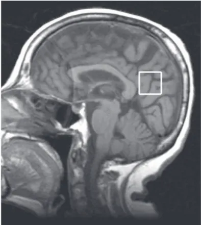
Figure 1. Sagittal T1-weighted image (TR=450 ms, TE=8 ms),
and 128 excitations, lasting approximately 3 minutes. The voxel was located in the posterior cingulate. Preceding the spectroscopy acquisition, automatic adjusting transmis-sion-reception, water suppression and fi eld homogeneity optimization were performed for the selected voxel.
The voxel measured 2x2x2 cm (8 cm3) and was located in the posterior cingulate in the median image of the sag-gital plane. It was positioned below the cingulate and above the parietoccipital sulcus, including the posterior cingulate as well as the inferior pre-cuneus, a location previously described in the literature8-12 (Figures 1 and 2).
We evaluated the N-acetyl-aspartate/creatine (NAA/ CR), Mio-inosytol/creatine (mI/CR) and Mio-inosytol/ N-acetyl-aspartate (mI/NAA)/ratios. Post-processing was carried out using the MRUI software (Magnetic Resonance User Interface), Java version, from Advanced signal process-ing for medical resonance imagprocess-ing and spectroscopy, TMR, FMRX-CT97-0160. The NAA/CR, mI/CR, and mI/NAA ratios obtained in the posterior cingulate for patients and controls were compared.
Two illiterate patients (12 and 13), together with a mild AD patient (patient 14), presenting with a marked compro-mise in language and a very low MMSE score (MMSE=6), disproportionate to the other cognitive functions which re-mained relatively preserved, were excluded from this
analy-sis. This lack of literacy and marked language impairment hampered the MMSE analysis and scoring which could have in turn introduced a bias in the association of the relationship between metabolites and the MMSE.
The Chi-square test was used to compare the gen-der distribution of the two groups (nominal data). The Student-t test was employed to compare the me-tabolite ratios in patient and control groups. Correla-tion between MMSE and metabolic ratios were cal-culated using Pearson’s linear coefficient (13). The descriptive level considered signifi cant was 5% (<0.05).
Results
The MMSE data was distributed as depicted in Table 1, which shows a lower MMSE and greater variance in the patient group.
Comparative analysis between the MMSE and meta-bolic ratios was carried out by calculating the indexes of Pearson’s linear correlation for each metabolite, consider-ing the entire sample and also patient and control groups separately. Separate group analysis revealed no signifi cant association. However, analysis of the pooled data (patients and controls) revealed a signifi cant correlation for some measures, being positive for the Naa/Cr ratio in the cingu-late (0.61), and negative for the mI/Naa ratio (–0.55). These
Figure 2. Representation of voxel in axial and coronal planes from volumetric images (SPGR).
Table 1. MMSE Scores in patients and controls.
Group Mean Median SD Minimum Maximum Samples
Controls 27.8 28 1.4 25 30 15
Patients 19.0 19 3.9 6 28 29
values indicated some degree of association, but evidenced large spread between the MMSE and the relationships among the metabolites. This data can be seen in Table 2.
The level of association is clearly illustrated in Figures 3 to 5, where each individual is represented by a point, and a line of correlation is presented for the global data.
Discussion
A signifi cant association was observed between Naa/ Cr and ml/Naa ratios in the posterior cingulate and the MMSE. The Naa/Cr ratio correlated positively while the ml/Naa ratio showed negative correlation. Naa and Naa/ Cr ratio values have correlated positively with the MMSE in earlier studies in the occipital and parietal gray mat-ter,14 parietoccipital gray matter15 occipital gray matter,16, temporal white matter17 and para-hippocampal gyrus (in a
post-mortem study).18 There is also evidence of an associa-tion between decreased Naa concentraassocia-tion and cognitive decline.19,20 Since NAA is considered a marker for neuronal density/viability, these associations indicate that the NAA/ Cr ratio obtained by proton spectroscopy could be consid-ered an indicator of cognitive decline in such patients.
The Naa/ml ratio also correlated positively with the MMSE.15 However, confl icting results have been found for this association regarding separate ml or ml/Cr. Several studies have demonstrated the presence of negative cor-Table 2. Association between MMSE and ratios among metabolites.
Measure
Global Patients Controls
Correlation p Correlation p Correlation p
Naa/Cr 0.61 <0.001 0.27 0.176 –0,46 0,086
mI/Cr –0,33 0,035 –0.29 0.149 –0.45 0.094
mI/Naa –0.55 <0.001 –0.36 0.071 –0.11 0.702
Naa/Cr, N-acetil-aspartate/creatine ratio; mI/Cr, mioinositol/creatine ratio; mI/Naa, mioinositol/n-acetil-aspartate ratio, and p, descriptive level.
Figure 3. Scatter plot for MMSE and Naa/Cr in the cingulated.
Naa/Cr, N-acetil-asparte/creatine ratio.
Figure 4. Scatter plot for MMSE and ml/Cr in the cingulate. ml/Cr,
mioinositol/creatine ratio.
Figure 5. Scatter plot for MMSE and ml/Naa in the cingulate. ml/
Naa, mioinositol/N-acetil-aspartate ratio. 1.4
1.5 1.6 1.7 1.8 1.9 2.0
12 16 20 24 28 32
MMSE
Patient Control
Naa/Cr
0.4 0.5 0.6 0.7 0.8 0.9 1.0 1.1 1.2
12 16 20 24 28 32
mI
/C
r
MMSE
Patient Control
0.3 0.3 0.4 0.4 0.5 0.5 0.6 0.6 0.7 0.7
12 16 20 24 28 32
mI
/N
a
a
MMSE
relation for the ml/Cr ratio in the posterior cingulate,21 or for ml concentration in frontal white matter.22 Neverthe-less, other authors14,15 observed no signifi cant correlation of the ml/Cr ratio or the ml concentration, with the MMSE. It is possible that the correlation of the Naa/ml or ml/Naa is due to the Naa component. Indeed, the present study showed a more signifi cant correlation for the Naa/Cr ratio than the ml/Naa ratio. In view of the fact that the increase in ml or the ml/Cr ratio found in this disease is early and associated with the accumulation of neurofi brillar tangles and astrocytic and glial proliferation, the absence of cor-relation between ml/Cr ratios and the MMSE test remains unclear and warrants future investigations involving a larger patient series.
Concluding, the analysis of proton spectroscopy studies on a single voxel within the posterior cingulate carried out in mild or moderate AD patients and controls evidenced positive correlation of the Naa/Cr ratio with the MMSE yet negative correlation of the ml/Naa ratio. These fi ndings corroborate a correlation between neuronal density/viabil-ity and the MMSE test.
References
1. Laakson MP, Soininen H, Partanen K, et al. MRI of the hip-pocampus in the Alzheimer’s disease: sensitivity, specifi city, and analysis of the incorrectly classifi ed subjects. Neurobiol Aging 1998;19:23-31.
2. Herminghaus S, Frolich L, Gorriz C, et al. Brain metabolism in Alzheimer disease and vascular dementia assessed by in vivo proton magnetic resonance spectroscopy. Psychiatry Res 2003;123:183-190
3. Fazekas F, Chawluk JB, Alavi A, Hurtig HI, Zimmerman RA. MR signal abnormalities at 1.5 T in Alzheimer’s dementia and normal aging. AJR Am J Roentgenol 1987;149:351-356. 4. Schmidt R, Fazekas F, Kleinert G, et al. Magnetic resonance
imaging signal hyperintensities in the deep and subcortical white matter. A comparative study between stroke patients and normal volunteers. Arch Neurol 1992;49:825-827. 5. Brucki SMD, Nitrini R, Caramelli P, Bertolucci PHF, Okamoto
IH. Sugestões para o uso do Mini-Exame do Estado Mental no Brasil. Arq Neuropsiquiatr 2003;61:777-781.
6. Nitrini R, Caramelli P, Herrera Junior E, et al. Performance of illiterate and literate nondemented elderly subjects in two tests of long-term memory. J Int Neuropsychol Soc 2004;10:634-638. 7. Caramelli P, Carthery MT, Porto CS, Charchat-Fichman H,
Bahia VS, Nitrini R. Teste de fl uência verbal no diagnóstico da doença de Alzheimer leve: notas de corte em função da escolaridade. Arq Neuropsiquiatr 2003;61:23.
8. Kantarci K, Jack Jr CR, Xu YC, et al. Regional metabolic pat-terns in mild cognitive impairment and Alzheimer’s disease: A 1H MRS study. Neurology 2000;55:210-217.
9. Kantarci K, Reynolds G, Petersen RC, et al. Proton MR spec-troscopy in mild cognitive impairment and Alzheimer dis-ease: comparison of 1.5 and 3 T. AJNR Am J Neuroradiol 2003;24:843-849.
10. Kantarci K, Xu Y, Shiung MM, et al. Comparative diagnostic utility of different MR modalities in mild cognitive impair-ment and Alzheimer’s disease. Deimpair-ment Geriatr Cogn Disord 2002;14:198-207.
11. Hattori N, Abe K, Sakoda S, Sawada T. Proton MR spectro-scopic study at 3 Tesla on glutamate/glutamine in Alzheimer’s disease. Neuroreport 2002;13:183-186.
12. Martinez-Bisbal MC, Arana E, Marti-Bonmati L, Molla E, Celda B. Cognitive impairment: classifi cation by 1H magnetic resonance spectroscopy. Eur J Neurol 2004;11:187-193. 13. Lee ET, Wang JW. Statistical methods for survival data
analy-sis. 3rd edition. New Jersey: John Wiley & Sons; 2003.
14. Huang W, Alexander GE, Chang L, et al. Brain metabolite concentration and dementia severity in Alzheimer’s disease: a (1)H MRS study. Neurology 2001;57:626-632.
15. Waldman AD, Rai GS. The relationship between cognitive impairment and in vivo metabolite ratios in patients with clinical Alzheimer’s disease and vascular dementia: a proton magnetic resonance spectroscopy study. Neuroradiology 2003;45:507-512.
16. Weiss U, Bacher R, Vonbank H, Kemmler G, Lingg A, Mark-steiner J. Cognitive impairment: assessment with brain mag-netic resonance imaging and proton magmag-netic resonance spectroscopy. J Clin Psychiatry 2003;64:235-242.
17. Frederick BD, Lyoo IK, Satlin A, et al. In vivo proton magnetic resonance spectroscopy of the temporal lobe in Alzheim-er’s disease. Prog Neuropsychopharmacol Biol Psychiatry 2004;28:1313-1322.
18. Kwo-On-Yuen PF, Newmark RD, Budinger TF, Kaye JA, Ball MJ, Jagust WJ. Brain N-acetyl-L-aspartic acid in Alzheimer’s disease: a proton magnetic resonance spectroscopy study. Brain Res 1994;667:167-174.
19. Kantarci K, Jack CR, Jr. Neuroimaging in Alzheimer dis-ease: an evidence-based review. Neuroimaging Clin N Am 2003;13:197-209.
20. Adalsteinsson E, Sullivan EV, Kleinhans N, Spielman DM, Pfefferbaum A. Longitudinal decline of the neuronal marker N-acetyl aspartate in Alzheimer’s disease. Lancet 2003;355(9216):1696-1697.
21. Rose SE, de Zubicaray GI, Wang D, Galloway GJ, Chalk JB, Ea-gle SC, et al. A 1H MRS study of probable Alzheimer’s disease and normal aging: implications for longitudinal monitoring of dementia progression. Magn Reson Imaging 1999;17:291-299. 22. Parnetti L, Tarducci R, Presciutti O, Lowenthal DT, Pippi M,