100
ELECTROMYOGRAPHIC STUDY OF THE DELTOID,
PECTORALIS MAJOR AND TRICEPS BRACHII
MUSCLES IN SWIMMERS DURING BILATERAL
CONTRACTIONS PERFORMED IN MULTI-JOINT
EXERCISE WITH DIFFERENT LOADS
Fernando Nazário-de-Rezende1, ,3
Gilmar da Cunha Sousa2
Eduardo G. Haddad2
Vanessa S. de Oliveira1
Robson da Silva Medeiros1
Guilherme Goulart de Agostini2
Moacir Marocolo3
1. FISIO2EX – Research and Physical
Evaluation in Human Performance Center of the President Antônio Carlos University – UNIPAC – Uberlândia, MG, Brazil.
2. CENESP – NIAFIS – FAEFI – UFU – Federal University of Uberlândia. 3. UFTM – Master’s Program in Physical Education of theFederal University of the Triangle of Minas Gerais– Uberaba – MG, Brazil.
Mailing address:
Rua Arlindo Sousa Monteiro, n.120 - B. Jardim Finotti
CEP 38408074 - Uberlândia-MG, Brasil. E-mail: [email protected]
ABSTRACT
The objective of this study was to compare the electrical activity of the deltoid (middle portion), pectoralis major (clavicular portion) and triceps (long head) muscles during bilateral contraction per-formed in a multi-articulated joint shoulder-press convergent machine with 40% and 80% maximum voluntary load (MVL) in 11 male swimmers (15 to 23 years, 70 ± 4 kg, 183 ± 6 cm and 10 ± 4 years’ time of practice in sport), trained in resistance exercise. Electromyographic signals (EMG) were obtained by placing surface active differential electrodes (20 x gain), composed of two parallel rectangular bars (EMG System, Brazil®
). A data acquisition system (EMG-Alc) which provided numerical data in RMS (Root Mean Square) to analyze the signals composed by a reference electrode (ground) and a signal conditioning module (EMG) with simultaneous acquisition of up to 8 differential channels (band-pass filter 5-20 Hz), adjustable amplifier stage, allowing gains between 100 and 4960 times, channel input impedance 10GΩ in differential modules and CMRR of 93 dB/60 Hz was used. Only the concentric phase (3 seconds duration) in each EMG signal collected was recorded. After the tests (Mann-Whitney U test, Friedman and Wilcoxon) were applied, it was concluded that for prescription and periodization of the neuromuscular training, bilateral contractions performed in the shoulder-press apparatus are efficient at aiming muscular recruitment (80%> 40% ) of middle portion of the deltoid, pectoralis major (clavicular portion), and triceps brachii (long head) muscles, evidencing differences between dominant and non-dominant limbs only for the dominant brachial triceps in 80% of MVL in swimmers trained in resistance exercises.
Keywords: EMG, development, resistance exercise, swimmers.
INTRODUCTION
In the past, resistance training programs were based on the ex-periences of the coach or athlete, and science as support in the resistance training programs was avoided, which made both begin-ner and experienced athletes confused1. In fact, science was slow in
validating the adopted practices in the resistance training.
Over the last years,many researchers have dedicated their time to the scientific study of the effects of different types of strength training in humans, with the aim to validate the basic exercises for physical fitness programs of athletes and non-athletes 2-6. In those
investigations, a series of comparisons between the deltoid, pec-toralis major and triceps brachii muscles as well as comparisons between the different portions of a single muscle have been performed2,7.
In the sports field, some electromyographic work has appro-ached the participation of the pectoralis major, latissimusdor-si, deltoid among other muscles, in a rowing simulator8 and in
swimmers from different categories, simulating backstroke and crawl unilateral exercises9,10. However, with the increasing use of
resistance exercises by swimming athletes with higher strength levels and performance improvement as goal, new investigations on this modality of exercises and action of different muscles du-ring their performance become necessary.
Thus, the aim of this study was to compare the electromyo-graphic signs emitted by the deltoid medialis, pectoralis major (clavicular portion) and triceps brachii muscles (long head) of the dominant and non-dominant limbs of 11 swimmers dur-ing bilateral contractions in the multi-articulated joint shoulder-pressconvergent machine.
METHODOLOGY
Sample
Participated in this study 11 male swimmers, practitioners of resistance exercise, aged between 19 ± 4 years, body mass 70 ± 4kg, height 183 ± 6cm and time of practice in the sport of 10 ± 4 years, participated in the study, and the resistance exercises were part of their training. The volunteers did not present history of osteomyo articular diseases which could interfere in the results.
Maximum Voluntary Load Test
All volunteers were submitted one day before the collec-tion to a test of concentric bilateral maximum voluntary load (MVL) performed according to Nazário-de-Rezendeet al.11. The
load adopted for the study was of 40% and 80% of MLV, an intensity to which all volunteers were submitted during the annual training sessions.
ORIGINAL ARTICLE LOCOMOTOR APPARATUS IN
101
B
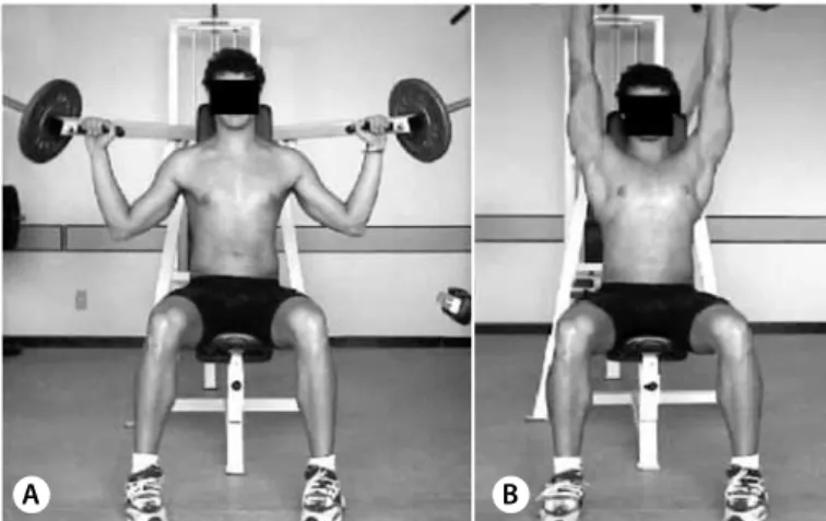
Figure 1. Multi-articulated joint shoulder-press convergent exercise: beginning of the bilateral movement (A) and end of the bilateral movement (B). General Procedures
Before performance of the electromyographic signs, the volun-teers received information about the research and were submitted to familiarization procedures. The volunteers underwent explanations and simulations on the most suitable posture for the performance of exercise, initial and final position of each movement, performance velocity and the verbal command given by the electromyography technician. Subsequently, they signed a consent form for participa-tion in the study and release of the results according to resoluparticipa-tion # 196/96 of the National Board of Health.
In order to establish specific muscular preparation, the vol-unteers performed three sets with 15 repetitions without load..
Electrode
Skin sanitation and shaving was performed for acquisition of the electric activity (EMG) of the muscles. The electrodes used were simple differential active surface ones (Lynx Eletronics Ltda., São Paulo, SP, Brazil), composed of two parallel rectangular bars of pure silver (Ag), each one 10mm-long, 1mm-wide and 10mm distant from each other;20mm-wide by 41mm-long and 5mm-thick acrylic resin capsule; 1m-long cable; gain of 20 times; CMRR (common mode rejection rate) of 84 dBn and Earth plate electrode (Bio-logic Systems Corp. – SP Médica, Científica e Comercial Ltda., São Paulo, SP, Brazil), composed of a stainless steel disc measuring 30mm in diameter and 1.5mm-thick an 1m cable attached, which was placed on the head of the ulna of the volunteers, with the aim to eliminate external interferences12.
The electrodes were attached to the skin; on the deltoid medialis muscles positioned approximately 4 ± 2cm away from the lateral border of the acromion, in a region where the higher volume of the muscle surface was clear. Concerning the triceps brachii (long head), the electrodes were attached according to Sousa et al.2, 10 ± 1cm above the olecranon. Regarding the
pectoralis major (clavicular portion) an activation maneuver was performed and the electrode was placed on the point of greatest muscular surface.
Electromyograph
EMG collection of the studied muscles was obtained through a sign conditioner module (electromyograph), with simultaneous acquisition of up to eight differential channels, entrance channel impedance of 10GΩin differential modules, 12 bits of resolution band-pass filter of 20Hz to 5Hz and RRMC of 93db to 60Hz, entrance range of –10 to +10v and a data acquisition system (Alc-EMG) which provided numerical data in RMS (root mean square) for analysis of the results. The electromyograph was adjusted with gain of 4,960 times, guaranteeing hence the necessary amplification to the an-alog-digital conversion process and sample number of 6,000 and channel frequency of 2,000Hz, resulting in total acquisition time of three seconds.
Multi-articulated joint shoulder-pressconvergent machine
A machine named multi-articulated joint shoulder-press con-vergent, brand name MASTER was used for determination of the load in one repetition maximum (1RM) and performance of the bilateral exercise in the study. Such machine simulates the movement performed with dumbbells.
Movement performance
The volunteers sat on the machine with their trunk and head resting on the back and feet on the ground. After load selection with the volunteer already positioned, the electrodes were at-tached on the studied muscles. The movement started with arms in semi-abduction, forearms in flexion on frontal plane, prone hands front (figure 1).
The movement occurred with arm abduction and forearm extension simultaneously following the path permitted by the machine, being this the concentric phase of the exercise which had duration of three seconds. The electric signs in the bilateral tests were firstly picked with 40% and immediately after at 80% of MVL only in the concentric phase. Five trials were performed for better reproducibility and maximization of collection accuracy and statistical analysis.
Recovery interval
The volunteers were told not to perform any type of training on the day before the EMG recordings to avoid possible fatigue effects and alterations in the results11.
The volunteers, after the end of the movement, remained seated, upper limbs facing down and parallel to the trunk and relaxed during five minutes of rest between trials, both for EMG recordings and for the MVL tests in order to avoid or minimize the fatigue effects13and
replace their energetic supplies14.
Goniometer
A plastic 35-cm long universal goniometer brand name CARCI, was used for measurement of the angles of the knee and elbow joints15, prior to the tests performance, when the volunteer was
already positioned at the machine.
Regarding the knee joint, the goniometer screw was placed on the lateral condyle of the femur, laterally aligned on the thigh longitudinal axis, from the trochanter major to the lateral condyle and on the axis between the fibula head until the lateral mal-leolus. On the elbow joint, the goniometer was aligned along the lateral medial line of the humerus, from the humerus head to the lateral epicondyle and the medial line of the radius until the radial styloid process.
The joint angles of the upper and lower limbs, at the beginning of the movement, have not been exactly delimited; however, the positionsof the knee joint (106º ± 5º) and elbow joint (105º ± 5º) were similar to those adopted in their training routines.
102
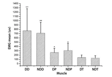
Figure 2. Values expressed in RMS (μν) of the summed mean electrical activity of the dominant deltoid with non-dominant deltoid, dominant pectoralis with non-dominant pectoralis and dominant triceps with non-dominant triceps muscles of the 11 swim-mers with 40% of MVL.
*significance of 0.05.
STATISTICAL ANALYSIS
The Wilcoxon and Student’s t tests were applied to the data under consideration with the goal to verify the existence or ab-sence of significant differences between the measures of the three muscles with 40% and 80% load for the 11 swimmers, with signifi-cance level adopted of 0.05 or 5% in a bilateral event.
RESULTS
The mean of the electric activity of the studied muscles is re-presented in tables 1, 2, 3 and 4. Note that when the 40% of 1RM load is analyzed (table 1 and figure 2), the mean values of the EMG signal of the sum of the dominant and non-dominant limbs for the deltoid, pectoralis and triceps muscles were 63.3%, 24% and 12.6%, respectively. Values significantly different are found when the EMG sum of the right and left deltoid is analyzed and compared with the sum of the EMG activity of the pectoralis activity. Significant diffe-rences were found in the 40% of 1RM load between the sum of the deltoid compared with the sum of the pectoralis sum (p = 0.004), between the deltoid sum with the triceps sum (p = 0,003) and the pectoralis sum with the triceps sum (p = 0.009). When the dominant and non-dominant limbs are compared, no significant difference was found (tables 2, 3 and figure 3, 4).
When the 80% of 1RM load (table 4 and figure 5) is analyzed, the mean values of the EMG sign of the sum of the dominant and non-dominant limbs for the deltoid, pectoralis and triceps muscles is50.5%, 35.9% and 13.5%, respectively, which are sig-nificantly different values. In the 80% loads we found significant differences between the EMG sum of the deltoid muscles com-pared with the pectoralis sum (p = 0.036), between the deltoid sum with the triceps sum (p = 0.000) and the pectoralis sum with the triceps sum (p < 0.001). As demonstrated in figure and table 2, when the dominant and non-dominant limbs were compared, no significant differences were found, except for the triceps, whose dominant side presented higher EMG sign (p = 0.016).
When the bilateral intermuscular kinesiologic activity is compared, the results presented in figures 3 and 4 were statis-tically the same with both loads (40% and 80%), and the deltoid muscle presented higher activity followed by the pectoralis and triceps (p = 0.003).
Table1. Values expressed in RMS (μν) of the electrical activity of the dominant deltoid (DD), dominant deltoid (NDD), dominant pectoralis(DP), non-dominant pectoralis (NDP), non-dominant triceps (DT) and non-non-dominant triceps (NDT) muscles of the 11 swimmers with 40% of MVL.
Muscular contraction (expressed in μν)
Volunteer DD NDD DP NDP DT NDT
1 620 893 115 170 136 111
2 776 409 306 581 238 256
3 616 667 302 216 212 160
4 710 420 295 295 350 169
5 604 1.137 105 151 106 166
6 1.241 439 200 195 111 102
7 415 556 447 644 53 58
8 407 583 321 173 171 116
9 376 785 285 372 86 95
10 2.134 965 261 269 204 127
11 531 962 225 251 113 93
Mean 766 ± 513 711 ± 253 260 ± 97 302 ± 167 162 ± 85 132 ± 54
Table 2. Values expressed in RMS (μν) of the electrical activity of the dominant deltoid (DD), non-dominant deltoid (NDD), dominant pectoralis(DP), non-dominant pectoralis(NDP), dominant triceps (DT) and non-dominant triceps muscles (NDT) of the 11 swimmers with 80% of MVL.
Muscualr contraction (expressed in μν) Volunteer DD NDD DP NDP DT NDT
1 988 653 826 746 276 223
2 1.153 551 639 949 429 323
3 784 1.070 713 674 404 261
4 851 673 784 907 312 282
5 987 1.417 396 415 210 240
6 1.239 751 554 434 176 215
7 756 532 795 1.070 99 91
8 756 774 573 774 340 275
9 578 1.245 588 789 273 171
10 2.068 1.124 661 630 324 205
11 1.146 913 569 450 268 242
Mean 1.028 ± 400 882 ± 295 645 ± 128 713 ± 218 283* ± 96 230 ± 62
*significance of 0.05.
Table 3. Values expressed in RMS (μν) of the summed mean electrical activity of the dominant deltoid with the dominant deltoid, dominant pectoralis with non-dominant pectoralis, non-dominant triceps (DT) and non-non-dominant triceps muscles of the 11 swimmers with 40% of MVL.
Muscles
Volunteer Deltoid Pectoralis Triceps
1 1.513 285 247
2 1.185 887 494
3 1.283 518 372
4 1.130 590 519
5 1.741 256 272
6 1.680 395 213
7 971 1.091 111
8 990 494 287
9 1.161 657 181
10 3.099 530 331
11 1.493 476 206
Mean/standard deviation 1.477** ± 598.3 562* ± 246.7 294 ± 127
*significance of 0.05.
EMG mean (uv)
103
Figure 3. Values expressed in RMS (μν) of the electrical activity of the dominant deltoid (DD), non-dominant deltoid (NDD), dominant pectoralis(DP), non-dominant pectoralis(NDP), dominant triceps (DT) and non-dominant triceps (NDT) muscles of the 11 swimmers with 40% of MVL.
Figure 4. Values expressed in RMS (μν) of the electrical activity of the dominant deltoid (DD), non-dominant deltoid (ND), dominant pectoralis (DP), non-dominant pectoralis (NDP), dominant triceps ( ) and non-dominant triceps (NDT)muscles of the 11 swimmers with 80% of MVL.
Figure 5. Values expressed in RMS (μν) of the summed electrical activity of the dominant deltoid with non-dominant deltoid, dominant pectoralis with non-dominant pectoralis and dominant triceps with non-dominant triceps muscles of the 11 swim-mers with 80% of MVL.
*significance of 0.05.
DISCUSSION
The muscular activity is always expressed through the joint activity of the muscles,where it is not possible that one move-ment occurs due to the action of a muscle in isolation. There-fore, to evaluate or analyze muscular actions in a multiarticular exercise by the observation of the action of agonists, antagonists and synergic muscles offers an interesting parameter concern-ing the comparison between the activities of the dominant and non-dominant limbs both uniand bilaterally.
Our data refer to the analyses of the shoulder-press exercise, whi-ch is crucial to the physical preparation of swimmers, with aim on increase of performance. When the bilateral intermuscular activity is analyzed, the deltoid muscle presented higher activity followed by the pectoralis major and tricepsbrachii both in the test with 40% and 80% of MVL. It is clear that the exercise studied can guarantee neural adaptations derived from the training applied to swimming, since it activates important swimming synergists, since the sport demands shoulder movements in arm circle movements above the head line15.
The findings by Bankoffand Vitti10andVittiand Bankoff9
sup-port ours, since they studied swimmers of different categories simulating unilateral exercises of backstroke and crawl styles approaching the participation of the pectoralis major and latis-simusdorsimuscles among other muscles. Generally speaking, the pectoralismajor muscle showed high electromyographic activity during the swimming practice of the backstroke and crawl styles of the individuals, either trained or not, being the signs concer-ning the trained categories more intense.
Our data agree with the ones by Kronberget al.16, Campos et
al.17and Oliveira et al.7, who reported that the medial and
ante-rior portions of the deltoid muscle play an important role in the arm abduction and that the EMG increase is proportional to the increase of the range of motion.
The significant increase in the electrical activity of the deltoid medial, pectoralis major (clavicular portion) and triceps brachii (long head) became evident in our study with 80% when com-pared 40% load of the MVL.
When using loads of 40% of MVL, the deltoid muscle acted with 63.3% of the EMG activity, the pectoralis major muscle 24%, and the triceps brachii12.6%. When the 40% load is doubled to 80% of MVL, the deltoid muscle decreased its relative participation to 50%
Table 4. Values expressed in RMS (μν) of the summed median electrical activity of the dominant deltoid with the dominant, dominant pectoralis with the non-dominant pectoralis, non-dominant triceps with the non-non-dominant triceps muscles of the 11 swimmers with 80% of MVL.
Muscles
Volunteer Deltoid Pectoralis Triceps
1 1.641 1.572 499
2 1.704 1.588 752
3 1.854 1.387 665
4 1.524 1.691 594
5 2.404 811 450
6 1.990 988 391
7 1.288 1.865 190
8 1.530 1.347 615
9 1.823 1.377 444
10 3.192 1.291 529
11 2.059 1.019 510
Mean/standard deviation 1.910** ± 521,7 1.358* ± 319,9 513 ± 150,2
*significance of 0.05.
DD NDD DP NDP DT NDT Muscle
EMG mean (uv)
Muscle
EMG mean (uv)
DD ND DP NDP DT NDT
EMG mean (uv)
104
of the EMG activity, followed by the pectoralis and triceps muscles to relative increase of 36% and 14%, respectively. The significant increase of the EMG activity of the pectoralis major muscles with 80% load may have decreased the production of relative recruiting strength that the deltoid medial muscle (primary motor) may generate; however, such increase may be a protection mechanism of the glenohumeral joint against possible injuries, occurring hence higher inter and intra-muscular strength distribution between the synergist, antagonist and stabilizer muscles.
In studies performed by Duarte Cintraand Furlani18 concerned with
uni and multiarticular exercises for lower limb, they verified that the increase in weight during the movements caused high level of activity and simultaneous contraction of all the studied muscles. Despite the impossibility of direct comparison, this statement is in agreement with our findings, since when the load factor in isolation is analyzed it was verified that the muscles analyzed presented higher electrical activity when the load was doubled from 40% to 80% of MVL.
The increase in muscular strength was possibly determined by the development of the adaptation alterations at the level of the central nervous system which led to the intensification of the motor centers capacity to recruit a large number of motor neurons, which were deactivated before, increasing the number of motor units which participated in the muscular contraction. This results is in agreement with the theory of muscular strength grading, which highlights that if there is simultaneous activation of a higher number of motor units, increase of muscular strength will occur as well, evidenced in the present study when 80% of MVL was used19.
For training purposes, this situation is favorable, since the mus-cle should act against some resistance it usually does not find so that the physiological alterations which result in the expected training effects can occur14.
Tassiet al.20, analyzed the bilateral behavior of a thigh muscle,
and contrary to our findings, they verified Strong potential of the dominant limb over the non-dominant one. In these authors’ opi-nion, the dominant limb is more demanded in daily situations, and it is also believed that the right muscles in right-handed in-dividuals present considerable development compared to the left-handed ones, and hence, contribute to the anatomic and functional asymmetry.
In our findings, when the myoelectric activity of the dominant and non-dominant limb is compared, it was visible that both present-ed similar electrical activities, except for the triceps brachii muscle, with 80% of the MVL. The resistance exercises correctly performed with individualized loads, adequate posture and guidance from a professional, can with the trainig progress, improve the recruiting pattern of the motor units (coordination), eliminating the diferences between the muscular contractions of opposite sides by the effect of transference (crossed education) mentioned by Moritaniand De Vries21, Sale22, Shi Zhou23, Simão et al.24and Brentano and Pinto25.
The prescription of uniarticular and multiarticular resistance exercises bilaterally performed for training helps in the develop-ment of the physical preparation of swimmers in and out the wa-ter. It became evident that the loads increase in a multiarticular exercise really boosts muscular activation through the increase of the neural drive with no significant compromising of the in-termuscular coordination of the synergists, offering safety for the athlete in his training routine.
CONCLUSION
The results presented added to the methodology used in this research let us conclude that in practical terms of neuromuscular training practical prescription and periodization, the bilateral con-tractions performed in the multi-articulated joint shoulder-press convergent machine are efficient in recruiting (80% > 40%) the deltoid medialis, pectoralis major (clavicular portion) and triceps brachii (long head) muscles, being differences between the do-minant and non-dodo-minant limbs only for the dodo-minant triceps brachii with load of 80% of MVL in these swimming athletes with eight training history.
ACKNOWLEDGEMENTS
We acknowledge the Coordination for the Improvement of Higher Level Personnel (CAPES) for the post-graduation scholar-ship and the Research Laboratory in Kinesiological Electromyo-graphy (LAPEC-UFU).
All authors have declared there is not any potential conflict of interests concerning this article.
REFERENCES
1. Benedic T. Manipulating resistance training program variables to optimize maximum strength in men: A Review. J Strength Cond Res 1999;13;289-304.
2. Sousa GC, Bérzin F, Silva Z, Negrão-Filho RF. Electromyographic study of the simultaneous action of the biceps brachii, triceps brachii, brachialis and brachiorradialis muscles in a semipronated position at different loads and angles. Braz J Morphol Sci São Paulo 2000;17;63-8.
3. Rodrigues JA, Büll ML, Dias GAR, Gonçalves M. Electromyographic analysis of the pectoralis major and deltoideus anterior muscle muscles in horizontal “flyer” exercises. Electromiographic. Electromyogr Kinesiol 2003;43:413-9. 4. Ferreira MI, Büll ML, Vitti M. Electromyographic validation of basic exercises for physical conditioning
programmes. IV. Analysis of the deltoid muscle (anterior portion) and pectoralis major muscle (clavicu-lar portion) in frontal-lateral cross, dumbbells exercises. Electromyogr Clin Neurophysiol 2003;43:67-74. 5. Ferreira MI, Büll ML, Vitti M. Electromyographic validation of basic exercises for physical conditioning programmes. V. The comparison of the response in the deltoid muscle (anterior portion) and pectoralis major muscle (clavicular portion) determined by the frontal-lateral cross, dumbbells and the rowing exercises. Electromyogr Clin Neurophysiol 2003;43:75-9.
6. Ferreira MI, Büll ML, Vitti M. Participation of the deltoid (anterior portion) and pectoralis major (clavicular portion) muscles in different modalitis of supine and frontal elevation exercises with different grips. Electromyogr Clin Neurophysiol 2003;43:131-40.
7. Oliveira AS, Rodrigues D, Berzin F. Atividade eletromiográfica das Porções Anterior, Média e Posterior do Músculo Deltóide na Abdução do Braço. Rev Bras Fisioter 2001;5:17-24.
8. Furlani J, Cerqueira EP, Scoarçoni M. Estudo eletromiográfico dos músculos peitoral maior, serrátil anterior e grande dorsal em movimentos de remo a seco. Rev Bras Ciênc Morfol 1987;4:40-4. 9. Vitti M, Bankoff ADP. Simultaneous EMG of latissimus dorsi and sternocostal part of pectoralis major
muscles during classic natatory stroke. Electromyogr Clin Neurophysiol 1979;19:505-10. 10. Bankoff ADP, Vitti M. Investigação eletromiográfica da ação conjugada dos músculos grande dorsal e
peitoral maior em movimentos natatórios durante o estilo “costa”. Rev Bras Ciênc Morfol 1986;3:95-9. 11. Nazario-de-Rezende F, Haddad EG, Sousa G C, Silva LFG, Goncalves A, Agostini GG, Silva D C O .
Electromyographic study of the rectus femoris and bíceps femoris (long head) muscle during bilateral
isotonic contraction in a 45o Leg Press Apparatus. Biosci Journal 2006;22:95-104.
12. De Luca CJ. The use of surface eletromyography in biomechanics. J Applied Biomech 1997;13:135-63. 13. Hakkinen K, Kalinen M, Linnamo V, Pastinen UN, Newton RU, Kraemer WJ. Neuromuscular adaptation during bilateral versus unilateral strenght training in middle-aged and elderly men and women. Acta Physiol Scand 1996;158:77-88.
14. Fleck SJ, Kraemer WJ. Princípios básicos do treinamento de força e prescrição de exercícios. In:Fundamentos do treinamento de força. 2. ed. Porto Alegre: Artes Médicas, 1999;19-26. 15. Vieira MSR, Lianza S. Estudo sobre o ombro doloroso do nadador. Medicina de Reabilitação 2001;57:12-5. 16. Kronberg M, Németh G, Broström LA. Muscle Activity and Coordination in the Normal Shoulder – an
Electromyographic Study. Clin Orthop Relat Res 1990;257:76-85.
17. Campos GER, Vitti M, Freitas V. Estudo eletromiográfico dos músculos trapézio e deltóide em movi-mentos do braço. Rev Bras Ciênc Morfol 1992;9:9-14.
18. Duarte Cintra AI, Furlani J. Eletromyographic study of quadriceps femoris in man. Electromyogr Clin Neurophysiol Louvain 1981;21:539-54.
19. Verkhoshanski YV. Capacidades de Força. In: Treinamento Desportivo: Teoria e Metodologia. 1. ed. Porto Alegre: ArtMed, 2001;163-74.
20. Tassi N, Filho JG, Gonçalves M, Vitti M, Krool LB. Electromyographic behavior of the biceps femoris mus-cle during knee extension and flexion performed on the leg press. Bras J Morphol Sci 1998;15:17-22. 21. Moritani T, De Vries HA. Neural factors versus hypertrophy in the time course of muscle strength gain.
Am J Phys Med 1979;58:115-30.
22. Sale DG. Neural adaptation to resistance training. Med Sci Sports Exerc 1988;20:S135-45. 23. Shi Zhou. Chronic neural adaptations to unilateral exercise: mechanisms of the cross education. Exerc
Sport Sci Rev 2000;28:177-84.
24. Simão R, Monteiro WD, Araújo CGS. Potência muscular máxima na flexão do cotovelo uni e bilateral. Rev Bras Med Esporte 2001;7:157-62.