Universidade Fernando Pessoa - Faculdade de Ciências da Saúde
Effects of cerium dioxide nanoparticles in Oncorhynchus mykiss liver
and kidney after an acute exposure: assessment of oxidative stress,
genotoxicity and histological alterations
Ana Cristina Taipa Pinto Nunes Porto, 2015
iii
Universidade Fernando Pessoa - Faculdade de Ciências da Saúde
Effects of cerium dioxide nanoparticles in Oncorhynchus mykiss liver
and kidney after an acute exposure: assessment of oxidative stress,
genotoxicity and histological alterations
Ana Cristina Taipa Pinto Nunes Porto, 2015
iv
Ana Cristina Taipa Pinto Nunes
Effects of cerium dioxide nanoparticles in Oncorhynchus mykiss liver and kidney after an acute exposure: assessment of oxidative stress, genotoxicity and
histological alterations
Orientador: Prof. Doutor Alberto Teodorico Correia
Coorientador: Prof. Doutora Sara Cristina Antunes
Projeto de Pós-Graduação/Dissertação apresentado à Universidade Fernando Pessoa como parte dos requisitos para obtenção do grau de Mestre em Análises Laboratoriais Especializadas, Ramo Análises Ambientais e Alimentares
__________________________________________
v
This work has been already presented as a poster communication in an international congress: Nunes AC, Antunes SC, Ribeiro MIA, Correia AT. 2015. Effects of cerium dioxide nanoparticles in Oncorhynchus mykiss liver after an acute exposure: assessment of oxidative stress, genotoxicity and histological alterations. XV European Congress of Ichthyology. 7th-11st September. Porto, Portugal.
vi
Resumo
Atualmente, as nanopartículas de dióxido de cério (CeO2-NPs) apresentam inúmeras
aplicações que vão desde a indústria até ao uso doméstico conduzindo à presença ubíqua deste composto no ambiente, em particular no compartimento aquático. Perante os poucos estudos existentes sobre o impacto destas nanopartículas no meio aquático, este trabalho teve como objetivo estudar a toxicidade de CeO2-NPs no fígado e rim de Oncorhynchus
mykiss (truta arco-íris) após exposição aguda (96h) a três diferentes concentrações (0,25; 2,50 e 25,00 mg/L) deste composto. Avaliou-se a genotoxicidade (comet assay), a resposta ao stress oxidativo (Catalase CAT, Glutationa-S-transferases GSTs), a peroxidação lipídica (substâncias reativas ao ácido tiobarbitúrico TBARS), os danos histopatológicos e o efeito na atividade da Na+/K+-ATPase no rim. A exposição ao CeO2
-NPs resultou em genotoxicidade nos hepatócitos em todos os grupos expostos ao composto, na inibição da CAT na concentração mais alta (25,00 mg/L) e em alterações histológicas em todos os grupos expostos, com predominância de alterações progressivas, circulatórias e inflamatórias. Porém TBARS, GSTs e Na+/K+-ATPase não apresentaram diferenças significativas comparativamente ao grupo controlo (indivíduos não expostos). Os resultados obtidos neste estudo sugerem assim que CeO2-NPs é capaz de causar
genotoxicidade, desequilíbrio da CAT e alterações histológicas em fígado de truta arco-íris.
vii
Abstract
At present cerium oxide nanoparticles (CeO2-NPs) have numerous applications ranging
from industry to the household, leading to its wide presence in the environment, namely in the aquatic compartment. The hereby study aimed to assess the toxic effects of CeO2
-NPs in Oncorhynchus mykiss liver and kidney after an acute exposure (96h) to three different concentrations (0.25, 2.50 and 25.00 mg/L) of the compound. Genotoxicity (comet assay), oxidative stress response (Catalase CAT; Glutathione S-Transferases GSTs), lipidic peroxidation (Thiobarbituric Acid Reactive Substances TBARS), histopathology and Na+/K+-ATPase activity were evaluated. CeO2-NPs exposure resulted
in genotoxic damage in all exposure treatments, inhibition of CAT in the highest concentration (25.00 mg/L) and histopathological changes in all exposure concentrations with predominance of progressive, circulatory and inflammatory alterations. However TBARS, GSTs and Na+/K+-ATPase activity showed no significant differences
comparatively to the control (unexposed) group. The results suggest that CeO2-NPs are
able to cause genotoxicity, CAT impairment and histological alterations in the liver of rainbow trout.
ix
Agradecimentos
A realização deste trabalho não teria sido possível sem a colaboração de várias pessoas, às quais não poderia deixar de expressar o meu profundo agradecimento.
Gostaria assim de agradecer, em primeiro lugar, ao meu orientador Professor Doutor Alberto Teodorico Correia, sem o qual este trabalho não teria sido possível. Obrigado pela confiança depositada, por sempre entender e me apoiar nas minhas limitações de tempo, enquanto trabalhadora-estudante, pela oportunidade única de integrar a sua equipa de investigadores num centro com o relevo do CIIMAR, e pelo desafio e oportunidade de expor o meu trabalho num congresso internacional da especialidade.
Tenho também de manifestar o meu grande agradecimento, à minha coorientadora, Professora Doutora Sara Antunes, por estar sempre disponível, pelo constante incentivo e pelo especial apoio que me deu na componente laboratorial realizada na FCUP.
Gostaria também de expressar o meu apreço à Professora Doutora Céu Costa pela sua disponibilidade e pelos ensinamentos transmitidos na área da histologia.
Não poderia deixar de expor a minha gratidão à aluna de doutoramento Sara Rodrigues que foi crucial na minha adaptação ao CIIMAR, porque a adaptação a um sítio novo é sempre mais fácil quando temos apoio incondicional. E também agradecer aos alunos de doutoramento Diogo Martins e Odete Gonçalves pela constante simpatia, paciência e total disponibilidade.
E por último e não menos importante aos meus amigos, noivo e família pelo apoio constante, por não censurarem as minhas ausências e pela paciência infindável.
x
“Para ser grande, sê inteiro: nada Teu exagera ou exclui. Sê todo em cada coisa. Põe quanto és No mínimo que fazes. Assim em cada lago a lua toda Brilha, porque alta vive.” Odes de Ricardo Reis, Fernando Pessoa
xi
Table of contents
1.Introduction ... 1
2. Material and methods ... 5
2.1. CeO2-NPs... 5
2.2. Test organisms ... 5
2.3. Acute exposure and fish sacrifice ... 6
2.4. Comet assay ... 6 2.5. Biomarkers analyses ... 7 2.6. Na+/K+-ATPase... 8 2.7. Histological analyses ... 8 2.8. Statistical analysis... 9 3. Results ... 9 3.1. Comet assay ... 9 3.2. Biomarkers analyses ... 10 3.3. Na+/K+-ATPase... 12 3.4. Histological analyses ... 12 4. Discussion ... 14 5. References ... 17
xii
Table of figures
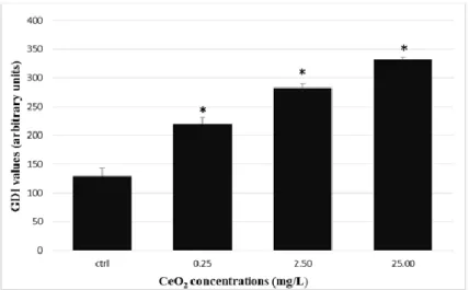
Figure 1- Genetic Damage Index (GDI) in hepatocytes after exposed to CeO2-NPs. Data
are expressed as means ± SE (n =15 fish/treatment).* Stands for significant differences among concentrations and control (p ≤ 0.05). ... 10
Figure 2- Catalase (CAT) activity in liver after exposed to CeO2-NPs. Data are Means ±
S.E. (n =15 fish/treatment).* Stands for significant differences among concentrations and control (p ≤ 0.05). ... 10
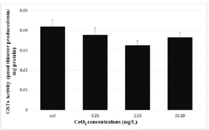
Figure 3- Glutationa S-transferase (GSTs) activity in liver after exposed to CeO2-NPs.
Data are Means ± S.E. (n=15 fish/treatment). ... 11
Figure 4 – Thiobarbituric Acid Reactie Substances (TBARS) in liver after exposed to
CeO2-NPs. Data are Means ± S.E. (n =15 fish/treatment). ... 11
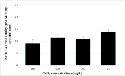
Figure 5- Na+/K+-ATPase activity in kidney after exposed to CeO2-NPs. Data are Means
± S.E. (n = 15 fish/treatment). ... 12
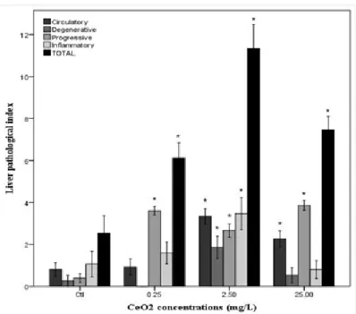
Figure 6 - Histological analyses in liver after exposed to CeO2-NPs. Lesion indices for
each reaction pattern and total lesion indices by concentration of CeO2-NPs. Data are
Means ± S.E. (n =15 fish/treatment). * Stands for significant differences among concentrations and control (p ≤ 0.05). ... 13
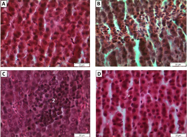
Figure 7 - Histological architecture of the liver of all experimental groups. (A) Control
(400x); (B) CeO2-NPs at 0.25 mg/L with hemorrhage (1) (400x); (C) CeO2-NPs 2.50
mg/L with leukocyte infiltration (2) (400x); and (D) CeO2-NPs 25.00 mg/L with
1
1. Introduction
Nanoparticles (NPs) are defined as particles with dimensions less than 100 nanometers (nm) which present new and improved characteristics compared to larger particles of the same material. NPs can be categorized, in general, into carbon and inorganic based
materials, such as metal oxides, metals and quantum dots (Ju-Nam & Lead 2008; Felix et al. 2013).
The wide interest in nanoparticles comes from their small particle size resulting on novel properties, as improved chemical reactivity, optical behavior and large surface area (Ju-Nam & Lead 2008; Gaiser et al. 2012; Xia et al. 2013). NPs actually have numerous applications including household products and equipment, food, medicine, optical devices, sun lotions, cosmetics, paints, textiles and electronics production and bioremediation (Handy & Shaw 2007; Gaiser et al. 2009; Dahle & Arai 2015). The increasingly widespread use and the great interest that industry demonstrates about NPs and consequently on nanomaterials, has led to an increasing distribution resulting in increased public and occupational exposure to NPs, and thus raising questions about the potential impact of these new materials in the environment and human health (Dreher 2004; Xia et al. 2013)
The cerium oxide is an example of nanoparticle (CeO2-NPs) which the main
application is as fuel additive to enhance the efficiency of combustion, to clean the engine and to decrease fuel consumption and emissions to the environment (Park et al. 2008). CeO2-NPs are also used as semiconductors, as catalysts in petroleum refining, in fuel
cells, as polishing agents in glass and as absorbers of ultraviolet radiation in sunscreens (Cassee et al. 2011; Dahle & Arai 2015). There is an urgent need to study the impact of the NPs metal oxides, such as CeO2-NPs, on the aquatic environment, and to assess the
potential toxicity of these compounds to the aquatic species. The NPs metal oxides can interact in the aquatic environment, at various stages of the species life cycles, and these interactions may lead to disruption of the organisms biochemical balance, and to the urgent need of the species to adapt to a changing environment (Xia et al. 2013)
NPs tend to sorb more than their bulk counterparts (larger sized particles with the same chemical composition) and exhibit multiple exposure routes for the organisms. However, information on the metabolism and excretion is limited, although hepatic excretion is suggested as the most likely route (Handy et al. 2008; Dahle & Arai 2015).
2
As toxicity mechanisms, it is proposed the production of reactive oxygen species (ROS) and free radicals as a primary mechanism of NPs toxicity (Dahle & Arai 2015).
The studies about ecotoxicological effects of CeO2-NPs exposure have been
shown both beneficial and toxic effects on biological systems (Tarnuzzer et al. 2005; Schubert et al. 2006; Heckert et al. 2008; Park et al. 2008; Nalabotu et al. 2011; Arnold et al. 2013; Xia et al. 2013). Among the positive effects, researchers enhance their antioxidant capacity providing protection against free radicals and ROS, by acting as a mimetic of superoxide dismutase (SOD) (Heckert et al. 2008). This ability was demonstrated after treating CeO2-NPs with hydrogen peroxide that create a surface
oxidation state which will mimetic SOD activity (Heckert et al. 2008). CeO2-NPs also
demonstrated protective capacity against oxidative stress in nerve cells culture exposed to CeO2-NPs independent of the particle size (Schubert et al. 2006). Furthermore CeO2
-NPs in anticancer therapy protects the radiation to induced damage in the normal cells (Tarnuzzer et al. 2005). Moreover, some researches also reported toxic effects, as liver damage, after 28 days exposure, in rats that receive intratracheal instillation of CeO2-NPs
(Nalabotu et al. 2011), and toxicity in human lung epithelial cells (Park et al. 2008). However, about the potential effects of CeO2-NPs in aquatic organisms the studies are
scarce. Arnold et al. (2013) exposed Caenorhabditis elegans and Danio rerio during 3 days to concentrations ranging 2.5 to 93.75 mg/L of CeO2-NPs and observed growth
inhibition in C. elegans and did not obtained developmental defects in Danio rerio. Felix et al. (2013) proposed to determine the effects of coated NPs, between them CeO2-NPs,
on zebrafish development. For this purpose, embryos of Danio rerio were exposed from 1 to 2000 mg/L of CeO2-NPs, over 72 hours. At the end of the experiment authors
suggested that the LC50 values were higher than 2000 mg/L to CeO2-NPs. For values
below or equal to 200 mg/L of CeO2-NPs, shown no effect in the survival of embryos.
Consistent with these findings Van Hoecke et al. (2009) also obtained no toxicity after exposed Danio rerio embryos to the same concentration (200 mg/L) of CeO2-NPs.
Furthermore Gaiser et al. (2009) had not mortality on Daphnia magna after acute exposure to concentrations of 0-10 μg/mL or cytotoxicity in primary trout hepatocytes after exposed to concentrations ranging 0-1000 μg/ml for 24h. Lee et al. (2009) exposed
Daphnia magna and Chironomus riparius to a 1 mg/L of CeO2-NPs of 15 and 30 nm, and
observed genotoxic effects in both species exposed, suggesting a correlation between genotoxic effects and increased mortality only for C. riparius species. Finally, Xia et al.
3
(2013) after exposing, for 4 days, Carassius auratus to CeO2-NPs reported enzymatic
changes in brain (acetylcholinesterase), liver (catalase (CAT) and SOD) and in gills (Na+/K+-ATPase).
Aquatic organisms are in continuous contact with many substances found in the water capable of causing oxidative stress via free radicals and ROS mechanisms (Valavanidis et al. 2006). ROS can occur by several mechanisms such as interference in the electron transport in the mitochondrial membrane (with consequent accumulation of reactive intermediates), inactivation of antioxidant enzymes, depletion of non-enzymatic antioxidants and membrane lipid peroxidation (Modesto & Martinez 2010). Oxidative stress is a consequence of the imbalance between generation of ROS such as hydrogen peroxide (H2O2), superoxide radicals (O𝟐•−) and hydroxyl radicals (HO•), and
neutralization of ROS by antioxidant mechanisms (Valavanidis et al. 2006). This situation could lead to oxidative damage of macromolecules (nucleic acids, proteins, carbohydrates and lipids) and consequently inhibiting their normal function (Limón-Pacheco & Gonsebatt 2009). Organisms are however able to give adaptative responses such as an increase of antioxidant defenses and repair mechanisms, but severe oxidative damage can lead to cellular death (Limón-Pacheco & Gonsebatt 2009).
Lipid peroxidation (LPO), formation of carbonyls of proteins, enzymes of the antioxidant defense system that act as defense mechanisms in tissues are response parameters that can be used to evaluate the oxidative stress state (Loro et al. 2012). Antioxidant defense system involve enzymes such as CAT, present in peroxisomes, which degrades hydrogen peroxide into water and molecular oxygen. Glutathione S-transferase (GSTs), for instance, catalyzes the conjugation of a variety of compounds (such as xenobiotic metabolites and lipoperoxidation products) with electrophilic centers with glutathione (GSH) facilitating their excretion (Modesto & Martinez 2010). Although the efficiency of antioxidant defense system, peroxidative damage can occur. LPO extent can be accessed by the amount of malondialdehyde (MDA), product of oxidative degradation of membrane lipids (Nunes et al. 2014). The spread of lipid peroxidation induced by oxidative stress severely affects Na+/K+-ATPase, a transmembrane protein, composed of α and β subunits, responsible for the active transport of Na+ and K+ across the plasma membranes (Kaplan 2002; Maiti et al. 2010). In teleost fish, kidney and gills contain a control ion transport mechanisms to guarantee a successful osmoregulation. Na+/K+-ATPase is found in the epithelial cells and in the basolateral membrane in the
4
gills and kidney, respectively and is responsible of reabsorption of Na+ (Tipsmark &
Madsen 2003).
ROS presents high chemical reactivity because they contain unpaired valence electrons what leads to react with most of cellular components and also with deoxyribose and bases in DNA which generate lesions in DNA base and strand breaks (Ramana et al. 1998). In aquatic organisms DNA damage has been associated with atypical growth and development and reduced survival of fish (Lee & Steinert 2003). Genotoxicity can be assessed using the comet assay that act as a biomarker of genetic toxicity allowing to measure DNA damage in individual cells. This method needs a small number of cells with the advantage of being a fast and very sensitive method. (Lee & Steinert 2003). Furthermore, response to sub-lethal stressors can be assessed by the appearance of histological changes in the tissues and organs, which can be used as a biomarker of intermediate level of biological organization (Bernet et al. 1999).
There are many xenobiotics in the aquatic environment that fish are exposed and can causing damage in various organs. The fish skin and gills, because of their location and extent surface, are in constantly contact with potential contaminants, although both organs have mucus cells as the main barrier of defense against pathogens and toxic substances (Shephard 1994). Liver is the main organ in the metabolism and excretion of xenobiotics and kidney is extremely important in regulation of water and salt concentrations with the aim of maintain stable internal environment and intervenes partially in xenobiotic metabolism (Bucher & Hofer 1993; Jobling & Sumpter 1993).
Oncorhynchus mykiss (rainbow trout) is a freshwater fish species, used for human consumption, abundant, easily maintained in laboratory, and considered a standard species for toxicological testing (OECD 2012). Rainbow trout is one of the most sensitive species for ecotoxicological studies since it is an appropriate species to understand the relationship between biological systems and the effects of anthropogenic compounds (Bailey et al. 1996; Nunes et al. 2013). Furthermore rainbow trout is appointed was an adequate and standard model to quantify antioxidant defense system (Ramos et al. 2014) The aim of the present study was to make the toxicological assessment of the effect of CeO2-NPs in O. mykiss after an acute exposure to this compound. Liver was
5
GSTs), lipidic peroxidation (TBARS) and histological damage. Kidney was used to assessed Na+/K+-ATPase activity.
2. Material and methods
2.1. CeO2-NPs
The test material was 99.95% cerium (IV) oxide nanopowder with a particle size less than 50 nm and were purchased from Sigma-Aldrich (CAS Number 1306-38-3).
The range of concentrations of CeO2-NPs used in this study were 0.25, 2.50 and
25.00 mg/L. This concentrations are ecotoxicologically relevant and within the reported concentrations found in the aquatic environment as suggested by Velzeboer et al. (2008). After testing 100 mg/L of various types of NPs in four ecotoxicological tests, did not observed any effect, suggesting that this may be due to decreased of active compound due to rapid aggregation of free NPs. They also suggested that in natural environment the process is similar where the larger particles will tend to settle and decrease bioactive concentration of nanoparticles (Velzeboer et al. 2008).
2.2. Test organisms
Juvenile O. mykiss (9.7±0.1 cm and 12.4±0.4 g) were obtained from an aquaculture facility in northern Portugal (Posto Aquícola do Torno-Marão). Animals were captured using hand nets and were transported in plastic bags with cold freshwater and air supply until the laboratory facilities. The specimens were acclimatized in 500 L tanks with dechlorinated tap water, continuous aeration and an artificial photoperiod (12h light /12 h
dark) for 2 months before exposure. Individuals were fed daily ad libitum with commercial pellets, except in the day before the onset of exposure. During this period, were made two daily inspections to the tank’s room to discard all diseased and dead animals.
6 2.3. Acute exposure and fish sacrifice
The acute exposure occurred for a period of 96 hours according to the standard testing guideline, no. 203 (OECD 1992). O. mykiss were exposed to three different concentrations of CeO2-NPs, 0.25, 2.50 and 25.00 mg/L, with an additional control
(unexposed) group. The experimental design included 3 replicates per group with 5 fish per replicate. All experiment was conducted in 50 L tanks. Each tank was distributed randomly in the exposure room. Water was renovated 80% after 48h of the beginning of exposure. Each tank had circulating water pump to avoid sedimentation of CeO2-NPs.
Animals were not fed during the exposure period.
For monitoring water quality, ammonia (NH4+) and nitrites (NO2-) a photometer
(YSI, 9300 Photometer) with water test tablets (Palintest) equipment were used. Temperature, Oxygen (O2) and pH were also evaluated with a multiparameter probe (YSI,
556 MPS).
At the end of exposure, immediately after capture, animals were euthanized by rapid cooling, with immediate immersion in a tank containing approximately equal amounts of ice and water (≤ 4°C) until animals presented inability to swim and to right themselves and lost opercular movements. This procedure shown to be more effective euthanasia being more rapid and less distressful method with no evidence of producing histological abnormalities (Wilson et al. 2009). Liver and posterior kidney were removed from fish. Tissues were divided in aliquots and properly preserved for different analysis.
2.4. Comet assay
For comet assay a section of liver was immediately cut in phosphate buffered saline (PBS). Comet assay in alkaline version was performed according to the protocol described in Collins (2004) and was adopted a system of ten gels per slide based on a model of Shaposhnikov et al. (2010) to increase the yield. Genetic Damage Index (GDI) was determined by visual scoring with observation of stained slides (ethidium bromide) using a fluorescence microscope. For each sample were analyzed 100 comets and each one was assigned one of five classes of damage, where class 0 is no damage and no visible tail and class 4 is almost all of the DNA in the tail and insignificant DNA in head. GDI was calculated, according to the equation:
7
GDI = (% of nucleoids in class 0 x 0) + (% of nucleoids in class 1 x 1) + (% of nucleoids
in class 2 x 2) + (% of nucleoids in class 3 x 3) + (% of nucleoids in class 4 x 4).
GDI results were expressed as arbitrary units on a scale of 0–400 per 100 scored nucleoids (as average value for the two gels observed per fish) (Collins 2004; Azqueta et al. 2011). As positive controls, blood cells of control animals were treated with 50 µM of H2O2 for 5 min.
2.5. Biomarkers analyses
After sacrifice, liver samples were preserved in liquid nitrogen (−196 °C) before storage at −80 °C until they were analyzed.
For CAT, GSTs and TBARS, liver samples were homogenized in 2 mL of homogenization buffer (200 mM, pH = 7.0 with Triton X-100 0.1%) and centrifuged at 14000 g for 10 min at 4ºC.
CAT activity was determined by monitoring spectrophotometrically the consumption of H2O2 and consequently a decreased absorbance, monitored at 240 nm for
5 minutes with 10 seconds intervals, as described by Aebi (1984). Results were expressed as µmol of H2O2 consumed per min, per mg protein.
GSTs activity was determined spectrophotometrically according to the protocol by Habig (1974). The conjugation of glutathione and the substrate 1-chloro-2,4-dinitrobenzene (CDNB) was catalyzed by GSTs with formation of a thioether that can be monitored by the increase absorbance at 340 nm. Enzymatic activity was expressed as μmol of thioeter produced per min, per mg protein.
The extent of lipid peroxidation was determined by the quantification of TBARS described by the protocol of Buege & Aust (1978) adapted to microplate. ROS formation leads to deterioration of the polyunsaturated lipids membranes with formation of MDA-like compounds that react with 2-thiobarbituric acid (TBA) forming a colored complex which can be measured spectrophotometrically at 535 nm. TBARS concentration were expressed as mmol of MDA equivalents, calculated using an extinction coefficient of 1.56 × 10⁵ M−1cm−1.
8
Protein sample concentration was determined for each sample according to Bradford (1976) adapted to microplate.
2.6. Na+/K+-ATPase
After sacrifice, kidney samples with 100µL of SEI buffer (150 mM sucrose, 10 mM Na2EDTA and 50 mM Imidazole) were preserved in liquid nitrogen (−196 °C) before storage at −80 °C until they were analyzed.
To start the assay, samples were thawed on ice, were added 50µl of 0.5% SEID (0.1 g Na deoxycholic acid in 20 mL SEI) and were sonicated. The homogenates were centrifuged at 16000 g for 5 min at 4ºC and the supernatant were used for the Na+/K+ -ATPase assay. The Na+/K+-ATPase assay is a kinetic assay and were realized at 25ºC at a wavelength of 340 nm for 10 minutes with 30 seconds intervals as described by McCormick (1993). Protein sample concentration was determined for each sample according to Bradford (1976) adapted to microplate. The difference between the ATP hydrolysis in presence and in absence of ouabain allows to know Na+/K+-ATPase activity and was expressed as µmol ADP per mg protein, per hour.
2.7. Histological analyses
For histological analyses, a portion of the liver, per treatment, was fixed in Bouin liquid,
dehydrated in increasing concentrations of alcohol (70%, 80%, 90% and 100%), diaphonized by xylene and impregnated in paraffin. The paraffin blocks were sectioned at 5–8 µm on a microtome and sections were stained with haematoxylin–eosin (H.E.). Examination were made in light microscope (Olympus CX 41) at 400x and microphotographs were taken with digital camera (Olympus SC 30).
Identification of the histological alterations in the liver was based on standard protocols (Takashima & Takashi 1995). Semi-quantitative evaluation of histological alterations were performed according to proposed by Bernet et al. (1999). Liver changes were classified into five reaction patterns (circulatory disturbances, regressive and progressive changes, inflammation and tumour) and each reaction pattern includes a number of changes that relate to each of the functional units of the organ. Every change
9
are classified into three importance factors and every alteration is assessed using a score ranging 0 to 6 conform the degree ant extent of the alteration. The mathematical calculation of total lesion indices consist in sum of the multiplied score value and the importance factor of the alteration.
2.8. Statistical analysis
Data were checked for normality and homoscedasticity prior to statistical analysis. One-Way Analysis of Variance was used to discriminate significant differences among treatments groups. Dunnett’s test was used, if needed, to compare the differences between the exposed individuals and the control group. Data were analyzed using the SigmaPlot 11.0 software. The statistical level of significance (p) was 0.05.
3. Results
3.1. Comet assayThe comet assays preformed in liver showed significant differences for the GDI between experimental groups (One-Way Anova: F[3, 56] = 76.253, p<0.001). Furthermore, all CeO2
-NPs exposed groups showed a significant higher GDI comparatively to the control group (Dunnett test, p<0.05) (Figure 1).
10
Figure 1- Genetic Damage Index (GDI) in hepatocytes after exposed to
CeO2-NPs. Data are expressed as means ± SE (n =15 fish/treatment).* Stands for significant differences among concentrations and control (p ≤ 0.05).
3.2. Biomarkers analyses
Exposure to CeO2-NPs revealed significant differences in CAT activity between
experimental groups (One-Way Anova: F[3, 55] = 4.479, p=0.007) caused a significant
inhibition on CAT activity at the highest CeO2-NPs concentration tested (25.00 mg/L)
(Dunnett test, p<0.05) (Figure 2). In all groups tested no significant differences in GSTs activity were observed (One-Way Anova: F[3, 55] = 1.628, p=0.193) (Figure 3). TBARS
levels were not affected by CeO2-NPs exposure for all concentrations studied (One-Way
Anova: F[3, 55] = 0.281, p=0.839) (Figure 4).
Figure 2- Catalase (CAT) activity in liver after exposed to CeO2-NPs. Data are
Means ± S.E. (n =15 fish/treatment).* Stands for significant differences among concentrations and control (p ≤ 0.05).
11
Figure 3-Glutationa S-transferase (GSTs) activity in liver after exposed to CeO2
-NPs. Data are Means ± S.E. (n=15 fish/treatment).
Figure 4 – Thiobarbituric Acid Reactive Substances (TBARS) in liver after
12
3.3. Na+/K+-ATPase
Data obtained for Na+/K+-ATPase activity in kidney revealed no significant differences between experimental groups (One-Way Anova: F[3, 53] = 2.688, p=0.056) (Figure 5).
Figure 5- Na+/K+-ATPase activity in kidney after exposed to CeO
2-NPs. Data
are Means ± S.E. (n = 15 fish/treatment).
3.4. Histological analyses
The histological evaluation of the liver showed a normal parenchyma in the control group. However, the groups exposed to the CeO2-NPs showed a dose-dependent incidence of
progressive alterations, circulatory disturbances and inflammatory changes (Figures 6 and 7).
At the lowest concentration of CeO2-NPs tested (0.25 mg/L) the predominant
reaction was progressive changes particularly with the enlargement of nuclear volume of hepatocytes and were also observed at the highest concentration (Figure 7D). Hemorrhage signals were also seen as shown in Figure 7B. Progressive changes and total lesion indices were significantly increased from control group for all concentrations tested (Dunnett test, p<0.05) (Figure 6).
The CeO2-NPs concentration of 2.50 mg/L presented the higher lesion indices
with predominance of inflammatory changes and circulatory disturbances, in particular leukocyte infiltration (Figure 7C) and hemorrhage signals. Progressive and degenerative
13
changes also had expression namely enlargement of nuclear volume and nuclear alterations, respectively. Here-obtained data showed significant increase (Dunnett test,
p<0.05) in organisms exposed to 2.5mg/L of CeO2-NPs in all evaluated changes and in
total liver index compared to unexposed group (Figure 6).
Results for the highest concentration (25.00 mg/L) showed the same trend of previous concentrations with prevalence of progressive changes and circulatory disorders (Figure 6 and Figure 7D). Significant increase from control group (Dunnett test, p<0.05) were obtained in circulatory disorders, progressive changes and total lesion index (Figure 6).
Figure 6 - Histological analyses in liver after exposed to CeO2-NPs.
Lesion indices for each reaction pattern and total lesion indices by concentration of CeO2-NPs. Data are Means ± S.E. (n =15 fish/treatment).
* Stands for significant differences among concentrations and control (p ≤ 0.05).
14
4. Discussion
Being the aquatic environment, very often, the last station of all contaminants (Reeves et al. 2008), it becomes urgent to understand the potential harmful of a recent xenobiotics such as nanoparticles on the aquatic environment.
Prior to this study, only a few studies have accessed the toxicity of CeO2-NPs in
aquatic organisms (Gaiser et al. 2009; Lee et al. 2009; Van Hoecke et al. 2009; Arnold et al. 2013; Felix et al. 2013; Xia et al. 2013) but none using a freshwater fish, Oncorhynkus mykiss. The above mentioned studies did not reported negative effects after exposing the organisms to CeO2-NPs (Gaiser et al. 2009; Van Hoecke et al. 2009; Felix et al. 2013).
However, same studies present results with negative effects; growth inhibition of C.
elegans (Arnold et al. 2013), genotoxic effects in Daphnia magna and C. riparius
increasingly mortality (Lee et al. 2009) and biochemical imbalance in Carassius auratus (Xia et al. 2013).
Figure 7 - Histological architecture of the liver of all experimental groups. (A) Control (400x); (B) CeO2-NPs at
0.25 mg/L with hemorrhage (1) (400x); (C) CeO2-NPs 2.50 mg/L with leukocyte infiltration (2) (400x); and (D)
15
The here-obtained data demonstrate that CeO2-NPs are able to cause DNA
damage in hepatocytes. All treatments showed a significant increase of genetic damage index in a dose dependent manner. This is in agreement with Lee et al. (2009) who reported similar results, namely the increase of strand breaks after exposing Daphnia
magna and Chironomus riparius to a 1 mg/L of 15 and 30 nm CeO2-NPs, respectively.
Reeves et al. (2008) also reported a significant increase in DNA damage after exposed gold fish skin cells (GFSk-s1) to 1, 10 and 100 µg/mL of TiO2-NPs, for 24h. Strand breaks
are included in one of DNA lesions induced by xenobiotics and can be introduced directly by genotoxic compounds, through the induction of apoptosis or necrosis, by the interaction with ROS or as a consequence of excision repair enzymes (Lee & Steinert 2003).
As previously mentioned oxidative stress can be established through the imbalance between production of ROS and antioxidant defense (Valavanidis et al. 2006). To analyze the capacity of CeO2-NPs to generate oxidative stress (Modesto & Martinez
2010), some antioxidant defenses such as CAT, GSTs and LPO extent, were evaluated. Exposure to CeO2-NPs caused a significant inhibition of CAT activity at the highest
concentration tested (25.00 mg/L). This reduction in enzyme activity can be due to depletion of the responsiveness of the enzyme, damage to the enzyme structure or may be by inactivation of the enzyme by excess of oxidant, particularly superoxide anion (Modesto & Martinez 2010; Loro et al. 2012). The results of CAT activity obtain in this study show the same trend to those obtained by Xia et al. (2013), that presented inhibition in CAT activity in liver of Carassius auratus after 4 days exposure to CeO2-NPs (20 to
320 mg/L) and those obtained by Hao et al. (2009) after exposing juvenile carps to concentrations ranging from 10 to 200 mg/L of TiO2-NPs during 20 days. All groups
exposed to here-concentrations of CeO2-NPs did not presented significant differences in
GSTs activity and TBARS levels. Federici et al. (2007) exposed rainbow trout to TiO2
-NPs to investigate oxidative stress and reported a significant decrease in total glutathione associated to the absence of increased TBARS levels, suggesting that a non-change in an enzyme of the antioxidant defense may be due to liver capacity to use other enzyme to preventing oxidative stress.
Histological evaluation could allow us to identify cellular and tissue changes that arise in response to sub-lethal stressors (Bernet et al. 1999). In this study, groups exposed to the CeO2-NPs mainly presented progressive changes, circulatory disturbances and
16
inflammatory alterations, such as the enlargement of nuclear volume, hemorrhage and leukocyte infiltration, respectively.
The predominant process described as pathological effects of nanoparticles is inflammation, nanoparticles appear to have the capacity to initiate, prolong or worsen inflammation on multiple systems (Seaton et al. 2010). Exposure of adult medaka during 14 days to Ag-NPs (from 0.3 to 4.8 mg/L) produced similar alterations as obtained in liver in present study, such as hemocyte overfilling in blood vessels, hepatocyte enlargement, focal lymphocytic infiltration (at higher concentration) and other changes as global basophilia, ballooning degeneration, loosened liver parenchyma, disorganization of hepatocytes and focal necrosis (Wu & Zhou 2013). In mammals, Nalabotu et al. (2011) observed enlargement of the hepatocytes, enlargement of the nucleus in the hepatocyte and occasional inflammation areas after intratracheal instillation of CeO2-NPs in rats consistent with principally changes observed in current
study. Other works with nanoparticles (Choi et al. 2010) observed in liver cellular alterations including disruption of hepatic cell cords and apoptotic changes after exposing adults of zebrafish to 30 and 120 mg/L of Ag-NPs, during 24 hours. In rainbow trout exposed to TiO2-NPs, liver cells showed lipidic alterations and some condensed nuclear
bodies (Federici et al. 2007).
Na+/K+-ATPase activity in kidney was performed in order to evaluate the potential
effect of the CeO2-NPs on osmoregulation capacity of the kidney. However the results
obtained in this study revealed no significantly effects in the Na/K pump activity. Actually, there are no studies to corroborate our findings.
In summary, and for the first time, the hereby results suggest that CeO2-NPs are
able to cause genotoxicity, CAT impairment and histological alterations in the liver of rainbow trout. However, further studies, with different exposure periods and species will be conducted to corroborate the here-obtained results.
17
5. References
Aebi, H. (1984). Catalase in vitro. Methods in Enzymology, 6, pp.105–121.
Apel, K. & Hirt, H. (2004). Reactive oxygen species: metabolism, oxidative stress, and signal transduction. Annual Review of Plant Biology, 55, pp.373–399.
Arnold, M.C., Badireddy, a. R., Wiesner, M.R., Di Giulio, R.T. & Meyer, J.N. (2013). Cerium oxide nanoparticles are more toxic than equimolar bulk cerium oxide in Caenorhabditis elegans. Archives of Environmental Contamination and Toxicology, 65(2), pp.224–233.
Azqueta, A., Gutzkow, K.B., Brunborg, G. & Collins, A.R. (2011). Towards a more reliable comet assay: Optimising agarose concentration, unwinding time and electrophoresis conditions. Mutation Research/Genetic Toxicology and Environmental Mutagenesis, 724(1-2), pp.41–45.
Bailey, G.S., Williams, D.E. & Hendricks, J.D. (1996). Fish Models for Environmental Carcinogenesis : The Rainbow trout. Environmental Health Perspectives, 104(March), pp.5–21.
Bernet, D., Schmidt, H., Meier, W., Burkhardt-Holm, P. & Wahli, T. (1999). Histopathology in fish: Proposal for a protocol to assess aquatic pollution. Journal of Fish Diseases, 22(1), pp.25–34.
Bradford, M.M. (1976). A rapid and sensitive method for the quantitation of microgram quantities of protein utilizing the principle of protein-dye binding. Analytical Biochemistry, 72, pp.248–254.
Bucher, F. & Hofer, R. (1993). The effects of treated domestic sewage on three organs (gills, kidney, liver) of brown trout (Salmo trutta). Water Research, 27(2), pp.255– 261.
Buege, J.A. & Aust, S.D. (1978). Microsomal lipid peroxidation. Methods in Enzymology, 52(C), pp.302–310.
Cassee, F.R., Van Balen, E.C., Singh, C., Green, D., Muijser, H., Weinstein, J. & Dreher, K. (2011). Exposure, health and ecological effects review of engineered nanoscale cerium and cerium oxide associated with its use as a fuel additive. Critical Reviews in Toxicology, 41(3), pp.213–229.
Choi, J.E., Kim, S., Ahn, J.H., Youn, P., Kang, J.S., Park, K., Yi, J. & Ryu, D.Y. (2010). Induction of oxidative stress and apoptosis by silver nanoparticles in the liver of adult zebrafish. Aquatic Toxicology, 100(2), pp.151–159.
Collins, A.R. (2004). The comet assay for DNA damage and repair: principles, applications, and limitations. Molecular Biotechnology, 26(3), pp.249–261.
18
Dahle, J. & Arai, Y. (2015). Environmental Geochemistry of Cerium: Applications and Toxicology of Cerium Oxide Nanoparticles. International Journal of Environmental Research and Public Health, 12(2), pp.1253–1278.
Dreher, K.L. (2004). Health and environmental impact of nanotechnology: Toxicological assessment of manufactured nanoparticles. Toxicological Sciences, 77(1), pp.3–5. Federici, G., Shaw, B.J. & Handy, R.D. (2007). Toxicity of titanium dioxide nanoparticles
to rainbow trout (Oncorhynchus mykiss): Gill injury, oxidative stress, and other physiological effects. Aquatic Toxicology, 84(4), pp.415–430.
Felix, L.C., Ortega, V. a., Ede, J.D. & Goss, G.G. (2013). Physicochemical characteristics of polymer-coated metal-oxide nanoparticles and their toxicological effects on zebrafish (Danio rerio) development. Environmental Science and Technology, 47(12), pp.6589–6596.
Gaiser, B.K., Fernandes, T.F., Jepson, M. a., Lead, J.R., Tyler, C.R., Baalousha, M., Biswas, A., Britton, G.J., Cole, P. a., Johnston, B.D., Ju-Nam, Y., Rosenkranz, P., Scown, T.M. & Stone, V. (2012). Interspecies comparisons on the uptake and toxicity of silver and cerium dioxide nanoparticles. Environmental Toxicology and Chemistry, 31(1), pp.144–154.
Gaiser, B.K., Fernandes, T.F., Jepson, M., Lead, J.R., Tyler, C.R. & Stone, V. (2009). Assessing exposure, uptake and toxicity of silver and cerium dioxide nanoparticles from contaminated environments. Environmental Health : a global access science source, 8 Suppl 1, p.S2.
Habig, W. (1974). Glutathione S-Transferases: : the first enzymatic step in mercapturic acid formation. The Journal of Biological Chemistry, 249(22), pp.7130–7140. Handy, R.D., Henry, T.B., Scown, T.M., Johnston, B.D. & Tyler, C.R. (2008).
Manufactured nanoparticles: Their uptake and effects on fish - A mechanistic analysis. Ecotoxicology, 17(5), pp.396–409.
Handy, R.D. & Shaw, B.J. (2007). Toxic effects of nanoparticles and nanomaterials: Implications for public health, risk assessment and the public perception of nanotechnology. Health, Risk & Society, 9(2), pp.125–144.
Hao, L., Wang, Z. & Xing, B. (2009). Effect of sub-acute exposure to TiO2 nanoparticles on oxidative stress and histopathological changes in Juvenile Carp (Cyprinus carpio). Journal of Environmental Sciences, 21(10), pp.1459–1466.
Heckert, E., Karakoti, A., Seal, S. & T. Self, W. (2008). The role of cerium redox state in the SOD mimetic activity of nanoceria. Biomaterials, 29(18), pp.2705–2709. Jobling, S. & Sumpter, J.P. (1993). Detergent components in sewage effluent are weakly
oestrogenic to fish: An in vitro study using rainbow trout (Oncorhynchus mykiss) hepatocytes. Aquatic Toxicology, 27(3-4), pp.361–372.
19
Ju-Nam, Y. & Lead, J.R. (2008). Manufactured nanoparticles: An overview of their chemistry, interactions and potential environmental implications. Science of the Total Environment, 400(1-3), pp.396–414.
Kaplan, J.H. (2002). Biochemistry of Na,K-ATPase. Annual Review of Biochemistry, 71, pp.511–535.
Lee, R.F. & Steinert, S. (2003). Use of the single cell gel electrophoresis/comet assay for detecting DNA damage in aquatic (marine and freshwater) animals. Mutation Research - Reviews in Mutation Research, 544(1), pp.43–64.
Lee, S.W., Kim, S.M. & Choi, J. (2009). Genotoxicity and ecotoxicity assays using the freshwater crustacean Daphnia magna and the larva of the aquatic midge Chironomus riparius to screen the ecological risks of nanoparticle exposure. Environmental Toxicology and Pharmacology, 28(1), pp.86–91.
Limón-Pacheco, J. & Gonsebatt, M.E. (2009). The role of antioxidants and antioxidant-related enzymes in protective responses to environmentally induced oxidative stress. Mutation Research - Genetic Toxicology and Environmental Mutagenesis, 674(1-2), pp.137–147.
Loro, V.L., Jorge, M.B., Silva, K.R. Da & Wood, C.M. (2012). Oxidative stress parameters and antioxidant response to sublethal waterborne zinc in a euryhaline teleost Fundulus heteroclitus: Protective effects of salinity. Aquatic Toxicology, 110-111, pp.187–193.
Maiti, A.K., Saha, N.C. & Paul, G. (2010). Effect of lead on oxidative stress, Na+K+ATPase activity and mitochondrial electron transport chain activity of the brain of Clarias batrachus L. Bulletin of Environmental Contamination and Toxicology, 84(6), pp.672–676.
McCormick, S.D. (1993). Methods for Nonlethal Gill Biopsy and Measurement of Na+, K+- ATPase Activity. Can. J. Fish. Aquat. Sci., 50, pp.656–658.
Modesto, K.A. & Martinez, C.B. (2010). Roundup® causes oxidative stress in liver and inhibits acetylcholinesterase in muscle and brain of the fish Prochilodus lineatus. Chemosphere, 78(3), pp.294–299.
Nalabotu, S.K., Kolli, M.B., Triest, W.E., Ma, J.Y., Manne, N.D.P.K., Katta, A., Addagarla, H.S., Rice, K.M. & Blough, E.R. (2011). Intratracheal instillation of cerium oxide nanoparticles induces hepatic toxicity in male Sprague-Dawley rats. International Journal of Nanomedicine, 6, pp.2327–2335.
Nunes, B., Capela, R.C., Sérgio, T., Caldeira, C., Gonçalves, F. & Correia, A.T. (2014). Effects of chronic exposure to lead, copper, zinc, and cadmium on biomarkers of the European eel, Anguilla anguilla. Environmental Science and Pollution Research International, 21(8), pp.5689–700.
Nunes, B., Miranda, A.F., Ozório, R.O, Gonçalves, F., Gonçalves, J.F.M. & Correia, A.T. (2013). Modulation of neuronal activity and hepatic metabolism by ploidy and l
-20
carnitine supplement in rainbow trout (Oncorhynchus mykiss). Aquaculture Nutrition, 20(3), pp.242–252.
OECD. (2012). Fish Toxicity Testing Framework. ENV/JM/MONO, 16(171), pp.71–73. OECD. (1992). Guidelines for the Testing of Chemicals: 203 - Fish, Acute Toxicity Test.
OECD iLibrary, (July), pp.1–8.
Park, B., Donaldson, K., Duffin, R., Tran, L., Kelly, F., Mudway, I., Morin, J.-P., Guest, R., Jenkinson, P., Samaras, Z., Giannouli, M., Kouridis, H. & Martin, P. (2008). Hazard and risk assessment of a nanoparticulate cerium oxide-based diesel fuel additive - a case study. Inhalation toxicology, 20(6), pp.547–566.
Park, E.J., Choi, J., Park, Y.K. & Park, K. (2008). Oxidative stress induced by cerium oxide nanoparticles in cultured BEAS-2B cells. Toxicology, 245(1-2), pp.90–100. Ramana, C. V, Boldogh, I., Izumi, T. & Mitra, S. (1998). Activation of
apurinic/apyrimidinic endonuclease in human cells by reactive oxygen species and its correlation with their adaptive response to genotoxicity of free radicals. Proceedings of the National Academy of Sciences of the United States of America, 95(9), pp.5061–5066.
Ramos, A.S., Correia, A.T., Antunes, S.C., Gonçalves, F. & Nunes, B. (2014). Effect of acetaminophen exposure in Oncorhynchus mykiss gills and liver: Detoxification mechanisms, oxidative defence system and peroxidative damage. Environmental Toxicology and Pharmacology, 37(3), pp.1221–1228.
Reeves, J.F., Davies, S.J., Dodd, N.J.F. & Jha, A.N. (2008). Hydroxyl radicals ({radical dot}OH) are associated with titanium dioxide (TiO2) nanoparticle-induced cytotoxicity and oxidative DNA damage in fish cells. Mutation Research - Fundamental and Molecular Mechanisms of Mutagenesis, 640(1-2), pp.113–122. Schubert, D., Dargusch, R., Raitano, J. & Chan, S.W. (2006). Cerium and yttrium oxide
nanoparticles are neuroprotective. Biochemical and Biophysical Research Communications, 342(1), pp.86–91.
Seaton, A., Tran, L., Aitken, R. & Donaldson, K. (2010). Nanoparticles, human health hazard and regulation. Journal of the Royal Society, Interface / the Royal Society, 7 Suppl 1(September 2009), pp.S119–S129.
Shaposhnikov, S., Azqueta, A., Henriksson, S., Meier, S., Gaivão, I., Huskisson, N.H., Smart, A., Brunborg, G., Nilsson, M. & Collins, A.R. (2010). Twelve-gel slide format optimised for comet assay and fluorescent in situ hybridisation. Toxicology Letters, 195(1), pp.31–34.
Shephard, K.L. (1994). Functions for fish mucus. Reviews in Fish Biology and Fisheries, 4(4), pp.401–429.
Takashima, F. & Takashi, H. (1995). An atlas of fish histology. Normal and pathogical features. Tokyo. 2 nd. Kodansha Ltd.
21
Tarnuzzer, R.W., Colon, J., Patil, S. & Seal, S. (2005). Vacancy Engineered Ceria Nanostructures for Protection from Radiation-Induced Cellular Damage. Nano Letters, 5(12), pp.2573–2577.
Tipsmark, C.K. & Madsen, S.S. (2003). Regulation of Na+/K+-ATPase activity by nitric oxide in the kidney and gill of the brown trout (Salmo trutta). The Journal of Experimental Biology, 206(Pt 9), pp.1503–1510.
Valavanidis, A., Vlahogianni, T., Dassenakis, M. & Scoullos, M. (2006). Molecular biomarkers of oxidative stress in aquatic organisms in relation to toxic environmental pollutants. Ecotoxicology and Environmental Safety, 64(2), pp.178– 189.
Van Hoecke, K., Quik, J.T.K., Mankiewicz-Boczek, J., De Schamphelaere, K. a C., Elsaesser, A., Van Der Meeren, P., Barnes, C., Mckerr, G., Howard, C.V., Van De Meent, D., Rydzyński, K., Dawson, K. a., Salvati, A., Lesniak, A., Lynch, I., Silversmit, G., De Samber, B., Vincze, L. & Janssen, C.R. (2009). Fate and effects of CeO2 nanoparticles in aquatic ecotoxicity tests. Environmental Science and Technology, 43(12), pp.4537–4546.
Velzeboer, I., Hendriks, a J., Ragas, A.M.J. & Van de Meent, D. (2008). Aquatic ecotoxicity tests of some nanomaterials. Environmental Toxicology and Chemistry, 27(9), pp.1942–1947.
Wilson, J.M., Bunte, R.M. & Carty, A.J. (2009). Evaluation of rapid cooling and tricaine methanesulfonate (MS222) as methods of euthanasia in zebrafish (Danio rerio). Journal of the American Association for Laboratory Animal Science, 48(6), pp.785– 789.
Wu, Y. & Zhou, Q. (2013). Silver nanoparticles cause oxidative damage and histological changes in medaka (Oryzias latipes) after 14 days of exposure. Environmental Toxicology and Chemistry, 32(1), pp.165–173.
Xia, J., Zhao, H.Z. & Lu, G.H. (2013). Effects of selected metal oxide nanoparticles on multiple biomarkers in Carassius auratus. Biomedical and Environmental Sciences, 26(9), pp.742–9.