RESUMO.- [Enteropatia proliferativa em um haras bra-sileiro.] Descreve-se a infecção por Lawsonia intracellula-ris em uma propriedade na região Oeste do Brasil. Em um rebanho de 300 equinos, 39 potros com poucos dias de vida à 21 meses apresentaram diarreia de características e intensidades variáveis, com perda de peso e desidratação. Em potros com três a seis meses de idade, hipoproteinemia associada a edema submandibular também foram frequen-tes. Fragmentos intestinais de um potro de 7 meses foram enviados ao laboratório de patologia animal para diagnós-tico. Na macroscopia foi observada hiperemia de serosa e moderado espessamento de parede intestinal. Na histolo-gia do intestino delgado existia hiperplasia de enterócitos de criptas difusa intensa com redução marcante de células caliciformes e marcação positiva na imuno-histoquímica
para L. intracellularis. Na sorologia de 11 animais da mes-ma propriedade, três foram positivos. Já a PCR foi negativa para todos os animais. Com base nos sinais clínicos e nos
achados patológicos confirmou-se o diagnóstico de ente -ropatia proliferativa equina, associada a sorologia positiva que demonstrava circulação do agente na propriedade.
TERMOS DE INDEXAÇÃO: Enteropatia proliferativa equina, Law-sonia intracellularis, potro, IPMC.
INTRODUCTION
Lawsonia intracellularis, an obligate intracellular pathogen, is the etiological agent of proliferative enteritis (PE)
(Pus-terla & Gebhart 2013). The first reported incidence in hor -ses was described by Duhamel & Wheeldon in 1982, but it was not until 1996 that Williams et al.(1996) characterized the disease and associated lesions with L. intracellularis in-fection using immunohistochemistry and electron micros-copy (Williams et al. 1996).
Equine proliferative enteropathy (EPE) is an emerging disease in foals and has a higher incidence in 2- to 9-month--old animals. Infected animals become lethargic rapid and severe weight loss, cramps, diarrhea, ascites, and swelling
Equine proliferative enteropathy on a Brazilian farm
1Michelle P. Gabardo2, José P.H. Sato2, Talita P. Resende2 and Roberto M.C. Guedes2*
ABSTRACT.- Gabardo M.P., Sato J.P.H., Resende T.P. & Guedes R.M.C. 2015. Equine prolife-rative enteropathy on a Brazilian farm. Pesquisa Veterinária Brasileira 35(5):443-447. Departamento de Clinica e Cirurgia Veterinária, Escola de Medicina Veterinária, Universi-dade de Federal de Minas Gerais, Av. Antônio Carlos 6627, Pampulha, Belo Horizonte, MG 31270-901, Brazil. E-mail: [email protected]
Lawsonia intracellularis infection on a horse farm in the Midwest region of Brazil is des-cribed. Thirty-nine foals a few days to months old from a herd with 300 horses, experien-ced diarrhea with variable characteristics and intensities, weight loss, hyperemic mucous membranes and dehydration. In foals 3 to 6 months of age, hypoproteinemia associated with submandibular edema were also common. Intestinal fragments of a 7-month-old foal were sent to an animal disease laboratory for diagnosis. The observed macroscopic lesions were hyperemic serosa, thickening of the intestinal wall with a corrugation, thickening of the mucosa folds and reduction of intestinal lumen. Histological analysis of the small and large intestine revealed enterocyte hyperplasia of the crypts associated with diffuse ma-rked decrease in the number of goblet cells and positive L. intracellularis antigen labeling by immunohistochemistry. Three out of 11 animals of the same property were seropositive for L. intracellularis, demonstrating the circulation of the agent throughout the farm, but
none were PCR positive in fecal samples. Based on clinical signs and pathological findings, the diagnosis of equine proliferative enteropathy was confirmed.
INDEX TERMS: EPE, Equine proliferative enteropathy, Lawsonia intracellularis, foals, diar-rhea, IPMA.
1 Received on April 16, 2015.
Accepted for publication on May 12, 2015.
2 Departamento de Clinica e Cirurgia Veterinária, Escola de Medicina
in the submandibular region and the forelimb distal por-tion. Lesions compatible with EPE are more often restric-ted to the small intestine. Necropsies reveal the most cha-racteristic lesion to be a thickening of the bowel wall due to the thickening of the mucosa. Microscopically, hyperplasia of immature crypt enterocytes in the absence of goblet cells are observed.
The number of EPE cases described in the literature is increasing, with reports of occurrence in North America, Europe, South Africa and Japan(Pusterla & Gebhart 2013). Currently, there have been only 2 reports of the presence of L. intracellularis in horses in Brazil: the first was a serolo -gical and PCR study that determined the prevalence of the disease on farms in the state of Minas Gerais (Guimarães--Ladeira et al. 2009). Although there were serological and PCR positive results, no clinical symptoms of the disease were observed among the studied animals. The second
report was in the state of Rio de Janeiro, which confirmed
the presence of EPE through clinical symptoms and positi-ve PCR results from stool samples; therapeutic treatment and cure of the infected animal followed (Guttmannet al. 2014). Therefore, even though circulation of L. intracellu-laris and clinical cases of EPE have been observed, no asso-ciated mortality has yet been detected in equine in Brazil.
Here, we describe the first case in Brazil of EPE with clinical
signs, serological results, gross and histological lesions and
immunohistochemistry confirmation of L. intracellularis infection.
MATERIALS AND METHODS
The farm had a herd of approximately 300 horses of different ages and sex. Foals were generally weaned between 4 and 7 months of age, and then grouped in batches of approximately 35 individu-als sorted according to age. The foindividu-als were handled routinely in the same facilities as other individuals of various ages. Pregnant mares, non-pregnant mares with foals and weanling foals were frequently bought without prior examination for Lawsonia intra-cellularis. However, none of the animals introduced to the farm over the past several years showed signs of having diarrhea. All animals were dewormed 5 to 6 times annually using an alterna-ting program of doramectin and association of ivermectin and pyrantel pamoate or praziquantel. In this farm cases of diarrhea
of variable severity and characteristics were reported every year and involved foals of different ages.
In May of 2013, fragments of the small and large intestines, of a 7-month-old horse that died, were fixed in 10% formalin and submitted for histopathology to Laboratory of Veterinary Patho-logy in Veterinary School, Universidade Federal de Minas Gerais. Samples were processed according to routine histological metho-ds and stained by hematoxylin-eosin. An immunohistochemical assay was performed using labeled streptavidin (DAKO, LSAB 675), and polyclonal anti-L. intracellularis at a concentration of 1:30,000 (Guedes et al. 2009).
Eleven sera and fecal samples from this group of foals were re-ceived previously for serological analysis and PCR testing, respec-tively, for detection of L. intracellularis (Jones et al 1993). Serum samples were tested with an immunoperoxidase monolayer assay (IPMA) (Guedes et al. 2009) with serum dilution of 1:60. Stool samples from the same animals were also submitted to other la-boratories for PCR analysis to search for Clostridium spp., Clostri-dium perfringens, and Salmonella sp., scanning electron microsco-py analysis to search for coronavirus and parasitological testing and PCR analysis to search for Cryptosporidium spp.
RESULTS
Among 39 foals, between the ages of a few days to 21 mon-ths, 9 animals born in 2011 and 30 born in 2012 exhibi-ted clinical signs of semisolid evolving to watery diarrhea, body temperature varying between 39.5°C to 41.0°C in the
first 48 hours, a lack of appetite and dehydration. In foals 3
to 6 months of age, hypoproteinemia associated with sub-mandibular edema were frequently observed. Clinical signs lasted from a few days to several weeks.
In animals with clinical signs of diarrhea, treatment with antibiotics (gentamicin), antispasmodic with anti-pyretic (Buscopan®) combined with supportive treatment with serotherapy (Lactated Ringer’s solution, 5% dextrose
and potassium chloride) and omeprazole® was performed. Animals showed improvement in clinical symptoms, inclu-ding decreasing fever and mucosal hyperemia, following
treatment, but there were no significant changes in diar -rheal signs. In foals with hypoproteinemia, erythromycin was administered in doses of 25 mg/Kg b/w every 6 hours for 28 days and, in severe cases, new applications were
made with an interval of 7 days. Approximately 30% of ani
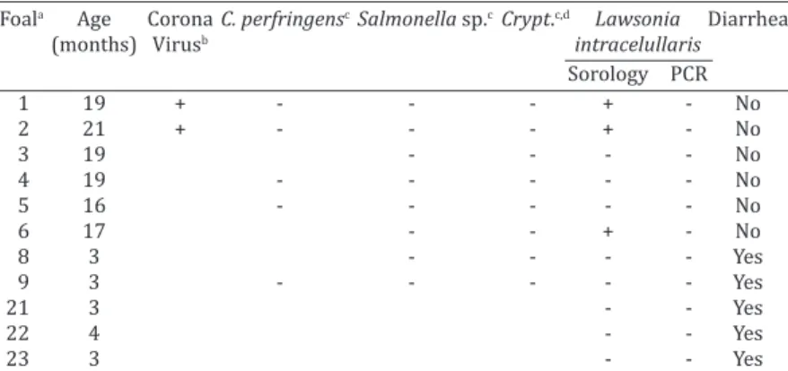
-Table 1. Examinations performed for certain causative agents of diarrhea in stool and serum samples of 11 foals
Foala Age Corona C. perfringenscSalmonella sp.cCrypt.c,d Lawsonia Diarrhea
(months) Virusb intracelullaris
Sorology PCR
1 19 + - - - + - No
2 21 + - - - + - No
3 19 - - - - No
4 19 - - - No
5 16 - - - No
6 17 - - + - No
8 3 - - - - Yes
9 3 - - - Yes
21 3 - - Yes
22 4 - - Yes
23 3 - - Yes
a Foals 9, 21, 22 and 23 were being treated in the period of feces collection, b Detected by
mals showed signs of clinical improvement following this treatment.
Three foals were found to be seropositive for L. intra-cellularis. All foals tested negative for L. intracellularis in PCR analysis of fecal samples. The results of the other agen-tes are shown in Table 1.
Seven of the 39 foals had clinical signs of diarrhea, and 3 of these animals died. A 7-month-old foal died 4 days af-ter the onset of clinical signs, and intestinal samples from this foal were submitted to the Laboratory.
Macroscopi-cally, a large volume of blood-tinged fluid was observed in
the peritoneal cavity. Small intestine serosa was hyperemic (Fig.1A). Thickening of the intestinal wall was associated with a clear corrugation and thickening of the mucosa folds and reduction of intestinal lumen (Fig.1B). The mucosa was heavily corrugated, characterizing thickening of intestinal folds (Fig.1C).
Histological analysis of the duodenum, jejunum, ileum and large intestine demonstrated enterocyte hyperplasia of the crypts associated with intense diffuse decrease in abun-dance (Fig.2A). Rare crypts were dilated and the lumen
found to be filled with cell debris and neutrophils (crypt
abscess). In addition, crypts were present in some areas of the submucosa (Fig.2B). Immunohistochemistry staining demonstrated antigen labeling at the cytoplasmatic apex of enterocytes and in macrophages in the lamina propria of the duodenum, ileum and large intestine (Fig.2C).
DISCUSSION
Reported clinical signs and macro- and microscopic findin -gs, all consistent with the literature (Lester 2001,Pusterla et al. 2010, Vannucci et al. 2012)associated with positive im-mune staining for Lawsonia intracellularis allowed us to re-ach the diagnosis of equine proliferative enteropathy (EPE). Unlike pigs, in which lesions and immunostaining are more
highly concentrated in the final third of the small intestine,
in horses, these lesions can also be found in the duodenum, as reported in this case. The lesions of the large intestine are less frequent, but in our case, histologic lesions compatible with EPE were found in the large intestine. The ages of affec-ted animals ranged from a few days to 21 months of age, although diarrhea associated with hypoproteinemia and submandibular edema had been observed more frequently in foals 3 to 6 months old, an age group in which animals are more susceptible to and affected by L. intracellularis (Pus-terla et al. 2010, Vannucci et al. 2012). This predisposition is most likely associated with the decline of maternal antibo-dies (Pusterla et al. 2008a), as well as stressors such as we-aning, moving to new paddocks and barns, deworming and/ or vaccination programs and/or early conditioning progra-ms (Frazer 2008), which were performed at this farm.
Three and 4 month old foals (animals 8, 9, 21, 22 and 23) had diarrhea at the time of feces collection, but sero-logy and PCR showed them to be negative. PCR cannot, ho-wever, detect the low bacterial elimination in feces of re-cently infected or subclinical animals, or in foals that have experienced prolonged disease or are under antibiotic tre-atment (Dauvillier et al. 2006, Pusterla & Gebhart 2013). According to the reports, L. intracellularis fecal excretion detection stops or ceases 4-6 days after the beginning of doxycycline (Pusterla & Gebhart 2013) and erythromy-cin (Dauvillier et al. 2006)administration. In inoculated animals, elimination of L. intracellularis in feces was first
detected by PCR analysis 12 to 18 days post-inoculation and continued for 7 to 21 days (Pusterla & Gebhart 2013,
Pusterla et al. 2010). Positive serologic results occur 14 to 21 days after exposure (Pusterla & Gebhart 2013, Pusterla et al. 2010), as negative results are expected in the early stage of the disease, when the humoral immune response is too insubstantial to be detected by serology (Pusterla & Gebhart 2013). Thus, the period during which blood
sam-ples were collected may also have influenced the results.
Seropositivity for L. intracellularis in animals that tested negative in PCR analysis of fecal samples can be explained by the sensitivity of the last technique and/or by the course of the infection. There are many PCR inhibitors in fecal ma-terial that reduce the sensitivity of the technique and, as a result, the amount of shed bacteria in the feces could be be-low the detection threshold. In addition, serum antibodies against L. intracellularis last much longer than the bacteria fecal shedding (Pusterla et al. 2008a).
Despite the absence of a definitive EPE diagnosis, the de -ath of several foals that exhibited signs of diarrhea can be explained by the application of the antibiotic gentamycin. This antimicrobial is not effective in the treatment of EPE (Schumacher et al. 2000, Vannucci et al. 2012, Wuersch et al. 2006) because its active property has a high minimum inhi-bitory concentration (MIC) for L. intracellularis (McOrist et al. 1995). In animals with hypoproteinemia, erythromycin doses were followed by clinical improvement in some infec-ted animals, and treatment with erythromycin was effective for EPE in other reports (Bihr 2003, Lavoie et al. 2000, Schu-macher et al. 2000) and is generally considered the drug of choice for the treatments of suspected cases. Disease pro-gression varies from days to weeks, and the prognosis for recovery is good when diagnosis is timely and appropria-te treatment programs are performed. Laappropria-te diagnosed or untreated cases, on the other hand, generally result in the animal’s death (Pusterla & Gebhart 2013). The
seropositivi-ty rate in our study was 27%, a rate lower than that of other studies (Pusterla et al. 2008a) (29.7% and 33.8%) conduc
-ted at farms with EPE cases. However, the first serum dilu -tion used by that author was 1:30, while serological tests in our lab were 1:60, as this dilution showed the least
nonspe-cific labeling. Epidemiological investigations on farms with clinical cases indicate that 10–65% of healthy adult horses
and foals are seropositive for L. intracellularis (Frazer 2008, Pusterla et al. 2009). The percentage of positive animals
ran-ged from 3.57% to 16.67% in Minas Gerais herds, while the properties had no cases of clinical disease, which justifies
lower rates of seropositive (Guimarães-Ladeira et al. 2009). The infection of the foals discussed in this report may have occurred through contact with the feces of other hor-ses infected with L. intracellularis due to the daily use of the same facilities by horses of all ages and/or from the intro-duction of new animals without prior examination for the agent. None of the animals entering the property during the past year exhibited clinical signs of EPE, but the absen-ce of clinical signs in equine does not rule out the chanabsen-ce of a possible carrier and thus a possible source of contamina-tion for other animals on the farm (Guimarães-Ladeira et al. 2009, Page et al. 2011, Pusterla et al. 2010). EPE is not included on differentials enteric diseases in horses in Bra-zil, and there are many neglected positive animals.
Wild and domestic animals such as dogs, cats, opossu-ms (Didelphis sp.), and bush dog (Canis thous) known to inhabit or visit the property may also be sources of con-tamination. Bacterial DNA has been detected in the feces of domestic and wild animals trapped in farms where EPE has occurred (Pusterla et al. 2008b, Pusterla et al. 2012). Lavoie et al. (2000) reported the ingress of the agent in a farm where new horses were not introduced and resident horses did not come into contact with pigs, suggesting con-tamination by wild animals. However, the role of these ani-mals in the disease epidemiology is not well understood. Although the animals have no direct contact with pigs, the role of pigs in the introduction of the agent to farms and the subsequent contamination of horses is controversial. Al-Ghamdi et al. (2012)reproduced proliferative enteropa-thy(EP) in 2-month-old foals using a pure culture of L. in-tracellularis and intestinal mucosa homogenates obtained from experimentally infected pigs. However, Vannucci et al. (2012) did not observe the same results, as pigs infected with equine isolated and foals infected with L. intracellu-laris isolated from pigs showed no clinical signs, decline in performance, pathological changes or hypoproteinemia, suggesting adaptation to the host.
CONCLUSIONS
Equine proliferative enteropathy is present in horses in Midwest Brazil, and despite the report of a clinical case of the disease and the detection of Lawsonia intracellularis in feces and via serological analysis in foals in other Brazi-lian states, the disease remains neglected in the differential diagnosis of other enteric diseases in foals.
This report describes the death of a foal caused by the disease and contributes to the understanding of the
beha-vior of the disease in the field, with the description of cli -nical signs, lesions, treatment and epidemiology, as well as presenting different diagnostic techniques that can be em-ployed to detect the agent.
Declaration of conflicting interests.- The author(s) declare no potential
conflicts of interest with respect to the research, authorship, and/or pu
-blication of this article.
Acknowledgements.- The authors thank CNPq, CAPES and Fapemig for
their financial support. RMCG is a recipient of a fellowship from CNPq.
REFERENCES
Al-Ghamdi G.M., Guedes R.M.C., Sage A.M., Hayden D.W., Neubauer A. & Ames T.R. 2012. Reproduction of proliferative enteropathy in foals using porcine intestinal mucosal homogenate. Bulgarian J. Vet. Med. 15(4):273-282.
Bihr T.P. 2003. Protein-losing enteropathy caused by Lawsonia intracellu-laris in a weanling foal. Can. Vet. J. 44(1):65-66.
Dauvillier J., Picandet V., Harel J., Gottschalk M., Desrosiers R., Jean D. & Lavoie J.-P. 2006. Diagnostic and epidemiological features of Lawsonia intracellularis enteropathy in 2 foals. Can. Vet. J. 47(7):689-691.
Frazer M.L. 2008. Lawsonia intracellularis infection in horses: 2005-2007. J. Vet. Intern. Med.22(5):1243-1248.
Guedes R.M.C., Franca S.A., Machado G.S., Blumer M.A. & Da Costa Cruz Jr E.C. 2009. Use of tylvalosin-medicated feed to control porcine prolifera-tive enteropathy. Vet. Rec. 165(12):342-345.
Guimarães-Ladeira C.V., Palhares M.S., Oliveira J.S., Ramirez M.A. & Guedes R.M.C. 2009. Shedding and serological cross-sectional study of Lawsonia intracellularis in horses in the state of Minas Gerais. Brazil. Equine Vet. J. 41(6):593-596.
Guttmann P.M., Viscardi V., Lessa D.A.B. & Guedes R.M.C. 2014. Equine Proliferative Enteropathy Caused by Lawsonia intracellularis in a Foal in Brazil. J. Equine Vet. Sci. 34(5):701-703.
Jones G.F., Ward G.E., Murtaugh M.P., Lin G. & Gebhart C.J. 1993. Enhanced detection of intracellular organism of swine proliferative enteritis, Ileal symbiont intracellularis, in faeces by polymerase chain reaction. J. Clin. Microbiol. 31(10):2611-2615.
Lavoie J.P., Drolet R., Parsons D., Leguillette R., Sauvageau R., Shapiro J., Houle L., Hallé G. & Gebhart C.J. 2000. Equine proliferative enteropathy: a cause of weight loss, colic, diarrhea and hypoproteinaemia in foals on the breeding farms in Canada. Equine Vet. J. 32(5):418-425.
Lester G.D. 2001. Infectious diarrhea in foals. Proceedings of the 47th AARP
Annual Convention, San Diego, CA, p.468-471.
McOrist S., Mackie R.A. & Lawson G.H. 1995. Antimicrobial susceptibility of ileal symbiont intracellularis isolated from pigs with proliferative en-teropathy. J. Clin. Microbiol. 33(5):1314-1317.
Page A.E., Slovis N.M., Gebhart C.J., Wolfsdorf K., Mapes S.M. & Pusterla N.
2011. Serial use of serologic assays and fecal PCR assays to aid in identifica -tion of subclinical Lawsonia intracellularis infection for targeted treatment of Thoroughbred foals and weanlings. J. Am. Vet. Med. 238(11):1482-1489. Pusterla N. & Gebhart C. 2013, Lawsonia intracellularis infection and
proli-ferative enteropathy in foals. Vet. Microbiol. 167:34-41.
Pusterla N., Higgins J.C., Smith P., Mapes S. & Gebhart C. 2008a. Epidemiolo-gical survey on farms with documented occurrence of equine prolifera-tive enteropathy due to Lawsonia intracellularis. Vet. Rec. 163(5):156-8. Pusterla N., Jackson R., Wilson R., Collier J., Mapes S. & Gebhart C. 2009.
Temporal detection of Lawsonia intracellularis using serology and real--time PCR in Thoroughbred horses residing on a farm endemic for equi-ne proliferative enteropathy. Vet. Microbiol. 136(1/2):173-176. Pusterla N., Mapes S. & Gebhart C. 2012. Further investigation of exposure
to Lawsonia intracellularis in wild and feral animals captured on horse properties with equine proliferative enteropathy.Vet. J. 194(2):253-255. Pusterla N., Mapes S., Rejmanek D. & Gebhart C. 2008b. Detection of Law-sonia intracellularis by Real-time PCR in the feces of free-living animals from equine farms with documented occurrence of Equine proliferative enteropathy. J. Wildl. Dis. 44(4):992-998.
Pusterla N., Wattanaphansak S., Mapes S., Collier J., Hill J., Difrancesco M. & Gebhart C. 2010. Oral infection of weanling foals with an equine isolate of Lawsonia intracellularis, agent of Equine proliferative enteropathy. J. Vet. Intern. Med. 24:622-627.
Schumacher J., Schumacher J., Rolsma M., Brock K.V. & Gebhart C.J. 2000.
Sur-gical and medical treatment of an Arabian filly with proliferative enteropa
-thy caused by Lawsonia intracellularis. J. Vet. Intern. Med. 14(6):630-632. Vannucci F.A., Pusterla N., Mapes S.M. & Gebhart C. 2012. Evidence of host
adaptation in Lawsonia intracellularis infections. Vet. Res. 43:53. Williams N.M., Harrison L.R. & Gebhart C.J. 1996. Proliferative enteropathy
in a foal caused by Lawsonia intracellularis-like bacterium. J. Vet. Diagn. Invest. 8(2):254-256
Wuersch K., Huessy D., Koch C. & Oevermann A. 2006, Lawsonia intracellu-laris proliferative enteropathy in a filly. J. Vet. Med. A, Physiol. Pathol.