RESUMO.-
[
Detecção de genes associados à virulência
em cepas de
Salmonella
Enteritidis isoladas de frangos
na região sul do Brasil.
]
Salmonella
spp. estão entre os
principais agentes causadores de doenças transmitidas por
alimentos, e o sorovar
Salmonella
Enteritidis é o mais
fre-quentemente isolado no mundo. A virulência de
Salmonella
spp. e a sua interação com o hospedeiro são processos
com-plexos que envolvem fatores de virulência para sobreviver
às defesas do hospedeiro. O objetivo deste estudo foi
detec-tar genes de virulência em cepas de
S.
Enteritidis isoladas
a partir de fontes avícolas no sul do Brasil. Ensaios de PCR
foram desenvolvidos para a detecção de nove genes (
lpfA
,
agfA, sefA, invA, hilA, avrA, sopE, sivH
e
spvC
) associados à
virulência em oitenta e quatro amostras de
S.
Enteritidis.
Os genes
invA
,
hilA, sivH, sefA
e
avrA
estavam presentes em
100% dos isolados;
lpfA
e
sopE
estavam presentes em 99%;
agfA
em 96%; e o gene
spvC
estava presente em 92%. Foi
possível caracterizar os isolados em quatro perfis genéti
-cos distintos (P1, P2, P3 e P4), sendo P1 positivo para todos
os genes; P2 negativo apenas para
spvC
; P3 negativo para
agfA
e P4 negativo para
lpfA
,
spvC
e
sopE
. O perfil de maior
frequência foi P1, presente em 88% dos isolados. Apesar de
Detection of virulence-associated genes in
Salmonella
Enteritidis isolates from chicken in South of Brazil
1Karen A. Borges
2*, Thales Q. Furian
2, Anderlise Borsoi
3, Hamilton L.S. Moraes
2,
Carlos T.P. Salle
2e Vladimir P. Nascimento
2ABSTRACT.
- Borges K.A., Furian T.Q., Borsoi A., Moraes H.L.S., Salle C.T.P. & Nascimento V.P.
2013.
Detection of virulence-associated genes in S
almonella
Enteritidis isolates from
chicken in Southern Brazil. Pesquisa Veterinária Brasileira 33(12):1416-1422
. Centro de
Diagnóstico e Pesquisa em Patologia Aviária, Faculdade de Veterinária, Universidade
Fe-deral do Rio Grande do Sul, Avenida Bento Gonçalves 8824, Porto Alegre, RS 91540-000,
Brazil. E-mail: [email protected]
Salmonella
spp. are considered the main agents of foodborne disease and
Salmonella
Enteritidis is one of the most frequently isolated serovars worldwide. The virulence of
Sal-monella
spp. and their interaction with the host are complex processes involving virulence
factors to overcome host defenses. The purpose of this study was to detect virulence genes
in
S.
Enteritidis isolates from poultry in the South of Brazil. PCR-based assays were
deve-loped in order to detect nine genes (
lpfA
,
agfA, sefA, invA, hilA, avrA, sopE, sivH
and
spvC
)
associated with the virulence in eighty-four isolates of
S.
Enteritidis isolated from poultry.
The
invA
,
hilA, sivH, sefA
and
avrA
genes
were present in 100% of the isolates;
lpfA
and
sopE
were present in 99%;
agfA
was present in 96%; and the
spvC
gene was present in 92%. It
was possible to characterize the isolates with four different genetic profiles (P1, P2, P3 and
P4), as it follows: P1, positive for all genes; P2, negative only for
spvC
; P3, negative for
agfA
;
and P4, negative for
lpfA
,
spvC
and
sopE
. The most prevalent profile was P1, which was pre
-sent in 88% of the isolates. Although all isolates belong to the same serovar, it was possible
to observe variations in the presence of these virulence-associated genes between different
isolates. The characterization of the mechanisms of virulence circulating in the population
of
Salmonella
Enteritidis is important for a better understanding of its biology and
patho-genicity. The frequency of these genes and the establishment of genetic profiles can be used
to determine patterns of virulence. These patterns, associated with
in vivo
studies, may
help develop tools to predict the ability of virulence of different strains.
INDEX TERMS: Salmonella Enteritidis, PCR, virulence profile, poultry.
1 Received on June 27, 2013.
Accepted for publication on October 26, 2013.
2 Centro de Diagnóstico e Pesquisa em Patologia Aviária (CDPA),
Facul-dade de Veterinária, UniversiFacul-dade Federal do Rio Grande do Sul (UFRGS), Av. Bento Gonçalves 8824, Porto Alegre, RS 91540-000, Brazil. *Corres-ponding author: [email protected]
3 Tuiuti Universidade do Paraná, Rua Sydnei Antônio Rangel 238,
todos os isolados pertencerem ao mesmo sorovar, foi
pos-sível observar variações na presença de genes associados à
virulência entre os mesmos. A caracterização dos
mecanis-mos de virulência circulantes na população de
Salmonella
Enteritidis é importante para um maior entendimento da
sua biologia e patogenicidade. A frequência destes genes e
o estabelecimento de perfis genéticos podem ser utilizados
para determinar os padrões de virulência dos isolados.
Es-tes padrões, associados a estudos
in vivo
, podem auxiliar na
elaboração de ferramentas que permitam predizer a
capa-cidade de virulência das diferentes cepas.
TERMOS DE INDEXAÇÃO: Salmonella Enteritidis, PCR, perfil de
virulência, frangos.
INTRODUCTION
Salmonella
spp. are considered the major cause of
food-borne disease in humans, and
Salmonella
Enteritidis is the
most frequently isolated serovar in Europe, South America
and Asia (Vieira et al. 2009). Phylogenetic analyses show
the influence of different factors in the existence and per
-sistence of
Salmonella
spp in animals, such as
cross-con-tamination among animals, environment and feed (Mello
et al. 2011). The virulence of
Salmonella
spp. is associated
with a combination of chromosomal and plasmid factors
(Oliveira et al. 2003), and many studies have identified ge
-nes that encode these factors. Some virulence factors are
associated with the cellular structure of the bacteria, such
as fimbriae (Edwards & Puente 1998). The long polar fim
-bria (
lpf operon
) contributes to the affinity of the bacteria
for Peyer’s patches and adhesion to intestinal M cells
(Bäu-mler & Heffron 1995, Bäu(Bäu-mler et al. 1996). One of the main
functions of aggregative fimbria (
agf operon
) is to promote
the initial interaction of the bacteria with the intestine of
the host and stimulate bacterial self-aggregation, resulting
in higher rates of survival (Collinson et al. 1992, 1993). The
Salmonella-
encoded fimbria (
sef operon
) promotes a
bet-ter inbet-teraction between the bacbet-teria and the macrophages
(Collinson et al. 1996).
Salmonella
spp. pathogenicity islands (SPI) are of critical
importance for
Salmonella
spp. virulence, once they encode
a molecular apparatus called the type III secretion system
(TTSS), which is able to inject bacterial effector proteins
through bacterial and host membranes to interact with host
cells (Marcus et al. 2000). The
hilA
gene encodes the central
regulator HilA, which is necessary for the expression of the
TTSS components. HilA is also required to invade
epithe-lial cells and induce apoptosis of macrophages (Bajaj et al.
1996). The protein InvA is essential for epithelial invasion
(Galán & Curtis III 1989) and AvrA is an effector protein of
the TTSS complex that contributes to the virulence of
Sal-monella
spp. by limiting the host’s inflammatory respon
-ses through the inducement of cell apoptosis, especially
of macrophages, and by the innibition of IL-8 and TNF-α
(Collier-Hyames et al. 2002, Ben-Barak et al. 2006).
Salmo-nella
spp.’s outer proteins (Sops) contribute to the invasion
by these bacteria through the generation of membrane
de-formations (Hardt et al. 1998) and the rearrangement of
the cytoskeleton of the host cells (Galán & Zhou 2000). The
sivH
gene encodes an outer membrane protein associated
with intestinal colonization (Kingsley et al. 2003). Other
important
Salmonella
spp. virulence factors are found on
virulence plasmids. All of the virulence plasmids share a
highly conserved region designated
spv
RABCD (
Salmonella
plasmid virulence). The spv region promotes rapid growth
and survival of
Salmonella
spp. within the host cells and it
is important for systemic infection (Libby et al. 1997). The
purpose of this study was to evaluate the virulence
poten-tial of
S
. Enteritidis isolates from poultry by detecting the
presence of nine virulence-associated genes by polymerase
chain reactions (PCRs), as well as to determine the
distri-bution patterns of these genes.
MATERIALS AND METHODS
Bacterial isolates. This stduy was developed at the Diagnos-tic Center and Research in Avian Pathology (CDPA) of Federal University of Rio Grande do Sul (UFRGS). Eighty-four isolates of
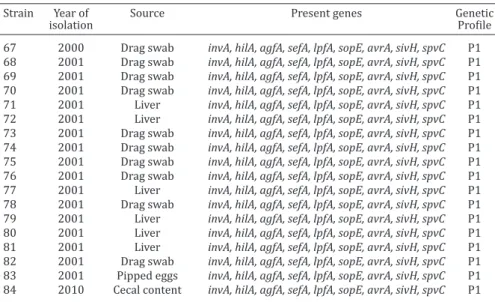
Salmonella Enteritidis were selected from CDPA collection. The-se bacteria were isolated between 1996 and 2010 from different avian sources in Rio Grande do Sul state in the south of Brazil. The sources included broiler systems and slaughterhouses; in addi-tion we also used one sample of hatchery. Addiaddi-tional data of iso-lates (year and source of isolation) are shown in Table 1. A
com-plete antigenic characterization and serovar identification were
performed by the Enteric Pathogens Laboratory in the Oswaldo Cruz Institute Foundation (Fiocruz, Rio de Janeiro, Brazil). The bacterial isolates were kept frozen at -70°C in brain heart infusion broth and glycerol.
DNA extraction. The bacteria were retrieved from frozen cul-ture stocks and culcul-tured overnight at 37°C in brain heart infusion broth (Oxoid; Cambridge, United Kingdom). An aliquot of 1 mL of each bacterial culture was separated for DNA extraction by heat treatment as described by Borsoi et al. (2009).
Polymerase chain reaction (PCR). The PCRs were conduc-ted in individual reactions using primers for the following genes:
invA, hilA, avrA, agfA, lpfA, sefA, sopE, spvC and sivH. The sets of primer pairs, sizes of the PCR products and references used in the
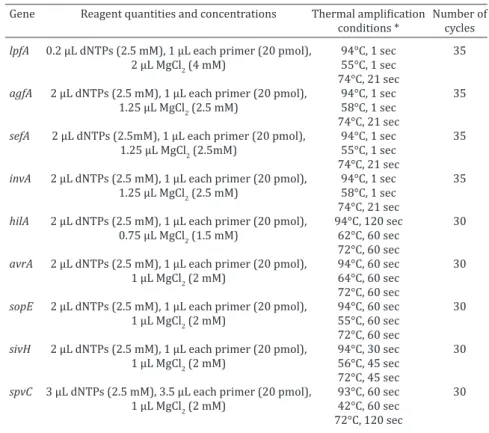
PCR assay are described in Table 2. All PCR mixtures (25μL) were performed with 2.5μL of 10X PCR buffer (Centro de Biotecnolo -gia UFRGS; Porto Alegre, Rio Grande do Sul, Brazil), 1 U of Taq DNA polymerase (Centro de Biotecnologia UFRGS; Porto Alegre,
Rio Grande do Sul, Brazil) and 2 μL of template DNA. The reagent concentrations, amplification conditions and number of cycles
are described in Table 3. The cycling program was perfomed in the Esco Swift MaxPro thermal cycler (Esco, Singapore). The
amplified products were separated by electrophoresis in a 1.2%
agarose gel and stained with ethidium bromide. Fragments were transilluminated with UV light. Escherichia coli ATCC 25922 and
Salmonella Enteritidis ATCC 13076 were used as negative and po-sitive controls, respectively, for all PCR reactions, except for that of the agfA gene, for which Salmonella Typhimurium ATCC 14028 was used as a positive control. In all PCRs, a mixture of all cons-tituents of the PCR mixed without the addition of extracted DNA was used as PCR control.
RESULTS
The
Salmonella
Enteritidis strains had different
frequen-cies of the target genes, regardless the year and the
sour-ce of isolation (Table 1). The
invA
,
hilA, sivH, sefA
and
avrA
genes
were present in 100% (84/84) of the isolates.
lpfA
pre-Table 1. Virulence genes and genetic profile of Salmonella Enteritidis isolates from chicken in South of Brazil
Strain Year of Source Present genes Genetic
isolation Profile
1 1996 Broiler carcass invA, hilA, agfA, sefA, lpfA, sopE, avrA, sivH, spvC P1 2 1996 Broiler carcass invA, hilA, agfA, sefA, lpfA, sopE, avrA, sivH, spvC P1 3 1996 Broiler carcass invA, hilA, agfA, sefA, lpfA, sopE, avrA, sivH, spvC P1 4 1996 Broiler carcass invA, hilA, agfA, sefA, lpfA, sopE, avrA, sivH, spvC P1 5 1996 Broiler carcass invA, hilA, agfA, sefA, lpfA, sopE, avrA, sivH, spvC P1 6 1996 Broiler carcass invA, hilA, agfA, sefA, lpfA, sopE, avrA, sivH, spvC P1 7 1996 Broiler carcass invA, hilA, agfA, sefA, lpfA, sopE, avrA, sivH, spvC P1 8 1996 Broiler carcass invA, hilA, agfA, sefA, lpfA, sopE, avrA, sivH, spvC P1 9 1996 Broiler carcass invA, hilA, sefA, lpfA, sopE, avrA, sivH, spvC P3 10 1996 Broiler carcass invA, hilA, sefA, lpfA, sopE, avrA, sivH, spvC P3 11 1996 Broiler carcass invA, hilA, agfA, sefA, lpfA, sopE, avrA, sivH P2 12 1996 Broiler carcass invA, hilA, agfA, sefA, lpfA, sopE, avrA, sivH, spvC P1 13 1996 Broiler carcass invA, hilA, agfA, sefA, lpfA, sopE, avrA, sivH P2 14 1996 Broiler carcass invA, hilA, agfA, sefA, lpfA, sopE, avrA, sivH, spvC P1 15 1996 Broiler carcass invA, hilA, agfA, sefA, lpfA, sopE, avrA, sivH, spvC P1 16 1996 Broiler carcass invA, hilA, agfA, sefA, lpfA, sopE, avrA, sivH, spvC P1 17 1999 Broiler carcass invA, hilA, agfA, sefA, lpfA, sopE, avrA, sivH, spvC P1 18 1999 Drag swab invA, hilA, agfA, sefA, lpfA, sopE, avrA, sivH, spvC P1 19 1999 Drag swab invA, hilA, agfA, sefA, lpfA, sopE, avrA, sivH, spvC P1 20 1999 Drag swab invA, hilA, agfA, sefA, lpfA, sopE, avrA, sivH, spvC P1 21 1999 Drag swab invA, hilA, agfA, sefA, lpfA, sopE, avrA, sivH, spvC P1 22 1999 Drag swab invA, hilA, agfA, sefA, lpfA, sopE, avrA, sivH P2 23 1999 Drag swab invA, hilA, agfA, sefA, lpfA, sopE, avrA, sivH, spvC P1 24 1999 Drag swab invA, hilA, agfA, sefA, lpfA, sopE, avrA, sivH, spvC P1 25 1999 Liver invA, hilA, agfA, sefA, lpfA, sopE, avrA, sivH, spvC P1 26 1999 Liver invA, hilA, agfA, sefA, lpfA, sopE, avrA, sivH, spvC P1 27 1999 Liver invA, hilA, agfA, sefA, lpfA, sopE, avrA, sivH, spvC P1 28 1999 Liver invA, hilA, agfA, sefA, lpfA, sopE, avrA, sivH, spvC P1 29 1999 Liver invA, hilA, agfA, sefA, lpfA, sopE, avrA, sivH, spvC P1 30 1999 Liver invA, hilA, agfA, sefA, lpfA, sopE, avrA, sivH, spvC P1 31 1999 Liver invA, hilA, agfA, sefA, lpfA, sopE, avrA, sivH, spvC P1 32 1999 Liver invA, hilA, agfA, sefA, lpfA, sopE, avrA, sivH, spvC P1 33 1999 Liver invA, hilA, agfA, sefA, lpfA, sopE, avrA, sivH, spvC P1 34 1999 Liver invA, hilA, agfA, sefA, lpfA, sopE, avrA, sivH, spvC P1 35 1999 Liver invA, hilA, agfA, sefA, lpfA, sopE, avrA, sivH, spvC P1 36 1999 Liver invA, hilA, agfA, sefA, lpfA, sopE, avrA, sivH, spvC P1 37 1999 Liver invA, hilA, agfA, sefA, lpfA, sopE, avrA, sivH, spvC P1
38 1999 Liver invA, hilA, agfA, sefA, avrA, sivH P4
sent in 96% (3/84) and the
spvC
gene was present in 92%
(7/84). All isolates showed at least five virulence-associa
-ted genes. The results of the PCRs are summarized in Table
4. Based on the combination of present and absent genes,
the
S.
Enteritidis were divided in four different gene
pro-files. In order to facilitate the analysis, these profiles were
named P1, P2, P3 and P4. Regarding the profiles, among the
84 isolates analyzed, 88% (74/84) were categorized as P1
(positive for all genes), 7% (6/84) as P2 (
spvC
absent), 4%
(3/84) as P3 (
agfA
absent) and 1% (1/84) as P4 (
lpfA
,
sopE
and
spvC
absent).
DISCUSSION
All South Brazilian
Salmonella
Enteritidis isolates were
positive for
invA
and
hilA;
similar observations have been
reported by other studies around the world (Amini et al.
2010, Campioni et al. 2012, Craciunas et al. 2012). It was
expected that these genes would be detected in all of the
isolates due to their importance for cell invasion. PCR is a
useful tool for the rapid detection of
Salmonella
spp., and
inv
A and
hilA
genes can be considered target genes for the
detection of this genus.
Although results for the
sopE
and
avrA
genes were
si-milar to the 100% frequency found in previous studies on
Salmonella
Enteritidis (Hopkins & Threlfall 2004), other
works have found lower frequencies (Rahman et al. 2004,
Streckel et al. 2004, Zou et al. 2011, Liu et al. 2012). This
frequency variation could be caused by the
recombina-tions that frequently occur in the location of these genes
(Hopkins & Threlfall 2004). These findings are important,
since changes in the repertoire of proteins, such as SopE
and AvrA, can lead to changes in the ability of this serovar
to adapt to new hosts and, consequently, the emergence of
novel virulent strains (Prager et al. 2000). In our study, a
high percentage (99%) of isolates had the
avrA
and
sopE
genes. However, only 17.1% and 9.7% of
Salmonella
Hadar
isolates had the
avrA
and
sopE
genes, respectively (Cesco
2010). When comparing this data, it is clear that there is
Table 1 (cont.). Virulence genes and genetic profile of Salmonella Enteritidis
isolates from chicken in South of Brazil
Strain Year of Source Present genes Genetic
isolation Profile
67 2000 Drag swab invA, hilA, agfA, sefA, lpfA, sopE, avrA, sivH, spvC P1 68 2001 Drag swab invA, hilA, agfA, sefA, lpfA, sopE, avrA, sivH, spvC P1 69 2001 Drag swab invA, hilA, agfA, sefA, lpfA, sopE, avrA, sivH, spvC P1 70 2001 Drag swab invA, hilA, agfA, sefA, lpfA, sopE, avrA, sivH, spvC P1 71 2001 Liver invA, hilA, agfA, sefA, lpfA, sopE, avrA, sivH, spvC P1 72 2001 Liver invA, hilA, agfA, sefA, lpfA, sopE, avrA, sivH, spvC P1 73 2001 Drag swab invA, hilA, agfA, sefA, lpfA, sopE, avrA, sivH, spvC P1 74 2001 Drag swab invA, hilA, agfA, sefA, lpfA, sopE, avrA, sivH, spvC P1 75 2001 Drag swab invA, hilA, agfA, sefA, lpfA, sopE, avrA, sivH, spvC P1 76 2001 Drag swab invA, hilA, agfA, sefA, lpfA, sopE, avrA, sivH, spvC P1 77 2001 Liver invA, hilA, agfA, sefA, lpfA, sopE, avrA, sivH, spvC P1 78 2001 Drag swab invA, hilA, agfA, sefA, lpfA, sopE, avrA, sivH, spvC P1 79 2001 Liver invA, hilA, agfA, sefA, lpfA, sopE, avrA, sivH, spvC P1 80 2001 Liver invA, hilA, agfA, sefA, lpfA, sopE, avrA, sivH, spvC P1 81 2001 Liver invA, hilA, agfA, sefA, lpfA, sopE, avrA, sivH, spvC P1 82 2001 Drag swab invA, hilA, agfA, sefA, lpfA, sopE, avrA, sivH, spvC P1 83 2001 Pipped eggs invA, hilA, agfA, sefA, lpfA, sopE, avrA, sivH, spvC P1 84 2010 Cecal content invA, hilA, agfA, sefA, lpfA, sopE, avrA, sivH, spvC P1
Table 2. Virulence factors genes identified in Salmonella Enteritidis from avian
origin in South Brazil
Gene Virulence factor Primer sequence (5’-3’) Base pair Reference
lpfA Fimbria CTTTCGCTGCTGAATCTGGT
CAGTGTTAACAGAAACCAGT 250 Bäumler &
agfA Fimbria TCCACAATGGGGCGGCGGCG Heffron 1995
CCTGACGCACCATTACGCTG 350 Cesco et al. 2008
sefA Fimbria GATACTGCTGAACGTAGAAGG
GCGTAAATCAGCATCTGCAGTAGC 488 Oliveira et al. 2002
invA Invasion GTGAAATTATCGCCACGTTCGGGCAA
TCATCGCACCGTCAAAGGAACC 284 Oliveira et al. 2002
hilA Invasion CTGCCGCAGTGTTAAGGATA
CTGTCGCCTTAATCGCATGT 497 Guo et al. 2000
avrA Effector protein GTTATGGACGGAACGACATCGG
ATTCTGCTTCCCGCCGCC 385 Prager et al. 2003
sopE Effector protein ACACACTTTCACCGAGGAAGCG
GGATGCCTTCTGATGTTGACTGG 398 Prager et al. 2003
sivH Invasion CAGAATGCGAATCCTTCGCAC
GTATGCGAACAAGCGTAACAC 763 Kingsley et al. 2003
spvC Plasmid - virulence CGGAAATACCATCTACAAATA
a difference in the pattern of proteins among different
se-rovars. Some authors consider this high frequency of
avrA
gene is present only in serovars that are considered to be
the most important etiological agents of salmonellosis
(Ben-Barak et al. 2006). All of these isolates of
S
.
Enteriti-dis were
sivH
gene positive. Although there are few studies
on the frequency of this gene in populations of
Salmonella
spp., our results are similar to previous works (Kingsley et
al. 2003). Many of these effectors proteins were shown to
play an important role in
Salmonella
virulence. However,
their absence in some isolates, such as
sopE
, suggests that
they are not essential for invasive manifestation in the
hu-man host (Suez et al. 2013).
It was verified that all isolates presented had at least
two of the fimbrial genes analyzed in this study, highli
-ghting the importance of fimbriae in the infection process.
It is possible that there are additive effects of the adhesins
Lpf and Agf in the colonization of the intestine and systemic
virulence (Wagner & Hensel 2011). The high frequency of
lpfA
and
agfA
were similar to other data obtained by
pre-vious works that studied different serovars (Doran et al.
1993, Borsoi et al. 2009, Cesco 2010). Besides being
impor-tant in the adhesion during the infection process, the
agfA
gene is also associated with biofilm formation (Barnhart
& Chapman 2006, Yoo et al. 2013). Our results show this
gene is present in isolates from carcasses, which can pose
greater risk of contamination on the slaughterhouses. The
negative isolates may have lost the gene during their
evo-lution. Studies concerning the frequency of these genes are
important in tracking the adaptation of different serovars
of
Salmonella
spp. to an increasing number of hosts
becau-se the acquisition and loss of fimbrial genes are involved in
this process (Bäumler et al. 1997). The high frequency of
sefA
is consistent with previous findings (Amini et al. 2010,
Craciunas et al. 2012), and it can be considered a target
gene to identify the serovar
S.
Enteritidis by PCR (Amini et
al. 2010).
Our results for the virulence plasmid gene
spvC
were
si-milar to those found by other authors (Oliveira et al. 2003,
Castilla et al. 2006, Amini et al. 2010). There are also other
studies that have found lower frequencies for this gene in
strains of avian origin (Okamoto et al. 2009,
Derakhshan-deh et al. 2013, Moussa et al. 2013). It is possible that the
presence of this gene is related to the host from which the
sample was isolated (Amini et al. 2010). Amini et al. (2010)
compared the frequencies of
spvC
in strains isolated from
Table 3. PCR assay conditions used in this study to detect thevirulence-associated genes in Salmonella Enteritidis isolates
Gene Reagent quantities and concentrations Thermal amplification Number of
conditions * cycles
lpfA 0.2 µL dNTPs (2.5 mM), 1 µL each primer (20 pmol), 94°C, 1 sec 35
2 µL MgCl2 (4 mM) 55°C, 1 sec
74°C, 21 sec
agfA 2 µL dNTPs (2.5 mM), 1 µL each primer (20 pmol), 94°C, 1 sec 35
1.25 µL MgCl2 (2.5 mM) 58°C, 1 sec
74°C, 21 sec
sefA 2 µL dNTPs (2.5mM), 1 µL each primer (20 pmol), 94°C, 1 sec 35
1.25 µL MgCl2 (2.5mM) 55°C, 1 sec
74°C, 21 sec
invA 2 µL dNTPs (2.5 mM), 1 µL each primer (20 pmol), 94°C, 1 sec 35
1.25 µL MgCl2 (2.5 mM) 58°C, 1 sec
74°C, 21 sec
hilA 2 µL dNTPs (2.5 mM), 1 µL each primer (20 pmol), 94°C, 120 sec 30
0.75 µL MgCl2 (1.5 mM) 62°C, 60 sec
72°C, 60 sec
avrA 2 µL dNTPs (2.5 mM), 1 µL each primer (20 pmol), 94°C, 60 sec 30
1 µL MgCl2 (2 mM) 64°C, 60 sec
72°C, 60 sec
sopE 2 µL dNTPs (2.5 mM), 1 µL each primer (20 pmol), 94°C, 60 sec 30
1 µL MgCl2 (2 mM) 55°C, 60 sec
72°C, 60 sec
sivH 2 µL dNTPs (2.5 mM), 1 µL each primer (20 pmol), 94°C, 30 sec 30
1 µL MgCl2 (2 mM) 56°C, 45 sec
72°C, 45 sec
spvC 3 µL dNTPs (2.5 mM), 3.5 µL each primer (20 pmol), 93°C, 60 sec 30
1 µL MgCl2 (2 mM) 42°C, 60 sec
72°C, 120 sec
* sec = seconds.
Table 4. Frequency of detection of virulence-associated genes in isolates of Salmonella Enteritidis from avian in South of
Brazil
Gene Positive strains
Total (n=84) Total (%)
lpfA 83 99
agfA 81 96
sefA 84 100
invA 84 100
hilA 84 100
avrA 84 100
sopE 83 99
sivH 84 100
humans (100%) and cattle (90%), which are not similar to
those found in
S.
Enteritidis strains isolated from poultry.
Comparing different serovars, it was observed that 92% of
S.
Enteritidis strains had the
spvC
gene, whereas only 28%
of
S.
Typhimurium strains (Moussa et al. 2013) and 0% of
S.
Hadar strains (Cesco 2010) were positive for the gene.
The different frequencies found for this gene showed that
the virulence of
Salmonella
Enteritids alternates among the
plasmid and chromosomal factors, according to the genetic
profile of each isolate.
Despite the antigenic similarities among the
S.
Enteri-tidis isolates used in this study, the gene pattern was not
the same for all bacteria. Although all the serovars of
Sal-monella
spp. can be considered as potentially pathogenic,
there are some differences in their virulence (Karasova et
al. 2009). The highest frequency of P1 profile demonstrate
that these genes are widely distributed in the population of
Salmonella
spp. The presence of more than one genetic
pro-file may suggest acquisitions or deletions of genes in diffe
-rent clones, which could promote diffe-rent levels of strain
adaptation to the host (Bäumler et al. 1997, Prager et al.
2000, Moussa et al. 2013).
It is known that
Salmonella
spp. isolates lost and
acqui-re new virulence factors over time in order to adapt to new
hosts or to the environment (Bäumler et al. 1997, Suez et
al. 2013). Currently, the foremost challenges are
determi-ning how
Salmonella
acquires virulence factors and what
the most important genetic traits conferring virulence to
Salmonella
spp. The knowledge of these characteristics
allows a better approach in the study of
Salmonella
viru-lence and hence the development of strategies to reduce
this virulence. Studies involving the exact involvement of
each gene in the pathogenesis of this bacterium would be
possible to establish criteria for predicting the virulence of
this microorganism.
CONCLUSION
The understanding of the virulence of
Salmonella
spp.
re-quires several steps. Nevertheless, the results of this study
support, through the provision of protocols and gene
pro-files, the premise that there is a genetic differentiation in
isolates from the same serovar, which provides a basis for
criteria to determine possible variations for
in vivo
viru-lence of different strains, as well as for further studies in
phylogenetic analysis.
REFERENCES
Amini K., Salehi T.Z., Nikbakht G., Ranjbar R., Amini J. & Ashrafganjooei S.B. 2010. Molecular detection of invA and spv virulence genes in Salmonella Enteritidis isolated from human and animals in Iran. Afr. J. Microbiol. Res. 4(21):2202-2210.
Bajaj V., Lucas R.L., Hwang C. & Lee C.A. 1996. Co-ordinate regulation of Salmonella typhimurium invasion genes by environmental and regula-tory factors is mediated by control of hilA expression. Mol. Microbiol. 22(4):703-714.
Barnhart M.M. & Chapman M. 2006. Curli biogenesis and function. Annu. Rev. Microbiol. 60:131-147.
Bäumler A.J. & Heffron F. 1995. Identification and sequence analysis of lpfABCDE, a putative fimbrial operon of Salmonella typhimurium. J. Bac-teriol.177(8):2087-2097.
Bäumler A.J., Tsolis R.M. & Heffron F. 1996. Contribution of fimbrial ope -rons to attachment to and invasion of epithelial cell lines by Salmonella Typhimurium. Infect. Immun. 64(5):1862-1865.
Bäumler A.J., Gilde A.J., Tsolis R.M., Van der Velden W.M., Ahmer B.M.M. & Heffron F. 1997. Contribution of horizontal gene transfer and deletion
events to development of distinctive patterns of fimbrial operon during
evolution of Salmonella serovars. J. Bacteriol. 179(2):317-322.
Ben-Barak Z., Streckel W., Yarona S., Cohenc S., Prager R. & Tschape H. 2006. The expression of the virulence-associated effector protein gene
avrA is dependent on a Salmonella enterica-specific regulatory function.
Int. J. Med. Microbiol. 296:25-38.
Borsoi A., Santin E., Santos L.R., Salle C.T.P., Moraes H.L.S. & Nascimento V.P.2009. Inoculation of newly hatched broiler chicks with two brazilian isolates of Salmonella Heidelberg strains with different virulence gene
profile, antimicrobial resistance and pulsed field gel electrophoresis
pattern to intestinal changes evaluation. Poult. Sci. 88:750-758. Campioni F., Bergamini A.M.M. & Falcão J.P. 2012. Genetic diversity,
viru-lence genes and antimicrobial resistance of Salmonella Enteritidis iso-lated from food and humans over a 24-year period in Brazil. Food Mi-crobiol. 32:254-264.
Castilla K.S., Ferreira C.S.A., Moreno A.M., Nunes I.A. & Ferreira A.J.F. 2006. Distribution of virulence genes sefC, pefA e spvC in Salmonella Enteri-tidis phage type 4 strains isolated in Brazil. Braz. J. Microbiol. 37:135-139.
Cesco M.A.O. 2010. Pesquisa de Fatores Associados à Virulência de Salmo-nella Hadar através da Reação em Cadeia da Polimerase (PCR). Disser-tação de Mestrado em Ciências Veterinárias, Faculdade de Veterinária, Universidade Federal do Rio Grande do Sul, Porto Alegre, RS. 84p. Cesco M.A.O., Zimermann F.C., Giotto D.B., Guayba J., Borsoi A., Rocha S.L.S.,
Camilotti E., DalMolin J., Moraes H.L.S. & Nascimento V.P. 2008. Pesquisa de genes de virulência em Salmonella Hadar em amostras provenientes de material avícola. Anais 35º Congresso Brasileiro de Medicina Veteri-nária, Gramado/RS, R0701-0.
Collier-Hyames L.S., Zeng H., Sun J., Tomlinson A.D., Bao Z.Q., Chen H., Ma-dara J.L., Orth K., Chen H. & Neish A.S. 2002. Cutting edge: Salmonella
AvrA effector inhibits the key proinflammatory, anti-apoptotic NF-kB
pathway. J. Immunol. 169:2846-2850.
Collinson S.K., Emody L., Trust T.J. & Kay W.W. 1992. Thin aggregative fim -briae from Diarrheagenic Escherichia coli. J. Bact. 174(13):4490-4495. Collinson K., Doig P.C., Doran J.L., Clouthier S., Trust T.J. & Kay W.W. 1993.
Thin aggregative fimbriae mediate binding of Salmonella Enteritidis to
fibronectin. J. Bacteriol. 175(1):12-18.
Collinson K., Liu S.L., Clouthier S.C., Banser P.A., Doran J.L., Sanderson K.E.
& Kay W.W. 1996. The location of four fimbrin-enconding genes, agfA, fimA, sefA and sefD, on the Salmonella Enteritidis and/or S. Typhimu-rium XbalI-BlnI genomic restriction maps. Gene 169:75-80.
Craciunas C., Keul A.L., Flonta M. & Cristea M. 2012. DNA-based diagnostic tests for Salmonella strains targeting hilA, agfA. spvC and sefC genes. J. Env. Man. 95:512-218.
Derakhshandeh A., Firouzi R. & Khoshbakht R. 2013. Association of three plasmid-encoded spv genes among different Salmonella serotypes isola-ted from different origins. Indian J. Microbiol. 53(1):106-110.
Doran J.L., Collinson S.K., Burian J., Sarlos G., Todd E.C.D., Munro C.K., Kay C.M., Banser P.A., Peterkin P.I. & Kay W.W. 1993. DNA-based diagnostic tests for Salmonella species targeting agfA, the structural gene for thin,
aggregative fimbriae. J. Clin. Microbiol. 31(9):2263-2273.
Edwards R.A. & Puente J.L. 1998. Fimbrial expression in enteric bacteria: a critical step in intestinal pathogenesis. Trends Microbiol. 6(7):282-287. Galán J.E. & Curtis III R. 1989. Cloning and molecular characterization of
genes whose products allow Salmonella typhimurium to penetrate tis-sue culture cells. Proc. Natl Acad. Sci. USA 86:6383-6387.
Galán J.E. & Zhou D. 2000. Striking a balance: modulation of the actin cytoskeleton by Salmonella. PNAS 97(16):8754-8761.
Hardt W.D., Urlab H. & Galán J.E. 1998. A substrate of the centisome 63 type III protein secretion system of Salmonella typhimurium is encoded by a cryptic bacteriophage. Proc. Natl Acad. Sci. USA 95:2574-2579. Hopkins K.L. & Threlfall E.J. 2004. Frequency and polymorphism of sopE
in isolates of Salmonella enterica belonging to the ten most prevalent serovars in England and Wales. J. Med. Microbiol. 53:539-543.
Karasova D., Havlickova H., Sisak F. & Rychlik I. 2009. Deletion of sodCI and spvBC in Salmonella enterica serovar Enteritidis reduced its viru-lence to the natural viruviru-lence of serovars Agona, Hadar and Infantis for mice but not for chickens early after infection. Vet. Microbiol. 139:304-309.
Kingsley R.A., Humphries A.D., Weening E.H., Zoete M.R., Winter S., Papa-constantinopoulou A., Dougan G. & Bäumler A.J. 2003. Molecular and phenotypic analysis of the CS54 island of Salmonella enterica serovar
Typhimurium: identification of intestinal colonization and persistence
determinants. Infect. Immun. 71(2):629-640.
Libby S.J., Adams G., Ficht T.A., Allen C., Whitford H.A., Buchmeier N.A., Bos-sie S. & Guiney D.G. 1997. The spv genes on the Salmonella Dublin viru-lence plasmid are required for severe enteritis and systemic infection in the natural host. Infect. Immun. 65(5):1786-1792.
Liu Y., Yang Y., Liao X., Li L., Lei C., Li L., Sun J. & Liu B. 2012. Antimicrobial resistance, resistance genes and virulence genes in Salmonella isolates from chicken. J. Anim. Vet. Adv. 11(23):4423-4427.
Marcus S.L., Brumell J.H., Pfeifer C.G. & Finlay B.B. 2000. Salmonella pa-thogenicity islands: big virulence in small packages. Microbes Infect. 2:145-156.
Mello R.T., Guimarães A.R., Mendonça E.P., Coelho L.R., Monteiro G.P.,
Fon-seca B.B. & Rossi D.A. 2011. Identificação sorológica e relação filogené -tica de Salmonella spp. de origem suína. Pesq. Vet. Bras. 31(12):1039-1044.
Moussa I.M., Aleslamboly Y.S., Al-Arfaj A.A., Hessain A.M., Gouda A.S. & Kamal R.M. 2013. Molecular characterization of Salmonella virulence genes isolated from different sources relevant to human health. J. Food Agrc. Environ. 11(2):197-201.
Okamoto A.S., Andreatti Filho R.L., Rocha T.S., Menconi A. & Marietto-Gon-çalves G.A. 2009. Relation between the spvC and invA genes and resis-tance of Salmonella Enteritidis isolated from avian material. Int. J. Poult. Sci. 8(6):279-582.
Oliveira S.D., Santos L.R., Schucha D.M.T., Silva A.B., Salle C.T.P. & Canal C.W.
2002. Detection and identification of salmonellas from poultry-related
samples by PCR. Vet. Microbiol. 87:25-35.
Oliveira S.D., Rodenbusch C.R., Michael G.B., Cardoso M.I.R., Canal C.W. & Brandelli A.2003. Detection of virulence genes in Salmonella Enteritidis isolates from different sources. Braz. J. Microbiol. 34(1):123-124. Prager R., Mirold S., Tietze E., Strutz U., Knüppel B., Rabsch W., Hardt W.D.
& Tschäpe H. 2000. Prevalence and polymorphism of genes encoding translocated effector proteins among clinical isolates of Salmonella en-terica. Int. J. Med. Microbiol. 290(7):605-617.
Prager R., Rabsch W., Streckel W., Voigt W., Tietze E. & Tschäpe H. 2003. Molecular properties of Salmonella enterica serovar Paratyphi B distin-guish between its systemic and its enteric pathovars. J. Clin. Microbiol. 41(9):4270-7278.
Rahman H., Streckel W., Prager R. & Tschäpe H. 2004. Presence of sopE gene and its phenotypic expression among different serovars of Salmo-nella isolated from man and animals. Indian J. Med. Res. 120:35-38. Streckel W., Wolff A.C., Prager R., Tietze E. & Tschäpe H. 2004. Expression
profiles of effector proteins SopB, SopD1, SopE1 and AvrA differ with
systemic, enteric and epidemic strains of Salmonella enterica. Mol. Nutr. Food Res. 48:496-503.
Suez J., Porwollik S., Dagan A., Marzel A., Schorr Y.I., Desai P.T., Agmon V.,
McClelland M., Rahav G. & Gal-Mor O. 2013. Virulence gene profiling and
pathogenicity characterization of non-typhoidal Salmonella accounted for invasive disease in humans. PLoS ONE 8(3):e58449.
Swamy S.C., Barnhart H.M., Lee M.D. & Dreesen D.W. 1996. Virulence de-terminants invA and spvC in Salmonellae isolated from poultry products, wastewater and human sources. Appl. Environ. Microbiol. 62(10):3768-3771.
Vieira A., Jensen A.R., Pires S.M., Karlsmose S., Wegener H.C. & Wong D.L.F. 2009. WHO global foodborne infections network country databank: a resource to link human and non-human sources of Salmonella. Proc. 12th
Symposium of the International Society for Veterinary Epidemiology and Economics, Durban, South Africa, p.1.
Wagner C. & Hensel M. 2011. Adhesive mechanisms of Salmonella enterica, p.17-34. In: Linke D. & Goldman A.(Eds), Bacterial Adhesion - Chemis-try, Biology and Physics. Springer, New York.
Yoo A.Y., Yu J.E., Yoo H., Lee T.H., Lee W.H., Oha J.I. & Kang H.Y. 2013. Role of sigma factor E in regulation of Salmonella Agf expression. Biochem. Biophys. Res. Commun. 430:131-136.
Zou W., Al-Khaldi S.F., Branham W.S., Han T., Fuscoe J.C., Han J., Foley S.L., Xu J., Fang H., Cerniglia C.E. & Nayak R. 2011. Microarray analysis of