UNIVERSIDADE DE LISBOA
FACULDADE DE CIÊNCIAS
DEPARTAMENTO DE BIOLOGIA VEGETAL
Crosstalk between NF-κB and PI3K-Akt-mTOR signalling in
thyroid cancer: the pursuit of novel therapeutic options
Mestrado em Biologia Molecular e Genética
Dissertação orientada por:
Doutora Ana Luísa Silva
Prof. Doutora Margarida Telhada
2018
I “You can’t connect the dots looking forward. You can only connect the dots looking backwards. So, you have to trust that the dots will somehow connect in your future” (Steve Jobs)
III
Agradecimentos
Antes de tudo, devo um especial e grande agradecimento à minha orientadora Doutora Ana Luísa Silva, por ter aceite orientar-me, por me ter guiado ao longo deste ano e sobretudo pela paciência ao lidar com os meus dilemas e inseguranças. Não posso também deixar de agradecer toda a dedicação e disponibilidade que teve em me ensinar e ajudar sempre que necessitei. Saio daqui com uma bagagem cheia de ensinamentos e acima de tudo com uma postura crítica e autónoma, perante o trabalho científico.
Agradeço à Professora Doutora Margarida Telhada por ter aceite ser minha orientadora interna e pelo encorajamento inicial.
Agradeço a todos os elementos do grupo. À Rita por ser incansável connosco e estar sempre disponível para ajudar e ensinar. À Márcia por todo o apoio que me deu, pelos ensinamentos e sobretudo por também ter sido incansável e me ter ajudado sempre que precisei. E à Francisca, por todos os momentos de apoio e descontração.
Agradeço aos meus colegas de mestrado, pelos momentos de discussão de trabalhos, pela força e por me fazerem ver que uma das coisas que mais amo na vida é a ciência. Não posso deixar de fazer um especial agradecimento à Cristiana pelo apoio incondicional e por toda as horas que passou comigo a discutir ciência. Muito obrigado, mesmo! Devo também um muito obrigado à Andreia por me ajudar a descomplicar e pela animação diária.
Agradeço à minha família por ser tão divertida e unida. É sempre uma fonte de inspiração. Agradeço à minha mãe por todo o apoio e por me incentivar a lutar pelo que quero. Como ela diz ‘’o não está sempre garantido, mas pode haver um sim algures’’. Agradeço ao meu pai por todo o suporte. Sem ele e sem a minha mãe, nada disto teria sido possível. Por isso, agradeço do fundo do meu coração por me terem conferido esta sorte de poder estudar e seguir os meus sonhos. Agradeço também à minha irmã que nunca deixou de me dar força.
Agradeço aos meus amigos, que mesmo estando longe, nunca deixaram de me dar uma palavra de apoio, força e amor. Sem eles, tudo seria muito mais difícil. Agradeço especialmente ao Cláudio, pelos almoços, pelos desabafos, por tudo.
Companheiro da minha vida, sem ti não sei como me teria aguentado. A vida tem-me pregado algumas partidas e só tu sabes o quanto foi difícil. As perdas, as dificuldades, as complicações… Não sei por onde começar. Sei que tu melhor do que ninguém me entende. Por isso, muito obrigada por nunca me deixares ir abaixo, por não me deixares desistir, por me incentivares e por me fazeres crescer todos os dias. Sei que ao teu lado, todas as batalhas podem ser vencidas. Muito obrigada.
Por fim, seria ingrato não agradecer à minha Tia Paula. Quando soube que ia estudar para Lisboa, acolheste-me e trataste-me como se fosse tua filha. Nunca te faltou uma palavra de apoio e a tua força era algo de inspirador. Acredita que se cheguei até aqui, muito se deve a ti. E partires sem eu te poder agradecer devidamente e sem ter dito o quanto importante foste na minha vida, vai ser difícil superar. Vais ser sempre uma inspiração. Obrigada por tudo.
V
Resumo
As células cancerígenas são o resultado de um processo gradual e complexo chamado oncogénese. Durante este processo, as células normais transformam-se progressivamente em células cancerígenas através da acumulação de diversas alterações genéticas, que eventualmente culminam numa ou mais características definidas como ‘’hallmarks of cancer’’. Estas características foram definidas como propriedades essenciais ao desenvolvimento cancerígeno por Hanahan and Weinberg e correspondem a: sustentar sinalização proliferativa, escapar aos supressores tumorais, resistir à morte celular, possibilitar imortalidade replicativa, induzir angiogénese e ativar processos de invasão e metastastização. Este conjunto de características foi mais tarde alargado, considerando igualmente a importância da instabilidade genómica e da inflamação, bem como da reprogramação do metabolismo e do escape à vigilância imunitária no desenvolvimento dos processos tumorais.
O carcinoma da tiroide é a neoplasia maligna mais frequente do sistema endócrino e a sua incidência tem vindo a aumentar ao longo dos últimos anos. De acordo com as suas características histológicas e morfológicas, o carcinoma da tiroide pode ser subdividido em quatro subtipos principais: carcinoma medular da tiroide (MTC, medullary thyroid cancer), carcinoma papilar da tiroide (PTC, papillary thyroid cancer), carcinoma folicular da tiroide (FTC, follicular thyroid cancer) e carcinoma anaplástico da tiroide (ATC, anaplastic thyroid cancer). Os subtipos PTC, FTC e ATC desenvolvem-se a partir das células epiteliais foliculares da glândula da tiroide, enquanto que o subtipo MTC deriva das células parafoliculares. Para além disso, dentro dos grupos que se desenvolvem a partir das células foliculares, os subtipos papilar e folicular são considerados carcinomas da tiroide bem diferenciados (WDTC, well-differentiated thyroid cancer), enquanto que o subtipo anaplástico corresponde a um tipo de carcinoma da tiroide indiferenciado.
O carcinoma papilar da tiroide é o subtipo mais frequentemente diagnosticado, correspondendo a cerca de 80% dos casos dos carcinomas da tiroide. Normalmente, os doentes com estas formas apresentam um prognóstico favorável após remoção total ou parcial da glândula da tiroide e, quando se justifique, terapia com iodo radioativo. No entanto, existe um subconjunto de doentes que apresentam formas agressivas da doença, frequentemente associadas a resistência à radioterapia com iodo e para os quais não existem alternativas terapêuticas eficazes, sendo por isto essencial o desenvolvimento de novas estratégicas terapêuticas. As alterações genéticas mais frequentemente associadas ao carcinoma papilar da tiroide incluem mutações pontuais no gene BRAF ou rearranjos RET/PTC. Uma vez que estas alterações promovem a ativação constitutiva da via de sinalização MAPK (mitogen activated protein kinase), esta por sua vez é considerada essencial ao desenvolvimento do cancro da tiroide. Por outro lado, mutações pontuais no gene RAS também podem ser encontradas em doentes com o subtipo papilar. À semelhança dos rearranjos RET/PTC, as mutações em RAS têm a capacidade de ativar tanto a via de sinalização das MAPK, como a via PI3K/Akt/mTOR. Neste sentido, também a via de sinalização PI3K/Akt/mTOR tem vindo a ser considerada um elemento importante durante o desenvolvimento e progressão do cancro da tiroide. Sendo o carcinoma papilar da tiroide, um cancro que envolve frequentemente a ativação constitutiva da via MAPK, uma terapêutica dirigida à inibição da mesma poderia ser uma opção. No entanto, efeitos secundários indesejados associados ao uso de inibidores desta via, têm vindo a ser reportados em doentes com diferentes formas de carcinoma da tiroide, bem como o escape à terapêutica após longos períodos de tratamento. Desta forma, a compreensão dos mecanismos moleculares subjacentes à oncogénese do subtipo papilar e, em particular, da interação entre diferentes vias de sinalização implicadas, poderá ser uma mais valia no desenvolvimento de novas terapias dirigidas aos doentes com as variantes agressivas.
A via de sinalização PI3K/Akt/mTOR é umas das vias mais estudadas no contexto da tumorigénese, devido ao seu papel determinante na proliferação e sobrevivência celular. No carcinoma da tiroide, mutações que afetam esta via costumam ser mais comuns nos tipos foliculares e anaplásticos. No
VI
entanto, pensa-se que esta via tem um papel importante na progressão de PTC para formas mais agressivas. Para além disso, como algumas das mutações associadas ao carcinoma papilar da tiroide também têm a capacidade de promover uma ativação da via de sinalização PI3K/Akt/mTOR, também esta via acaba por representar um alvo apelativo ao desenvolvimento de novas terapêuticas dirigidas, visando as formas agressivas.
O NF-κB é um fator de transcrição, cuja desregulação pode facilmente promover condições favoráveis ao desenvolvimento cancerígeno, devido ao controlo que exerce sob diversas funções biológicas, tais como na inflamação ou em mecanismos associados à apoptose, crescimento e proliferação celular. No contexto do cancro da tiroide, este fator de transcrição tem sido descrito como um elemento envolvido na resistência à terapêutica, o que leva a suspeitar da presença de algum tipo de interação entre a via de sinalização do NF-κB e as vias de sinalização mais relevantes ao processo oncogénico da tiroide. De facto, uma relação entre a via de sinalização MAPK e a via canónica do NF-κB, foi já descrita por vários autores em diferentes modelos de carcinoma da tiroide, incluindo o subtipo papilar. No entanto, uma interação entre as vias NF-κB e PI3K/Akt/mTOR não se encontra ainda descrita no contexto das neoplasias da tiroide. O principal objetivo deste trabalho foi investigar esta interação em modelos celulares de carcinoma papilar da tiroide. Neste sentido, foram estabelecidas três abordagens experimentais que consistiam na avaliação da atividade do NF-κB: i) na presença de inibidores químicos da via PI3K/Akt/mTOR, ii) na presença de inibidores químicos da via PI3K/Akt/mTOR e com estimulação exógena da via canónica do NF-κB e iii) na presença combinada de inibidores químicos da via PI3K/Akt/mTOR e da via canónica do NF-κB. Os efeitos observados foram ainda comparados entre modelos celulares de PTC com diferentes contextos genéticos.
A nível da análise da atividade transcricional do NF-κB, foi verificado um aumento da expressão de um alvo transcricional, em resposta à inibição química da via de sinalização PI3K/Akt/mTOR. Curiosamente, o mesmo não se verifica na presença de estimulação exógena da via canónica do NF-κB, onde a inibição da via PI3K/Akt/mTOR parece não ter impacto na atividade transcricional do NF-κB. Foi no entanto observada uma aparente inconsistência entre a avaliação da ativação de NF-κB com base na sua atividade transcricional e a avaliada através da análise da translocação nuclear da subunidade p65 deste fator de transcrição. Nesta última situação, os resultados indicam um decréscimo da translocação nuclear da subunidade p65 do NF-κB, em resposta à inibição da via de sinalização PI3K/Akt/mTOR. Este fenómeno ocorre tanto na ausência de estímulos exógenos da via canónica do NF-κB, como na presença dos mesmos.
No seu conjunto, os resultados deste trabalho sugerem que a via de sinalização PI3K/Akt/mTOR poderá influenciar o estado de ativação do fator de transcrição NF-κB. No entanto, devido à aparente inconsistência entre a atividade transcricional e a translocação nuclear do NF-κB, não foi possível esclarecer se o resultado final do impacto da via de sinalização PI3K/Akt/mTOR no estado de ativação deste fator de transcrição é no sentido de inibir ou estimular a sua atividade. Assim, experiências futuras serão necessárias de forma a compreender e clarificar esta interação, bem como as suas implicações biológicas no contexto do cancro da tiroide.
Compreender as possíveis interações entre diferentes vias de sinalização envolvidas na tumorigénese da tiroide será uma mais valia para o desenvolvimento e adequação de terapêuticas dirigidas, particularmente relevante na gestão de doentes com formas agressivas da doença.
Palavras-chave:
Cancro papilar da tiroide; via de sinalização PI3K/Akt/mTOR; NF-κB; resistência ao tratamento; comunicação entre vias de sinalização.
VIII
Abstract
Thyroid cancer is the most frequent endocrine malignancy and its incidence has been rising over the past few years.
Accounting for more than 80% of the cases, the papillary thyroid carcinoma (PTC) is the most common subtype of thyroid cancer. In general, PTC patients have a good prognosis after surgery which, in specific cases, is followed by radioiodine therapy. However, a subset of patients present advanced forms of the disease, with lesions that are frequently unresectable or unresponsive to radioiodine therapy. For these patients, no effective alternative treatment exists and new therapeutic options are needed in order to increase patients’ survival rate and lifespan.
Throughout cancer development, several genetic changes occur that deregulate different signalling pathways controlling cancer survival, progression and invasion. The most common genetic alterations involved in papillary thyroid cancer include BRAFV600E point mutation and RET/PTC rearrangements, affecting positively the activity of the pro-tumorigenic MAPK pathway. Nonetheless, RET/PTC rearrangements can also activate the PI3K/Akt/mTOR pathway. Besides, RAS activating mutations have been detected in PTC patients and, similar to RET/PTC, can signal through both MAPK and PI3K/Akt/mTOR pathways. Thus, despite MAPK being considered the main signalling pathway involved in thyroid cancer oncogenesis, PI3K/Akt/mTOR can be expected to play an important role during this process. Therefore, targeting the PI3K/Akt/mTOR pathway becomes an attractive therapeutic option, also in the context of thyroid cancer.
NF-κB transcription factor has been described as an important anti-apoptotic factor in thyroid carcinomas as well as being involved in acquired resistance to therapy. The interplay of NF-κB with both MAPK and PI3K/Akt/mTOR pathways has been described in several cancers. Considering that in thyroid carcinomas, an interplay between NF-κB and MAPK has been described it may also be relevant to analyse a possible crosstalk between NF-κB and PI3K/Akt/mTOR pathways. Thus, aiming to address this potential crosstalk, the impact of PI3K/Akt/mTOR in NF-κB activation status was analysed in PTC cellular models. NF-κB activity was evaluated in three different conditions: i) upon inhibition of PI3K signalling; ii) upon inhibition of PI3K signalling in the presence of exogenous stimulation of the NF-κB canonical pathway and iii) upon inhibition of both PI3K and NF-κB signalling.
Altogether our results suggest the existence of a crosstalk between NF-κB and PI3K/Akt/mTOR signalling. However, whether PI3K/Akt/mTOR pathway exerts a positive or negative impact in the overall NF-κB activation status as well as the molecular mechanisms behind this interplay and its biological significance, require further clarification.
Keywords:
Papillary thyroid cancer; PI3K/Akt/mTOR pathway; NF-κB; treatment resistance; signalling pathways’ crosstalk
X Índex Agradecimentos... III Resumo ... V Abstract ... VIII Índex ... X List of Figures ... XII List of tables ... XII List of abbreviations and acronyms ... XIII
1. Introduction ... 1
1.1. Oncogenesis ... 1
1.2. Thyroid cancer... 2
1.3. Gene alterations in thyroid cancer ... 3
1.3.1. BRAF mutations ... 3
1.3.2. RET/PTC rearrangements ... 3
1.3.3. RAS point mutations ... 4
1.3.4. PAX8/PPARγ rearrangements ... 4
1.4. Signalling pathways in thyroid cancer... 4
1.4.1. MAPK signalling pathway ... 4
1.4.2. PI3K/Akt/mTOR signalling pathway ... 6
1.5. NF-κB (Nuclear factor kappa-light-chain-enhancer of activated B cells) ... 8
1.5.1. NF-κB in thyroid cancer ... 10
1.6. Crosstalk between signalling pathways in papillary thyroid carcinomas ... 10
2. Aim of the project ... 11
3. Materials and Methods ... 12
3.1. Cell culture and reagents ... 12
3.2. RNA extraction and complementary DNA (cDNA) synthesis ... 12
3.3. Reverse transcription polymerase chain reaction (RT-PCR) ... 13
3.4. Quantitative reverse transcription polymerase chain reaction (RT-qPCR) ... 13
3.5. Western Blot ... 14
3.6. Immunofluorescence ... 14
3.7. Statistical analysis ... 14
4. Results ... 15
4.1. PI3K signalling inhibition is associated with an increase of NF-κB activation readout in K1 cells ... 15
4.2. Inhibition of PI3K signalling decreases p65 nuclear translocation in K1 cells ... 16
4.3. Inhibition of PI3K signalling has no effect on NF-κB transcriptional readout in the presence of NF-κB exogenous stimulation ... 18
4.4. Inhibition of PI3K signalling in the presence of exogenous activation of NF-κB canonical pathway, decreases p65 nuclear translocation in K1 cells ... 19
4.5. Inhibition of NF-κB canonical pathway hampers the effect of PI3K inhibition on NF-κB activational readout in K1 cells ... 21
4.6. PI3K signalling inhibition has differential impact on NF-κB transcriptional readout among PTC cell lines with different genetic backgrounds ... 22
5. Discussion ... 24
5.1. Main conclusions ... 26
5.2. Future Perspectives ... 27
XII
List of Figures
Figure 1.1- Hallmarks of cancer 2
Figure 1.2- Classic MAPK signalling 5
Figure 1.3–PI3K/Akt/mTOR signalling pathway 8
Figure 1.4– Canonical and non-canonical pathways of NF-κB 9
Figure 4.1- PI3K signalling inhibition is associated with an increase of NF-κB activation
readout in K1 cells 16
Figure 4.2- PI3K signalling inhibition decreases p65 nuclear translocation in K1 cells 18
Figure 4.3- Inhibition of PI3K signalling has no effect on NF-κB transcriptional readout in the
presence of NF-κB exogenous stimulation 19
Figure 4.4- Inhibition of PI3K signalling in the presence of exogenous activation of NF-κB
canonical pathway, induces a decrease on p65 nuclear translocation in K1 cells 21
Figure 4.5- Inhibition of κB canonical pathway hampers the effect of PI3K inhibition on
NF-κB activational readout in K1 cells 22
Figure 4.6- PI3K signalling inhibition has differential impact on NF-κB transcriptional readout
among PTC cell lines with different genetic backgrounds 23
List of tables
Table 3.1- PTC-derived cell lines major genetic alterations………... 12
Table 3.II - PI3K/Akt/mTOR signalling pathway inhibitors conditions 12
Figure 3.III– cDNA synthesis conditions 13
Figure 3.IV–IκBα RT-PCR amplification conditions 13
Supplementary table I– PCR buffer 10x composition 32
XIII
List of abbreviations and acronyms
Akt- protein kinase B (PKB) ATC- anaplastic thyroid cancer BAFF- B-cell activating factor
BRAF- rapidly accelerated fibrosarcoma type-B cDNA – complementary DNA
DAPI- 4′,6-Diamidine-2′-phenylindole dihydrochloride DEPTOR- DEP domain containing mTOR interacting protein DMEM- dulbecco’s modified eagle medium
DMSO- dimethyl sulfoxide DNA- deoxyribonucleic acid dNTPs – deoxynucleotides
DPBS- dulbecco's phosphate-buffered saline ECL- enhanced chemiluminescence
EDTA- ethylenediamine tetraacetic acid ERK- extracellular signal-regulated kinase FBS- fetal bovine serum
FOXO- forkhead box O FTC – follicular thyroid cancer GDP- guanosine diphosphate
GEF- guanine nucleotide exchange factor GSK- glycogen synthase kinase
GTP- guanosine triphosphate HRP- horseradish peroxidase Hsp70- 70 kD heat shock protein IκB- inhibitor of κB
IKK- IκB kinase
IRS- insulin receptor substrate LTβ- Linfotoxin β
MAPK- mitogen activated protein kinase MEK- MAPK/ kinase
mLST8- mammalian lethal with SEC13 protein 8 mRNA- messenger RNA
mSIN- stress-activated map kinase-interacting protein 1 MTC- medullary thyroid cancer
mTOR- mammalian target of rapamycin mTORC1- mTOR complex 1
mTORC2- mTOR complex 2
NF-κB- nuclear factor kappa-light-chain-enhancer of activated B cells NIK- NF-κB-inducing kinase
NLS- nuclear localization signal PAX8- paired box gene 8 PBS- phosphate-buffered saline PBST – PBS-triton
PDK1- 3-phosphoinositide-dependent protein kinase 1 PDTC- poorly differentiated thyroid cancer
XIV
PIP2- phosphatidylinositol 3,4,5-diphosphate PIP3- phosphatidylinositol 3,4,5-triphosphate
PI3K- phosphatidylinositol-4,5-bisphosphate 3-kinase PKC– protein kinase C
PPARγ- peroxisome proliferator-activated receptor γ PRAS40- proline-rich Akt substrate of 40 kDa PROTOR- protein observed with rictor p-S6 – phosphorylated S6
PTC- papillary thyroid cancer
PTEN- phosphatase and tensin homolog 10 PVDF- polyvinylidene difluoride
p70S6K- 70 kDa ribosomal protein S6 kinase, also known as S6K RAF- rapidly accelerated fibrosarcoma
RAPTOR- regulatory-associated protein of mTOR RAS- rat sarcoma
RHD- REL homology domain Rheb- ras homolog enriched in brain
RICTOR- rapamycin-insensitive companion of mTOR RNA- ribonucleic acid
RT-qPCR- quantitative reverse transcription PCR RTK- receptor tyrosine kinase
SDS- sodium dodecyl sulphate
SDS-PAGE- SDS-polyacrylamide gel electrophoresis TBE- Tris-borate EDTA
TBP- TATA-binding protein TBS- tris-buffered saline TBST- TBS-triton
TNFα- tumour necrosis factor α TSC2- tuberous sclerosis protein 2
WDTC- well differentiated thyroid carcinoma w/ - with
w/o- without
1
1. Introduction
1.1. Oncogenesis
Normal cells evolve progressively into a neoplastic state, through a multistep process called oncogenesis. During this process, homeostatic control mechanisms fail, cells grow faster than normal, and growth restrains are circumvented. Besides, several genetic changes occur promoting a Darwinian advantage to cancer cells clones.1
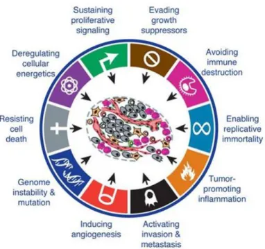
By the year of 2000, Hanahan and Weinberg proposed six features, described as hallmarks of cancer, which define six essential properties shared by most cancers: sustaining proliferative signalling, evading growth suppressors, resisting cell death, enabling replicative immortality, inducing angiogenesis and activating invasion and metastasis (figure 1.1). Indeed, cancer cells are characterized by excessive cell growth and, therefore, they must sustain proliferative signalling to keep chronic proliferation. Throughout this process an excess of oncogene activation occurs and an antiproliferative response is induced. Thus, to proliferate without restrictions, cancer cells must evade growth suppressors to circumvent anti-proliferative signalling. Furthermore, hyperactive proliferation induces pro-apoptotic responses, which need to be circumvented by cancer cells in order to resist cell death. Taking into account that cells have a limited number of cell divisions, at a certain time genetic material lost is inevitable and cells enter in senescence or even crisis. However, cancer cells can avoid this limitation by increasing telomerase activity, which will allow them to replicate endlessly. As cancer cells grow, they start to form disorganized tissues, which, like normal tissues, need to evacuate their wastes and obtain nutrients and oxygen. Therefore, tumours develop strategies to induce angiogenesis and form new vasculature to support their needs. Moreover, at a certain point nutrients and space become limited, and cellular stress increases. Thus, cancer cells can also activate a process of invasion and metastasis to colonize new places where conditions are more favourable. 2,3
In 2011 Hanahan and Weinberg revisited their previous report and spread the concept of hallmarks of cancer introducing two new emerging hallmarks: reprograming energy metabolism and evading immune destruction; and two enabling characteristics: genome instability and mutation and tumour-promoting inflammation (figure 1.1). The emerging hallmark of reprograming energy metabolism reinforced the notion that cancer cells can reprogram their energy metabolism, adapting their needs to the environments they are exposed to. In this way, cancer cells can direct their metabolism for glycolysis, even in the presence of oxygen, obtaining energy faster than normal cells, as well as the building blocks needed to grow and proliferate. The second emerging hallmark proposed, evading immune destruction, explains why cancer cells can escape immune surveillance and grow as if immune cells forgot how to limit tumour formation. In fact, tumour cells can become invisible to the immune systems and corrupt immune cells, so they can cooperate with them, by a process called immunoediting. The emerging characteristics are described as essential properties for tumour formation. Accordingly, tumours need some genetic instability to promote expansion of clones with selective advantage, as well as a certain degree of inflammation, which is a major driver of mutagenic events that could accelerate clones’ evolution and also provide bioactive molecules such as growth factors, essential for tumour proliferation. 3
During the multistep oncogenesis, cancer cells adapt a tumour microenvironment composed by several elements essential for tumour formation, progression and invasion. This microenvironment is different between organs and is composed by cells that support and communicate with cancer cells to support their needs.1–3
2
1.2. Thyroid cancer
Within the endocrine system, thyroid cancer is the most frequent malignancy and its incidence has been rising over the past years. 4–9
The thyroid gland is composed of two different types of cells: the parafollicular C cells responsible for calcitonin production; and the epithelial follicular cells responsible for iodide uptake and thyroid hormone synthesis.6,10 Thyroid cancer usually develops from the follicular cells, whereas only 3-5%
arise from the parafollicular cells originating a different type of cancer known as medullary thyroid cancer (MTC). 6,10–17
Follicular cells give rise to three main different groups of cancer, distinguished by their histological architecture and cellular morphology features.6,8,12,17,18 The most common, accounting for more than
80% of the cases, is called papillary thyroid cancer (PTC) 6,17,19,20 and it is defined for being a
well-differentiated carcinoma (WDTC) and for having a good prognosis.7,21,22 Less frequent, follicular thyroid
cancer (FTC) is also a well-differentiated carcinoma, however it has a worse prognosis than PTC.12,23
The anaplastic thyroid cancer (ATC), an undifferentiated carcinoma, is the rarest form of thyroid cancer (around 1-3% of all cases), but it is the most aggressive form of all subtypes. 6,8,12,21,24 Moreover, an
intermediate form between the WDTC and ATC called poorly differentiated thyroid carcinoma (PDTC) can also be found, accounting for less than 6% of the cases. Like ATC, PDTC represents an aggressive form of thyroid cancer6,10,11,21
The majority of patients with papillary thyroid cancer are managed successfully and present low mortality, after surgery and eventually radioiodine therapy. However, a subset of these patients harbours advanced aggressive forms, which are frequently unresectable or unresponsive to radioiodine therapy.
4,12,17,22,25–27. For these patients new therapeutic options with more efficient treatments are needed. Thus,
understanding the molecular mechanisms behind thyroid tumorigenesis would be a relevant asset to identify new targets and develop better therapeutic strategies.4–6,9,12,15,22,28
Figure 1.1- Hallmarks of cancer. This figure encompasses the classic hallmarks of cancer along with the two new
emerging hallmarks and enabling characteristics. The hallmarks of cancer define essential properties for cancer formation, development and progression shared by most cancers. Adapted from Hanahan and Weinberg (2011) 3
3 1.3. Gene alterations in thyroid cancer
Throughout the multistep cancer formation, several genetic changes occur affecting genes responsible for major cell survival and proliferation signalling. In fact, thyroid cancer is not an exception to the former and results from a gradual accumulation of genetic alterations which frequently lead to an excessive activation of the pro-survival MAPK (mitogen-activated protein kinase) pathway, mostly due to point mutations or gene rearrangements.4,6,7,9,11,19.
Different subtypes of thyroid cancer are composed by distinct patterns of gene alterations. The PTC group, mutations are mostly related to BRAF (rapidly accelerated fibrosarcoma type-B) point mutations (40-60% of the cases) or RET/PTC rearrangements (20% of the cases).24 On the other hand, 40-55% of
FTC cases are linked with RAS (rat sarcoma) point mutations or in a smaller fraction (30-35%) related to PAX8/PPARG rearrangements, or even with PTEN (phosphatase and tensin homolog) or PI3KCA gene alterations.7,18,19,21,24,25 Contrasting with the other two subtypes, in ATC, gene rearrangements are
rare but, in addition to the mutations described above, they also can harbour TP53 and CTNNB1 mutations.11,24
1.3.1. BRAF mutations
BRAF is an intracellular serine-threonine kinase and is related to MAPK pathway.7,11 When
activated, this protein is translocated to the cell membrane where it will activate its downstream effectors.11 In thyroid cancer, this kinase is frequently mutated and associated with the diagnosis of PTC.
Indeed, the majority of PTC harbours a BRAF point mutation called BRAFV600E
, which is characterized
by a substitution of a thymine for an adenine at codon 600 at BRAF gene. This results in an amino acid substitution of a valine-to-glutamine, which in terms of protein activity, leads to constitutive activation of BRAF protein, resulting in a downstream activation of MAPK pathway independent of RAS activation.6,7,9,11,21,28 Furthermore, negative feedback mechanisms responsible for controlling this
pathway are ineffective in the presence of this downstream activation.17
Several lines of evidence suggest BRAF mutations are involved in initiation of thyroid cancer. The fact that BRAF mutations could be found in microcarcinomas as well as in more advanced forms, supports this idea that BRAF mutations are an early event in the process of thyroid oncogenesis. 6,11
1.3.2. RET/PTC rearrangements
RET is a proto-oncogene well-conserved between species that encodes a tyrosine kinase receptor.7,29
In thyroid cells, RET gene is normally expressed in parafollicular C cells, but not in the follicular ones.7
However, a hybrid protein can be formed through the fusion between oncogenic RET protein and a protein that is constitutively expressed in thyroid follicular cells, leading to the ectopic RET expression in these cells. RET/PTC rearrangements occur through this process, due to a chromosomal translocation. More specifically, the carboxyl domain of RET kinase domain is fused to the amino domain of a protein constitutively expressed in follicular thyrocytes. This phenomenon results in autophosphorylation of RET protein in thyroid follicular cells and, consequently, leads to its constitutive activation.18,24,29
RET/PTC gene rearrangements by allowing constitutive activation of RET tyrosine kinase domain in thyroid follicular cells, promote an activation of both MAPK and PI3K/Akt/mTOR pathways in these cells.7,11,25,29
These rearrangements are more frequent in PTC subtype, being the RET/PTC1 and RET/PTC3 the most common.7,18,21
4
1.3.3. RAS point mutations
RAS is a family of proto-oncogenes, encoding small GTPases.18,21,30 These proteins are located at
the cytoplasmatic surface of the cell membrane and after receiving signals from tyrosine kinase receptors, for example, they transmit a signal to its downstream effectors that frequently activate MAPK and PI3K/Akt/mTOR pathways.6,9,11,18,21
In thyroid cancer, activating point mutations of all three RAS genes (HRAS, KRAS, NRAS) were detected, being those in NRAS gene the most frequent. Contrary to BRAF or RET/PTC alterations, RAS point mutations are mostly related to FTC. However, 10-20% of PTC cases, also harbour these alterations. 7,10,11,18,21
1.3.4. PAX8/PPARγ rearrangements
PAX8 gene encodes for a transcription factor that belongs to the paired box family. 11,12,31This gene
is critical for normal thyroid development and function, as well as for the expression of many thyroid-specific genes. On the other hand, PPARG gene encodes PPARγ protein, which is also a transcription factor but belonging to the nuclear receptor family. Its main function is related with lipid metabolism and adipogenesis, though there are pieces of evidences pointing out PPARγ as a tumour suppressor.31
The fusion between PAX8 and PPARG genes leads to PAX8/PPARγ rearrangements, which like RET/PTC rearrangements are also a result of a chromosomal translocation. However, contrary to RET/PTC, PAX8/ PPARγ rearrangements are more frequently found in FTC patients.11,21,31
1.4. Signalling pathways in thyroid cancer
Thyroid cancer has been described by several researchers as a “MAPK cancer”.4,6,7,9,11,32 This
signalling pathway plays the most important role in thyroid tumorigenesis, since early events in thyroid cancer are frequently related with MAPK signalling alterations.4,6,10,11 Nonetheless, thyroid cancer is
characterized by mutations with the ability to activate both MAPK and PI3K/Akt/mTOR signalling, as previously described. In fact, in the past years with the advances in genetics and molecular biology, PI3K/Akt/mTOR pathway has also been recognized as an important player in thyroid cancer pathogenesis and progression, particularly in FTC and ATC but also in PTC.6,7,12,15,16,25,33,34
Constitutive activation of PI3K/Akt/mTOR pathway has been shown to confer predisposition for thyroid cancer in Cowden’s syndrome.35 However, PI3K signalling activation by itself is not sufficient
to promote thyroid transformation, suggesting that other alterations are needed.6,36 Even though,
mutations affecting PI3KCA gene are relatively common in FTC and ATC, as well as amplifications of this gene. In PTC, mutations in PI3KCA gene, or in other factors affecting different components of PI3K signalling, are rare.6,7,10,17,25,33 Nevertheless, it is believed that this signalling pathway is involved in PTC
progression to more advanced forms, or even in the transition of ATC from PTC. 9 Moreover, PTC is
characterized by harbouring alterations with the ability to affect both MAPK and PI3K/Akt/mTOR pathways.12 These facts, along with the fact of MAPK pathway being able to communicate and activate
PI3K signalling, led researchers in the past years to look at PI3K as an appealing target in the development of novel therapies, toward advanced forms of thyroid cancer.17
1.4.1. MAPK signalling pathway
Like many other signalling pathways, MAPK signalling is a double-edge sword for being essential for normal cell survival and maintenance as well as for tumour formation and progression.
MAPK constitute a family of serine/threonine protein kinases with four parallel and independent pathways.9,37 The classical MAPK pathway is composed by the protein kinases RAF (rapidly accelerated
5 as MAPK kinase kinase, MAPK kinase and MAPK, respectively. This pathway is considered a conserved intracellular signal-transduction pathway, which is often hyperactivated in cancer. 6,38,39
Upon external stimuli, activation of plasma membrane receptors occurs, leading to the recruitment of GEF (guanine nucleotide exchange factors) by adaptor proteins. GEF will in turn, activate RAS proteins by exchanging GDP (guanosine diphosphate) for GTP (guanosine triphosphate). Active RAS (GTP-bound) recruits RAF proteins to the cell membrane where they will become active. Once RAF proteins become active, a series of phosphorylation events take place resulting in ERK activation, which will phosphorylate several substrates in the cytoplasm, mitochondria, Golgi, endoplasmic reticulum and nucleus (figure 1.2). The result of this signalling cascade will be the regulation of several proteins and transcription factors related with cell proliferation, survival, differentiation, apoptosis, metabolism and immune response.6,17,38,39
MAPK pathway is tightly regulated under non-pathological conditions by phosphatases and by bidirectional communications with other signalling pathways such as PI3K/Akt/mTOR. Furthermore, highly complex regulatory events present in both cytoplasm and nucleus allow a spatial and temporal fine tune of MAPK signalling intensity. However, during cancer formation and progression, this regulation is corrupted and MAPK signalling suffers an abnormal hyperactivation, feeding the needs of cancerous cells.37
Figure 1.2- Classic MAPK signalling pathway. MAPK pathway is a signalling pathway essential for normal cell
maintenance and survival and its abnormal activity is frequently involved in cancer. The classic pathway involves RAS activation and signals through three main proteins: RAF, MEK, ERK. After ERK phosphorylation, this protein becomes active and regulates the activity of several proteins responsible for cellular functions such as survival, proliferation and apoptosis. Adapted from Burotto et al. (2014).37
6
1.4.2. PI3K/Akt/mTOR signalling pathway
PI3K/Akt/mTOR, like MAPK pathway, an evolutionary conserved signalling pathway, is recognized for being crucial in both normal cell function and survival and cancer development. 15,40 This
pathway controls many cellular processes including growth, proliferation, survival, metabolism, apoptosis, cell motility and migration. Interestingly, many of these functions are related with the essential features for tumour formation and progression, previously defined as hallmarks of cancer. Indeed, alterations in normal PI3K/Akt/mTOR signalling pathway, hereafter called PI3K signalling, are frequently connected with cell transformation, tumour development and progression and also with metastasis.6,9,25,15,40,41
As its name suggests, this signalling pathway is composed by three main proteins: PI3K (phosphatidylinositol-4,5-bisphosphate 3-kinase), Akt (also known as protein kinase B) and mTOR (mammalian target of rapamycin).15 PI3K is a family of intracellular lipid kinases responsible for
catalysing the phosphorylation of phosphatidylinositols and phosphoinositides.9,25,15,40,41 According to
lipid substrate specificity and structure regulation, PI3K proteins can be divided into three different classes, in which class I is the most studied.6,40,42 Class I PI3K can be further divided into two different
subclasses according to the signal receptors that activate them.40 However, besides all classes of PI3K
being related with cell growth and regulation, once class IA is the most related to cancer development, for the scope of this work class IA will be the only PI3K class considered.6,15,40,42
Class IA PI3K is composed by heterodimers that are normally activated in response to RTK (receptor tyrosine kinase), by direct interaction or through adaptor proteins such as IRS (insulin receptor substrate).25 Moreover, this class of PI3K could also be activated by direct interaction with active RAS
since they have a RAS binding domain.6 These heterodimers are composed by a regulatory and a
catalytic subunit. 6,41,42 The regulatory subunit controls activation of catalytic subunit and has three
isoforms: p85α, p85β and p55γ. The catalytic subunit is responsible for the production of PIP3 (phosphatidylinositol 3,4,5-triphosphate) and also exists as three isoforms: p110α, p110β and p110δ.40
Notably, in cancer context, constitutive activating mutations of the PI3K class IA subunit genes have been described, namely in the PI3KCA gene which encodes for the p110α catalytic isoform. More specifically, PI3KCA is frequently mutated in human cancer, in which around 80% of mutations occurs in one of the three hot spot regions: E542K, E545K or K1047R. This results in catalytic subunit activation, independently of the regulatory one, leading to an increase of PI3K signalling activity.6,25,40
Upon activation, PI3K heterodimers are recruited to their lipid substrates in the plasma membrane where the catalytic subunit will be activated, leading to the production of PIP3 second messenger.6,41
This action is reversed by the phosphatase PTEN, which dephosphorylates PIP3 into PIP2 (phosphatidylinositol 3,4,5-diphosphate). After being produced, PIP3 binds to PH (pleckstrin homology) domains of different target proteins, recruiting them to the plasma membrane. Two of those proteins are Akt and PDK1 (3-phosphoinositide-dependent protein kinase 1), which are important downstream effectors of PI3K signalling. Co-recruitment of Akt and PDK1 will promote Akt phosphorylation by PDK1 at threonine 308 (Thr308), which along with phosphorylation at serine 473 (Ser473) by mTORC2 (mammalian target of rapamycin complex 2), induce full activation of Akt.
6,25,30,33,40
Akt is a serine/threonine kinase that presents three isoforms with distinct patterns of expression. Akt1 and Akt2 are expressed in almost all cells, whereas Akt3 is only found in the brain, heart and kidneys.6,25,33,40 After being fully activated, Akt phosphorylate several substrates within the cytoplasm
or into the nucleus.9 Some of these substrates are GSK-3 (glycogen synthase kinase), FOXO (forkhead
box O), PRAS40 (proline-rich Akt substrate of 40 kDa) and TSC2 (tuberous sclerosis protein 2).15,30,33,40
Phosphorylation of FOXO and GSK-3 will prevent cell cycle arrest and pro-apoptotic signalling.40 TSC2
phosphorylation will relief its inhibition on Rheb (ras homolog enriched in brain) GTPase, which in turn leads to mTORC1 (mammalian target of rapamycin complex 1) activation. Furthermore,
7 phosphorylation of PRAS40 avoids its negative regulation on mTORC1, favouring once again mTORC1 activity.15,33,40
mTOR is a serine threonine/serine kinase ubiquitously expressed in mammals responsible for controlling protein synthesis necessary for cell growth and metabolism.6,41 mTOR kinase is present in
two functionally different complexes: mTORC1 and mTORC2.6,30 mTORC1 is characterized for being
sensitive to Rapamycin and besides mTOR kinase, it is composed by RAPTOR (regulatory-associated protein of mTOR), mLST8( mammalian lethal with SEC13 protein 8) and negative regulators PRAS40 and DEPTOR (DEP domain-containing mTOR-interacting protein).30,41 This complex is activated by
PI3K/Akt signalling and regulates protein biosynthesis through the phosphorylation of S6K (70 kDa ribosomal protein S6 kinase, also known as p70S6K) and 4E-BP1 (eIF-4E binding protein 1).30,33,41
Phosphorylation of S6K will promote its activity, leading to phosphorylation and activation of S6 protein, responsible for promoting protein synthesis of some elements important for cell growth such as ribosomal proteins and elongation factors. For this reason, phospho-S6 (p-S6) is frequently used in experiments as an indicator of mTORC1 activation. On the other hand, phosphorylation of 4E-BP1 blocks its activity, preventing its effects on protein translation inhibition.30 mTORC2 activation seems
to be more related with growth factors, and its best characterized activity is the phosphorylation of Akt at Ser473.25,30 However, it seems that mTORC2 is also responsible for phosphorylating PKCα (protein
kinase c) and paxillin, as well as for regulating small GTPases .22,30 In terms of composition, like
mTORC1 , mTORC2 is composed by mTOR kinase, mLST8 and DEPTOR. Additionally, mTORC2 is further composed by RICTOR (rapamycin-insensitive companion of mTOR), mSIN1 (stress-activated map kinase-interacting protein 1), PROTOR (protein observed with rictor) and Hsp70 (70 kD heat shock protein).25,30,41
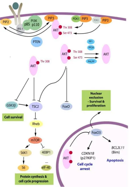
In summary, activation of PI3K leads to the production of a second messenger PIP3, which in turn recruits Akt to the plasmatic membrane allowing its activation by PDK1 and mTORC2. Once activated, Akt phosphorylates and inhibits TSC2, relieving Rheb GTPase activity and promoting mTORC1 activity. With this, activity of S6K is promoted and 4E-BP1 is blocked, resulting in translation of several proteins essential for cell growth and metabolism. (figure 1.3). 9,15,25,30,33,41
8
Figure 1.3- PI3K/Akt/mTOR signalling pathway. PI3K/Akt/mTOR pathway is essential for normal cell function and
survival and its dysregulation is often seen in cancer. After a stimulatory event, PI3K is activated and through the production of PIP3, recruits Akt to the cell’s membrane, where it will be activated. After activation, Akt relieves Rheb inhibition, promoting mTOR activation. Once activated, mTOR will regulate protein translation and cell’s growth, through S6K stimulation and 4E-BP1 inhibition. Adapted from Robbins et al. (2016).33
1.5. NF-κB (Nuclear factor kappa-light-chain-enhancer of activated B cells)
Since its discovery in 1986, NF-κB has been extensively studied due to its involvement in a multitude of diseases, including cancer.5,43 This transcription factor is responsible for the control of
several biological aspects such as immune and inflammatory responses or even cell growth and apoptosis. 13,14,44,45
NF-κB is conserved among species and is ubiquitously expressed, although its function varies according the cell type it is expressed.43,45 Moreover, in mammals NF-κB is frequently viewed as a
family of transcription factors composed by five members that form homodimers or heterodimers among each other: p65 (RELA), RELB, c-Rel, NF-κB1 (also known as p105) and NF-κB2 (also known as p100).46–48 These members can be further divided into two classes according to their mode of synthesis
9 forms and includes p65, RELB and c-Rel. The second class is composed by NF-κB1 and NF-κB2 which are synthetized as large precursors and are further processed into p50 and p52, respectively.49 In
common, both classes contain a highly conserved REL homology domain (RHD) essential for dimerization, DNA binding and association with inhibitory proteins.48,50
The way NF-κB dimers are activated can be viewed as two main pathways: the canonical and the non-canonical pathway. Canonical pathway is triggered in response to numerous stimuli, involving pro-inflammatory cytokines such as TNFα (tumour necrosis factor α) and depends on the activity of IKKγ regulatory subunit as well as the catalytic IKKβ subunit. This pathway involves preferentially the heterodimer p65/p50 and is responsible for controlling several aspects of cell growth and inflammation.13,14,46–49,51 On the other hand, non-canonical pathway involves preferentially p52/RELB
heterodimers, being triggered in a more restricted way by some cytokines of TNF super family and depending on the activity of NF-κB-inducing kinase (NIK) and IKKα.46–49,52. Also, non-canonical
pathway functions are more related with immunity aspects, including the regulation of lymphoid organogenesis and B-cell maturation, for example.51,52 Furthermore, while canonical NF-κB activation
is dependent on degradation of its inhibitor ( inhibitor of κB, IκB), the non-canonical NF-κB is related with a mechanism involving p100 processing.52
In the absence of stimuli, NF-κB canonical dimers are normally present in the cytoplasm as an inactive form for being held to their inhibitor IκB.5,13,14,44,47 After a stimulatory event, activation of a
protein kinase complex called IKK (IκB kinase) occurs. This complex is composed by IKKα and IKKβ catalytic subunits in combination with a regulatory subunit IKKγ (also called NEMO) and its activation leads to IκB phosphorylation and, consequently, to its degradation, resulting in the release and activation of NF-κB dimers.13,14,45–47,49 Upon release, the nuclear localization signal (NLS) of NF-κB dimers is
exposed and NF-κB is translocated into the nucleus promoting the transcription of several target genes (figure 1.4). 49
Figure 1.4- Canonical and non-canonical pathways of NF-κB. NF-κB transcription factor regulates several biological
aspects such as inflammation and cell’s growth and apoptosis, which are essential during cancer’s formation. NF-κB is normally retained in the cytoplasm for being bound to its repressor IκB. Upon stimuli, IκB is phosphorylated by IKK, resulting in its proteasomal degradation. Canonical pathway involves the heterodimer p65/p50 and depends on IKKγ regulatory subunit activity. Non-canonical pathway is more related with NIK and IKKα activity and involves preferentially the heterodimer p52/ReLB. Adapted from Shao-Cong Sun (2011). 52
10
1.5.1. NF-κB in thyroid cancer
Dysregulation of NF-κB signalling has been implicated in many cancers. In the particular case of thyroid cancer, activation of NF-κB was found in PTC, FTC and ATC, suggesting that this transcription factor has an important role during thyroid cancer formation.9,13,35 In fact, many authors have gathered
evidence that NF-κB contributes to thyroid tumorigenesis. The first evidence was exposed by Visconti et al. in 1997 53, where they demonstrated that NF-κB canonical activation occurs during thyroid cancer
development.14,53 Later, in 2004, Pacifico 13 and his group demonstrated that NF-κB contributes to
thyroid oncogenesis, by inhibiting the apoptotic program. Moreover, this group also proposed that chronic activation of NF-κB in thyroid cancer cells could be due to defects in IκBα regulation or even by production of autocrine factors that stimulate this signalling.13 In line with the previous references,
in 2017 Faria et al.4 proposed that in PTC cell lines RAC1b overexpression signals through NF-κB,
resulting in apoptosis resistance.
In the context of normal thyroid physiology, in 2016, a publication from Reale’s group 45 has shown
that NF-κB signalling is required for normal function and structure of thyroid cells. Thus, despite being crucial for normal thyroid cell survival and maintenance, NF-κB signalling dysregulation plays an important role during thyroid cancer formation, leading to an anti-apoptotic behaviour.4,5,9,13,14,45
Furthermore, NF-κB is frequently related with treatment resistance and with aggressive behaviour of thyroid carcinomas.5,35
1.6. Crosstalk between signalling pathways in papillary thyroid carcinomas
Once mutations characterizing PTC are mostly related with aberrant activation of MAPK signalling6,12,17, inhibition of this pathway would be an obvious therapeutic choice for those patients with
advanced disease. However, the use of MAPK pathway inhibitors, such as MEK or BRAF inhibitors, are related with undesirable side effects and most patients develop resistance to treatment after a short period of time.26,54,55 Therefore, other molecular pathways must be acting in concert to promote tumour
survival and resistance to therapy, in these cases.
Roelli et al. (2017) 20 have shown that in aggressive PTC, activation of PI3K signalling conferred
resistance to BRAF inhibitors and that this resistance was overcome with a PI3K inhibitor combination. In line with that, another study had established that thyroid carcinoma’s migration and proliferation is in part mediated by both Ras/MAPK and PI3K/Akt/mTOR pathways and that these pathways communicate with each other. Moreover, the same authors suggested that the interplay between both pathways results in cross-activation or inhibition processes, depending on the cell type. 34
When treatment resistance is discussed in the context of thyroid cancer one of the most mentioned elements is the NF-κB transcription factor. In 2006, a study has demonstrated that accumulation of BRAFV600E protein promote an increase of IκBα degradation, and consequently to NF-κB activation.
Also, CRAF, a protein that transmits signals from RAS to MEK, has been indicated as an activator of NF-κB.56
In summary, it seems that MAPK can communicate with both PI3K/Akt/mTOR and NF-κB, being this phenomenon intrinsically related with treatment resistance. However, potential crosstalk between PI3K/Akt/mTOR and NF-κB pathways remains to be elucidated.
11
2. Aim of the project
This work aims to address the potential interplay between the transcription factor NF-κB and the signalling pathway PI3K/Akt/mTOR in papillary thyroid cancer cell lines. For this purpose, a PTC cell line harbouring a mutation in PI3KCA gene that leads to constitutive activation of PI3K protein was used.
The main tasks were to evaluate NF-κB activity upon inhibition of PI3K/Akt/mTOR pathway: (i) by using chemical inhibitors for PI3K and/or mTORC1.
(ii) by blocking PI3K signalling in the presence of exogenous stimulation of the canonical NF-κB pathway.
(iii) by addressing the impact of PI3K signalling on NF-κB activity in the presence of chemical inhibition of NF-κB canonical pathway.
To further evaluate the impact of PI3K signalling on NF-κB activity, cellular systems with different signalling backgrounds were used and results were compared.
12
3. Materials and Methods
3.1. Cell culture and reagents
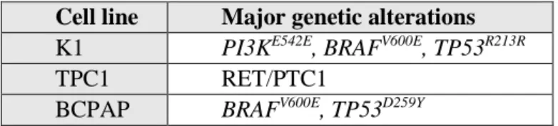
In this study, the human papillary thyroid carcinoma cell lines K1, TPC1 and BCPAP were used. These cell lines harbour different genetic backgrounds as shown in table 3.I.
K1 cells were maintained in Dulbecco’s modified eagle medium/nutrient mixture F-12 (DMEM:F12 1:1, Lonza). TPC1 and BCPAP cells were cultured in RPMI 1640 medium (Lonza). All mediums were supplemented with 10% (v/v) of foetal bovine serum (FBS, Biochrom) and 1% (v/v) glutamine (Gibco). All cells were maintained at 37ºC in an humified environment with a 5% CO2
atmosphere. When cells reached the optimum confluence (80-100%), they were washed with Dulbecco’s phosphate buffered saline 1x (DPBS1x, Lonza), detached by incubation at 37ºC with trypsin-EDTA (ethylenediamine tetraacetic acid, Invitrogen) and subcultured in a new flask at a confluency of 25%.
Cells were treated with PI3K inhibitor Ly294002 (Sigma-Aldrich), mTORC1 inhibitor Rapamycin, PI3K/mTOR dual inhibitor BEZ235 (Sigma-Aldrich), IKKα/β inhibitor BMS-345541 and human Tumour Factor Necrosis α (hTNFα, R&D Systems), using the conditions described in Table 3.II. To perform these treatments, cells were seeded in 12-well plates (Thermo Scientific) at 60% confluency and synchronized for 24 h in starvation (serum free) medium, after they became adherent. Then, medium was replaced with fresh supplemented medium or fresh starvation medium, depending on the intended treatment. At the end, cells were used for RNA extraction (3.2) or for western blot analysis (3.5).
Table 3.1- PTC-derived cell lines major genetic alterations
Table 3.II - PI3K/Akt/mTOR signalling pathway inhibitors conditions
Reagents Final concentration Time of treatment (hours)
Ly294002 50 µM 6
Rapamycin 100 nM 6 or 24
BEZ235 100 nM 6
TNFα 100 nM 1
BMS-34554 10 µM 7
3.2. RNA extraction and complementary DNA (cDNA) synthesis
Cells were lysed with tripleXtractor reagent (Grisp) and RNA was extracted according to the manufacturer’s protocol. During protocol procedures, an Eppendorf centrifuge 5415R was used. For each sample, RNA concentration was measured in nanodrop™ 2000 spectrophotometer (Thermo Scientific).
After RNA extraction, cDNA was synthetized from 1 µg of RNA. Firstly, the RNA sample, 0.1 µL of random primers (3 µg/µL) (Roche), 0.8 µL of deoxynucleotides (dNTPs mix: dATP, dCTP, dGTP, dTTP, 25 mM each) and purified water (ddH2O) up to 15 µL were mixed together and incubated at 65ºC
during 10 min. This initial step aimed the removal of RNA secondary structures which could affect cDNA synthesis efficiency. After this step, a mix made with 4 µL of reverse transcriptase buffer 5x (Thermo Scientific), 0.5 µL reverse transcriptase (200 U/µL) (Thermo Scientific) and 0.5 µL RNAaseOut™ ribonuclease (40 U/µL) (Invitrogen), was added to each sample. Then, cDNA synthesis
Cell line Major genetic alterations
K1 PI3KE542E, BRAFV600E, TP53R213R
TPC1 RET/PTC1
13 was performed using the basic program described in Table 3.III, in a 2720 thermal cycler (Applied Biosystems).
Table 3.III - cDNA synthesis conditions
3.3. Reverse transcription polymerase chain reaction (RT-PCR)
RT-PCR amplification was performed to control IκBα gene expression. Specific primers for IκBα amplification (F-forward, R-reverse) IκBα human F (5’- CTACACCTTGCCTGTGAGCA), IκBα human R (5’-CCCCACACTTCAACAGGAGT), were used, originating a 280 base pairs (bp) PCR product.
A reaction mixture containing 0.25 µL of specific primers, along with 12.5 µL of PCR buffer (see supplementary table I), 0.1 µL of Taq polymerase (5 U/µL) (Nzytech) and 1 µL of cDNA sample, was prepared. The reaction took place in a 2720 thermal cycler (Applied Biosystems) using the basic program described in Table 3.IV.
Table 3.IV - IκBα RT-PCR amplification conditions
Stage Temperature ºC Time Cycles
Initial denaturation 95 5 min 1
Denaturation 95 30 seconds
24
Annealing 60 30 seconds
Elongation 72 30 seconds
Final elongation 72 3 min 1
Inactivation and Cooling 4 ∞ 1
The RT-PCR products were analysed by electrophoresis on a 2% agarose gel made with Tris-Borate-EDTA buffer 1x (TBE 1x) diluted in distilled water from TBE 10x (Grisp) and stained with 0,05% (v/v) ethidium bromide (Invitrogen). Gel electrophoresis was performed in a Biorad sub-cell GT electrophoresis chamber at 130 mV for 30 minutes, and the gel was visualized upon exposer to UV light in VWR Genosmart 1107 transilluminator.
3.4. Quantitative reverse transcription polymerase chain reaction (RT-qPCR)
IκBα (target gene) mRNA expression, was quantified by RT-qPCR in the LightCycler® 480 II (Roche) using the xpert fast SYBR mastermix (Grisp). TATA-binding protein (TBP) expression was used as reference. RT-qPCR was performed according to manufacturer’s protocol and reaction mixtures were prepared using 5 µL of xpert fast SYBR mastermix, 0.25 µL of each primer, 1 µL of cDNA sample in ddH2O to a final volume of 10 µL. The sequence of specific primers used for IκBα and TBP
amplification were as follow: IκBα human F CTACACCTTGCCTGTGAGCA) IκBα human R (5’-GACACGTGTGGCCATTGTAG), TBP human F (5’-TGCACAGGAGCCAAGAGTGAA), TBP human R (5’-CACATCACAGCTCCCCACCA).
IκBα expression was normalized to TBP and mRNA relative quantification was determined using the 2-∆∆Ct method (the efficiency of both target and reference genes were similar, nearly 100%).
Stage Temperature ºC Time
Annealing 25 10 min
Elongation 42 60 min
Inactivation 70 10 min
14
3.5. Western Blot
PI3K/Akt/mTOR pathway activation status was evaluated by western blot, through monitorization of phospho-S6 (p-S6) protein. β-actin was used as an endogenous control.
Protein extracts were obtained using 50 µL of lysis buffer (see supplementary table II) and denatured during 10 min at 95ºC.
SDS-polyacrylamide gel electrophoresis (SDS-PAGE) was performed to separate proteins of each sample, by adding equivalent amounts of protein extracts to the gel. The SDS-PAGE gel consisted of two distinct gels: a lower 10% polyacrylamide gel (resolving) and an upper 4% polyacrylamide gel (stacking) (see supplementary table II). Electrophoresis was carried in SDS-PAGE buffer 1x at 20 mA per gel, during approximately 1h. Subsequently, proteins were transferred into polyvinylidene difluoride (PVDF) membranes (Bio-Rad), previously activated in methanol, using a blot electrophoresis transfer cell (Bio-Rad) for 1 h at 100 V. Then, membranes were stained with coomassie blue and washed: firstly, with a destain solution and then with tris-buffered saline 0.05% triton x-100 (TBST) (see supplementary table II).
Membranes aimed for detection of the endogenous control β-actin, were incubated for 1 h in a solution of TBST with 5 % (w/v) non-fat milk (TBST milk) to avoid unspecific bindings and then, incubated overnight at 4 ºC with mouse anti-β-actin (Sigma-Aldrich) in a 1:10000 dilution in TBST milk. Membranes aimed for detection of phosphorylated S6 protein were incubated overnight at 4 ºC with the primary antibody rabbit anti-pS6 (Cell Signaling) in TBST in a 1:2000 dilution. Finally, after a washing step with TBST, membranes were incubated for 1h at room temperature (RT) with horseradish peroxidase-conjugated (HRP) specific secondary antibodies: anti-mouse (1:5000) (Thermo Scientific) and anti-rabbit (1:5000) (Thermo Scientific), all diluted in TBST milk.
Protein bands were detected by exposure on autoradiographic films after luminol-based enhanced chemiluminescence (ECL) (see supplementary table II).
3.6. Immunofluorescence
Analyses of NF-κB nuclear translocation were assessed by immunofluorescence technique. For this purpose, K1 cells grown on coverslips (10 mm x 10 mm) in a 12-well plate and were subjected to treatment with different drugs as described above.
Cells were washed in PBS 1x (Lonza) and fixed in paraformaldehyde 4% (v/v) for 30 min at RT. Two steps of permeabilization were performed aiming the permeabilization of plasmatic and nuclear membranes, respectively: first cells were incubated with PBS-triton x-100 (PBST) 0,5% (v/v) for 15 min at RT and then were followed by an incubation with methanol for 10 min at -20ºC. Subsequently, cells were washed three times during 5min with PBST 0.05% (v/v) and incubated overnight at 4 ºC with the MUL1 polyclonal primary antibody rabbit anti-NF-κB p65 NLS (Thermo Scientific) at a 1:750 dilution. Then, cells were incubated with secondary antibody goat anti-rabbit Alexa Fluor 532 (Life Technologies; 1:500 dilution) for 30 min, after being washed with PBST 0.05% (v/v) three times for 5 min. Cells were washed again three times for 5 min with PBST 0.05% (v/v) and nucleus were stained with DAPI, following a final fixation step with paraformaldehyde 4% (v/v) for 15min. Coverslips were washed with PBST 0.05% (v/v), mounted in a microscope slide with VectaShield medium and sealed with nail polish. Images were recorded in a Leica TCS-SPE confocal microscope with 405 nm and 532 nm laser lines and processed in ImageJ software.
3.7. Statistical analysis
Graphpad Prim 5 software (San Diego, USA) was used to perform statistical analysis. The statistical analysis and the evaluation of statistical significance between results were performed using an unpaired one-tailed or two-tailed Student’s t-test. Values were expressed as mean with its respective standard deviation (SD). Significant results were accepted as p<0,05.
15
4. Results
4.1. PI3K signalling inhibition is associated with an increase of NF-κB activation readout in K1 cells In order to evaluate if there was a potential interplay between NF-κB and PI3K/Akt/mTOR pathway in K1 cells, we first analysed the effects of PI3K/Akt/mTOR pathway inhibition on IκBα mRNA levels, here used as a readout for κB transcriptional activity. Among several approaches to monitor the NF-κB activation status, we monitored the transcriptional levels of a target gene by RT-qPCR. Bottero et. al showed that IκBα mRNA levels perfectly correlate with the activation of NF-κB in several cellular models, indicating that quantification of IκBα is a valid readout of NF-κB activation. Indeed, besides being NF-κB repressor, IκBα is under NF-κB transcriptional control and is one of the genes most rapidly transcribed after NF-κB is activated.52 For this reason, we decided to use IκBα as a reporter for NF-κB
activation.
Inhibition of PI3K signalling was performed using three different approaches. The first consisted in the use of Ly294002, which is considered a potent chemical inhibitor of PI3K protein.36 The second,
was directed to a downstream PI3K signalling event and was performed with Rapamycin, which is a mTORC1 chemical inhibitor.57,58 Finally, the last approach consisted in a dual inhibition of PI3K and
mTOR proteins, using the dual inhibitor BEZ23559 or a combination of Ly294002 and Rapamycin.
Controls were obtained from cells cultured in the same conditions but treated in the absence of drugs with dimethyl sulfoxide (DMSO), the vehicle used for drug’s dilution.
The effects of chemical inhibitors on PI3K/Akt/mTOR pathway were monitored by western blot. For this purpose, β-actin was used as a control to document equal amounts of protein loaded on the gel and p-S6 as an indicator of PI3K/Akt/mTOR pathway activation status. As shown in figure 4.1A, all inhibitors decreased p-S6 expression, being this effect more notorious with Rapamycin inhibitor.
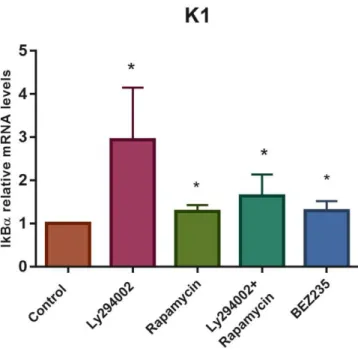
Under these experimental conditions, a RT-qPCR was performed to measure the relative mRNA levels of IκBα in response to the chemical inhibitors of PI3K signalling. For each assay, data obtained with drug treatments were normalized to the corresponding control condition. As shown in figure 4.1B, all PI3K signalling inhibitors promoted a statistically significant increase in IκBα mRNA levels. Comparing to control, PI3K protein inhibition with Ly294002 resulted in a significant (p<0,05) 2,9-fold increase of IκBα mRNA levels, which was the greatest increase verified. Inhibition of mTORC1 with Rapamycin, resulted in a significant (p<0,05) 1,28-fold increase of IκBα mRNA levels, being the same observed with dual inhibition of PI3K and mTOR with either BEZ235 (1.29-fold increase; p<0,05) or the combination of Ly294002 and Rapamycin (1.63-fold increase; p<0,05). Collectively, inhibition of PI3K/Akt/mTOR pathway increased IκBα mRNA levels, suggesting an increase in NF-κB transcriptional activity, which was higher with upstream signalling inhibition.
1 2 3 4 5 Anti-β-actin 1 – Control 2 – Ly294002 3 – Rapamycin 4 – Ly294002+Rapamycin 5 – BEZ235 Anti-p-S6 A
16
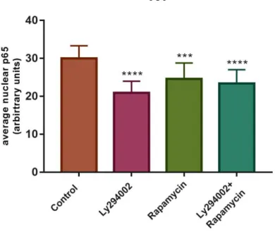
4.2. Inhibition of PI3K signalling decreases p65 nuclear translocation in K1 cells
Since NF-κB transcriptional activity depends on its translocation to the nucleus, we decided to further investigate if NF-κB nuclear translocation was affected by the presence of PI3K signalling inhibitors, in K1 cells. For that, an immunofluorescence was performed using an antibody that binds to uncovered NLS region of p65 protein, which is exposed after IκB repressor degradation. p65 protein is involved in the canonical pathway of NF-κB, where it usually forms p65/p50 heterodimers. Nuclei were stained with DAPI (blue).
As shown in figure 4.2A, the red signal intensity present in cells’ nuclei appears to be lower upon drug treatment, compared to control conditions. This fact is supported in figure 4.2B quantification of fluorescence intensity. Moreover, the decrease in nuclear red staining is slightly more pronounced with the PI3K inhibitor Ly294002 than with mTORC1 inhibitor Rapamycin or even with the combination of both drugs. This suggests that inhibition of PI3K signalling led to a decrease in the active p65 at the nucleus, which is more evident when triggered by pathway inhibition upstream of mTOR. However, data correspond to one single experiment and further assays are needed to clarify these results.
Figure 4.1- PI3K signalling inhibition is associated with an increase of NF-κB activation readout in K1 cells. Cells were
treated with Ly294002 (50 µM), Rapamycin (100 nM), BEZ235 (100 nM) or with a Ly294002 and Rapamycin combination, for 6h. (A) Western Blot analysis was performed to monitor PI3K/Akt/mTOR pathway activation status in the presence of its chemical inhibitors. Detection of endogenous β-actin served as control of protein expression. (B) Results of IκBα mRNA levels by RT-qPCR. The mRNA levels were measured after cells were treated with chemical inhibitors of the PI3K/Akt/mTOR pathway. Controls were obtained in the same conditions but treated with DMSO and in the absence of inhibitors. Collectively, results show an increase in NF-κB activation readout after PI3K signalling inhibition. Data are mean ± error bars (SD) of at least three independent experiments. p-values were calculated comparing control with drug treatments, using an unpaired one-tailed Student’s t-test. *p≤0.05.