A R T I G O O R I G I N A L
N A I L F O L D C A P I L L A R O S C O P Y A B N O R M A L I T I E S
C O R R E L A T E W I T H C U T A N E O U S A N D V I S C E R A L
I N V O L V E M E N T I N S Y S T E M I C S C L E R O S I S P A T I E N T S
Lucy T Sato,
*Cristiane Kayser,
*Luís E C Andrade
*taneous involvement, the number of affected tracts, and the presence of anti-Scl-70 antibodies.
Keywords: Microcirculation; Nailfold
capillaros-copy; Raynaud’s phenomenon; Systemic sclerosis.
Resumo
Objectivo: Correlacionar parâmetros quantitativos
e semiquantitativos da capilaroscopia perinun-gueal (CPU) com a extensão do acometimento cu-tâneo, envolvimento visceral e alterações sorológi-cas em pacientes com esclerose sistêmica (ES).
Métodos: A presença de alterações clínicas e
soro-lógicas foi avaliada retrospectivamente e correlacio-nada com os achados capilaroscópicos (número de
capilares/mm,score de deleção vascular e número
de capilares ectasiados e megacapilares). Para ava-liação da extensão da doença, cinco manifestações foram analisadas: lesões em polpa digital, acometi-mento cutâneo, envolviacometi-mento esofagiano, doença pulmonar intersticial e hipertensão pulmonar.
Resultados: Foram avaliadas 105 CPUs realizadas
em 92 pacientes, 13 dos quais foram avaliados em dois tempos diferentes. Pacientes com ES forma
cutânea difusa apresentaram maior score de
dele-ção vascular do que pacientes com ES forma
cutâ-nea limitada, ES sine scleroderma SSc e em
super-posição (1,67±0,91 vs 0,99±0,82; p=0,0005). O score
cutâneo de Rodnan modificado se correlacionou positivamente com desvascularização capilar,
ava-liado pelo score de deleção vascular e pelo número
de capilares/mm (p<0,001, p=0,012; respectiva-mente). Pacientes com três ou mais órgãos acome-tidos apresentaram menor número de
capila-res/mm (8,00±1,69 vs 9,23±1,31 capilares/mm;
p=0,025) e maior score de deleção vascular
(1,41±0,95 vs 0,73±0,76; p=0,027) em comparação
com pacientes com menos de três órgãos
acome-tidos. O score de deleção vascular foi
significativa-*Rheumatology Division, Universidade Federal de São Paulo, São Paulo, Brazil
Abstract
Objective: The aim of this study was to correlate
quantitative and semiquantitative nailfold capilla-roscopy (NFC) parameters with the extent of cuta-neous and visceral involvement in systemic sclero-sis (SSc) patients.
Methods: The presence of clinical and serological
alterations was evaluated retrospectively and cor-related with NFC findings (number of capillary loops/mm, vascular deletion score and number of enlarged and giant capillary loops). For evaluation of disease extension five manifestations were ana-lyzed: finger pad lesions, skin involvement, esop-hageal involvement, interstitial lung disease, and pulmonary hypertension.
Results: There were 105 NFC examinations in
92 patients, 13 of whom were evaluated at two different time points. Patients with diffuse cutane-ous SSc had a higher vascular deletion score than
patients with limited cutaneous SSc, sine
sclero-derma SSc, and overlap syndrome (1.67±0.91
vs 0.99±0.82; p=0.0005). Modified Rodnan’s skin score correlated positively with capillary deletion, evaluated by the vascular deletion score and the number of capillary loops/mm (p<0.001 and p=0.012; respectively). Patients with three or more involved tracts presented lower number of capillary loops/mm (8.00±1.69 vs 9.23±1.31 capillary loops/mm; p=0.025) and a higher vascular deletion score (1.41±0.95 vs 0.73±0.76; p=0.027) when compared to patients with less than three affected tracts. Vascular deletion score was significantly higher in patients with anti-Scl-70 antibodies that in patients without anti-Scl-70 antibodies (p=0.02).
Conclusions: NFC abnormalities correlated
cu-N A I L F O L D C A P I L L A R O S C O P Y C O R R E L AT E S W I T H C U TA cu-N E O U S A cu-N D V I S C E R A L I cu-N V O LV E M E cu-N T I cu-N S S C
mente maior nos pacientes com anticorpos anti-Scl-70 quando comparados com pacientes com au-sência destes anticorpos (p=0,02).
Conclusões: Alterações na CPU se correlacionaram
positivamente com a forma cutânea difusa da ES, o grau de acometimento cutâneo, o número de ór-gãos envolvidos e a presença de anticorpos anti-Scl-70.
Palavras-Chave: Microcirculação; Capilaroscopia
Periungueal; Fenômeno de Raynaud; Esclerose Sis-têmica.
Introduction
Systemic Sclerosis (SSc) is an autoimmune rheu-matic disease of unknown etiology characterized by vascular alterations and tissue fibrosis of multiple organs. It affects mostly the skin, musculoskeletal system, heart, kidneys, lungs and gastrointestinal
tract.1Vascular structural alterations and lesions
are detected early and seem to play an important role in SSc pathogenesis. Dysfunction of vascular tone control, endothelial activation/lesion, and myointimal cell proliferation of small blood ves-sels and capillaries result in progressive reduction of vessel lumen, decreased blood flow, and a state
of chronic hypoxia.2,3In the periphery,
microvascu-lar damage is characterized by enmicrovascu-largement and distortion of capillary loops, micro-haemorrhages
and progressive devascularization.4,5
Peripheral microangiopathy can be easily recog-nized by widefield nailfold capillaroscopy (NFC), a noninvasive and safe method, that is well establi-shed in the investigation of patients with Raynaud’s
phenomenon and SSc.5,6 Patients with SSc exhibit a
typical pattern at NFC, designated “SD pattern” by Maricq et al., and characterized by enlargement of capillary loops, loss of capillaries, disruption of the orderly appearance of the capillary bed and
distor-tion of capillaries.7Maricq et al., defined also two
major nailfold capillary patterns, namely the “ac-tive” (extensive loss of capillaries and minimal ca-pillary enlargement) and the “slow” (caca-pillary en-largement and/or extremely enlarged capillaries
with no or minimal capillary loss) pattern.5
Re-cently, Cutolo et al., have reclassified the scleroder-ma microangiopathy into 3 different patterns:
“ear-ly”, “active” and “late” pattern.8The early pattern is
characterized by few enlarged/giant capillaries, few hemorrhages, relatively well-preserved capillary
distribution and no evident loss of capillaries; in the “active” pattern there are frequent giant capil-laries and hemorrhages, moderate loss of capilla-ries, mild disorganization of the capillary architec-ture, absence or mild ramified capillaries, and pre-sence of edema; the “late” pattern is characterized by irregular enlargement of the capillaries, few or no giant capillaries and hemorrhages, severe loss of capillaries with extensive avascular areas, disor-ganization of the normal capillary array, and
pre-sence of ramified/bushy capillaries.8The SD
pat-tern has been observed in 84-97% of SSc patients but is not strictly limited to patients with SSc and can also be found in dermatomyositis, mixed con-nective tissue disease, and overlap syndromes with SSc.4,5,9,10Several studies have addressed a possible
relationship between NFC alterations and the
ex-tent of skin and visceral involvement in SSc.11-15The
apparently controversial findings of some of these studies are possibly due to small and heterogene-ous samples of patients and also to heterogeneheterogene-ous methodology in the interpretation of NFC findings. Our group has previously reported on a repro-ducible NFC method, comprehending several mi-croangiopathic parameters assessed in quantitati-ve and semiquantitatiquantitati-ve manner, evaluating all 10
fingers of the hands.6The aim of the present study
was to report the morphological alterations in the peripheral microcirculation, evaluated by panora-mic nailfold capillaroscopy (NFC) using the
me-thod proposed by Andrade et al.,6and to correlate
these microvascular abnormalities with cutaneous and visceral involvement, in a large sample of pa-tients with SSc.
Patients and Methods
Patients
This was a retrospective study which included all SSc patients seen in a two-year interval (2003-2004) at the Scleroderma Spectrum Outpatient Clinic at Federal University of São Paulo (UNIFESP) Medi-cal School Hospital. All patients with at least one NFC exam performed in the last 15 years were in-cluded in the study. SSc diagnosis was based on the American College of Rheumatology (ACR)
crite-ria.16Disease subtype was classified as follows:
dif-fuse cutaneous SSc (skin thickening on the trunk, face, in addition to proximal and distal extremi-ties), limited cutaneous SSc (skin thickening loca-lized to the face and neck, and distal to the elbows
L U C Y T S AT O E C O L.
and knees), sine scleroderma SSc, and in overlap
syndromes.17,18For further clinical, serological and
capillaroscopic analysis patients were divided into two groups: diffuse SSc and non-diffuse SSc. The latter category comprised patients with limited
cu-taneous SSc, sine scleroderma SSc and patients
with overlap syndromes.
Nailfold capillaroscopy
All NFCs were performed in a stereomicroscope (Olympus - SZ40) under 10-20 x magnification according to the protocol proposed by Andrade et
al.6All the ten digits of the hands were examined,
except when prevented by extremely poor visibi-lity or amputation. The following parameters were analyzed: (1) number of capillary loops/mm, (2) vascular deletion score, (3) number of enlarged loops (about four times the normal afferent, tran-sition, and efferent limbs width), and (4) number of giant capillary loops (10 or more times the nor-mal width of capillary limbs). Enlarged and giant loops were counted together. The vascular dele-tion score was assessed according to Lee’s
me-thod,19in which a deletion area is defined as the
ab-sence of two or more consecutive loops. Each fin-ger was rated from 0 to 3: grade 0 – no deletion area; 1 – one or two discrete deletion areas; 2 – more than two discrete deletion areas; 3 – extensi-ve and confluent deletion areas. For each patient the NFC parameters were calculated as the avera-ge obtained in all analyzed digits.
During the 15-year period NFCs were perfor-med by three investigators with similar NFC trai-ning. All exams were performed according to the same NFC protocol, and examiners had no infor-mation about the patient’s clinical condition. Re-producibility of the NFC examination was tested as
previously described.6
Clinical and laboratory evaluation
All clinical and laboratory data were collected re-trospectively from medical charts according to a standardized protocol routinely filled out at each visit to the Scleroderma Spectrum Outpatient Cli-nic during the period of 1989 to 2004.
The presence and extent of cutaneous involve-ment was assessed by the modified Rodnan’s skin
score.20Finger pad lesions were defined as the
pre-sence of active digital ulcerations, digital pitting
scars, resorption and/or digital amputations. Eso
-phageal involvement was evaluated by the presen-ce of esophageal dismotility on barium contrast
roentgenographic study of the upper gastrointes-tinal tract. The presence of lung interstitial disea-se was defined by forced vital capacity (FVC) < 75% of the predicted values and/or bibasal interstitial pulmonary infiltrate on chest radiogram and/or high resolution computed tomography (HRCT) showing ground-glass opacities, reticular or ho-neycombing pattern. Pulmonary hypertension was defined by pulmonary systolic arterial pressu-re (PSAP) >35mmHg, estimated on Doppler echo-cardiography. Finally, renal scleroderma crisis was defined by the presence of malignant hyperten-sion and renal failure.
For evaluation of disease extension five manifes-tations were analyzed: (1) finger pad lesions, (2) presence of skin thickness, (3) presence of esopha-geal dismotility on barium study, (4) interstitial lung disease, and (5) presence of pulmonary hyper-tension. Only patients with available information on these five manifestations were included.
Antinuclear antibodies (ANA) were determined by indirect immunofluorescence using HEp-2 cell as substrate. Anticentromere antibodies were iden-tified by the typical pattern on the indirect immu-nofluorescence HEp-2 cell assay and anti-topoi-somerase I (anti-Scl-70) antibodies were determi-ned by the double immunodiffusion method against rabbit thymus extract.
For all included patients, the clinical-laboratory evaluation was not more than 12 months apart the NFC evaluation. Patients with more than one NFC made more than 12 months apart were handled in two alternative ways. If the NFCs conclusions were different (normal NFC or nonspecific capillary al-terations on the first exam and presence of SD pat-tern on the second, or the contrary), the clinical and laboratorial findings were analyzed at each time and compared to the relevant NFC exam. Al-ternatively, if the NFCs conclusions were similar in both occasions, only the NFC with closest availa-ble clinical-laboratory evaluation was taken into consideration. However, if the interval between NFCs was higher than three years each NFC/clini-cal/laboratory set was individually analyzed. Pati-ents with no NFC record were excluded from the study.
Statistical analysis
Results are presented as mean and standard devia-tion. Correlations between NFC, clinical and labo-ratory parameters were evaluated by Pearson’s or Spearman’s correlation analysis. Associations
between qualitative parameters were analyzed by the Chi square test. Mann-Whitney’s test was used
to compare quantitative variables. p <0.05 was
con-sidered significant.
Results
One hundred and five NFCs were evaluated in 92 patients, 13 of whom were evaluated at two diffe-rent moments within a time interval varying from 2.5 to 12 years. The mean age of patients (79 wo-men and 13 wo-men) at each NFC evaluation was 47.4 years (ranging from 19 to 71) and the mean age at disease onset was 40.1 ± 13.9 years (ranging from 11 to 68). Thirty-two patients (35%) had diffuse cu-taneous SSc and 60 (65%) had non-diffuse SSc, comprising 42 (45.65%) with limited cutaneous
SSc, 8 (8.7%) with SSc sine scleroderma, and 10
(10.9%) with SSc in the context of overlap syndro-me (one with rheumatoid arthritis, five with
N A I L F O L D C A P I L L A R O S C O P Y C O R R E L AT E S W I T H C U TA N E O U S A N D V I S C E R A L I N V O LV E M E N T I N S S C
polymyositis and four with systemic lupus erythe-matosus) and with cutaneous involvement limi-ted to the face and distal extremities. Among the 105 evaluations, 35 were in patients with diffuse cu-taneous SSc, and 70 in patients with non-diffuse SSc. The clinical and laboratory characteristics of the patients in each evaluation are depicted in Ta-ble I. The duration of RP at time of the NFC evalua-tion was significantly longer in patients with non-diffuse SSc as compared with those with non-diffuse cutaneous disease (p=0.008). The mean modified Rodnan’s skin score was 20.86 ± 11.64 in patients with diffuse SSc and 6.81 ± 6.45 in patients with non-diffuse SSc.
SD pattern was found in 84% of the NFCs. The mean number of capillary loops/mm was 8.06 ± 1.72. The mean number of enlarged and giant ca-pillary loops in each digit was 3.38 ± 2.92 capilla-ries, and the mean vascular deletion score was 1.22 ± 0.91.
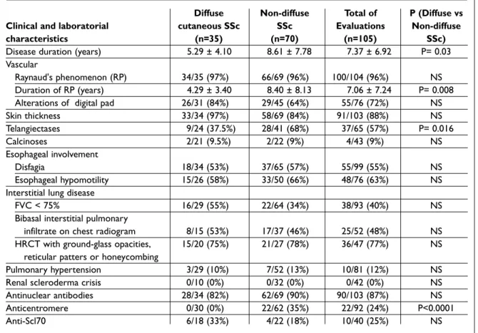
Patients with diffuse cutaneous SSc showed
hi-Table I. Clinical and laboratory characteristics of Systemic Sclerosis (SSc) patients
Diffuse Non-diffuse Total of P (Diffuse vs
Clinical and laboratorial cutaneous SSc SSc Evaluations Non-diffuse
characteristics (n=35) (n=70) (n=105) SSc)
Disease duration (years) 5.29 ± 4.10 8.61 ± 7.78 7.37 ± 6.92 P= 0.03
Vascular
Raynaud's phenomenon (RP) 34/35 (97%) 66/69 (96%) 100/104 (96%) NS
Duration of RP (years) 4.29 ± 3.40 8.40 ± 8.13 7.06 ± 7.24 P= 0.008
Alterations of digital pad 26/31 (84%) 29/45 (64%) 55/76 (72%) NS
Skin thickness 33/34 (97%) 58/69 (84%) 91/103 (88%) NS Telangiectases 9/24 (37.5%) 28/41 (68%) 37/65 (57%) P= 0.016 Calcinoses 2/21 (9.5%) 2/22 (9%) 4/43 (9%) NS Esophageal involvement Disfagia 18/34 (53%) 37/65 (57%) 55/99 (55%) NS Esophageal hypomotility 15/26 (58%) 33/50 (66%) 48/76 (63%) NS
Interstitial lung disease
FVC < 75% 16/29 (55%) 22/64 (34%) 38/93 (40%) NS
Bibasal interstitial pulmonary
infiltrate on chest radiogram 8/15 (53%) 17/37 (46%) 25/52 (48%) NS
HRCT with ground-glass opacities, 15/20 (75%) 21/27 (78%) 36/47 (77%) NS
reticular patters or honeycombing
Pulmonary hypertension 3/29 (10%) 7/52 (13%) 10/81 (12%) NS
Renal scleroderma crisis 0/10 (0%) 0/32 (0%) 0/42 (0%) NS
Antinuclear antibodies 28/34 (82%) 62/69 (90%) 90/103 (87%) NS
Anticentromere 0/30 (0%) 22/62 (35%) 22/92 (24%) P<0.0001
Anti-Scl70 6/18 (33%) 4/22 (18%) 10/40 (25%) NS
L U C Y T S AT O E C O L.
gher vascular deletion scores than those with
non-diffuse disease (1.67±0.91 vs 0.99±0.82; p=0.0005).
In contrast, no statistically significant difference was found between patients with diffuse cutaneous SSc and those with non-diffuse disease concerning
the number of capillary loops/mm (7.75±1.80 vs
8.21±1.67 loops/mm; p=0.225) and the number of
enlarged and giant capillary loops (3.19±2.37 vs
3.46±3.17; p=0.852) (Figure 1). Modified Rodnan’s skin score showed a moderate positive correlation with vascular deletion score (r= 0.52; p<0.001) and a modest negative correlation with the number of capillary loops/mm (r = -0.26; p=0.012).
Patients with presence of finger pad lesions showed higher vascular deletion scores and decrea-sed number of capillary loops/mm than those with absence of finger pad lesions (1.49 ± 0.90 vs 0.86 ±
0.82, p=0.01; 7.71 ± 1.79 vs 8.64 ± 1.15, p=0.03,
res-pectively) (Table II). Among patients with esopha-geal dismotility, we found higher number of enlar-ged and giant capillary loops than those with ab-sence of esophageal dismotility (4.28 ± 3.27 vs 2.82 ± 2.42, p=0.04). No difference was detected in the three NFC parameters analyzed between patients with pulmonary hypertension or interstitial lung disease and patients without these manifestations (Table II).
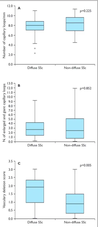
Visceral involvement was studied in 50 patients who had simultaneous records for the five manifes-tations evaluated. These patients were classified into those presenting up to two involved tracts (n=10) and those presenting three or more involved tracts (n=40). Patients with three or more involved tracts showed lower number of capillary loops/mm and higher vascular deletion score than patients with less than three involved tracts (8.00±1.69 vs 9.23±1.31 loops/mm; p=0.025; and 1.41±0.95 vs 0.73±0.76; p=0.027, respectively) (Figure 2). There was no difference in the number of enlarged and gi-ant capillary loops between the two groups
(3.39±2.94 vs 3.24±2.34; p=0.98) (Figure 2).
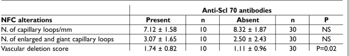
The presence of anti-Scl-70 antibodies was stu-died in 40 patients at the time of the NFC. The vas-cular deletion score was significantly higher in pa-tients with anti-Scl-70 antibodies (1.74±0.82) as compared with patients without anti-Scl-70 antibo-dies (1.11±0.96) (p=0.02). No significant difference was found in the number of capillary loops/ /mm or in the number of enlarged and giant capillary lo-ops between patients with and without anti-Scl-70 antibodies (Table III). No patient with diffuse dise-ase had anti-centromere antibody. Therefore, the
2.0 0.0 12.0 4.0 6.0 Numbe r o f ca pilla ry loo ps /mm10.0 8.0 A Diffuse SSc Non-diffuse SSc p=0.225 * * 2.0 0.0 13.0 1.0 5.0 4.0 3.0 6.0 N. of enla rge d and g iant ca pillar y lo op s 7.0 12.0 11.0 10.0 9.0 8.0 B Diffuse SSc Non-diffuse SSc p=0.852 0,5 0.0 3.5 1.5 1.0 Va scular y de le tio n sco re 2.0 3.0 2.5 C Diffuse SSc Non-diffuse SSc p=0.005
Figure 1. Distribution of the number of capillary
loops/mm (a), number of enlarged and giant capillary loops (b) and, vascular deletion score (c) on nailfold capillaroscopy in patients with diffuse systemic sclerosis (SSc) and non-diffuse SSc.
Box-Plot graph: Rectangles depict 50% of the sample; thick horizontal bar corresponds to median; upper and lower horizontal bars represent highest and lowest figures.The symbol (*) represents outliers. N: number.
association between anti-centromere antibodies and NFC abnormalities was performed only for the non-diffuse SSc patients. Among patients with non-diffuse disease no significant difference in the three NFC parameters was found between the 22 patients with anti-centromere antibodies and the 40 patients without anti-centromere antibodies (Data not shown).
Discussion
The results herein obtained were derived from a large sample of SSc patients retrospectively ana-lyzed for NFC, clinical and laboratory variables. It represents a retrospective analysis gathering the cumulated 15-year experience using the same NFC
protocol in our center.6This NFC protocol has the
advantage of gathering well-established quantita-tive and semiquantitaquantita-tive parameters, such as the number of capillary loops, the vascular deletion score, and the number of enlarged loops analyzed in all 10 fingers of the hands. The present results showed a clear-cut association between the degree of microangiopathic alterations detected by NFC and the extent of cutaneous and visceral involve-ment of the disease. Among the several nailfold microvascular abnormalities observed in SSc the
parameters related to devascularization (vascular deletion scores and/or number of capillary loops/ /mm), but not those related to capillary enlarge-ment, were significantly associated with more ex-tensive skin involvement, diffuse cutaneous SSc, presence of finger pad lesions and with a more ex-tensive visceral involvement. In addition, patients with anti-Scl-70 antibodies had also higher vascu-lar deletion scores.
The microangiopathic abnormalities characte-rized by devascularization and capillary enlarge-ment are specifically observed in SSc and related diseases and tend to occur at early stages of the di-sease, what has granted a place for NFC in the cli-nical investigation of SSc and in the differential
di-agnosis of Raynaud’s phenomenon.21,22This is
es-pecially relevant for the evaluation of patients at early stages of the disease, in which isolated Ray-naud’s phenomenon may be the only clinical ma-nifestation. In that way, NFC has been found to bee especially helpful in distinguishing primary Ray-naud’s phenomenon (functional, not related to
di-sease) from secondary Raynaud’s phenomenon.23
In a meta-analysis study, it has been found that among patients with presumed primary Raynaud’s phenomenon, a secondary disorder developed in
12% of patients.24In patients with Raynaud’s
phe-nomenon NFC can be very helpful to exclude a
se-N A I L F O L D C A P I L L A R O S C O P Y C O R R E L AT E S W I T H C U TA se-N E O U S A se-N D V I S C E R A L I se-N V O LV E M E se-N T I se-N S S C
Table II. Nailfold capillaroscopy (NFC) alterations and correlation with finger pad lesions, esophageal dismotility, pulmonary hypertension and interstitial lung disease
Finger Pad Lesions
NFC alterations Present n Absent n P
N. of capillary loops/mm 7.71 ± 1.79 55 8.64 ± 1.15 21 0.03
N. of enlarged and giant capillary loops 3.89 ± 2.79 55 2.58 ± 2.18 21 NS
Vascular deletion score 1.49 ± 0.90 55 0.86 ± 0.82 21 0.01
Esophagel Dismotility
N. of capillary loops/mm 7.80 ± 1.44 48 8.51 ± 1.74 28 NS
N. of enlarged and giant capillary loops 4.28 ± 3.27 48 2.82 ± 2.42 28 0.04
Vascular deletion score 1.30 ± 0.86 48 1.04 ± 0.80 28 NS
Pulmonary Hypertension
N. of capillary loops/mm 8.93 ± 1.15 10 8.06 ± 1.69 71 NS
N. of enlarged and giant capillary loops 1.82 ± 1.91 10 3.68 ± 2.91 71 NS
Vascular deletion score 1.0 ± 0.82 10 1.31 ± 0.95 71 NS
Interstitial Lung Disease
N. of capillary loops/mm 8.18 ± 1.69 62 8.01 ± 1.62 38 NS
N. of enlarged and giant capillary loops 3.38 ± 2.99 62 3.97 ± 3.50 38 NS
Vascular deletion score 1.28 ± 0.95 62 1.12 ± 0.84 38 NS
condary disorder, since it has a high negative pre-dictive value for the development of a connective tissue disease (>90%). On the other hand, its posi-tive predicposi-tive value is about 50%, which is higher
than any other single screening test.23,24 The
pos-sibility that the NFC abnormalities might also cor-relate with the extent of cutaneous and visceral in-volvement in SSc is intriguing and could have im-plications for the understanding of the disease pa-thophysiology and for the clinical assessment of patients.
The results obtained in the present study con-firm previous reports on a positive correlation between the degree of abnormalities at NFC and the extent of cutaneous and visceral involvement in SSc.11,12,25-27Maricq et al, in 1976, first studied this
issue by analyzing capillary abnormalities in 28 SSc patients, 13 with Raynaud’s phenomenon and
3 with dermatomyositis.11A positive correlation
was found between the degree of microvascular abnormalities and the number of affected systems. In another study of the same group, a positive as-sociation was founded between the presence of the “active” pattern on NFC and more extensive skin involvement. They also presented a higher fre-quency of involvement of all organ systems than
those with a “slow” pattern.12Recently, Ostojic and
Damjanov,25also found more extensive capillary
loss in patients with diffuse cutaneous SSc compa-ring to patients with limited SSc. Also in
accordan-ce with our findings, Bredemeier et al.,15found a
positive correlation between the vascular deletion score in NFC and higher Rodnan’s skin score, pre-sence of anti-Scl-70 antibodies, signs of periphe-ral ischemia, esophageal dysfunction and pulmo-nary disease. On the other hand, some other
stu-dies could not find such association.14,28
Heteroge-neity in patient selection and NFC methodology may contribute to the lack of association observed in the latter studies.
In our study anti-Scl-70 antibodies were deter-mined only in 40 patients at the time of the analy-sis due to non-availability of anti-Scl-70 at the ins-titution. Despite this limitation we found higher vascular deletion scores in patients with anti-Scl--70 antibodies in the sera. The latter association supports a possible relationship between disease severity and NFC abnormalities since this autoan-tibody has been previously demonstrated to be
as-sociated with more severe and extensive disease.29
The analysis of the present series of patients dis-closed epidemiological features similar to those
L U C Y T S AT O E C O L. 2.0 0.0 12.0 4.0 6.0 Numbe r o f ca pilla ry loo ps /mm Number of involvements Number of involvements Number of involvements 10.0 8.0 A
Up to two Three or more
p=0.025 2.0 0.0 10.0 1.0 5.0 4.0 3.0 6.0 N. of enla rge d and g iant ca pillar y lo op s 9.0 8.0 7.0 B
Up to two Three or more
p=0.98 0,5 0.0 3.5 1.5 1.0 Va scular y de le tio n sco re 2.0 3.0 2.5 C
Up to two Three or more
p=0.027
Figure 2. Distribution of patients with up to two
involvements or three or more involvements according the number of capillary loops/mm (a), number of enlarged and giant capillary loops (b) and, vascular deletion score (c) on nailfold capillaroscopy.
Box-Plot graph: Rectangles depict 50% of the sample; thick horizontal bar corresponds to median; upper and lower horizontal bars represent highest and lowest figures. N. number.
registered in the literature in that there was fema-le predominance, disease onset around 40 year old in average, and the predominance of the
cutane-ous limited disease.17,30As for the distribution of
clinical manifestation it is noteworthy that there was no case of renal scleroderma crisis, what may be due to bias of the retrospective study.
It is relevant to point out that the present study represents a retrospective analysis of the cumula-tive 15-year experience with NFC uniformly perfor-med according to the protocol proposed by
An-drade et al.6This protocol uses established
quan-titative and semi quanquan-titative techniques, allowing the concomitant determination of several parame-ters that are strongly associated with SSc, such as the number of capillary loops, the number of en-larged loops and the degree of avascular score. It should also be emphasized that conventional NFC is a simple and low cost method, and therefore fea-sible in any average Rheumatology Division. More recently studies using videocapillaroscopy has
been also published with interesting results.26,31
However it should be noticed that this is a much more expensive method and is restricted to specia-list centers.
Nonetheless, the retrospective design of the study bears some intrinsic limitations. For exam-ple, not all patients followed in our hospital had the necessary records for NFC, clinical and laboratory data within a one-year interval and therefore could not be included in the study. In addition only 50 pa-tients could be included for the analysis of corre-lation of NFC abnormalities and the number of in-volved tracts, since the other ones did not have the required simultaneous records for the five clinical involvements. On the other hand, this particular retrospective analysis is strengthened by the fact that these patients have been followed by the same standard protocol for clinical, laboratory and NFC evaluation.
Despite these limitations, the results obtained
showed a clear-cut association of the degree of NFC microangiopathy with the extension of the cutaneous and visceral involvement as well as with the presence of anti-Scl-70 antibodies. Therefore, it is appropriate to consider the NFC approach for the evaluation of the extension and severity of SSc. Prospective studies with long follow-up are war-ranted to investigate a possible role for nailfold ca-pillary microscopy in the prediction of disease ex-tension and severity.
Acknowledgement
Lucy Tiemi Sato was supported by the National Council for Scientific and Technological Develop-ment (CNPq). This study was partially supported by Research Funds from the Brazilian Society of Rheumatology. The authors state that there is no conflict of interest regarding this manuscript.
Correspondence to
Luís E. C. Andrade
Universidade Federal de São Paulo (UNIFESP), Rheumatology Division
Rua Botucatu 740, 3º andar CEP 04023-062, São Paulo, SP, Brazil E-mail: [email protected]
References
1. LeRoy EC. Systemic Sclerosis. A vascular perspective. Rheum Dis Clin North Am 1996;22:675-694.
2. Campbell PM, LeRoy EC Pathogenesis of systemic sclerosis: a vascular hypothesis. Semin Arthritis Rheum 1975;4:351-368.
3. Dabich L, Bookstein JJ, Zweifler A et al. Digital arter-ies in patients with scleroderma. Arch Intern Med 1972;130:708-714.
4. Maricq HR, LeRoy EC. Patterns of finger capillary ab-normalities in connective tissue disease by “wide-field” microscopy. Arthritis Rheum 1973;16:619-628. 5. Maricq KR, LeRoy EC, D'Angelo WA et al. Diagnostic
potential of in vivo capillary microscopy in scleroder-ma and related disorders. Arthritis Rheum 1980;23:183-189.
6. Andrade LEC, Gabriel Jr. A, Assad RL et al. Panoramic
N A I L F O L D C A P I L L A R O S C O P Y C O R R E L AT E S W I T H C U TA N E O U S A N D V I S C E R A L I N V O LV E M E N T I N S S C
Table III. Nailfold capillaroscopy (NFC) alterations and correlation with anti-Scl 70 antibodies
Anti-Scl 70 antibodies
NFC alterations Present n Absent n P
N. of capillary loops/mm 7.12 ± 1.58 10 8.32 ± 1.87 30 NS
N. of enlarged and giant capillary loops 3.07 ± 1.65 10 2.50 ± 2.43 30 NS
Vascular deletion score 1.74 ± 0.82 10 1.11 ± 0.96 30 P=0.02
nailfold capillaroscopy: a new reading method and normal range. Semin Arthritis Rheum 1990;20:21-31. 7. Maricq HR. Wide-field capillary microscopy. Arthritis
Rheum 1981;24:1159-1165.
8. Cutolo M, Grassi W, Matucci Cerinic M. Raynaud's phenomenon and the role of capillaroscopy. Arthritis Rheum 2003;48:3023-3030.
9. Carpentier P, Franco A, Béani JC et al. Intérêt de la capillaroscopie périunguéale dans le diagnostic pré-coce de la sclérodermie systémique. Ann Dermatol Venereol 1983;110:11-20.
10. Ganczarczyk ML, Lee P, Armstrong SK. Nailfold capil-lary microscopy in polymyositis and dermatomyosi-tis. Arthritis Rheum 1988;31:116-119.
11. Maricq HR, Spencer-Green G, LeRoy EC. Skin capil-lary abnormalities as indicators of organ involve-ment in scleroderma, Raynaud's syndrome and der-matomyositis. Am J Med 1976;61:862-870.
12. Chen Ze-Y I, Silver RM, Ainsworth SK et al. Associa-tion between fluorescent antinuclear antibodies, capillary patterns, and clinical features in scleroder-ma spectrum disorders. Am J Med 1984;77:817-822. 13. Joyal F, Choquette D, Roussin A et al. Evaluation of
the severity of systemic sclerosis by nailfold capillary microscopy in 112 patients. Angiology 1992;43:203--210.
14. Lovy M, MacCarter D, Steigerwald JC. Relationship between nailfold capillary abnormalities and organ involvement in systemic sclerosis. Arthritis Rheum 1985;28:496-501.
15. Bredemeier M, Xavier RM, Capobianco KG et al. Nail-fold capillary microscopy can suggest pulmonary disease activity in systemic sclerosis. J Rheumatol 2004;31:286-294.
16. Masi AT, Rodnan GP, Medsger Jr TA et al. Preliminary Criteria for the classification of systemic sclerosis (scleroderma). Arthritis Rheum 1980;23:581-590. 17. Seibold JR. Scleroderma. In: Kelley WN, Ruddy S,
Harris ED, Sledge CB. Textbook of Rheumatology, 5th ed. WB Saunders, Philadelphia-USA, 1997:1133-1163. 18. Le Roy EC, Black C, Fleischmajer R et al. Scleroderma (systemic sclerosis): classification, subsets and pathogenesis. J Rheumatol 1988;15:202-205.
19. Lee P, Leung F, Alderdice C et al. Nailfold capillary mi-croscopy in the connective tissue diseases: a semi-quantitative assessment. J Rheumatology 1983;10: 930-938.
20. Brennan P, Silman A, Black C et al. Reliability of skin involvement measures in scleroderma. The UK Scle-roderma Study Group. Br J Rheumatol 1992;31:457--460.
21. Maricq HR, Harper FE, Khan MM. Microvascular ab-normalities as possible predictors of disease subsets in Raynaud's phenomenon and early connective tis-sue disease. Clin Exp Rheum 1983;1:195-205. 22. Priollet P, Vayssairat M, Housset E. How to classify
Raynaud,s phenomenon. Long-term follow-up study of 73 cases. Am J Med 1987;83:494-498.
23. Cortes S, Cutolo M. Capillaroscopic patterns in rheumatic diseases. Acta Reumatol Port 2007;32:29--36.
24. Spencer Spencer-Green G. Outcomes in primary Raynaud phenomenon. A meta-analysis of the fre-quency, rates, and predictors of transition to sec-ondary diseases. Arch Intern Med 1998;158:595-600. 25. Ostojic P, Damjanov N. Different clinical features in
patients with limited and diffuse cutaneous systemic sclerosis. Clin Rheumatol 2006;25:453-457.
26. Cutolo M, Sulli A, Pizzorni C et al. Nailfold videocap-illaroscopy assessment of microvascular damage in systemic sclerosis. J Rheumatol 2000;27:155-160. 27. Houtman PM, Kallenberg CG, Wouda AA et al.
De-creased nailfold capillary density in Raynaud's phe-nomenon: a reflection of immunologically mediated local and systemic vascular disease? Ann Rheum Dis 1985;44:603-609.
28. Statham BN, Rowell NR. Quantification of the nail fold capillary abnormalities in systemic sclerosis and Raynaud's syndrome. Acta Derm Venereol 1986;66:139-143.
29. Giordano M, Valentini G, Migliaresi S et al. Different antibody patterns and different prognoses in pa-tients with scleroderma with various extent of skin sclerosis. J Rheumatol 1986;13:911-916.
30. Medsger TA Jr, Masi AT. The epidemiology of sys-temic sclerosis (scleroderma). Ann Intern Med 1971;74:714-721.
31. Ingegnoli F, Gualtierotti R, Lubatti C et al. Feasibility of Different Capillaroscopic Measures for Identifying Nailfold Microvascular Alterations. Semin Arthritis Rheum In Press, Available online, December 2007.