p3 – InTegraTed longITudInal analysIs InCreases preCIsIon and reduCes bIas: a ComparaTIve 5-year analysIs In The desIr CohorT
Alexandre Sepriano1, 2, Sofia Ramiro1, Robert Landewé3, Dougados M4, Pascal Claudepierre5, Antoine Feydy6, Monique Reijnierse7, Damien Loeuille8,
Désirée van der Heijde1
1. Rheumatology, Leiden University Medical Center, Leiden, Netherlands
2. CEDOC, NOVA Medical School, Nova University, Lisboa, Portugal
3. Amsterdam Rheumatology and Immunology Center (ARC), Amsterdam, Netherlands
4. Department of Rheumatology, Cochin Hospital, Paris, France
5. Radiology, Hôpital Henri-Mondor, Créteil, Paris, France 6. Radiology, Cochin Hospital, Paris, France
7. Radiology, Leiden University Medical Center, Leiden, Netherlands
8. Rheumatology, Hospital Brabois, Nancy, France
Background: Evaluation of imaging is important in
spondyloarthritis (SpA) research, but loss to follow up often jeopardizes interpretation of the evaluation. The Interpretation may further be challenged by the fact that often different readers have contributed to scores, in multiple read ‘waves’. A common approach is to eva-luate patients (pts) with complete follow up (comple-ters analysis), and aggregate scores of individual readers (eg. agreement ≥ 2 out of 3 reareaders). These approa -ches are not assumption-free, may cause non-random data loss, and may as such provide spurious estimates and loss of external validity.
Objectives: We aimed to investigate if the use of all
data in an assumption-free manner (a so called ‘inte-grated analysis’) affects the precision of estimates for imaging outcomes in pts with axial SpA (axSpA), with completers analysis as reference standard.
Methods: Pts from the DESIR cohort fulfilling the ASAS
axSpA criteria were included. Radiographs and MRIs of the SIJ and spine were obtained at baseline (BL), 1, 2 and 5 years. Each film was scored by 2 or 3 readers in
Posters
ACTA REUMATOL PORT. 2018:43:47-141 (SUP)
BME – bone marrow edema; CI – confidence interval; N – number
Table I. Change per year In The perCenTage of posITIve Cases for bInary ImagIng ouTComes over 5-years of follow-up, aCCordIng To 3 dIfferenT
analyTICal meThods, In early axspa paTIenTs fulfIllIng The asas axspa CrITerIa from The desIr-CohorT
3 ‘reading-waves’ (wave 1: BL only; wave 2: BL, 1, 2 years; wave 3: BL, 2, 5 years). Each outcome was ana-lyzed in two ways: i. according to a ‘combination algo-rithm’ (‘2 out of 3’ for binary and mean of 3 readers for continuous variables); and ii. per individual reader. The change of each outcome was analyzed by generalized estimating equations (GEE)) with ‘time’ as explanato-ry variable. Three analytical approaches were pursued: i) ‘integrated-analysis’ (including all pts with ≥1 score from ≥1 reader from all waves); ii) completers--only analysis (including only pts with complete 5-year fol-low-up, using scores from individual readers from wave 3); iii) aggregated completers analysis using a combi-nation algorithm (the same as ii but using combined scores).
Results: In total, 413 pts were included (mean (SD)
symptom duration: 1.6 (0.9) years) and 366 comple-ted the 5-year follow up. An analysis with all data from different readers and ‘waves’ (‘integrated analysis’) was more inclusive, but did not result in a meaningful loss of precision (width of 95%CIs) of the change-estima-tes as compared to both completers analyses (table). In fact, for low-incident outcomes (e.g. % of mNY-positi-ve omNY-positi-ver 5-years), a similar incidence was ‘captured’, with more precision, by the ‘integrated analysis’ com-pared to the completers analysis with combined scores (% change/year (95% CI): 1.1 (0.7; 1.5) vs 1.2 (0.5; 1.8), respectively). The same results were seen using continuous outcomes.
Conclusions: An efficient and entirely assumption-free
usage of all data from different readers and ‘read-waves’ does not compromise precision of the estimates of change in imaging parameters, and may yield increa-sed statistical power for detecting changes with low in-cidence. In addition, integrated analysis may protect against attrition bias and avoid bias by ‘convenient choices’.
p4 – whICh ImagIng ouTComes for axspa are mosT sensITIve To Change? a 5-year analysIs of The desIr CohorT Alexandre Sepriano1, 2, Sofia Ramiro1,
Robert Landewé3, Dougados M4, Pascal Claudepierre5, Antoine Feydy6, Monique Reijnierse7, Damien Loeuille8, Désirée van der Heijde1
1. Rheumatology, Leiden University Medical Center, Leiden, Netherlands
2. CEDOC, NOVA Medical School, Nova University, Lisboa, Portugal
3. Amsterdam Rheumatology and Immunology Center (ARC), Amsterdam, Netherlands
4. Department of Rheumatology, Cochin Hospital, Paris, France
5. Radiology, Hôpital Henri-Mondor, Créteil, Paris, France 6. Radiology, Cochin Hospital, Paris, France
7. Radiology, Leiden University Medical Center, Leiden, Netherlands
8. Rheumatology, Hospital Brabois, Nancy, France
Background: Several imaging outcomes have become
available to assess inflammation and structural da -mag e over time in patients with axial spondyloarthris (axSpA). However, no formal comparison of their sen-sitivity to change has been made in the early phases of the disease.
Objectives: We aimed to compare the sensitivity to
change of different MRI and radiographic scoring me -thods in patients with early axSpA.
Methods: Patients from the DESIR cohort fulfilling the
ASAS axSpA criteria were included. Radiographs and MRI of the sacroiliac joints and spine were obtained at baseline, 1 year, 2 years and 5 years. Each film was scored by 2 or 3 readers in 3 ‘reading-waves’ (wave 1: baseline; wave 2: baseline, 1 year, 2 years; wave 3: base-line, 2 years, 5 years). Outcomes measuring inflam-mation and structural damage both on MRI and radio-graphs in the spine and SIJ were assessed (Table). The analysis of change captured over time was performed using generalized estimating equations (GEE) longitu-dinal models separately for each outcome, taking into account data from all readers and waves (‘integrated analysis’). To allow comparisons across outcomes, these were standardized (difference between the individual score and the mean of all scores divided by the standard deviation, per reader, wave and time-point) before run-ning the models. The higher the standardized coeffi-cient the more change in inflammation/damage is captu red.
Results: In total, 345 patients were included (mean
(SD) symptom duration: 1.6 (0.9) years; 53% males; 89% HLA-B27 positive). Inflammation on MRI-SIJ (accor ding to both the ASAS definition of sacroiliitis and the continuous SPARCC score) was more sensitive to change as compared to inflammation on the spine that remained essentially unchanged regardless of the outcome (table). Structural damage on the SIJ was found to increase over time, but with a higher stan-dardized yearly rate of change on MRI-SIJ (range: 0.015-0.274) as compared to X-SIJ (range:
0.043--0.126). Notably, ≥3 Fatty lesions on MRI-SIJ was the structural outcome in the SIJ with highest sensitive to change (0.274), while ≥3 erosions was the least sensi-tive (0.015). Spine structural damage slowly progressed over time but, in contrast to SIJ, radiographic outcomes (i.e. ≥ 1 syndesmophytes and mSASSS) were more sen-sitive to change than MRI structural outcomes.
Conclusion: Our data adds to the body of evidence
showing that structural damage assessed in pelvic ra-diographs only has low sensitivity to change. MRI-SIJ is a promising alternative (especially fatty lesions) captu ring more structural changes. In contrast, in de-tecting structural change in early axSpA radiographic outcomes outperform MRI outcomes.
p9 – anTIbody profIle and sysTemIC sClerosIs ClInICal feaTures – myTh or realITy?
Ana Catarina Duarte1, Ana Cordeiro1,
Maria José Santos1, José António Canas da Silva1
1. Rheumatology Department, Hospital Garcia de Orta, Almada, Portugal
Background: Antinuclear antibodies(ANA) occur in
80-98% of systemic sclerosis(SSc) patients (pts), with different specificities. Anticentromere antibody(ACA), antitopoisomerase I(anti-Scl70) and anti-RNA poly-merase III are the commonest and are included in the new SSc ACR/EULAR classification criteria. According to literature, ANA specificities are associated with clini -cal features of the disease.
Objectives: Evaluate the relationship between
anti-body profile and clinical manifestations in a cohort of SSc pts.
Methods: We conducted a retrospective analysis of SSc
pts followed in our department. Demographic data, disease duration, ANA specificities and clinical mani-festations were collected. Mann-Whitney U test and Chi-square were used for comparisons between pts who tested positive or negative for different ANA speci-ficities.
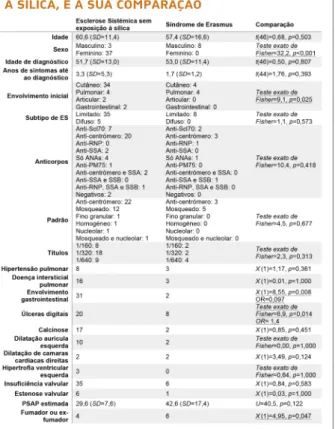
Results: In total, 117 pts were included, 91.5% female
with mean age of 60.7±15.2 years and mean disease duration of 11.9±10.7 years. Seventy-five pts (64.1%) had limited cutaneous SSc(lcSSC), 26(22.2%) diffuse cutaneous SSc(dcSSc), 8(6.8%) very early diagnosis SSc, 7(6%) overlap syndromes and 1(0.9%) SSc sine scleroderma. Most (92.3%) were ANA positive, with 53.8% having ACA, 26.5% anti-Scl70, 3.4% anti-U3 RNP, 2.6% anti-U1 RNP, 1.7% anti-PM/Scl and 0.9% anti-RNA polymerase III and 0.9% anti-Th/To.
Positivity for ACA was significantly associated with female gender (OR: 1.18 95%CI 1.04-1.34) and lcSSc phenotype (OR: 9.43 95%CI 3.86-23.03). ACA was also associated with older age at disease onset (p=0.008). Vascular involvement, defined by cur-rent/previous digital ulcers and/or telangiectasias, was also more prevalent in this group (OR: 5.59 95%CI 2.47-12.66). Pulmonary arterial hypertension (group 1 ERS/ECS 2013 classification) was present in 6.3% of pts with ACA. Oesophageal involvement was the se -cond commonest manifestation and occurred in 57.1% of pts with ACA, although this association was not sta-tistically significant. ACA seemed to have a protective effect for interstitial lung disease (ILD) (OR: 0.027 Table I. sTandardIzed raTe of Change of
ImagIng ouTComes over 5 years of follow-up In early axspa paTIenTs from The desIr-CohorT who fulfIl The asas axspa ClassIfICaTIon CrITerIa
95%CI 0.004-0.213).
Anti-Scl70 positivity was associated with dcSSc phe-notype (OR: 9.29 95%CI 3.26-26.5) and ILD (OR: 10.39 95%CI 3.86-27.92).
From the 4 pts with anti-U3 RNP, 3 had dcSSC subty pe. The only patient with renal manifestations was anti-U3 RNP positive and had rapidly progressive cutaneous involvement.
Anti-U1 RNP was associated with muscle-skeletal ma nifestations (OR: 10.7 95%CI 0.92-20.44) and with overlap syndromes (OR: 15.2 95%CI 4.7-29.1).
Pts with anti-Th/To and anti-RNA-polymerase III had lcSSc subtype. Vascular manifestations, oeso -phageal involvement and calcinosis cutis were the main manifestations, respectively.
Table 1 shows detailed clinical manifestations and antibody profile.
Conclusions: In our cohort, ACA and anti-Scl70 were
the commonest antibodies and were associated with lcSSC and dsSSc phenotype, respectively. ACA posi-tivity conferred a higher risk of vascular disease and had a protective effect for ILD, while anti- Scl70 was as-sociated with ILD.
Pts with anti-U1 RNP and anti-PM/Scl had mainly muscle-skeletal manifestations.
This study confirms an association between im-munological profile and clinical manifestations, rein-forcing the importance of antibody profile and raising awareness for possible disease complications. Larger national studies would be desirable, specially for a better understanding of major organ involvement associa ted with least common antibodies.
p10 – o paradIgma das neuropaTIas perIfÉrICas nas doenÇas reumÁTICas sIsTÉmICas – doIs anos em revIsTa Ana Catarina Duarte1, Sandra Sousa1, Ana Cordeiro1
1. Serviço de Reumatologia, Hospital Garcia de Orta, Almada, Portugal
Introdução: A neuropatia periférica (NP) está descrita
em várias doenças reumáticas sistémicas (DRS), po-dendo ser causada por vasculite, compressão ou toxi-cidade farmacológica. A síndrome de Sjögren (SS) é a DRS com maior prevalência de NP.
Objetivos: Partilhar a experiência do nosso centro no
diagnóstico e tratamento da NP nas DRS nos últimos 2 anos.
Material e métodos: Incluímos doentes internados no
serviço de Reumatologia ou observados por esta espe-cialidade durante o internamento noutros serviços por NP em contexto de DRS, em 2016 e 2017. Foram ex-cluídos os que apresentavam apenas neuropatia por compressão ou toxicidade farmacológica.
Foram colhidos dados demográficos e clínicos, in-cluindo DRS subjacente e respetiva duração, relação temporal entre o diagnóstico da doença e da NP, o tipo de NP, o tratamento instituído e a resposta obtida.
Resultados: Foram identificados 9 doentes, 2 homens e
7 mulheres, com idade média 67,4 ± 7,4 anos. Dois ti-nham diagnóstico de SS e 7 de vasculite de pequenos va-sos (1 poliangeíte microscópica, 2 granulomatose com po-liangeíte e 4 granulomatose eosinofílica com popo-liangeíte). Nos doentes com SS, a DRS foi identificada no de-correr da investigação etiológica da NP (em média 90,5 meses depois), enquanto nos restantes a NP foi parte do quadro inicial, à exceção de 1 em que se manifestou 6 meses após o diagnóstico de vasculite.
O eletromiograma (EMG) inicial demonstrou uma mononeuropatia múltipla (MM) nos doentes com vas-culite e polineuropatia (PNP) sensitiva e sensitivo-mo-tora nos doentes com SS.
Todos os doentes foram medicados com predniso-lona (PDN;1mg/kg/dia), na sua maioria em associação com outros fármacos, incluindo ciclofosfamida (CYC), rituximab (RTX) ou imunoglobulina humana endove-nosa (IVIG). Em 8 dos 9 doentes verificou-se uma me-lhoria sintomática, corroborada em EMG, quando dis-ponível. Duas das doentes medicadas com CYC apre-sentaram eventos adversos graves, uma com neutrope-nia febril e outra com sépsis de ponto de partida abdominal, tendo ambas falecido. O tipo de NP, bem como o tratamento instituído e a resposta obtida en-Table I, ClInICal feaTures aCCordIng To
contram-se apresentados na Tabela 1.
Discussão/Conclusões: A NP está descrita em várias
DRS, podendo ser a primeira/única manifestação da doença. A MM é a apresentação mais comum (35--65%), seguida da PNP simétrica, distal.
Uma história clínica cuidada é essencial, sobretudo no que diz respeito a outras comorbilidades, e tera-pêuticas em curso. O EMG permite confirmar a pre-sença de NP e avaliar a sua gravidade; a biópsia de ner-vo deve ser feita em caso de dúvida diagnóstica.
Em 7 dos 9 doentes apresentados a NP fez parte da clínica inicial, embora em alguns o diagnóstico da DRS tenha sido feito meses/anos após a NP. Deste modo, a va-lorização das queixas de parestesias, dor neuropática ou fraqueza muscular referidas pelos doentes é essencial.
A PDN foi prescrita a todos os doentes, na maioria dos casos em associação com outras terapêuticas de in-dução, nomeadamente CYC, RTX ou IVIG. A azatio-prina foi usada como terapêutica de manutenção em 2 doentes. A escolha do fármaco teve por base as caracte -rísticas da doença e do doente, nomeadamente o
con-texto infecioso (IVIG foi usada em doentes coloniza-dos por microrganismos multirresistentes). Os doentes medicados com CYC ou IVIG obtiveram uma melho-ria clinica e, quando disponível, em EMG. Os doentes sob RTX têm à data pouco tempo de “follow-up” para se poder tirar conclusões de eficácia, embora este fár-maco seja considerado uma alternativa no tratamento das vasculites anticorpo anti-citoplasma do neutrófilo positivo, com a mesma eficácia que a CYC.
p11 – naIlfold CapIllarosCopy In sysTemIC sClerosIs – sIx years In revIew
Ana Catarina Duarte1, Ana Cordeiro1, Maria José Santos1
1. Rheumatology Department, Hospital Garcia de Orta, Almada, Portugal
Background: Microvascular dysfunction is a dynamic
process that plays a central role in systemic sclerosis’ (SSc) pathogenesis.
Tabela I. resumo das CaraCTerÍsTICas dos doenTes Com dIagnÓsTICo de neuropaTIa perIfÉrICa
Legenda: PDN – prednisolona; RTX – rituximab; CYC – ciclofosfamida; AZA – azatioprina; IVIG – imunoglobulina endovenosa; EMG – eletromiograma
Nailfold capillaroscopy (NCP) is a rapid, non-invasive exam that can illustrate the early capillary changes in SSc and monitor their evolution. It is an extremely useful tool in daily clinical practice and that has been recognized in 2013ACR/EULAR classification criteria for SSc.
Objetives: To evaluate the prevalence and the
evolu-tion of NCP scleroderma pattern in SSc patients and analyse possible associations with their disease-phe-notype.
Methods: NCP of all SSc patients followed in our
cen-tre were reviewed and clinical and demographic fea-tures were collected.
A descriptive analysis was performed and nonpara-metric tests were used to compare patients with and without specific SSc pattern.
Results: In total, 70 out of 117 SSc patients had at least
1 NCP available during the last 6 years. Most of these patients (62.9%) had limited cutaneous SSc, 21.4% dif-fuse cutaneous SSc, 11.4% very early diagnosis SSc and 4.3% overlap syndromes; mean disease duration was 10.7 ± 9.6 years.
At the moment of the NCP first evaluation, 46
pa-tients (39.4%) had a scleroderma pattern, 12 (10.3%) had non-specific (NS) NCP abnormalities and 12 (10.3%) had a normal NCP. During the 6 years follow--up, NCP changed in 5 patients (7.1%) as illustrated in figure 1. However, none of these patients had con-comitant development/worsening of other clinical manifestations.
At the end of the follow-up period, 49 patients (70%) had a NCP with a scleroderma pattern. From these, 13 (26.5%) had an early pattern, with one of them having a previous normal NCP and other a NS NCP; the mean time of progression was 11 and 34 months, respectively. The active pattern was present in 21 patients (42.8%), with 1 of them having a previous NS NCP 10 months before, and the late pattern was described in 12 patients (24.5%), with only one of them having a previous early pattern 20 months before (this patient had a diffuse cutaneous SSc subtype). Three patients had abnormalities that could be framed into an active/late pattern.
When comparing patients with and without sclero-derma specific patterns (table 1), the presence of scle-roderma pattern was associated with the presence of current/previous digital ulcers (OR 1.49 95%CI 1.17--1.92). However, this difference was not confirmed be-tween the different scleroderma patterns.
Regarding, major organ involvement, although there were no statistical differences between both groups, pa-tients with scleroderma pattern had a higher prevalence of oesophageal involvement.
Conclusions: This study demonstrates how NCP can
be useful in illustrating the dynamic vascular damage that occurs in SSc.
In our data, a NCP scleroderma pattern was signifi-cantly associated with a higher number of digital ul-cers and these patients had a higher percentage of oe-sophageal involvement.
In daily clinical practice, NCP is useful not only for corroborating the diagnosis of SSc, but also for moni-toring endothelial injury with consequent potential macrovascular/systemic damage. Although our sample is too small to demonstrate possible associations be-tween specific NCP alterations and internal organ in-volvement, some studies have already identify NCP patterns as predictive factors for severe organ damage. p12 – InflammaTIon on mrI of spIne and saCroIlI1In axIal spondyloarThrITIs: The 5 years daTa of The desIr CohorT Alexandre Sepriano1, 2, Sofia Ramiro1,
fIgure 1.Progression of naifold capillaroscopy alterations during follow-up
Table I. ComparIson beTween paTIenTs wITh and wIThouT sCleroderma paTTern
Legend – NCP: nailfold capillaroscopy; ILD: interstitial lung disease; ANAs: antinuclear antibodies; ACA: anticentromere antibody; Scl 70: antitopoisomerase I
Robert Landewé3, Dougados M4, Désirée van der Heijde1
1. Rheumatology, Leiden University Medical Center, Leiden, Netherlands
2. CEDOC, NOVA Medical School, Nova University, Lisboa, Portugal
3. Amsterdam Rheumatology and Immunology Center (ARC), Amsterdam, Netherlands
4. Department of Rheumatology, Cochin Hospital, Paris, France
Background: The effect of local inflammation on
struc-tural damage in patients (pts) with axial spondy-loarthritis is not well known.
Objectives: We aimed to test the possible effect of
in-flammation on structural damage both assessed by MRI and at the level of the spine and the SIJ.
Methods: Pts with recent onset (≤3 years) axSpA
(accor ding to the treating rheumatologist) from the DE-SIR cohort were included. MRI of the SIJ (MRI-SIJ) and spine (MRI-spine) were obtained at baseline (BL), 2 and 5 years and scored by 3 trained central readers un-aware of the chronology. Bone Marrow Edema (BME) at MRI-SIJ was assessed according to ASAS definition and at the MRI-spine by the presence of ≥ 3 lesions. Structural damage in the SIJ (MRI-SIJ-STR) and in the spine (MRI-spine-STR) was defined by ≥ 3 fatty lesions. The % of structural net progression (number of ‘pro-gressors’ minus the number of ‘re‘pro-gressors’ divided by the total number of pts) was assessed in subgroups ac-cording to CRP and BME status at BL. The effect of BME on MRI-SIJ on MRI-SIJ-STR and of BME on MRI-spine on MRI-spine-STR was evaluated using two types of binomial generalized estimating equations (GEE) mo -dels: i. effect at BL on 5 years incorporating measure-ments from all readers (GEE adjusted for reader); ii.
ef-fect of BME over 5 years (longitudinal time-lagged models with auto-regression). The final models were adjusted for variables proved to confound the associa-tion of interest (variables tested: age, gender, HLA-B27, smoking status, CRP, BASDAI, ASDAS, treatment with NSAIDs and TNFi).
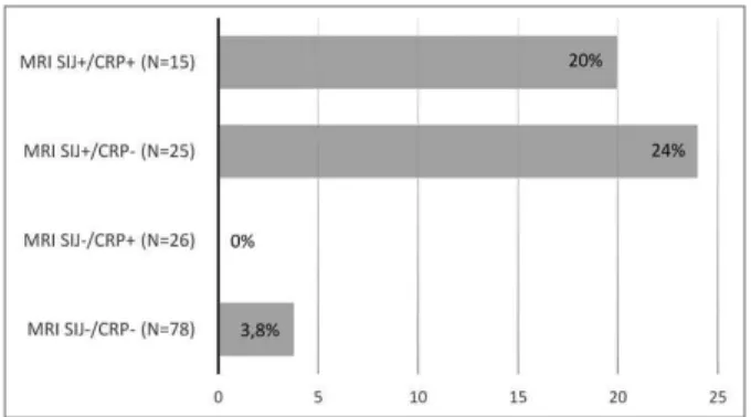
Results: In total, 151 and 145 pts had complete 5-year
MRI-SIJ and MRI-spine data available from 3 readers, respectively. Of the 151 pts with complete MRI-SIJ data, the net % pts who switched from MRI-SIJ-STR negative to positive ranged from 3.8% to 24% accor -ding to the presence of objective signs of inflammation at BL (Figure 1). Low number of pts did not allow for simi lar analysis in the spine. In the multivariable ana -lysis, both the presence of BME at MRI-SIJ (OR=4.2 [95% CI: 2.4-7.3]), and BME at MRI-spine (OR=8.9 [95% CI: 2.1-38.7]) at baseline were highly predictive of MRISIJ and MRIspine structural progression res -pectively 5 years later, adjusting for CRP (only factor found to confound the association of interest). Similar positive associations were found in the longitudinal models testing the effect of BME on MRI-SIJ-STR and
Table I. effeCT of InflammaTIon on mrI (asas defInITIon of saCroIlIITIs and bme In The spIne) on bInary mrI sTruCTural ouTComes
fIgure 1.Net progression from MRI-SU-STR negative to MRI-SIJ-STR positive (≥3 fatty lesions) according to baseline objective inflammatory markers
MRI-spine-STR over 5 years (table).
Conclusion:Our results show that local inflammation
is strongly associated with the development of struc-tural damage over 5 years both in the SIJ and spine in early axSpA and that this effect is independent of sys-temic inflammation.
p19 – renal and overall survIval analysIs In a CohorT of paTIenTs wITh lupus nephrITIs wITh up To 40 years of follow up
Filipa Farinha1, Brett Sydney Bernstein1,
Ruth Pepper2, David A Isenberg1, Anisur Rahman1
1. Centre for Rheumatology, University College of London, London, United Kingdom
2. Centre for Nephrology, University College London, London, United Kingdom
Background: Although the prognosis has improved in
the last decades, Lupus patients still have a 3-fold in-crease in mortality, compared with the general popu-lation1. Lupus nephritis (LN) is one of the most severe
manifestations of this complex systemic disease, occur -ring in up to 60% of patients.
Objectives: 1) To obtain the overall and renal survival
curves for a LN cohort; 2) To investigate factors affect-ing survival; 3) To identify the causes of death in this cohort.
Methods: Single-centre retrospective observational
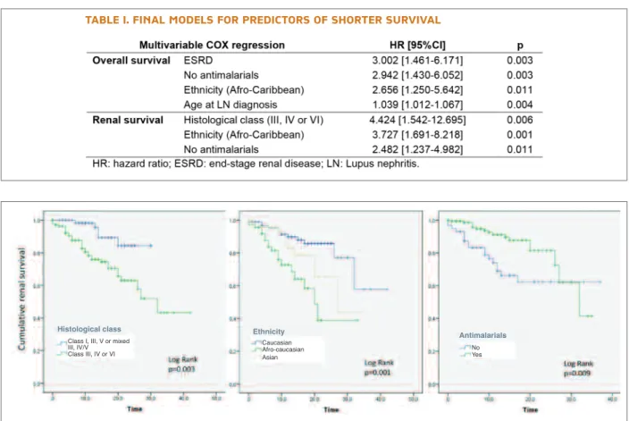
study, including all patients with biopsy-proven LN, followed at our Rheumatology department from 1975 to 2017. Individual clinical files were reviewed to obtain demographic, clinical, laboratory and patho-logical data. We also recorded data on treatment with corticosteroids, immunosuppressants and antimala -rials. We analysed overall survival and renal survival through the Kaplan-Meier method. COX regression analyses were conducted to investigate possible pre-dictors of shorter survival. Significance level was de-fined at 0.05.
Results: 209 patients were included (90% females),
with diverse ethnic background – 44% Caucasian, 33% Afro-Caribbean and 23% Asian. There were 38 deaths during the time of follow-up. Cumulative survival at 5,
Class I, III, V or mixed III, IV/V Class III, IV or VI Caucasian No Yes Afro-caucasian Asian
Histological class Ethnicity
Antimalarials Table I. fInal models for predICTors of shorTer survIval
10, 15 and 20 years after the diagnosis of LN was 92%, 86%, 81% and 76%, respectively. Main causes of death were infection (29%), malignancy (21%) and cardio-vascular (21%). Regarding progression to end-stage re-nal disease (ESRD), which occurred in 40 patients, cu-mulative renal survival at 5, 10, 15 and 20 years was 94%, 86%, 79% and 72%, respectively. Table 1 shows the predictors of shorter survival identified for this co-hort. Image 1 represents the Kaplan-Meier curves ac-cording to the factors affecting renal survival.
Conclusions: Cumulative survival rates and causes of
death for this cohort are comparable with other cohorts of LN2. ESRD confers the higher risk for death; African or Caribbean ethnicities and not taking antimalarials predict shorter overall and renal survival among these patients.
referenCes
1. Yurkovich M, Vostretsova K, Chen W, Avina-Zubieta JA. Overall and cause-specific mortality in patients with systemic lupus erythematosus: a meta-analysis of observational studies. Arth-ritis Care Res (Hoboken). 2014;66(4):608-616.
2. Siso A, Ramos-Casals M, Bove A, Brito-Zeron P, Soria N, Nardi N, et al. Outcomes in biopsy-proven lupus nephritis: evaluation of 190 white patients from a single center. Medicine (Baltimo-re). 2010;89(5):300-307.
p21 – arTrITe reumaTÓIde vs polImIalgIa reumÁTICa – ComparaÇÃo das
alTeraÇÕes eCogrÁfICas em doenTes Com omalgIa bIlaTeral de novo João Pedro Freitas1, Flávio Campos Costa1, Mary Lucy Marques1, Sara Serra1, João Rovisco1
1. Rheumatology Department, Centro Hospitalar e Universitário de Coimbra, Coimbra, Portugal
Introdução: O ombro doloroso é uma das condições
mais comuns em Reumatologia, sendo responsável por um grande número de referenciações de consulta. A omalgia pode ter uma origem periarticular ou articular e pode estar presente numa grande variedade de doen-ças inflamatórias como a Polimialgia Reumática (PMR), Artrite Reumatoide (AR) e patologia degenerativa.
Dor e rigidez da cintura escapular com afetação dos ombros bilateralmente é uma das principais caracte-rísticas da PMR. Diferentes estudos de imagem de-mostraram a existência de bursite subacromio-subdel-toidea (SAD) em associação com sinovite da gleno-umeral (GU) e tenossinovite da cabeça longa do bici-pite (LB) em doentes com PMR. No entanto, estes achados podem também estar presentes em doentes com AR, nomeadamente no idoso e não devem ser
usa-dos para diferenciar estas duas patologias. Alguns doentes com PMR e AR podem apresentar queixas de ombro doloroso bilateral durante flares. Os estudos de ecografia nestas situações são escassos.
O Objetivo do estudo foi avaliar retrospectivamen-te os achados ecográficos das estruturas anatómicas afe-tadas aquando de um novo episódio de ombro doloro-so bilateral, em doentes com PMR e AR e comparar os achados entre estes dois grupos de doentes.
Métodos: Foram avaliados retrospectivamente os
re-latórios de ecografia de doentes com o diagnóstico clí-nico de AR e PMR com omalgia bilateral, de novo, num serviço de Reumatologia entre 2013 e 2016. Foram re-colhidos os seguintes achados numa avaliação dicotó-mica (presença /ausência): Bursite SAD; Tenossinovite LB; Sinovite GU; Tendinopatia ou ruptura parcial/total da coifa dos rotadores. As variáveis dicotómicas foram descritas em percentagens e foram comparadas pelo teste qui-quadrado de Pearson.
Resultados: Foram incluídos 17 doentes com PMR e
17 doentes com AR. Bursite SAD e tenossinovite da lon-ga porção do bicipite unilateral foram mais frequentes em doentes com AR quando comparados com doentes com PMR (p>0,05). Nenhum doente apresentou sino-vite gleno-umeral bilateral. Apenas três apresentaram sinovite gleno-umeral unilateral, todos com AR (p>0,05), quando comparado com os doentes com PMR. Bursite SAD bilateral foi mais frequente em doen-tes com PMR comparado com doendoen-tes com AR (p<0,05), assim como a tenossinovite da longa porção do bicipite (p<0,05).
Conclusão: Este trabalho demonstra algumas
diferen-ças ecográficas no envolvimento inflamatório entre doentes com AR e doentes com PMR com omalgia bi-lateral de novo. A patologia periarticular bibi-lateral (te-nossinovite da longa porção do bicípite e bursite SAD) foi mais frequente em doentes com PMR (p<0,05) e o envolvimento intra-articular mais comum em doentes com AR, embora não se tenha atingido significado es-tatístico.
p23 – early versus laTe-onseT sysTemIC sClerosIs: are There ClInICal and
ImmunologICal dIfferenCes?
Mariana Luis1, Flávio Campos Costa1, Anália Carmo2, João Ferreira3, Tânia Santiago1, Rosário Cunha2, Maria João Salvador1, JAP da Silva1
1. Rheumatology Department, Centro Hospitalar e Universitário de Coimbra, Coimbra, Portugal 2. Clinical Pathology Department, Centro Hospital e
Universitário de Coimbra, Coimbra, Portugal
3. Cardiology Department, Centro Hospital e Universitário de Coimbra, Coimbra, Portugal
Background: The clinical course of Systemic Sclerosis
(SSc) depends on subtype, organ involvement and age. Peak age at onset of SSc is between 30 and 50 years, al-though SSc may also start in both young and elderly pa-tients. Few data have been reported on patients suffer-ing from late-onset SSc.
Objective: To characterize clinical and immunological
features of early and late-onset SSc in a tertiary referral hospital.
Methods: We analyzed data from 178 patients followed
at our SSc clinic. All the patients fulfilled the ACR/EU-LAR 2013 classification criteria for SSc or the LeRoy’s criteria for the classification of early SSc.
Based on the mean of age of onset of the whole se-ries (50±15 years), ages extremes were defined as younger than 35 versus older than 65 years of age at on-set. Disease characteristics as well as clinical and im-munological features were evaluated.
Results: The early and the late-onset groups included
35 and 31 patients, respectively. Patients’ current mean age was 42.8±14.1 vs. 75.8±6.2 with a mean disease duration of 14.5±14.7 vs. 4.3±4.6 years. The most common first manifestation of disease was Raynaud phenomena followed by arthritis/inflammatory arthral-gia, in both groups. However, the time between clini-cal onset and SSc diagnosis was higher in the late-on-set group (p=0.034). A higher number of diffuse and preSS was observed in the early group but this diffe -rence didn’t prove statistically significant. There was a higher prevalence of centromere antibodies in the late--onset group (p=0.001). Clinical manifestations and target-organ damage didn’t differ between groups, ex-cept for a higher prevalence of heart conduction abnorma lities in the late-onset group (p=0.02). In mul-tivariate analyses, age alone (OR=1.04; 95%CI 1.0, 1.1), but not disease duration (OR=0.99; 95%CI 0.9--1.0), was an independent predictor for the presence of heart conduction abnormalities.
Conclusions: In line with findings from other studies,
late-onset SSc shows a distinct clinical and immuno-logical presentation. The present study confirms that late-onset is associated with longer diagnostic delay, positive centromere and heart conduction abnormali-ties. These observations may be due to age and poten-tial age-associated confounders, rather than the disease itself. Knowledge of these different characteristics can
help to improve the management of the disease.
referenCes
Alba M et al. Early-versus Late-Onset Systemic Sclerosis. Medicine. 2014; 93(2):73-81
Hugle T et al. Late-onset systemic sclerosis - a systemic survey of the EULAR scleroderma trials and research group database. Rheu-matology (Oxford). 2010. 50(1):161-5.
Manno R et al. Late-Age Onset Scleroderma. J. Rheumatol. 2011; 38(7): 1317–1325.
p24 – sysTemIC sClerosIs:
gender-assoCIaTed dIfferenCes In ClInICal and serologICal feaTures Mariana Luis1, Flávio Campos Costa1, Anália Carmo2, João Ferreira3, Tânia Santiago1, Rosário Cunha2, Maria João Salvador1, JAP da Silva1
1. Rheumatology Department, Centro Hospitalar e Universitário de Coimbra, Coimbra, Portugal 2. Clinical Pathology Department, Centro Hospital e Universitário de Coimbra, Coimbra, Portugal
Table I. demographIC, ClInICal and ImmunologICal feaTures of early and laTe-onseT ssc paTIenTs
Abbreviations: yr – years; SSc – Systemic Sclerosis. *confirmed by esophageal manometry. **Based on pulmonary function tests with diffusing capacity of lung for carbon monoxide. ***Diagnosed with echocardiography and confirmed by right heart catheterization wherever available.
3. Cardiology Department, Centro Hospital e Universitário de Coimbra, Coimbra, Portugal
Background: Systemic sclerosis (SSc), as many others
connective tissue diseases, is more prevalent in females. However, some studies suggest a more aggressive di -sease among male patients characterized by higher fre-quencies of digital ulcers, interstitial lung disease, scle-roderma renal crisis and worse prognosis.
Objective: To compare clinical and serological features
between female and male patients with SSc.
Methods: We analyzed data from 178 patients followed
by our SSc clinic. All patients fulfilled the ACR/EULAR 2013 classification criteria for SSc or the LeRoy’s criteria for the classification of early SSc. Demographic, cli -nical and serological characteristics were recorded. The survival and cumulative incidence of clinically signifi-cant organ complications in male and female patients were compared, while controlling for confounders.
Results: From the 178 patients, 29 were male (19.2%).
There were no differences regarding age at onset (M:48.2±15.8 vs. F:49.8±15.4) or disease duration (M:10.3±8.7 vs. F8.6±9.5) between male and female patients. Raynaud’s phenomenon as the first manifes-tation of disease was significantly more frequent among females (p=0.041). Time from the first manifestation until SSc diagnosis and the proportion of SSc subsets did not differ between genders. Anti-centromere antibody was significantly more common in females (66.7 vs. 27.9%, p=0.001). Regarding cumulative clini cal manifestations, male patients had more skin thi -cken ing (p=0.048), heart conduction abnormalities (p=0.022) and interstitial lung fibrosis (p=0.003). Diffu sing capacity for carbon monoxide was lower in males (p=0.050). Males were hospitalized twice as frequently as female patients (p=0.029) for diseaserela -ted causes and had a significantly shorter mean sur-vival time (p=0.023).
Conclusion: This study confirms that gender diffe
-rences are important clues to understand the natural history and pathogenesis of SSc. In our cohort, male gender was associated with both worst skin and lung involvement as well as poorer disease prognosis re-gardless of age of onset or disease duration.
p25 – physICIan awareness of rheumaTIC Immune-relaTed adverse evenTs In CanCer paTIenTs TreaTed wITh Immune CheCkpoInT InhIbITors Filipe Araújo1, 2, João Eurico Fonseca3, 4
1. Rheumatology and Osteoporosis Unit, Hospital de Sant’Ana, SCML, Cascais, Portugal
2. Institute of Microbiology, Faculdade de Medicina da Universidade de Lisboa, Lisboa, Portugal
3. Rheumatology Research Unit, Instituto de Medicina Molecular, Faculdade de Medicina da Universidade de Lisboa, Lisboa, Portugal
4. Rheumatology Department, Hospital de Santa Maria (CHLN), Lisbon Medical and Academic Centre, Lisboa, Portugal
Background: Immune checkpoint inhibitors (ICI) are
novel and promising therapies for the treatment of a range of cancer types, acting through stimulation of the patient’s immune system to engage on tumour cells. This enhanced immune system may potentially cross-react against any organ system, and reporting of rheumatic immune-related adverse events (irAE) has been growing.
Objective: To evaluate awareness of treatment with ICI
and rheumatic irAE among Portuguese rheumatolo-gists and oncolorheumatolo-gists.
Methods: A web-based questionnaire was sent in
November 2017 to members of the Portuguese Society of Rheumatology and Portuguese Society of Oncology. Aside from demographic variables, assessed domains included awareness and clinical experience with ICI and irAE, as well as educational needs on the topic and interest in participating in multidisciplinary ap-proaches.
Results: Response rates were 61/221 (27.6%) for rheu
matologists and 13/653 (2.0%) for oncologists. Demo -graphics were similar in both groups, including mean age (39.9 and 41.1 years) and female gender (59 and 53.8%), respectively; the majority were consultant physicians (67.2 and 69.2%) working at public hospi-tals (86.9 and 92.3%), respectively. Regarding ICI, most rheumatologists had heard of but were unfamiliar (63.9%) while most oncologists were at least modera -tely familiar (92.3%) with such therapies. Almost all physicians were aware but more oncologists reported having patients with rheumatic irAE (46.2 vs 4.9%); the most frequent were arthralgia and arthritis. These physicians were all moderately or very confident in managing these irAE. Most physicians considered that Rheumatology-Oncology multidisciplinary approa ches would be of benefit and were interested in partici -pating. Education on pathophysiology, epidemiology, clinical assessment and treatment was deemed neces-sary. Table I summarizes the main results.
Conclusions: Most rheumatologists had limited
knowledge of ICI and limited experience with rheumatic irAE, compared to oncologists. Both groups considered that the development of multidisciplinary teams would be beneficial to allow timely assessment and referral of these patients. Despite limited by the res ponse rate (particularly low for oncologists) and res -ponse bias, this study emphasizes the need for speci fic education on ICI and irAE, especially for Portuguese rheumatologists.
p26 – somaTosensory dysfunCTIon In rheumaToId arThrITIs – a quanTITaTIve sensory TesTIng assessmenT
Teresa Martins-Rocha1, Margarida Barbosa2, Sofia Pimenta1, Miguel Bernardes1,
Alexandra Bernardo1, Raquel Lucas3, Lúcia Costa1, Jan Vollert4, Prof. Christoph Maier4
1. Serviço de Reumatologia, Centro Hospitalar de São João, Porto, Portugal
2. Unidade de Dor Crónica, Centro Hospitalar de São João (CHSJ), Porto, Portugal
3. Epidemiologia Clínica, Medicina Preditiva e Saúde
Pública, Faculdade de Medicina da Universidade do Porto (FMUP), Porto, Portugal
4. Pain Management Unit, BG-University Hospital Bergmannsheil GmbH, Bochum, Germany
Background: Significant pain persists in a substantial
proportion of Rheumatoid Arthritis (RA) patients and features suggestive of neuropathic pain (NP) were des -cribed. Few studies applied quantitative sensory test-ing (QST) to evaluate the somatosensory phenotype of RA pain. Objectives: To explore the sensory abnorma -lities in RA and study its association with clinical and disease activity parameters.
Methods: Cross-sectional study was performed with
RA patients followed at our rheumatology department. QST was applied to patients classified with NP (accor -ding to LANSS and/or painDETECT scores) in both the most painful and non-painful contralateral joint areas. This evaluation followed the protocol of the German Research Network on Neuropathic Pain. Patients with diagnosed neuropathy or non-RA risk factors for NP were excluded. Proportions of abnormal detection/pain thresholds were calculated after z-transformation of QST data based on gender, age and site reference va -lues. Correlations were studied (Spearman correlation coefficient) and comparison between groups was performed (MannWhitney U and ChiSquare tests). Signi -ficance level was set as <0.05.
Results: From 112 evaluated RA patients, 47% were
classified as having NP and 39 performed QST. Thir-ty-four (87%) were females, with a mean (SD) age of 53.5 (11.8) years and median disease duration of 11 years (range: 2-31); 74% were seropositive for Rheuma-toid Factor and/or ACPA; 90% were treated with con-ventional synthetic Disease-Modifying Antirheumatic Drugs (DMARDs) and 39% with biological DMARDs (bDMARDs). Mean (SD) DAS28 CRP was 3.44 (0.7). For non-nociceptive parameters, 23 (59%) patients exhi bited sensory loss (Lo), 6 to thermal stimuli (L1), 10 to mechanical stimuli (L2) and the reminder for both (L3). Four (10%) had hyperesthesia to mechani-cal stimuli. Concerning nociceptive parameters, hy-peralgesia (Ga) was noted in almost all the patients (97%), 1 to thermal (G1), 20 to mechanical (G2) and 17 for both stimuli (G3). Twenty-two (60%) patients presented both Lo and Ga findings. Higher proportion of Lo was noted in patients under bDMARDs (86% vs 46%, p=0.02). Lo patients had significant lower median CRP mediand ESR levels (p=0.04 mediand p=0.03, respecti -vely), but no differences were observed concerning Table I. web-based quesTIonnaIre
disea se activity scores. Thermal Lo (L1 and L3) was also more frequent in the bDMARDs group (57% vs 21%, p=0.04) and cold Lo in previous/current hy-droxychloroquine (HCQ) treated patients (90% vs 21% p=0.02). Cold Ga was more frequent in patients under methotrexate (MTX) (48% vs 6%, p=0.04) and less fre-quent in the bDMARDs group (7% vs 46%, p<0.05). A weak positive correlation of Z cold detection and Z warm detection values with CRP and ESR levels was noted (r=0.34 and r=0.35, respectively, p=0.04). Time exposure to HCQ, MTX and bDMARDs was negative-ly correlated with Z cold detection (r=-0.34, p=0.03), Z pressure pain (r=-0.33, p=0.04) and Z vibration de-tection (r=-0.32, p=0.04), respectively.
Conclusions: Almost all RA patients presented hype
-ralgesia, but a sizable proportion also had sensory loss, most frequently involving Ab fibres. CRP and ESR le -vels possibly influence small fibre function, but no associa tion with disease activity was found. Possible association of bDMARDs and HCQ treatment with sen-sory detection loss and of MTX with lower pain thresh-olds was also pointed.
referenCes
Koop SM, et al. Arthritis Res Ther. 2015 Sep 3;17:237
Lee YC, et al. Arthritis Care Res (Hoboken). 2018 Feb; 70(2):197--204.
p27 – mulTIfaCTorIal explanaTory model of depressIon In paTIenTs wITh rheumaToId arThrITIs: a sTruCTural equaTIon approaCh
Eduardo Santos1,2,3, Cátia Duarte1,4, Ricardo J. O. Ferreira1, 2, Pinto, A.M.1,4, Rinie Geenen5, JAP da Silva1, 4
1. Rheumatology Department, Centro Hospitalar Universitário de Coimbra, Coimbra, Portugal
2. Health Sciences Research Unit: Nursing (UICiSA:E), Coimbra, Portugal
3. Instituto de Ciências Biomédicas Abel Salazar, Porto, Portugal
4. Faculdade de Medicina, Universidade de Coimbra, Coimbra, Portugal
5. Psychology Department, Utrecht University, Utrecht, Netherlands
Background: Rheumatoid arthritis (RA) can disturb all
aspects of the patient’s life, including social relation-ships, family life and psychological well-being. Not sur-prisingly, an ever-increasing body of evidence confirms that major depression disorder is common in patients
with RA, with a time-point prevalence of 10-20%. De-pression in patients with RA deserves a lot more atten-tion than it usually receives by health professionals, not only because it affects patient’s lives beyond disease control, but also because it hinders the success of the immunossupressive agents we manipulate to control the disease process.
Objectives: To foster the understand on the
intercon-nections between depression, personality traits, disease impact and disease activity.
Methods: This is an ancillary analysis of an
observational, crosssectional study. Structural equation mo -delling estimation was used to assess the associations between these dimensions, pursuing four hypotheses: H1 – Disease activity and perceived impact of disease are negatively associated to health-related QoL and positively associated to depressive symptoms; H2 – “Positive” personality traits are related with depressive symptoms, both directly and indirectly through per-ceived disease impact; H3 – Depressive symptoms have a mediating effect in the relation between impact of di -sease and health-related QoL; H4 – The proposed me-diational model varies in function of the presence/ /absence of identified comorbidities.
Results: Data from 254 patients was analysed. Results
obtained in the structural equation measurement mo -del indicated a good fit [ 2(51)=111.55, 2/df=2.19, p<.005; CFI=.96; GFI=.93; TLI=.95; RMSEA=.06, p=.04, 95% CI=.05-.09] and supported all three first driving hypotheses (Figure 1). Impact of disease showed a negative direct relation with QoL (b=-.55; p<.001) and a positive direct relation with depressive symptoms (b=.48; p<.001). Disease activity showed a positive direct relation with impact of disease (b=.37; p<.001) and a positive indirect effect of .18 (p=.002) on depressive symptoms, through the perception of im-pact of disease. “Positive” personality had a total effect of -.61 on depressive symptoms, being a direct effect of b=-.46 (p<.001) and an indirect effect of b=-.15 (p=.001) through impact of disease. “Positive” perso -nality also had a negative direct relation with impact of disease (b=-.30; p<.001), and an indirect effect of b=.33 (p=.002) on QoL, through the impact of disease. Im-pact of disease had a total effect of -.67 on QoL, of which b=-.12 (p=.001) was an indirect effect through QoL, indicating a mediating influence in this relation-ship. Nevertheless, there was a negative association be-tween QoL and depressive symptoms (b=-.27; p<.001). QoL also reflects the indirect effect of disease activity through the perception of impact of disease (b=-.25;
p=.003). Multigroup analysis showed that the existence of comorbidities has no relevance for the proposed model (d 2=7.34; df=12; p=.83).
Conclusions: Personality characteristics seem to have
a major influence upon the impact of disease and the patient’s adjustment to the condition, including the vul-nerability or resilience to depression. Individual personality traits deserve attention in personalized approa -ches to diagnosis and treatment in order to optimize outcomes.
p29 – deTermInanTs of happIness and qualITy of lIfe In paTIenTs wITh rheumaToId arThrITIs: a sTruCTural equaTIon modelIng approaCh
Eduardo Santos1, 2, 3, Cátia Duarte2, 4, Ricardo J. O. Ferreira2, 3, Pinto, A.M.2, 4, Rinie Geenen5, JAP da Silva2, 4
1. Instituto de Ciências Biomédicas Abel Salazar, Porto, Portugal
2. Rheumatology Department, Centro Hospitalar e Universitário de Coimbra, Coimbra, Portugal
3. Health Sciences Research Unit: Nursing (UICiSA:E), Coimbra, Portugal
4. Faculdade de Medicina, Universidade de Coimbra, Coimbra, Portugal, 5Psychology Department, Utrecht University, Utrecht, Netherlands
Background: Remission is the core target of disease
management in rheumatoid arthritis (RA), but the ul-timate goal of medical care is to improve patients’ en-joyment of life, a concept akin to happiness. What is the contribution of disease control towards happiness and what other means may the health professional consi der towards that goal?
Objectives: To examine the determinants of happiness
and quality of life (QoL) in patients with rheumatoid arthritis (RA), with emphasis on disease activity, di sease impact and personality traits.
Methods: This is an ancillary analysis of an
observa-tional, cross-sectional study. Consecutive patients were assessed on disease activity, disease impact, personali-ty, QoL and happiness. Structural equation modelling estimation was used to assess the associations between these dimensions, pursuing three hypotheses: H1 – Disease activity and perceived impact of disease are negatively associated to overall QoL and happiness in patients with RA; H2 – “Positive” personality traits are related to happiness both directly and indirectly through perceived disease impact; H3 - Happiness has a mediating effect in the relation between impact of di -sease and QoL.
Results: Data from 213 patients was analysed. Results
obtained in the structural equation measurement mo -del indicated a good fit [ 2/df=1.38; CFI=.98; GFI=.92; TLI=.97; RMSEA=.04] and supported all three driving hypotheses (Figure 1). Happiness was positively rela -ted to “positive” personality (total effect of .56, with a direct effect of b=.50, p<.001 and an indirect effect of b=.06, p=.03) and, to a lesser extent, negatively rela ted with perceived impact of disease (b=-.17; p=.02). This impact, in turn, was positively related to disease activi ty (b=.36; p<.001) and mitigated by “positive” persona -lity traits (b=-.37; p<.001). Impact of disease had a much stronger relation with QoL than with happiness (total effect of .72, of which b=.02, p=.04 was an indi -rect effect vs b=-.17; p=.02, respectively). Happiness mitigated the negative effect of disease impact upon QoL (b=.13; p=.01). Moreover, disease activity had a fIgure 1.Estimated standardised direct effects for the
proposed model
fIgure 1.Estimated standardised direct effects for the proposed model
negative indirect effect of -.26 (p=.003) on QoL and also a negative indirect effect of b=-.06 (p=.04) on hap-piness.
Conclusions: Optimization of QoL and happiness of
people with RA requires not only effective control of the disease process but also improvement of the disease impact domains. Personality, and its effects upon the patient’s perception and experience of life, seems to play a pivotal mediating role in these relations and should deserve paramount attention if happiness and enjoyment of life is taken as the ultimate goal of health care.
p33 – sepTIC arThrITIs: a realITy of a porTuguese deparTmenT of
rheumaTology
Flávio Campos Costa1, Luisa Brites1, João Freitas1, Mariana Santiago1, Margarida Coutinho1,
Armando Malcata1
1. Rheumatology Department, Centro Hospitalar e Universitário de Coimbra, Coimbra, Portugal
Introduction: Septic arthritis (SA) results from direct
invasion of articular space by different pathogens, most commonly by bacteria. It is considered one of the prin-cipal causes of destructive arthritis.
It is believed that 20 000 cases of SA occur each year in the United States (7.8 cases per 100 000 indivi -duals), with a similar incidence in Europe. Nowadays, SA is becoming more frequent in the elderly and im-munosuppressed individuals, with 45% being older than 65 year-old.
Objective: To evaluate the SA’s representability as a
cause of admission in a Rheumatology Department and to describe clinical features and potential risk factors.
Methods: All the patients were retrospectively
identi-fied between January 2000 and July 2017, according to the diagnosis codification of the hospital admission. Exclusion criteria were pediatric patients and incom-plete medical files.
Results: A total of 42 patients had suspected SA that
was confirmed in 33 of them, being 60.6% female with a mean age of 54.2±16 years. Twenty six patients (78.8%) had a concomitant rheumatic pathology, the most common being Rheumatoid Arthritis (30.3%). The majority of patients had comorbidities (n=26, 78.8%). The most prevalent were osteoporosis (n=11, 33.3%) and hypertension (n=8, 24.2%). In this cohort, 75.8% of patients were immunosuppressed. SA affect-ed mainly one joint (93.9%), being the knee and the
wrist the principal affected locations. About 97% of pa-tients were submitted to an arthrocentesis which iden-tified the microorganism in 63.3% of them (n=21). The most prevalent pathogen was Methicillin-sensitive Staphylococcus aureus (42,9%). Some patients had his-tory of previous corticosteroids injection (n=12, 28.6%). There was not a significant association be-tween SA and gender, surgery, prosthesis, corticosteroid injection or immunosuppression.
Discussion: Our results differ from previous published
data (A. Vivekanantham et al, Ann Rheum Dis. 2017;76). In this study, the majority of patients with SA were male and only 11 patients had a rheumatic di -sease. This may result from the difference in the sam-ple used: we only evaluate individuals that were ad-mitted in a department of Rheumatology, contributing to the higher percentage of concomitant rheumatic disea ses. On the other hand, our work has some simi-larities with previous published studies: the most com-mon pathogen was Staphylococcus aureus, which was expectable, because it is one of the skin commensal mi-croorganism.
Conclusion: SA is considered a rheumatic emergency
and its prognosis depends on an early and efficient approach in order to avoid the joint destruction. In rheumatic patients, the diagnosis of SA could be extre -mely difficult due to the many and potential risk factors inherent to this population. It is essential to perform a detailed clinical examination and an arthrocentesis in order to implement a timely and adequate the rapy. p34 – maTernal weIghT gaIn
durIng pregnanCy and offsprIng bone mass: dIfferenT assoCIaTIons In healThy weIghT versus overweIghT women
Teresa Monjardino1, Ana Henriques1, Teresa Rodrigues1,2, Luísa Nogueira3, Cyrus Cooper4, Ana Cristina Santos1,5, Raquel Lucas1, 5
1. EPIUnit, Institute of Public Health of the University of Porto, Porto, Portugal
2. Ginecologia e Obstetrícia, Centro Hospitalar de São João, Porto, Portugal
3. Radiologia, Escola Superior de Tecnologia da Saúde, Porto, Portugal
4. MRC Lifecourse Epidemiology Unit, Southampton, United Kingdom
5. Epidemiologia Clínica, Medicina Preditiva e Saúde Pública, Faculdade de Medicina da Universidade do Porto (FMUP), Porto, Portugal
Background: Weight management during pregnancy
has been shown to reduce child cardiometabolic risk. However, since maternal weight has an overall positi-ve correlation with offspring bone mass, pregnancy weight management may adversely affect bone health. We estimated associations between gestational weight gain (GWG) and bone mineral content (BMC) and areal density (aBMD) at 7 years (7y), and tested prepregnan -cy body mass index (BMI) as an effect modifier.
Methods: We analyzed 2140 mother-child pairs from
the Generation XXI birth cohort. GWG was analyzed as a continuous measure and using the Institute of Me-dicine (IOM) categories. Associations between GWG and offspring subtotal bone densitometry measure-ments at 7y were estimated through linear regression
coefficients (95% CI), crude and adjusted for maternal age, height, educational level, and child gestational age.
Results: Overall, we identified a quadratic relationship
between GWG and bone measures likely due to a bio-logical interaction between GWG and prepregnancy BMI: in under/normal weight mothers, GWG was as-sociated with increased bone measures at 7y [BMC: 0.07SD per 5kg weight gain (95% CI: 0.02, 0.12); aBMD: 0.10SD (0.05, 0.15) per 5kg], while in over-weight/obese mothers no beneficial effect of GWG on bone was observed [BMC: 0.04SD (-0.02, 0.10) per 5kg; aBMD: 0.04SD (-0.03, 0.10) per 5kg]. Also, no advantageous effect of gaining weight above the IOM recommendations was observed in either prepregnan-cy BMI group.
Conclusion: Adherence to IOM recommendations for
pregnancy weight gain is unlikely to have a negative repercussion on offspring bone health, particularly in women with excess weight before pregnancy.
p37 – CelasTrol ConTrols
InflammaTIon by deCreasIng human blood Cellular aCTIvaTIon In vITro Rita Cascão1, Rita A. Moura1, Susana Oliveira1, Alexandre Brito1, Luis Moita2, JE Fonseca1, 3
1. Rheumatology Research Unit, Instituto de Medicina Molecular, Faculdade de Medicina da Universidade de Lisboa, Lisboa, Portugal
2. Instituto Gulbenkian de Ciência, Oeiras, Portugal 3. Serviço de Reumatologia e Doenças Ósseas Metabólicas do Hospital de Santa Maria, Centro Hospitalar Lisboa Norte, Centro Académico de Medicina de Lisboa, Lisboa, Portugal Background: Celastrol is a bioactive component of the
Tripterygium wilfordii, a plant used in traditional Chi-nese medicine. We have previously characterized the ef-ficacy and safety of celastrol in an animal model of arthri-tis, showing a reduction of synovial leukocyte infiltration.
Objectives: The main goal of this work was to study
the in vitro effect of celastrol in the activation and sur-vival of human peripheral blood leukocytes.
Methods: Blood was collected from healthy indivi duals
(N=10). Red blood cells were lysed, leukocytes were then cultured for 4h at 37ºC, 5%CO2, stimulated with LPS (10mg/ml) and treated with(out) celastrol (0.3mM). After culture, cell viability was assessed using Alamar Blue by spectrophotometry assay, and an immunophe-notyping characterization of neutrophils, monocytes, B and T cells was performed by flow cytometry.
Results: Celastrol had no significant effect on CD66b+,
fIgure 1.Association between gestational weight gain and offspring size-corrected bone mineral content at 7 years BMC, bone mineral content, aBMD, areal bone mineral density; scBMC, size--corrected bone mineral content; BMI, body mass index; GWG, gestational weight gain. Models are adjusted for maternal age, height and educational level and gestational age at birth of the offspring.
*Statistically significant different compared to “Adequate GWG” group Table I. adjusTed means of bone mass properTIes aT 7 years of age, aCCordIng To maTernal gesTaTIonal weIghT gaIn CaTegorIes (InsTITuTe of medICIne, 2009), In under/normal weIghT and overweIghT/obese women before pregnanCy
CD14+, CD19+ and CD3+ cell levels. However, this compound was able to reduce CD62L frequency and restored (after LPS stimulation) CD11b and CXCR2 ex-pression to basal levels in neutrophils, and it was also able to diminish CD115 and restore CD86 and HLA--DR expression to basal levels in monocytes. Regarding adaptive immune system cells, celastrol restored RANKL and CD40L expression to basal levels in B and T cells, respectively, and induced a decrease in CXCR3 expression on T cells.
Conclusion: Celastrol seems to have a stronger effect
on innate immune system cells, reducing their overall activation, differentiation and migration potential. This observation has implications for the selection of po-tential diseases and disease stages where this com-pound might be particularly effective.
*RC and RAM equally contributed to this study.
p43 – predICTors of fragIlITy fraCTures and bone mIneral densITy varIaTIon In a spondyloarThrITIs CohorT
Teresa Martins-Rocha1, Francisca Aguiar1, Raquel Miriam Ferreira1, Sara Santos1, Ana Filipa Rocha Águeda2, Miguel Guerra3, Alexandra Bernardo1, Miguel Bernardes1, Raquel Lucas4, Lúcia Costa1
1. Serviço de Reumatologia, Centro Hospitalar de São João, Porto, Portugal
2. Serviço de Reumatologia, Centro Hospitalar do Baixo Vouga, E.P.E., Aveiro, Portugal
3. Rheumatology, Centro Hospitalar de Vila Nova de Gaia/Espinho, Vila Nova de Gaia, Portugal
4. Epidemiologia Clínica, Medicina Preditiva e Saúde Pública, Faculdade de Medicina da Universidade do Porto (FMUP), Porto, Portugal
Introduction: Spondyloarthritis (SpA) patients have an
increased risk of osteoporosis (OP) and vertebral fragili-ty fractures (FF). Bone loss is related with sustained in-flammation. We aimed to assess the variation of BMD in SpA and to study the predictors of these changes.
Methods: Observational, retrospective study was
per-formed including patients with the diagnosis of SpA (according to the modified New York and ASAS classi-fication criteria) followed at our Rheumatology Department. Demographic and clinical data were collec -ted from a national database (Reuma.pt). Two timesepa rated BMD (g/cm2) measurements (by dualener -gy X-ray absorptiometry - DXA) of the lumbar spine (LS) and total hip (TH) and the disease activity/functio
-nal scores from the respective date, were collected. Secon dary causes for OP were identified. Predictors of: D BMD/year, % BMD variation/year superior to the me-dian value (gain vs no gain-1yr) and of prevalent FF were studied using univariate/multivariate linear and logistic regression.
Results: Eighty-three SpA patients were included, 44
(53%) were male with a mean baseline (DXA1) age of 41.6 ± 11.5 years and median baseline disease duration of 14 years [0-37]. Fifty seven (67%) had Ankylosing Spondylitis (AS), 24% Psoriatic Arthritis (PsA), 6% Inflammatory bowel diseaseSpA and 1% had Undiffe rentiated SpA. Seventyfour patients (89%) were trea ted with TNFinhibitors (TNFi) and 45% had pre vious/current exposure to glucocorticoids (GC). Du -ring the study period, 21 (25%) patients had a FF. The mean time between the two DXA was 5.7 ± 2.8 years. Absolute mean D BMD for LS was 0.095 ± 0.15g/cm2 and 0.027 ± 0.07g/cm2 for TH; the mean D BMD/year was 0.020 ± 0.04 g/cm2/year and 0.007 ± 0.02 g/cm2/year, respectively. The median % BMD varia-tion/year was 1.1% per year [-5.0-18] for LS and 0.4% per year [-4.0-11] for TH. Male gender was a positive predictor of D BMD/year for LS (b= 0.021, p=0.04). Baseline age and disease duration were not significan -tly associated with the outcomes. Presence of syn-desmophytes was associated with absolute LS D BMD, but not after adjustment for time between the two DXA. Time exposure to GC was a negative predictor of LS D BMD/year (b= - 0.030 p=0.04). Comparing to AS, PsA had an inverse linear association with LS D BMD/year (b= -0.024, p=0.04), with no differences for other sub-types of SpA. Comparing to adalimumab, etanercept and infliximab groups were negative predictors of LS D BMD/year (b= -0.028 and -0.035, respectively, p<0.05), not remaining significant after adjustment for GC exposure time. No other statistically significant dif-ferences were found regarding TNFi treatment. BASFI and ASDAS CRP/ESR (DXA2) were all positive predic-tors of FF (gender and GC adjusted) (OR: 1.33, OR: 1.91 and OR: 2.43, respectively, p<0.05) and also pre-dicted the number of FF. ASDAS CRP/ESR (DXA2) also had lower odds of TH gain-1yr (OR: 0.53 and OR: 0.64, p<0.05). LS and TH gain-1yr significantly re-duced the risk of FF (gender adjusted) (OR: 0.32 for both, p<0.05).
Conclusions: Disease activity parameters were signi
-ficant predictors of FF and higher scores had lower odds of TH BMD gain at one year. Cut-offs of signifi-cant BMD increase at 1 year were suggested and were
associated with reduced FF risk. No differences were found concerning TNFi treatment possibly due to the small size of the control group.
referenCes
Durnez A, et al. Increase in bone density in patients with spondy-loarthritis during anti-tumor necrosis factor therapy: 6-year follow--up study. J Rheumatol. 2013;40(10):1712-8.
p44 – Is The radIographIC damage a rIsk faCTor for neuropaThIC paIn In rheumaToId arThrITIs paTIenTs? Teresa Martins-Rocha1, Sofia Pimenta1, Alexandra Bernardo1, Miguel Bernardes1, Margarida Barbosa2, Raquel Lucas3, Lúcia Costa1
1. Serviço de Reumatologia, Centro Hospitalar de São João, Porto, Portugal
2. Unidade de Dor Crónica, Centro Hospitalar de São João (CHSJ), Porto, Portugal
3. Epidemiologia Clínica, Medicina Preditiva e Saúde Pública, Faculdade de Medicina da Universidade do Porto (FMUP), Porto, Portugal
Introduction: Features suggestive of neuropathic pain
(NP) have been recently described in Rheumatoid Arthritis (RA) patients. The damage assessed on radio-graphs is a direct consequence and reflection of cu-mulative of disease activity and the association of struc-tural damage with NP has not been previously studied in RA.
Objectives: To determine whether existence and
in-tensity of neuropathic pain is associated with radiographic damage in RA patients. Methods: Crosssectio -nal study was performed with RA patients followed at our Rheumatology department. Patients with diagno -sed neuropathy or non-RA risk factors for NP were ex-cluded. Selected patients were evaluated in a medical visit. Demographic and clinical data were collected and two questionnaires were applied to assess NP: the Leeds Assessment of Neuropathic Symptoms (LANSS) and the painDETECT (PDQ). Wrist, hands and feet radio-graphic studies from the previous 12 months before the visit were classified according to the modified van der Heijde Sharp’s method by one trained reader, blind-ed for patient clinical variables and treatment alloca-tion. Correlation studies (spearman coefficient analy-sis) and univariate/multivariate logistic regression were performed. Significance level was set as <0.05. Results: Ninety one RA patients were included. Se venty (77%) were women, with a mean age of 55.6 ± 10.8 years and median disease duration of 12 years [2–41]; 84%
pa-tients were seropositive for Rheumatoid Factor and/or ACPA; 85 (93%) were treated with conventional syn-thetic Disease-Modifying Antirheu ma tic Drugs (DMARDs) and 41% with a biological DMARD (bD-MARDs). The mean DAS28 4V CRP was 3.15 ± 0.77 and the mean HAQ score was 1.04 ± 0.6. The median joint erosion score (JE) was 28 [3-143] and the medi-an joint space narrowing (JN) was 46 [10--133]. Forty-two (46%) patients had NP by the LANSS (≥12), 29% had a possible/likely NP in the PDQ (>12), and 13% had likely NP in the PDQ (>18). JN and glo -bal score (GS) had a negative weak correlation with LANSS (r=-0.21 and r=-0.25, respectively, p<0.05) and JN also correlated with PDQ (r=-0.23, p=0.03). No signi ficant correlations were observed with JE. Disease duration significantly correlated with all the radiogra -phic scores (r=0.48 for GS, r=0.43 for JN, r=0.44 for JE, p<0.05) and negatively correlated with LANSS (r=-0.28, p=0.009). Lower median GS values were obser ved in LANSS positive group (62 vs 79, p=0.01), but no significant differences were observed for PDQ. Patients under bDMARDs had significant higher me-dian GS (80 vs 61, p=0.03) but also significant higher disease duration (14 vs 10 years, p=0.01). No statisti-cally significant differences were observed for other variables. Disease duration was a negative predictor of LANSS NP (OR: 0.98 per year, p=0.03). JN was in-versely associated with LANSS NP (OR: 0.978, p=0.02) and remained significant after adjustments for bDMARDs treatment, but not for disease duration. JN was also a negative predictor of likely PDQ NP and remai ned significant after adjustment for bDMARDs (OR: 0.979, p=0.03), but not for disease duration. Ra-diographic scores were not significant predictors of possible/likely PDQ NP. Conclusions: In this cohort, JN score had a weak negative association with NP. Higher structural damage and disease duration do not seem to increase the risk of non-nociceptive RA pain. Further studies are needed to confirm these results.
referenCes
van der Heijde D, J Rheumatol. 2000 Jan;27(1):261-3 AW Christensen et al. Scand J Rheumatol 2016;1–9
p46 – The effeCT of CerTolIzumab pegol on skIn manIfesTaTIons of psorIaTIC arThrITIs over 4 years of TreaTmenT In paTIenTs wITh and wIThouT prIor anTI-Tnf exposure