ARTIGO ACEITE P ARA PUBLICAÇÃO DISPONÍVEL EM WWW .ACT AMEDICAPORTUGUESA.COM ARTIGO ORIGINAL
Prevalence of Cytogenetic Abnormalities and FMR1 Gene
Premutation in a Portuguese Population with Premature
Ovarian Insufficiency
Prevalência de Anomalias Citogenéticas e da
Pré-Mutação do Gene FMR1 numa População Portuguesa com
Insuficiência Ovárica Prematura
Ana Raquel NEVES1,2, Ana Sofia PAIS1,2, Susana Isabel FERREIRA3, Vera RAMOS1, Maria João CARVALHO1,2,
Alexandra ESTEVINHO4, Eunice MATOSO4, Fernanda GERALDES1, Isabel MARQUES CARREIRA3, Fernanda ÁGUAS1
Acta Med Port 2020 xxx;33(AOP):xxx-xxx ▪ https://doi.org/10.20344/amp.13490
ABSTRACT
Introduction: Chromosome abnormalities contribute to about 10% of cases of premature ovarian insufficiency. Most are associated
with X chromosome. Fragile mental retardation 1 (FMR1) gene premutation has an estimated prevalence of 1% - 7% in sporadic cases and up to 13% in familial cases. Our aim was to describe the clinical characteristics, cytogenetic and FMR1 testing of a Portuguese population with premature ovarian insufficiency.
Material and Methods: Women diagnosed with premature ovarian insufficiency in a Portuguese tertiary centre were retrospectively
analysed. Data were retrieved from electronic medical records including clinical characteristics, cytogenetic and FMR1 testing. The main outcome measures were the prevalence of chromosome abnormalities and FMR1 premutation in a Portuguese population with premature ovarian insufficiency.
Results: Ninety-four patients were included, with a median age at menopause of 36 years. The prevalence of chromosome
abnormali-ties was 16.5% (14/85) and most were X chromosome related (78.6%). The prevalence of FMR1 premutation was 6.7% (6/90). The prevalence of karyotypic abnormalities or FMR1 premutation did not differ significantly between familial and sporadic cases. Neither chromosome abnormalities nor FMR1 premutation influenced age at menopause or follicle stimulating hormone levels at diagnosis in premature ovarian insufficiency patients.
Discussion: This is the first study describing the clinical characteristics and both cytogenetic and FMR1 testing in a Portuguese
popu-lation with premature ovarian insufficiency. The rate of chromosome abnormalities in our sample was higher than in other popupopu-lations, while the prevalence of FMR1 premutation was similar to previous reports.
Conclusion: Our results underline the importance of genetic screening in premature ovarian insufficiency patients in both etiological
study and genetic counselling.
Keywords: Chromosome Abnormalities; Cytogenetic Analysis; Fragile X Mental Retardation Protein; Premature Ovarian Insufficiency
RESUMO
Introdução: As anomalias cromossómicas contribuem para 10% dos casos de insuficiência ovárica prematura estando
maioritaria-mente associadas ao cromossoma X. A pré-mutação do gene fragile mental retardation 1 (FMR1) tem uma prevalência estimada de 1% - 7% nos casos esporádicos e até 13% nos casos familiares. O nosso objetivo foi descrever as características clínicas e a análise citogenética e do gene FMR1 de uma população Portuguesa com insuficiência ovárica prematura.
Material e Métodos: Análise retrospetiva das mulheres com o diagnóstico de insuficiência ovárica prematura vigiadas num hospital
terciário Português. Recolha de dados através do processo médico eletrónico incluindo características clínicas, análise citogenética e análise do gene FMR1. Os desfechos principais foram a prevalência de anomalias cromossómicas e da pré-mutação FMR1 numa população Portuguesa com insuficiência ovárica prematura.
Resultados: Foram incluídas 94 doentes, com uma mediana de idade de menopausa de 36 anos. A prevalência de anomalias
cro-mossómicas foi 16,5% (14/85) e a maioria estavam relacionadas com o cromossoma X (78,6%, n = 11). A prevalência da pré-mutação
FMR1 foi de 6,7% (6/90). A prevalência de anomalias cromossómicas ou pré-mutação FMR1 não diferiu entre casos esporádicos e
fa-miliares. Nem as anomalias cromossómicas nem a pré-mutação FMR1 influenciaram a idade de menopausa ou os níveis da hormona estimulante dos folículos capilares aquando do diagnóstico na população com insuficiência ovárica prematura.
Discussão: Este é o primeiro estudo a descrever as características clínicas e a análise citogenética e do gene FMR1 numa população
Portuguesa com insuficiência ovárica prematura. A prevalência de anomalias cromossómicas na nossa amostra foi superior à descrita para outras populações, enquanto a prevalência da pré-mutação FMR1 foi semelhante à descrita em estudos anteriores.
Conclusão: Os nossos resultados sublinham a importância do rastreio genético em doentes com insuficiência ovárica prematura, quer
no estudo etiológico, quer no aconselhamento genético.
Palavras-chave: Análise Citogenética; Anomalias Cromossómicas; Insuficiência Ovárica Prematura; Proteína do X Frágil de Retardo
Mental
1. Department of Gynecology. Centro Hospitalar e Universitário de Coimbra. Coimbra. Portugal.
2. University Clinic of Gynecology. Faculty of Medicine. University of Coimbra. Clinical Academic Center of Coimbra. Coimbra. Portugal. 3. Cytogenetics and Genomics Laboratory. Faculty of Medicine. University of Coimbra. Polo Ciências da Saúde. Coimbra. Portugal.
4. Cytogenetics Laboratory. Department of Medical Genetics from the Pediatric Hospital. Centro Hospitalar e Universitário de Coimbra. Coimbra. Portugal. Autor correspondente: Ana Raquel Neves. [email protected]
ARTIGO ACEITE P ARA PUBLICAÇÃO DISPONÍVEL EM WWW .ACT AMEDICAPORTUGUESA.COM
ARTIGO ORIGINAL INTRODUCTION Premature ovarian insufficiency (POI) is defined as the loss of ovarian function before the age of 40 and affects
approxi-mately 1% of women.1 Clinically, patients may present with primary or secondary amenorrhea, or with olygomenorrhea.1 Several factors have been recognized as causes of POI, such as genetic factors, previous chemo- or radiotherapy, bilateral ovarian surgery, autoimmune or infectious diseases.1–3 In most cases, however, the underlying cause will remain unknown. In the last decades, an increasing interest has been drawn to the genetic causes of POI.4–6 Chromosome abnormalities are known to be present in 10% - 13% of patients with POI and most are associated with the X chromosome.4,7,8 Numer-ous karyotypic abnormalities have been reported, ranging from X chromosome deletions, X-autosome translocations or X-isochromosomes to numerical defects.4,9 X-monosomy, both with and without mosaicism, has been associated with an accelerated follicular atresia.4 Previous studies have reported that 47,XXX patients are also at risk for POI, with a preva-lence varying between 1.5% and 3.8%. The exact mechanism is still unclear but an association with autoimmune diseases or a meiotic disturbance caused by an extra X chromosome have been proposed.4,7,10 In 1973, Sarto et al defined a X chromosome critical region from Xq13-Xq21 to Xq23-q27.11 The implication of this region in translocations or deletions was associated with POI. Multiple studies have corroborated this finding.4
The fragile mental retardation 1 (FMR1) gene is the strongest genetic association with POI.6 The FMR1 premutation (FMR1-PM) has a prevalence of 1:130 - 250 in the female population.5,12 Carriers of premutated alleles, with 55 - 200 CGG repeats, are known to have a risk of developing POI as high as 34%.13,14 An association between the number of CGG re-peats and the development of POI has been reported, although the number of rere-peats associated with the highest risk is still a matter of debate.15–17 Contrary to what has been reported in the past, intermediate alleles (45 - 54 CGG repeats) do not seem to be associated with POI.18
Carriers of FMR1-PM are not only at risk of developping POI, but also have an increased risk of fragile-X-associated tremor/ataxia syndrome (FXTAS).1,19 This is a late onset neurodegenerative disorder, characterized by gait ataxia, demen-tia and intention tremor, which occurs in male carriers of FMR1-PM. The penetrance of symptoms increases with age, af-fecting more than one third of patients over 50 years of age and exceeding 50% for men aged 70 - 90 years. Females are also affected although to a lesser extent.19
Another reason to test for FMR1-PM is the increased risk of expanding to the full length mutation (over 200 CGG repeats) in the offspring, leading to the Fragile X Syndrome (FXS). This risk is directly associated with the number of the premutation carrier CGG repeats, increasing significantly with more than 65 - 70 repeats.20
These figures highlight the importance of the genetic characterization of these patients, both at the chromosomal and molecular level. This will contribute to a better understanding of the biological mechanisms associated with POI. Moreover, this knowledge will allow for an evaluation of their family risk of developing POI or having a fragile X or FXTAS descendent, identifying family members candidates for genetic evaluation, genetic counseling or prenatal diagnosis. In this regard, a multidisciplinary approach envolving gynecologists, obstetricians, geneticists and neurologists is of paramount importance in the correct counselling of these patients.
It is known that population characteristics, such as ethnicity, may affect POI prevalence and its genetic contribution.1 Therefore, our aim was to describe both cytogenetic abnormalities and FMR1 tests in a Portuguese population with POI.
MATERIAL AND METHODS Study design
Our group carried out a retrospective study regarding patients with the diagnosis of POI who attended their first visit in a tertiary university-affiliated hospital between January 2010 and December 2018. The study was performed in accordance with the 2013 Helsinki Declaration and with approval of the Institutional Ethics Committee (reg. 010-2020). Since the study involved completely anonymous data extraction from electronic medical records, patient consent was not required. The inclusion criteria were: primary or secondary idiopathic amenorrhea for at least four months in women under 40 years old and two serum follicle stimulating hormone (FSH) measurements over 25 mUI/mL obtained at least one month apart. Pa-tients with conditions known to induce POI (previous chemo- or radiotherapy, ovarian surgery and autoimmune diseases) were excluded. Patients with typical Turner syndrome stigmata were also ruled out. Family history of POI was considered when a history of first or second-degree relatives with POI was present. Family history of Fragile X syndrome was validated when a medical report confirming the diagnosis was available.
Electronic medical records were reviewed for gynecological and obstetric history (age at menarche and menopause, gravidity and parity, previous miscarriages and menstrual pattern), family history of POI and fragile X syndrome and labora-tory results (plasma serum FSH and estradiol levels at diagnosis, cytogenetic analysis and FMR1 test).
FSH and estradiol measurements
Plasma serum levels of estradiol and FSH were measured using a commercial chemiluminescence array (CMIA) using the Architect analyser (Abbot Diagnostics, Spain).
ARTIGO ACEITE P ARA PUBLICAÇÃO DISPONÍVEL EM WWW .ACT AMEDICAPORTUGUESA.COM ARTIGO ORIGINAL Cytogenetic analysis
Chromosomal analysis was performed on metaphases obtained from 72 h phytohemagglutinin (PHA) stimulated pe-ripheral blood lymphocyte cultures according to standard procedures. Analysis of GTG-banded chromosomes was done at a resolution of 700 bands per haploid genome, according to the International System for Human Cytogenetic Nomenclature (ISCN) 2016.21 A minimum of 30 cells were counted to rule out mosaicism, the common occurrence of age related sex chromosome losses and/or gains was considered before reporting sex chromosome mosaicism.22,23
FMR1 testing
Genomic DNA was extracted from peripheral blood lymphocytes using Jetquick blood and cell culture DNA Midi Spin kit (Genomed, Löhne, Germany) and DNA concentration and purity were evaluated using a NanoDrop1000 Spectropho-tometer (Thermo Scientific, Waltham, USA). FMR1 gene CGG repeat number was determined by conventional PCR using primers C and F described by Fu et al and by Triplet Repeat Primed PCR (TP PCR) using Asuragen AmplideX® FMR1 PCR Kit (Asuragen, Austin, USA), as previously described by Ferreira et al.24,25
Statistical analysis
Statistical analysis was performed using SPSS Statistics, Version 23.0 (IBM Corp., Armonk, NY, USA). Categorical variables were compared using the Fisher’s exact test according to the Cochrane rules. Quantitative non-normal variables were expressed as median (interquartile range) and the non-parametric Mann-Whitney U test was used for distribution comparisons. All tests were 2 tailed, and p < 0.05 was considered statistically significant.
RESULTS
Clinical characteristics
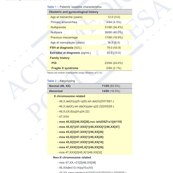
A total of 94 patients enrolled the study. Patients’ gynecological and family history is shown in Table 1. Median age at menopause was 36.0 (6.0) years. The majority of patients reported secondary amenorrhea (95.7%, n = 90).
Obstetric history was unavailable in four patients. Overall, the nulliparity rate was 40.0% (36/90) and 18.9% (17/90) of the patients had a history of previous spontaneous miscarriage.
Twenty-three patients presented a family history of POI. The prevalence of primary amenorrhea was 4.3% (1/23) in familial cases and 4.2% (3/71) in sporadic POI patients. A family history of fragile X syndrome was present in 2 patients. None of the cases with family history of fragile X syndrome presented with primary amenorrhea.
No statistically significant difference was found between the median FSH at diagnosis in patients with primary versus secondary amenorrhea [64.9 (56.0) vs 80.0 (39.0) IU/L, p = 0.392, Mann Whitney test].
Chromosomal abnormalities
Due to missing data, the karyotype was analysed in 85 patients (Table 2).
An abnormal karyotype was observed in 16.5% (n = 14), of which 78.6% (n = 11) involved the X chromosome. The most common abnormality was X chromosome mosaicism, which was found in 50.0% of our cohort (7/14). The four patients with primary amenorrhea presented a normal karyotype.
No statistically significant differences were found regarding age at menopause [35.5 (7.8) vs 36.0 (6.0) years, p = 0.691, Mann Whitney test] or FSH at diagnosis [83.0 (62.0) vs 78.1 (32.0) IU/L, p = 0.415, Mann Whitney test] between patients with (n = 14) or without (n = 71) an abnormal karyotype.
Also, no statistically significant difference was found regarding the prevalence of karyotypic abnormalities between the 23 patients with a family history of POI (8.7%, n = 2) and those without (19.4%, n = 12) (p = 0.333, Fisher’s exact test).
FMR1 testing
Due to missing data, FMR1 analysis was performed in 90 patients (Table 3). FMR1-PM was present in 6.7% (n = 6). The most frequent CGG number of repeats was 30 (n = 53), followed by 31 (n = 19) and 29 (n = 18).
None of the four patients with primary amenorrhea presented the FMR1-PM.
No significant difference was found between patients with and without FMR1-PM concerning age at menopause [38.0 (1.8) vs 36.0 (6.0) years, p = 0.092, Mann Whitney test] or FSH levels at diagnosis [84.7 (63.0.) vs 77.7 (40.0) IU/L, p = 0.340, Mann Whitney test].
There was a higher prevalence of FMR1-PM in patients with a family history of POI, but this difference was not statisti-cally significant [13.0% (3/23), vs 4.5% (3/67), p = 0.176, Fisher’s exact test].
Both patients with a family history of X fragile syndrome carried premutated alleles [(30,60) and (35,58)].
DISCUSSION
This is the first study describing the clinical characteristics and both cytogenetic and FMR1 testing in a Portuguese population with POI.
ARTIGO ACEITE P ARA PUBLICAÇÃO DISPONÍVEL EM WWW .ACT AMEDICAPORTUGUESA.COM
ARTIGO ORIGINAL The median age at menopause in our population was 36 years, similar to the results published by Murray et al in a UK population.26 In an Italian study, Baronchelli et al also reported a mean age at menopause of 34 years.27 However, in this study the authors considered patients with menopause before the age of 45 years.27 Janse et al described a median
age at menopause of 32 years in a POI Dutch population.28 Lower ages at menopause have been reported in POI
non-European populations, varying between 24 and 30 years.7,8,29–31 Although more studies are needed to consolidate this data, the available evidence seems to point towards a higher age at menopause in European populations with POI. This is in line with previous reports which suggest differences regarding age of natural menopause in different ethnic groups.32 Despite the controversy regarding race/ethnicity per se as a factor that influences age at menopause, a higher educational level, the prolonged use of oral contraceptives and a higher baseline weight seem to be associated with a higher age at natural menopause.32,33 The exact mechanism behind these associations is not completely understood. Although no epidemiologi-cal studies have been performed in POI populations, we hypothesize that these factors may also contribute to our results. The prevalence of primary amenorrhea in our population was 4.3% (95% CI 1.6% - 11.0%), which is lower than in other populations (13.2% - 51.0%).7,8,28,29,31,34 We hypothesized that the fact that our department attends to predominantly adult patients might have contributed to this bias.
The rate of previous spontaneous miscarriage was 18.9% (95% CI 12.0% - 28.5%), which is higher than the findings reported by Allen et al and Jansel et al in a POI population (5.0% - 13.9%), but similar to the expected rate in the general population.15,28,35
The prevalence of chromosomal abnormalities in our population was 16.5% (95% CI 9.9% - 26.1%). Most studies report a prevalence of karyotypic abnormalities varying between 9% and 14%.7,8,27,28,31,34 However, a higher prevalence, between 21% and 32%, has also been reported in Tunisian, American, Chilean and Turkish populations.29,36–38 Similarly to what has been previously published, most karyotypic abnormalities were X chromosome related.7,8,28,29 In our population, in accordance with the results of Lakahl et al and Janse et al, the most frequent were mosaic numerical X chromosome ab-normalities.28,31 Other authors reported X chromosome structural abnormalities as being the most frequent.7,8,27,29 Regard-ing X chromosome structural abnormalities, in our sample, all cases involved the Xq, which is in agreement with previous studies and with the critical regions previously defined for the development of POI (Xq13-Xq21 and Xq23-Xq27).4,7,11 Two patients presented Robertsonian translocations, which have also been previously reported in POI patients, although the autosomal role in POI remains unexplained.4,7 Finally, one patient, who was referred to our department due to secondary amenorrhea, presented one autosomal mosaic involving trisomy 21 in two different cell lines [47,XX,+21(2)/46,XX(38)]. Being a mosaic, we cannot predict the presence of the trisomy in other tissues and a causal effect with POI cannot be excluded, since women with Down syndrome have a higher chance of suffering from POI.39,40
Despite the fact that previous studies found a higher prevalence of chromosome abnormalities in patients with primary amenorrhea than in patients with secondary amenorrhea,8,29,31 none of the cases with primary amenorrhea presented karyotypic abnormalities in our study. Most certainly, the small size of the primary amenorrhea subgroup (n = 4) was under-powered to detect these differences. Similarly to what has been previously described, no difference was found regarding the prevalence of karyotypic abnormalities in familial and sporadic cases.8,31
The prevalence of FMR1-PM in our sample was 6.7% (96% CI 3.0% - 14.2%), similar to what has been previously described in non-Asian populations.1,29 In Asian populations, the prevalence seems to be lower (0.5% - 1.5%).30 This is in line with a previous study involving almost 135 000 women from an unselected pan-ethnic cohort, which also reported a lower incidence of FMR1-PM in Asian patients.41 In accordance with other studies, the prevalence was higher in familial cases of POI (13.6% vs 4.5%).5,14,26 The fact that this difference was not statistically significant in our sample may also be attributed to the small sample size. The number of CGG repeats has emerged as a possible predictor of risk and severity of FMR1-related POI. Despite still being a matter of debate, 80 - 100 repeat alleles seem to confer the highest risk.15,16 In our sample, among the six patients with FMR1-PM, only two presented alleles in the high-risk zone (respectively, 80 and 82).
The most frequent number of CGG repeats has been reported as 32,4 while in our sample the most frequent allele was 30. This probably reflects population related variations, which account for the importance of the genetic characterization of these patients on a population level. Considering that fragile X syndrome is the result of the expansion of the number of CGG repeats when transmitted from mother to offspring, the fact that both patients with family history of fragile X syndrome presented FMR1-PM [(30,60) and (35,58)] was an expected finding. In accordance with the findings by Bouali et al, all
patients with FMR1-PM presented with secondary amenorrhea.29
CONCLUSION
Taking into account the prevalence of chromosomal abnormalities and FMR1-PM in our cohort, these results demon-strate the importance of genetic screening for patients with POI and add new data on the different phenotypic and geno-typic patterns of this disorder in different populations. We highlight the higher prevalence of chromosome abnormalities in our Portuguese cohort. Chromosomal studies and FMR1 testing not only provide an etiological explanation for the POI patient, but they also bear important information for both reproductive and genetic counselling, both for the couple and
ARTIGO ACEITE P ARA PUBLICAÇÃO DISPONÍVEL EM WWW .ACT AMEDICAPORTUGUESA.COM ARTIGO ORIGINAL
other relatives. Taking into account the extra-reproductive risks conferred by FMR1-PM, namely FXS and FXTAS, the importance of a multidisciplinary approach for these patients, involving gynaecologists, obstetricians, neurologists and medical geneticists should not be disregarded.
PROTECTION OF HUMANS AND ANIMALS
The authors declare that the procedures were followed according to the regulations established by the Clinical Re-search and Ethics Committee and to the 2013 Helsinki Declaration of the World Medical Association.
DATA CONFIDENTIALITY
The authors declare having followed the protocols in use at their working center regarding patients’ data publication.
COMPETING INTERESTS
The authors have declared that no competing interests exist.
FUNDING SOURCES
This research did not receive any specific grant from funding agencies in the public, commercial, or not-for-profit sec-tors.
REFERENCES
1. European Society for Human Reproduction and Embryology (ESHRE) Guideline Group on POI, Webber L, Davies M, Anderson R, Bartlett J, Braat D, Cartwright B, et al. ESHRE Guideline: management of women with premature ovarian insufficiency. Hum Reprod. 2016;31:926-37.
2. Coccia ME, Rizzello F, Mariani G, Bulletti C, Palagiano A, Scarselli G. Ovarian surgery for bilateral endometriomas influences age at menopause. Hum Reprod. 2011;26:3000-7.
3. Silva C, Yamakami L, Aikawa N, Araujo D, Carvalho J, Bonfá E. Autoimmune primary ovarian insufficiency. Autoimmun Rev. 2014;13:427–30. 4. Qin Y, Jiao X, Simpson JL, Chen ZJ. Genetics of primary ovarian insufficiency: New developments and opportunities. Hum Reprod Update.
2015;21:787-808.
5. Rossetti R, Ferrari I, Bonomi M, Persani L. Genetics of primary ovarian insufficiency. Clin Genet. 2017;91:183-98.
6. Greene AD, Patounakis G, Segars JH. Genetic associations with diminished ovarian reserve: A systematic review of the literature. J Assist Reprod Genet. 2014;31:935-46.
7. Jiao X, Qin C, Li J, Qin Y, Gao X, Zhang B, et al. Cytogenetic analysis of 531 Chinese women with premature ovarian failure. Hum Reprod. 2012;27:2201-7.
8. Kalantari H, Madani T, Zari Moradi S, Mansouri Z, Almadani N, Gourabi H, et al. Cytogenetic analysis of 179 Iranian women with premature ovarian failure. Gynecol Endocrinol. 2013;29:588-91.
9. Liu J, Cox L. Primary ovarian insufficiency: an update. Int J Womens Health. 2014;6:235.
10. Goswami R, Goswami D, Kabra M, Gupta N, Dubey S, Dadhwal V. Prevalence of the triple X syndrome in phenotypically normal women with premature ovarian failure and its association with autoimmune thyroid disorders. Fertil Steril. 2003;4:1052–4.
11. Sarto G, Therman E, Patau K. X inactivation in man: a woman with t(Xq--;12q+). Am J Hum Genet. 1973;3:262–70. 12. Hagerman P. The fragile X prevalence paradox. J Med Genet. 2008;45:498-9.
13. Utine GE, Şimşek-Kiper PÖ, Akgün-Doğan Ö, Ürel-Demir G, Alanay Y, Aktaş D, et al. Fragile x-associated premature ovarian failure in a large Turkish cohort: Findings of Hacettepe Fragile X Registry. Eur J Obstet Gynecol Reprod Biol. 2018;221:76-80.
14. Wittenberger MD, Hagerman RJ, Sherman SL, McConckie-Rosell A, Welt C, Rebar RW, et al. The FMR1 premutation and reproduction. Fertil Steril. 2007;87:456-65.
15. Allen E, Sullivan A, Marcus M, Small C, Dominguez C, Epstein MP, et al. Examination of reproductive aging milestones among women who carry the FMR1 premutation. Hum Reprod. 2007;22:2142-52.
16. Sullivan AK, Marcus M, Epstein MP, Allen EG, Anido AE, Paquin JJ, et al. Association of FMR1 repeat size with ovarian dysfunction. Hum Reprod. 2005;20:402-12.
17. Spath M, Feuth T, Smits A, Yntema HG, Braat DD, Thomas CM, et al. Predictors and risk model development for menopausal age in fragile x premutation carriers. Genet Med. 2011;13:643-50.
18. Schufreider A, McQueen DB, Lee SM, Allen R, Uhler M, Davie J, et al. Diminished ovarian reserve is not observed in infertility patients with high normal CGG repeats on the fragile X mental retardation 1 (FMR1) gene. Hum Reprod. 2015;30:2686-92.
19. Jacquemont S, Hagerman RJ, Hagerman PJ, Leehey MA. Fragile-X syndrome and fragile X-associated tremor / ataxia syndrome : two faces of FMR1. Lancet Neurol. 2007;6:45-55.
20. Yrigollen C, Durbin-Johnson B, Gane L, Nelson DL, Hagerman R, Hagerman PJ, et al. AGG interruptions within the maternal FMR1 gene reduce the risk of offspring with fragile X syndrome. Genet Med. 2012;14:729-36.
21. McGowan-Jordan J, Simons A, Schmid M, editors. An international system for human cytogenomic nomenclature. Basel: S. Karger; 2016. 22. Gardner R, Sutherland G, Shaffer L. Chromosome abnormalities and genetic counselling. 4th ed. Oxford: Oxford University Press; 2012.
23. Guttenbach M, Koschorz B, Bernthaler U, Grimm T, Schmid M. Sex chromosome loss and aging: in situ hybridisation studies on human interphase nuclei. Am J Hum Genet. 1995;57:1143-50.
24. Fu Y, Kuhl D, Pizzuti A, Pieretti M, Sutcliffe JS, Richards S, et al. Variation of the CGG repeat at the fragile X site results in genetic instability: resolution of the Sherman paradox. Cell. 1991;67:1047-58.
25. Ferreira SI, Pires LM, Ferrão J, Sá J, Serra A, Carreira IM. Mosaicism for FMR1 gene full mutation and intermediate allele in a female foetus: a postzy-gotic retraction event. Gene. 2013;527:421-5.
26. Murray A, Schoemaker MJ, Bennett CE, Ennis S, MacPherson JN, Jones M, et al. Population-based estimates of the prevalence of FMR1 expansion mutations in women with early menopause and primary ovarian insufficiency. Genet Med. 2014;16:19-24.
27. Baronchelli S, Conconi D, Panzeri E, Bentivegna A, Redaelli S, Lissoni S, et al. Cytogenetics of premature ovarian failure: an investigation on 269 af-fected women. J Biomed Biotechnol. 2011;2011:20-2.
28. Janse F, Knauff E, Niermeijer M, Eijkemans MJ, Laven JS, Lambalk CB, et al. Similar phenotype characteristics comparing familial and sporadic prema-ture ovarian failure. Menopause. 2010;17:758-65.
ARTIGO ACEITE P ARA PUBLICAÇÃO DISPONÍVEL EM WWW .ACT AMEDICAPORTUGUESA.COM
ARTIGO ORIGINAL 30. Guo T, Qin Y, Jiao X, Li G, Simpson JL, Chen ZJ. FMR1 premutation is an uncommon explanation for premature ovarian failure in Han Chinese. PLoS in 100 Tunisian patients presenting premature ovarian failure. Ann Endocrinol. 2015;76:671-8. One. 2014;9:6-10.
31. Lakhal B, Braham R, Berguigua R, Bouali N, Zaouali M, Chaieb M, et al. Cytogenetic analyses of premature ovarian failure using karyotyping and inter-phase fluorescence in situ hybridization (FISH) in a group of 1000 patients. Clin Genet. 2010;78:181-5.
32. Gold EB. The timing of the age at which natural menopause occurs. Obs Gynecol Clin N Am. 2011;38:425-40.
33. Gold EB, Crawford SL, Avis NE, Crandall CJ, Matthews KA, Waetjen LE, et al. Original contribution factors related to age at natural menopause : longi-tudinal analyses from SWAN. Am J Epidemiol. 2013;178:70-83.
34. Portnoï MF, Aboura A, Tachdjian G, Bouchard P, Dewailly D, Bourcigaux N, et al. Molecular cytogenetic studies of Xq critical regions in premature ovarian failure patients. Hum Reprod. 2006;21:2329-34.
35. National Collaborating Centre for Women’s and Children’s Health (UK). Ectopic pregnancy and miscarriage: ectopic pregnancy and miscarriage: diagno-sis and initial management in early pregnancy of ectopic pregnancy and miscarriage. 2012. [accessed 2019 Jan 15]. Available rom: https://www.nice.org. uk/guidance/cg154/.
36. Ceylaner G, Altinkaya S, Mollamahmutoglu L, Ceylaner S. Genetic abnormalities in Turkish women with premature ovarian failure. Int J Gynaecol Obstet. 2010;110:122-4.
37. Castillo S, López F, Tobella L, Salazar S, Daher V. The cytogenetics of premature ovarian failure. Rev Chil Obs Ginecol. 1992;57:341-5. 38. Rebar R, Connolly H. Clinical features of young women with hypergonadotropic amenorrhea. Fertil Steril. 1990;53:804-10.
39. Romualdi D, Proto C, De Cicco S, Immediata V, Barone C, Romano C, et al. Low AMH levels as a marker of reduced ovarian reserve in young women affected by Down’s syndrome. Menopause. 2016;23:1247-51.
40. Parizot E, Dard R, Janel N, Vialard F. Down syndrome and infertility: what support should we provide? J Assist Reprod Genet. 2019;36:1063-1067. 41. Owens KM, Dohany L, Holland C, DaRe J, Mann T, Settler C, et al. FMR1 premutation frequency in a large, ethnically diverse population referred for
carrier testing. Am J Med Genet Part A. 2018;176:1304-8.
Table 1 – Patients’ baseline characteristics
Obstetric and gynecological history
Age at menarche (years) 12.0 (3.0) Primary amenorrhea 4/94 (4.3%) Nulligravida 31/90 (34.4%)
Nullipara 36/90 (40.0%)
Previous miscarriage 17/90 (18.9%) Age at menopause (years) 36.0 (6.0)
FSH at diagnosis (IU/L) 79.0 (43.9)
Estradiol at diagnosis (pg/mL) 20.0 (10.0)
Family history
POI 23/94 (24.4%)
Fragile X syndrome 2/94 (2.1%)
Values are median (interquartile range) deviation or n (%)
Table 2 – Karyotyping Normal (46, XX) 71/85 (83.5%) Abnormal 14/85 (16.5%) X chromosome related - 46,X,del(X)(q25~q26).ish del(X)(DXYS61-) - 46,X,del(X).ish del(X)(pter-q22.2)(DXS28-) - 46,X,t(X;8)(q24;q24.22) - 47,XXX
- mos 45,X[2]/46,XX[28].nuc ish(DXZ1x1)[4/110] - mos 45,X[1]/47,XXX[1]/48,XXXX[1]/46,XX[47] - mos 45,X[3]/47,XXX[1]/46,XX[26] - mos 45,X[3]/47,XXX[1]/46,XX[26] - mos 45,X[3]/47,XXX[1]/46,XX[16] - mos 47,XXX[3]/45,X[1]/46,XX[26] - mos 47,XXX[2]/45,X[1]/46,XX[32]
Non-X chromosome related
- mos 47,XX,+21[2]/46,XX[38] - 45,XXder(13;14)(q10;q10)
ARTIGO ACEITE P ARA PUBLICAÇÃO DISPONÍVEL EM WWW .ACT AMEDICAPORTUGUESA.COM ARTIGO ORIGINAL
Table 3 – Number of CGG repeats in FMR1 testing
Normal alleles 82 patients/90 (91.1%)
< 29 62
29 18
30 53
31 19
32 - 44 19
Intermediate zone alleles 2 patients/90 (2.2%)
53 1
54 1
Premutation alleles 6 patients/90 (6.7%)
58 1
60 2
69 1
80 1