Expression of RONIN and NANOG-associated proteins in goat parthenogenetic embryos
[Expressão de RONIN e de proteínas associadas ao NANOG em embriões partenogenéticos de
caprinos]
“Artigo Científico/Scientific Article”
Marcelo Tigre Moura
1*, Pamela Ramos-Deus
1, José Carlos Ferreira-Silva
1,
Priscila Germany Corrêa Silva
1, Ludymila Furtado Cantanhêde
1, Pábola Santos Nascimento
1,
Roberta Lane Oliveira Silva
2, Ana Maria Benko-Iseppon
2, Marcos Antonio Lemos Oliveira
1 1Laboratório de Biotécnicas Reprodutivas, Departamento de Medicina Veterinária, Universidade Federal Rural de Pernambuco, Recife-PE, Brasil.2Laboratório de Genética e Biotecnologia Vegetal, Departamento de Genética, Universidade Federal de Pernambuco, Brasil.
*Autor para correspondência/Corresponding author: E-mail: [email protected]
Abstract
The expression of a subset of transcription factors is enriched in early preimplantation embryos, which contributes to their cellular plasticity. RONIN, NANOG and its associated proteins are Pluripotency-Associated Transcription Factors (PATF) that control relevant downstream pathways in pluripotent stem cells, but their activity in early embryos remained less understood. The work was aimed to determine the expression of RONIN and four NANOG-associated PATFs in goat preimplantation embryos. Goat embryos were produced in vitro by parthenogenetic activation. Gene transcripts of cleavage-stage embryos were investigated by reverse transcriptase-polymerase chain reaction (PCR), while blastocysts were analyzed by both RT-PCR and quantitative RT-RT-PCR (RT-qRT-PCR) assays. Gene transcripts of ZFP281, NAC1, and NR0B1 were detected in cleavage-stage embryos, while RONIN and OCT4 were not found expressed. Detection in blastocysts by RT-PCR confirmed the activity of NR0B1, RONIN, and OCT4. Moreover, all five PATF were detected in blastocysts by RT-qPCR (ZFP281, NAC1, RONIN, OCT4, and NR0B1). In conclusion, RONIN and NANOG-associated proteins are active during goat parthenogenetic preimplantation development and hold stage-specific expression patterns.
Keywords: Capra hircus; caprine; embryogenesis; RT-PCR; RT-qPCR; mRNA.
Resumo
A expressão de um conjunto de fatores de transcrição é aumentada em embriões antes da implantação, fato que contribui para a sua plasticidade celular. RONIN, NANOG e suas proteínas associadas são Fatores de transcrição relacionados à pluripotência (FTRP) que controlam vias de sinalização importantes para células-tronco pluripotentes, embora suas atividades em embriões permaneçam menos compreendidas. O objetivo do trabalho foi determinar a expressão de RONIN e quatro FTRPs associados ao NANOG em embriões caprinos. Os embriões foram produzidos in vitro por ativação partenogenética. Embriões na fase de clivagem foram investigados por transcrição reversa, seguida da reação da reação em cadeia da polimerase (RT-PCR), enquanto que os blastocistos foram analisados por RT-PCR e por RT-PCR quantitativa (RT-qPCR). Os transcritos de ZFP281, NAC1 e NR0B1 foram detectados em embriões clivados, enquanto que RONIN e OCT4 não foram encontrados. A detecção em blastocistos pela RT-PCR confirmou a atividade de NR0B1, RONIN e OCT4. Além disso, os cinco FTRPS foram detectados em blastocistos pela RT-qPCR (ZFP281, NAC1, RONIN, OCT4 e NR0B1). Em conclusão, RONIN e os FTRP associados ao NANOG são ativos durante o desenvolvimento pré-implantacional de caprinos e apresentam padrão de expressão estádio-específica.
Introduction
Preimplantation development is initiated after a cascade of cellular events triggered by the fusion of a sperm cell and a metaphase II-arrested egg. Early preimplantation development is characterized by relatively fast mitotic cell divisions of symmetric blastomeres with progressively reduced size (Boroviak and Nichols, 2014; Frankenberg et al., 2016). This so called pre-compactation phase of development is a transcription-independent process in ruminants, where egg cytoplasm is responsible for its developmental coordination and timing (Dominko et al., 1999; Frankenberg et al., 2016; Onichtchouk and Driever, 2016; Silva et al., 2017).
The embryo compactation is concomitant with the emergence of dimorphic embryonic cells at the morulae stage, where outer cells become flatten and inner cells retain their sphere shape (Frankenberg et al., 2016). This initial cell differentiation event is resumed at the blastocyst stage. Outer cells become the trophoblast that ultimately gives rise to placental tissues, while inner cell mass differentiates into all cell types that make up the fetus (Moura, 2012; Boroviak and Nichols, 2014; Silva et al., 2017).
All the cellular events described above account for the first week of development in mammals, while several intrinsic factors contribute to the accurate timing of developmental cues throughout this period (Pesce and Schöler, 2001; Rossant, 2011; Moura, 2012; Goissis and Cibelli, 2014). Pluripotency-associated transcription factors (PATF) are transcription factors that play major roles in embryonic stem (ES) cells and embryos, such as cell-cycle coordination, modulation of cellular metabolism, and transcription initiation control (Dejosez et al., 2010; Young, 2011; Silva et al., 2017). The core pluripotency circuitry is formed by three PATFs, namely OCT4, SOX2, and NANOG (Boyer et al., 2005; Loh et al., 2006; Young, 2011; Moura, 2012). Collectively, these core PATFs control hundreds of downstream genes by reinforcing the expression of other PATFs and silencing of developmental regulators (Young, 2011), while ES cells remain poised for differentiation (Vastenhouw and Schier, 2012). RONIN has been described as an essential factor for mouse ES cell pluripotency and embryogenesis (Dejosez et al., 2008), thus forming a non-canonical circuitry in ES cells that regulates various cellular processes (Dejosez et al., 2010).
Despite the strikingly growing understanding of PATFs in ES cells (Young, 2011), limited progress has been made in preimplantation embryos, particularly in livestock species (Silva et al., 2017). The core PATFs have been studied during embryogenesis (Onichtchouk and Driever, 2016), but NANOG-associated proteins that were initially described in ES cells (Wang et al., 2006) and RONIN (Dejosez et al., 2008), remained poorly described. The work was aimed to determine the expression of RONIN and four NANOG-binding PATFs during goat early parthenogenetic preimplantation embryos.
Material and Methods
Oocyte recovery and in vitro maturation (IVM) Procedures were performed as previously described by Conceição et al. (2016) and Ferreira-Silva et al. (2017). Goat ovaries were collected at local slaughterhouses (Pernambuco, Brazil) and transported in saline solution at 35 ºC within three hours after slaughter. Cumulus-oocyte complexes (COC) were retrieved from 2-6 mm follicles with a 10 G needle into 10 mL syringes, and follicular content was deposited in Petri dishes containing HEPES-buffered IVM medium (H-IVM), consisting of TCM199 with Hank’s salts, 10 % fetal bovine serum (FBS), 50 IU mL-1 heparin and
50 µg mL-1 gentamicin (In vitro Brasil). The COCs
were recovered and selected those with a homogeneous cytoplasm granulation and three or more cumulus cell layers (Moura et al., 2008), and further washed in IVM medium. The IVM medium was composed of TCM-199 with Earle’s salts supplemented with 10 % FBS, 2 mM L-glutamine, 5.0 μg mL-1 FSH (Sigma®), and 50 µg
mL-1 gentamicin. Moreover, COCs were
incubated at 38.5 ºC in a humid atmosphere containing 5% CO2 during 24-27 hours.
Parthenogenetic activation
Parthenogenetic activation (PA) was performed as previously described by Moura et al. (2008). The COCs were denuded with 0.2 % hyaluronidase and gentle pipetting. The PA was performed with 5 μM ionomycin for five minutes and further cultivated in 2 mM 6-DMAP for four hours at 38.5 ºC in a humid atmosphere containing 5% CO2 (Moura et al., 2008; Malik et al., 2015).
Embryo culture
The presumptive zygotes were washed extensively from the 6-DMAP-containing media,
then transferred in groups of 10-15 to 150 μL droplets of Synthetic Oviduct Fluid (SOF) supplemented with 10 % FBS (In vitro Brasil) and co-cultured on a cumulus cell monolayer. Embryonic development was evaluated at 72 hours for cleavage-stage embryos (Day 3) and 168 hours (Day 7) for blastocysts after PA, respectively.
Total RNA extraction
One pool of 15 cleavage-stage embryos and another with two blastocysts were washed in 1X PBS (1.15 g Na2HPO4, 0.2 g KH2PO4, 8.0 g NaCl,
0.2 g KCl) and stored in RNA later® at -20 °C.
Total RNA extraction was performed using ReliaprepTM RNA Cell Miniprep as described by
the manufacturer. Total RNA of was not quantified due to its small quantity in such samples and were immediately used for reverse transcriptase (RT) reaction (cDNA synthesis).
The cDNA synthesis
The RT reaction was performed immediately after total RNA extraction. The
procedure was performed with quantitect®
Reverse Transcription Kit (Qiagen), as described by the manufacturer. Possible residual genomic DNA was removed by the Genomic DNA (gDNA) elimination reaction (2 µL gDNA wipeout buffer 7 x, and 12 µL of total RNA in a final volume of 14 µL), incubated at 42 °C for two minutes and transferred to 4 °C immediately. The gDNA elimination reaction was then added to the RT reaction (1 µL quantiscript reverse transcriptase, 4 µL quantiscript RT buffer 5X, 1 µL RT primer mix) and incubated at 42 °C for 20 minutes. Moreover, samples were incubated at 95 °C for three minutes and stored at -20 °C.
Primers
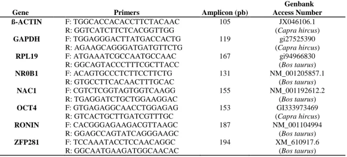
Primers were designed using Primer3 plus and Primer-BLAST Softwares (Ye et al., 2012; Hung and Weng, 2016), based on caprine EST sequences deposited in the Genbank (Table 1). If caprine EST sequences were not available, bovine EST sequences were used as templates for primer design (Table 1).
Table 1. Primers for both reverse transcriptase-polymerase chain reaction (PCR) and quantitative RT-PCR (RT-qRT-PCR) assays for detection of pluripotency-associated transcription factors in goat preimplantation embryos.
Gene Primers Amplicon (pb)
Genbank Access Number ß-ACTIN F: TGGCACCACACCTTCTACAAC R: GGTCATCTTCTCACGGTTGG 105 JX046106.1 (Capra hircus) GAPDH F: TGGAGGGACTTATGACCACTG R: AGAAGCAGGGATGATGTTCTG 119 gi27525390 (Capra hircus) RPL19 F: ATGAAATCGCCAATGCCAAC R: GGCAGTACCCTTTCGCTTACC 167 gi94966830 (Bos taurus) NR0B1 F: ACAGTGCCCTCTTCCTTCTG R: GTGCCTTCACAACTTTGCAC 131 NM_001205857.1 (Bos taurus) NAC1 F: CGTCTCGGTAGTGGTCAAGG R: TGAGGATCTGCTGGAAGGAC 155 NM_001192612.2 (Bos taurus) OCT4 F: GTGAGAGGCAACCTGGAGAG R: GTCACTGCTTGATCGTTTGC 153 GI333973469 (Capra hircus) RONIN F: CACGGGAGAAGACGTTAAGC R: GGAGCCAGTATCAGGGAAGC 187 NM_001104994 (Bos taurus) ZFP281 F: TCCAAATACCTCCAACAGGC R: GGCAATGAAGATGGCAACAC 194 XM_610917.6 (Bos taurus)
Reverse Transcriptase-Polymerase Chain Reaction (RT-PCR)
The PCR reactions were performed in a 96-well thermal cycler (Techne). The reaction was composed of 1 µL cDNA, 0.6 µL of each primer (0.25 µM), 2 µL 10 X PCR Buffer, 0.2 µL dNTP (10 mM), 0.6 µL 50 mM MgCl2, 0.1 µL Taq Polymerase (LGC Biotecnologia), and 14.9 µL
ultra-pure H2O in a final volume of 20 µL. The reaction was initiated at 94 °C for 5 minutes. Moreover, 35 cycles were performed with the following conditions: denaturation at 94 °C for 1 minute, annealing at 58 °C for 1 minute and extension at 72 °C for 1 minute. The final extension step was performed at 72 °C for 10 minutes. The PCR reactions had a final volume of
20 µL. Amplicon visualization was performed by electrophoresis in 1.0 % agarose gel, with 80 V, 120 A for 40 minutes. The housekeeping genes ß-ACTIN, GAPDH, and RPL19 were used as positive controls and reactions without template were used as negative controls.
Quantitative RT-PCR
Quantitative RT-PCR (RT-qPCR) reactions were performed in LineGene 9660 FQD-96A (Bioer®), using SYBR Green detection system.
The reaction was composed of 1 µL cDNA, 5 µL 2 x HotStart-IT SYBR Green qPCR Master Mix (USB®), 0.05 µL ROX® (USB), 0.3 µL each
primer (0.15 µM), and 3.35 µL ultra pure H2O in
a final volume of 10 µL. The embryo cDNA was used as undiluted samples. The reactions used the following conditions: initial denaturation at 95 ºC for 2 minutes, 40 cycles at 95 ºC for 15 seconds, 58 ºC for 30 seconds and 72 ºC for 30 seconds.
Melting curves were analyzed in 65 - 95 ºC for 20 minutes after 40 cycles. Each candidate gene was assessed by the quantification cycle (Cq) during the exponential (log) phase of the PCR reaction.
Results
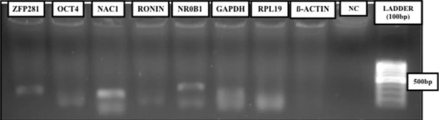
The detection of housekeeping genes GAPDH and RPL19 as single amplicons, albeit at low levels, allowed the analysis of cleavage-stage embryos (Figure 1). The fact that ß-ACTIN was not detected further suggests that improved total RNA extraction should be performed, or alternatively, greater embryo numbers should be used for the assay. More importantly, ZFP281, NR0B1, and NAC1 were detected as single amplicons in cleavage-stage embryos, , despite primer-dimer formation (Figure 1) that is removed after prolonged eletrocphoresis, while OCT4 and RONIN were not detected (Figura 1; Tabela 2).
Figure 1. Detection of pluripotency-associated transcrition factor in goat cleavage-stage embryos by reverse transcriptase-polymerase
chain reaction (RT-PCR). NC: Negative control. bp: base pairs.
Table 2. Detection of pluripotency-associated transcription factors in goat early preimplantation embryos reverse transcriptase-polymerase chain reaction (RT-PCR) and quantitative RT-PCR (RT-qPCR) assays.
Gene Cleavage-stage Blastocyst
ZFP281 +RT -RT+Q
OCT4 -RT -RT+Q
NAC1 +RT -RT+Q
RONIN -RT +RT+Q
NR0B1 +RT +RT+Q
GADPH, RPL19 and ß-ACTIN were used as positive controls. +RT:Detected by RT-PCR. +Q: Detected by quantitative RT-
PCR (RT-qPCR).
In order to determine if such gene expression pattern is maintained during goat preimplantation development, blastocyst stage embryos were analyzed by RT-PCR (Figure 2). The detection of housekeeping genes in blastocysts was effective, where GAPDH, RPL19,
and ß-ACTIN were detected as single amplicons of greater intensity when compared to cleavage-stage counterparts (Figure 2). Under such conditions, only NR0B1 and RONIN were detected as single amplicons by RT-PCR (Figure 2).
Figure 2. Expression of pluripotency-associated transcrition factor in goat parthenogenetic blastocysts by reverse
transcriptase-polymerase chain reaction (RT-PCR) assay. NC: Negative control. bp: base pairs.
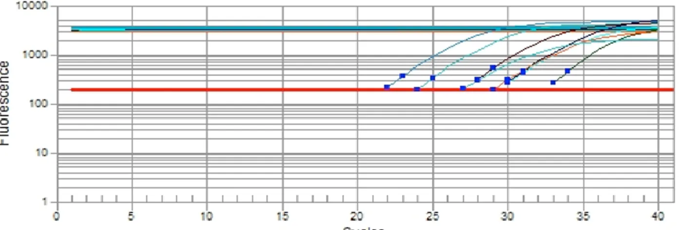
In order to better assess the expression of RONIN and NANOG-associated proteins in goat blastocysts, RT-qPCR reactions were also performed (Figure 3). The cycle of quantification (Cq) of RONIN and NANOG-associated proteins were quite high (26.95 - 32.43) compared to housekeeping genes (21.85 - 27.06) in blastocysts (Table 3), suggestive of low transcript abundance,
although primer efficiency would be required to directly test this hypothesis. These results highlight the necessity to improve the RNA extraction or increase the number of PCR cycles for RT-PCR. Moreover, melting curves values are according to each PCR product (Table 3), where melting curves are also indicative of single PCR products in each reaction (data not shown).
Figure 3. Expression of pluripotency-associated transcrition factor in goat parthenogenetic blastocysts by the quantitative reverse
transcriptase-polymerase chain reaction (RT-qPCR) assay.
Table 3. Melting curve and cycle of quantification determined by absolute reactions of the reverse transcriptase-polymerase chain reaction (RT-PCR) assay.
Cell Type ZFP281 OCT4 NAC1 RONIN NR0B1 GAPDH RPL19 ß-ACTIN
Blastocyst Cycle of quantification - Cq 26.95 29.00 29.43 29.33 32.43 27.06 24.00 21.85 Melting Curve 80.00 86.00 89.00 90.00 82.00 84.00 85.00 85.50 Discussion
The PATFs are a subset of transcription factors that control the establishment and maintenance of pluripotency in ES cells and during embryogenesis (Chambers and Tomlinson, 2009; Young, 2011; Moura, 2012; Yeo and Ng, 2013). Despite their contribution to the ES cell
state, these genes are subject to less intense investigation during early embryogenesis (Boroviak and Nichols, 2014; Silva et al., 2017), most notably due to technical issues.
RONIN was detected only in caprine blastocysts and by both PCR platforms. This is the first report of RONIN activity in ruminant species.
The gene is required for both embryogenesis and ES cell pluripotency in the mouse (Dejosez et al., 2008). RONIN binds to HCF-1 at its coiled coil, in order to regulate target genes in ES cells, since it lacks a true transactivation domain (Dejosez et al., 2008). This binding is also expected to form due to high homology of such genes (data not shown), as described earlier (Gervais et al., 2013). This fact later fact suggests that RONIN in mouse and goats may share similar functions in vivo. If this expression pattern holds true, RONIN may be involved in basal processes (Dejosez et al., 2010), such as energy metabolism and protein synthesis in post-compaction goat embryos.
All NANOG-associated proteins that were analyzed above were also detected in goat blastocysts, while only ZFP281, NAC1, and NR0B1 were detected in cleavage-stage embryos. NANOG was identified as a PATF by its restricted expression in ES cells (Chambers et al., 2003; Mitsui et al., 2003; Yates and Chambers, 2005). NANOG forms with OCT4 and SOX2 the core transcription circuitry in ES cells and regulates hundreds of downstream target genes (Boyer et al., 2005; Loh et al., 2006; Young, 2011). Moreover, NANOG and its associated proteins form large protein complexes in ES cells and regulate cell fate decisions (Wang et al., 2006; Kim et al., 2008). Despite the description of NANOG and OCT4 in goat embryos (He et al., 2006; Hosseinnia et al., 2016), most PATFs remained to be described. To our knowledge, no previous report addressed the expression transcript availability of ZFP281, NR0B1, and NAC1 during goat preimplantation development. The presence of all NANOG-associated proteins in goat blastocyst opens the possibility for investigation if these PATFs similar protein complexes during goat embryogenesis.
Some subtle differences were found in the gene expression of goat embryos described above, particularly for OCT4, which was not detected in cleavage-stage embryos. This PATF was previously detected throughout goat preimplantation development (He et al., 2006; Hosseinnia et al., 2016). This discrepancy may be due to an influence of embryo culture conditions (Purpera et al., 2009), or technical issues. The bare detection of housekeeping genes also suggests that some technical improvements are required for better investigation of such PATFs in goat embryos, while RT-qPCR was more efficient to detect some PATFs, possibly due to their low
transcript abundance which required an increased number of PCR cycles for their detection.
In conclusion, RONIN and NANOG-associated proteins are active during goat early parthenogenetic preimplantation development and may hold stage-specific expression patterns.
Animal Ethics
All procedures were approved by the Ethics committee at Universidade Federal Rural de Pernambuco (UFRPE), under the license number 035/2016 (Process 23082.002437/2016).
Conflict of interest
The authors declare that there is no conflict of interest in the study.
Acknowledgements
Authors would like to acknowledge CNPq for the financial support of the study.
References
Boroviak, T.; Nichols J. The birth of embryonic pluripotency. Philosophical transactions of the Royal Society of London. Series B, 369(1657): 1-10, 2014.
Boyer, L.A.; Lee, T.I.; Cole, M.F.; Johnstone, S.E.; Levine, S.S.; Zucker, J.P.; Guenther, M.G.; Kumar, R.M.; Murray, H.L.; Jenner, R.G.; Gifford, D.K.; Melton, D.A.; Jaenisch, R.; Young, R.A. Core transcriptional regulatory circuitry in human embryonic stem cells. Cell, 122(6): 947-956, 2005. Chambers, I.; Colby, D.; Robertson, M.; Nichols,
J.; Lee, S.; Tweedie, S.; Smith, A. Functional expression cloning of Nanog, a pluripotency sustaining factor in embryonic stem cells. Cell, 113(5): 643-655, 2003.
Chambers, I.; Tomlinson, S.R. The transcriptional foundation of pluripotency. Development, 136(14): 2311-2322, 2009.
Conceição, J.C.; Moura, M.T.; Ferreira-Silva, J.C.; Cantanhêde, L.F.; Chaves, R.M.; Lima, P.F.; Oliveira, M.A. Incidence of apoptosis after retinoids and insulin-like growth factor-I (factor-IGF-factor-I) supplementation during goat in vitro embryo production. Zygote, 24(6): 808-813, 2016.
Dejosez, M.; Krumenacker, J.S.; Zitur, L.J.; Passer, M.; Chu, L.F.; Songyang, Z.; Thomson, J.A.; Zwaka, T.P. Ronin is essential for embryogenesis and the
pluripotency of mouse embryonic stem cells. Cell, 133(7): 1162-1174, 2008.
Dejosez, M.; Levine, S.S.; Frampton, G.M.; Whyte, W.A.; Stratton, S.A.; Barton, M.C.; Gunaratne, P.H.; Young, R.A.; Zwaka, T.P. Ronin/Hcf-1 binds to a hyperconserved enhancer element and regulates genes involved in the growth of embryonic stem cells. Genes and Development, 24(14): 1479-1484, 2010.
Dominko, T.; Mitalipova, M.; Haley, B.; Beyhan, Z.; Memili, E.; McKusick, B.; First, N.L. Bovine oocyte cytoplasm supports development of embryos produced by nuclear transfer of somatic cell nuclei from various mammalian species. Biology of Reproduction, 60(6): 1496-1502, 1999. Ferreira-Silva, J.C.; Moura, M.T.; Silva, T.D.;
Oliveira, L.R.; Chiamenti, A.; Figueirêdo Freitas, V.J.; Oliveira, M.A. Full-term potential of goat in vitro produced embryos after different cryopreservation methods. Cryobiology, 75(X): 75-79, 2017.
Frankenberg, S.R.; de Barros, F.R.; Rossant, J.; Renfree, M.B. The mammalian blastocyst. Wiley Interdisciplinary Reviews: Developmental Biology, 5(2): 210-232, 2016.
Gervais, V.; Campagne, S.; Durand, J.; Muller, I.; Milon, A. NMR studies of a new family of DNA binding proteins: the THAP proteins. Journal of Biomolecular NMR, 56(1): 3-15, 2013.
Goissis, M.D.; Cibelli, J.B. Functional characterization of SOX2 in bovine preimplantation embryos. Biology of Reproduction, v.90, Artigo 30, 2014. Loh, Y.H.; Wu, Q.; Chew, J.L.; Vega, V.B.;
Zhang, W.; Chen, X.; Bourque, G.; George, J.; Leong, B.; Liu, J.; Wong, K.Y.; Sung, K.W.; Lee, C.W.; Zhao, X.D.; Chiu, K.P.; Lipovich, L.; Kuznetsov, V.A.; Robson, P.; Stanton, L.W.; Wei, C.L.; Ruan, Y.; Lim, B.; Ng, H.H. The Oct4 and Nanog transcription network regulates pluripotency in mouse embryonic stem cells. Nature Genetics, 38(4): 431-440, 2006.
He, S.; Pant, D.; Schiffmacher, A.; Bischoff, S.; Melican, D.; Gavin, W.; Keefer, C. Developmental expression of pluripotency determining factors in caprine embryos: novel pattern of NANOG protein localization
in the nucleolus. Molecular Reproduction and Development, 73(12): 1512-1522, 2006. HosseinNia, P.; Hajian, M.; Tahmoorespur, M.; Hosseini, S.M.; Ostadhosseini, S.; Nasiri, M.R.; Nasr-Esfahani, M.H. Expression Profile of Developmentally Important Genes in preand peri-Implantation Goat Embryos Produced In Vitro. International Journal of Fertility & Sterility, 10(3): 310-319, 2016. Hung, J.H.; Weng, Z. Designing Polymerase
Chain Reaction Primers Using Primer3Plus. Cold Spring Harbor Protocols, 2016(9): artigo pdb.prot093096, 2016.
Kim, J.; Chu, J.; Shen, X.; Wang, J.; Orkin, S.H. 2008. An extended transcriptional network for pluripotency of embryonic stem cells. Cell, 132(6): 1049-1061, 2008.
Malik, H.N.; Singhal, D.K.; Saugandhika, S.; Dubey, A.; Mukherjee, A.; Singhal, R.; Kumar, S.; Kaushik, J.K.; Mohanty, A.K.; Das, B.C.; Bag, S.; Bhanja, S.K.; Malakar, D. Generation of parthenogenetic goat blastocysts: effects of different activation methods and culture media. Zygote, 23(3): 327-335, 2015.
Mitsui, K.; Tokuzawa, Y.; Itoh, H.; Segawa, K.; Murakami, M.; Takahashi, K.; Maruyama, M.; Maeda, M.; Yamanaka, S. The homeoprotein Nanog is required for maintenance of pluripotency in mouse epiblast and ES cells. Cell, 113(5): 631-642, 2003.
Moura, M.T.; Sousa, R.V.; Oliveira Leme, L.; Rumpf, R. Analysis of actinomycin D treated cattle oocytes and their use for somatic cell nuclear transfer. Animal Reproduction Science, 109(1-4): 40-49, 2008.
Moura, M.T. Pluripotency and cellular reprogramming. Anais da Academia Pernambucana de Ciência Agronômica, 8(1): 138-168, 2012.
Onichtchouk, D.; Driever, W. Zygotic Genome Activators, Developmental Timing, and Pluripotency. Current Topics in Developmental Biology, 116: 273-297, 2016.
Pesce, M.; Schöler, H.R. Oct-4: gatekeeper in the beginnings of mammalian development. Stem Cells, 19(4): 271-278, 2001.
Purpera, M.N.; Giraldo, A.M.; Ballard, C.B.; Hylan, D.; Godke, R.A.; Bondioli, K.R. Effects of culture medium and protein supplementation on mRNA expression of in
vitro produced bovine embryos. Molecular Reproduction and Development, 76(8): 783-793, 2009.
Rossant, J. Developmental biology: A mouse is not a cow. Nature, 471(7339): 457-458, 2011.
Silva, P.G.C.; Moura, M.T.; Braga, V.A.A.; Ferreira-Silva, J.C.; Nascimento, P.S.; Cantanhêde, L.F.; Chaves, M.S.; Oliveira, M.A.L. 2017. Atividade dos genes relacionados à pluripotência em ovinos. Medicina Veterinária (UFRPE), 11(1): 000, 2017.
Vastenhouw, N.L.; Schier, A.F. Bivalent histone modifications in early embryogenesis. Current Opinion in Cell Biology, 24(3): 374-386, 2012.
Wang, J.; Rao, S.; Chu, J.; Shen, X.; Levasseur, D.N.; Theunissen, T.W.; Orkin, S.H. 2006. A
protein interaction network for pluripotency of embryonic stem cells. Nature, 444(7117): 364-368, 2006.
Yates, A.; Chambers, I. The homeodomain protein Nanog and pluripotency in mouse embryonic stem cells. Biochemical Society Transactions, 33(6): 1518-1521, 2005. Ye, J.; Coulouris, G.; Zaretskaya, I.; Cutcutache,
I.; Rozen, S.; Madden, T.L. Primer-BLAST: a tool to design target-specific primers for polymerase chain reaction. BMC Bioinformatics, 18 (13): artigo 134, 2012. Yeo, J.C.; Ng, H.H. The transcriptional regulation
of pluripotency. Cell Research, 23(1): 20-32, 2013.
Young, R.A. Control of the embryonic stem cell state. Cell, 144(6): 940-954, 2011.