U
NIVERSIDADE DE
L
ISBOA
F
ACULDADE DE
C
IÊNCIAS
D
EPARTAMENTO DE
F
ÍSICA
E
ASY
E
MBRYONIC
S
TEM
C
ELL
A
N
EW
T
OOL TO
M
ANIPULATE
G
ENE
E
XPRESSION IN
E
MBRYOID
B
ODIES
Mestrado Integrado em Engenharia Biomédica e Biofísica
Perfil em Engenharia Clínica e Instrumentação Médica
Felícia Margarida Correia da Fonseca Lopes
Dissertação Orientada por:
Dr. Cláudio Areias Franco Prof. Dr. Pedro Cavaleiro Miranda
iii
RESUMO
O sistema vascular permite a distribuição adequada de sangue e consequente fornecimento de oxigénio e nutrientes a todos os tecidos em vários organismos multicelulares. O sistema vascular é essencial para o desenvolvimento embrionário, o crescimento de tecidos e o processo de cicatrização. De especial interesse, muitas doenças que afectam o ser-humano, como por exemplo retinopatia diabética, isquemia, acidente vascular cerebral e crescimento de tumores sólidos, surgem devido a má formação e disfunção dos vasos sanguíneos.
A formação de uma rede vascular funcional requer a extensão dos vasos sanguíneos pré-existentes pelo processo de angiogénese de forma a alcançar os tecidos não vascularizados e, em simultâneo, a remodelação do plexo vascular primitivo amorfo criado por angiogénese numa rede hierarquizada de artérias, veias e capilares. Estes dois processos são fortemente regulados pela comunicação molecular entre as células dos tecidos e as células endoteliais (as células que revestem os vasos sanguíneos). De especial relevância, começa agora a ser evidente que as células endoteliais necessitam de coordenar os seus comportamentos individuais para permitir a organização de uma rede vascular de forma coerente e funcional. É, por isso, importante compreender os mecanismos moleculares de regulação do comportamento coordenando das células endoteliais durante as diferentes fases do processo de formação dos vasos sanguíneos, sendo que isso irá certamente criar novas possibilidades de tratamento médico de várias doenças vasculares e melhorar o impacto das terapias existentes na saúde humana.
Nos últimos anos temos assistido a um aumento significante tanto no desenvolvimento como no uso de tecnologias transgénicas, usadas em estudos que visam a nossa compreensão dos mecanismos biológicos, mas que têm também facilitado o desenvolvimento de uma vasta panóplia de modelos de doenças que têm grande impacto na abordagem das patologias humanas. As células estaminais embrionárias derivam de células totipotentes embrionárias e, como tal, servem como fonte putativa de vários tipos de células diferenciadas, dando origem a todos os tipos celulares do organismo. Existem duas características que distinguem as células estaminais embrionárias das restantes: a sua pluripotência e a sua capacidade de auto-renovação sob determinadas condições e, como tal, quando geneticamente modificadas in vitro, podem ser utilizadas no estudo de vários mecanismos biológicos em diferentes ensaios. Particularmente, as células estaminais embrionárias podem ser utilizadas em ensaios sobre angiogénese a partir da criação de corpos embrióides, que resultam da agregação in vitro destas células e que permitem testar o potencial das mesmas. A principal vantagem deste sistema, neste contexto, é poder ser manipulado a partir da utilização de células estaminais embrionárias distintas com diferentes modificações ou origem genética, criando quimeras que permitem a visualização da dinâmica das células endoteliais e o estudo da eficiência das diferentes fases do processo de formação dos vasos sanguíneos durante a angiogénese.
iv
As tecnologias correntemente desenvolvidas para inserir um gene numa célula viva estão limitadas pela natureza aleatória desta inserção no genoma da mesma. O novo gene, ao ser posicionado arbitrariamente, pode inactivar ou perturbar o funcionamento de genes endógenos, causando efeitos indesejados na célula. Adicionalmente, estas tecnologias não permitem a reprodutibilidade do processo uma vez que não há garantia de que a nova sequência seja inserida na mesma posição do genoma em duas células diferentes. Este problema pode ser contornado utilizando tecnologias que permitem recombinação homóloga, na inserção de transgenes em locais específicos ou em modificações in situ de genes existentes, mas que são muito morosas pela necessidade de alcançar e seleccionar os clones positivos.
De forma ultrapassar as dificuldades supramencionadas, pretendemos aumentar a eficiência e velocidade do processo de inserção de genes em células a partir do desenvolvimento de um método de fácil utilização para a manipulação de células estaminais embrionárias para o estudo em corpos embrióides. Para tal, pretendemos recorrer à actividade de um tipo de enzimas específico que reconhece curtas sequências de ADN que medeia a recombinação entre estes elementos, resultando em excisão, integração, inversão e substituição dos fragmentos de ADN, as chamadas site-specific recombinases (de sequência específica). Dentro desta família de enzimas, existe uma, a integrase PhiC31, que por recombinação homóloga e através de locais específicos de fixação, consegue integrar um fragmento de ADN de qualquer dimensão, de forma específica e irreversível. Com base neste mecanismo, desenvolvemos dois vectores, cada um deles com os locais de fixação específicos, sendo que um deles possui o genoma alvo e o segundo, o gene que pretendemos introduzir na célula. Através da expressão endógena de integrase PhiC31, a sequência de interesse será introduzida no genoma da célula de forma específica e eficiente, prevenindo a disrupção de genes endógenos e oferecendo perfeita reprodutibilidade.
A principal vantagem desta ferramenta para o nosso laboratório prende-se com o facto de facilitar a manipulação de células estaminais embrionárias, que permitem a criação de corpos embrióides quiméricos para a visualização da dinâmica das células endoteliais e a eficiência de migração durante o processo angiogénico. Até ao momento a nossa estratégia revelou ser promissora, tendo a expressão de integrase PhiC31 sido bem-sucedida em células cancerígenas em cultura mas, faltando ainda concluir a validação do vector de expressão, que revelou deformidades. A próxima fase será extrapolar a técnica para células estaminais embrionárias para que seja aprovada e posteriormente aplicada a diferentes ensaios em outras áreas da biologia.
Palavras-Chave: Vasos Sanguíneos, Rede Vascular, Angiogénese, Células Endoteliais, Células Estaminais Embrionárias, Corpos Embrióides, integrase PhiC31.
v
ABSTRACT
The established transgenesis methods used in embryonic stem cells allow efficient genomic integration of transgenes. However, the integration of multiple copies or transgenes at random genomic locations complicates comparative transgene analysis and makes long-term transgene stability unpredictable with variable expression. Targeted, site-directed transgene integration into pre-determined genomic loci can circumvent these issues, enabling direct comparison of different transgenes, thereby improving time and cost efficiency. The PhiC31 integrase catalyzes the unidirectional recombination reaction between heterotypic attP and attB sites and is an efficient platform for site-directed transgenesis.
Here, we implemented an efficient PhiC31-based site-specific transgenesis system for embryonic stem cells to be used in embryoid bodies’ studies. It allows the recombination of attB-containing transgene vectors into single genomic attP landing sites being this enzyme endogenously expressed by the cell, avoiding PhiC31 integrase encoding mRNA injection. Our strategy revealed a great potential as, cancer cells in culture were able to express PhiC31 integrase but, the validation of the expression vector is still ongoing due to deformities in the plasmid. The next step will be validating the technique in embryonic stem cells.
The major advantage of this system for our laboratory is that it can easily be manipulated by using different transgenic embryonic stem cells, creating chimeras to visualize the dynamics of endothelial cells and study the efficiency of sprouting and migration during angiogenesis.
This tool can also be applied to many other assays in transversal fields of biology. We hope that our results help researchers to optimize genome editing in their specific systems.
Keywords: Blood Vessels, Vascular Network, Angiogenesis, Endothelial Cells, Embryonic Stem Cells, Embryoid Bodies, PhiC31 integrase.
vii
ACKNOWLEDGEMENTS
There are a number of people to whom I am deeply grateful for guidance, advice, and support.
First, I would like to express my gratitude to my external supervisor, Cláudio Franco, for teaching me how to do science, for leading by example, and for pushing me to always become better. Cláudio is an exceptionally creative thinker and has continuously supported me in all my endeavors. Under his mentorship, inspired by his infectious enthusiasm, I have learned and grown more than I ever imagined.
I would also like to thank my internal supervisor, Pedro Cavaleiro Miranda, for dealing with all the paperwork implied in the realization of this thesis and for his valuable input in the written work.
Moreover, thanks to all the professors from Instituto de Biofísica e Engenharia Biomédica whom, during my academic life, were always very attentive and helpful in many different manners.
I am very grateful to all of the past and present members of the CFranco Lab, and our neighbors in the EGomes Lab, for the invaluable input, guidance and assistance, patience and friendship during my internship at Instituto de Medicina Molecular.
Importantly, I want to thank all my great friends and colleagues for all the joyful times that we spent together, for our travels and adventures, for the company during the countless nights spent studying and for being there when I most needed. I must also thank my sister for all the great moments, complicity and for reminding me to always have fun; despite the distance that separates us, you are always the closer you can be to my heart. Most of all, I am immensely grateful to my parents, who helped me to grow, personally and professionally, throughout my school years. Thank you for your emotional and financial support, for your friendship and guidance, for all the opportunities you have given me and, specially, for being always by my side. You are never too far away from my thoughts.
ix
TABLE OF CONTENTS
Resumo ... iii Abstract ... v Acknowledgements ... vii Table of Contents ... ixList of Figures ... xiii
List of Abbreviations ... xv
1. Introduction ... 1
2. Background ... 3
2.1. Sprouting Angiogenesis ... 3
2.1.1. VEGF and Notch/Dll4 Signaling Pathways ... 4
2.1.2. Competition between Tip and Stalk cells ... 5
2.1.3. Embryoid Body Sprouting Assay as a Tool to Measure Cell Competition ... 6
2.1.3.1. Assessing Sprouting Capacity, Dynamics and Competition of Cells ... 6
2.2. How to Manipulate Stem Cells ... 8
2.2.1. Genetic Engineering ... 8
2.2.1.1. Site-Specific Recombinases ... 8
2.2.1.2. CRISPR/Cas9 ... 10
2.3. Purpose of This Thesis ... 12
3. Materials & Methods ... 13
3.1. Polymerase Chain Reaction... 13
3.2. Agarose Gel Electrophoresis ... 14
3.3. DNA Extraction from Agarose Gel and Purification ... 14
3.4. EtOH Precipitation ... 14
3.5. Digestion of DNA with Restriction Enzymes ... 15
3.6. Ligation of DNA Fragments ... 15
3.7. Gateway® Technology ... 16
3.8. Transformation of DNA into Competent Cells ... 16
x
3.10. Transfection of U2OS Cells ... 17
3.11. RNA Extraction ... 17
3.12. Real-Time PCR ... 17
4. Results ... 19
4.1. Targeting Vector ... 19
4.1.1. p5E MCS CAGGS AttP PhiC31 ... 19
4.1.2. pME PhiC31 2A ... 20
4.1.3. p3E Neo PolyA ... 21
4.1.4. pROSA26 DV2 ... 22
4.1.5. LR Reaction ... 22
4.2. Expression Vector ... 23
4.3. Test Constructs in U2OS Cells ... 26
5. Discussion ... 31
References ... 35
Appendix ... 41
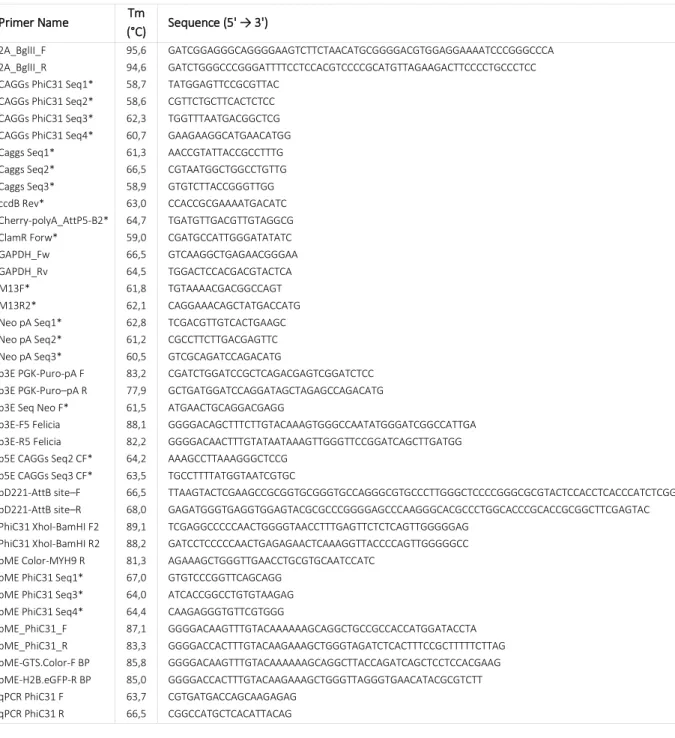
Primers ... 41
Restriction Enzymes ... 42
Cloning Process Overview ... 43
xi
LIST OF TABLES
Table 1. Component and cycling instructions for the PCR reactions. ... 13 Table 2. Reaction mixtures and conditions that were used for either sticky- or blunt-end ligations in the present study. ... 15 Table 3. Optimized reaction mixtures and conditions for both BP and LR reactions. ... 16
xiii
LIST OF FIGURES
Figure 1. Schematic model of sprout initiation, vessel branching, and maturation. ... 4
Figure 2. Tip to stalk cell lateral inhibition during sprouting angiogenesis. ... 5
Figure 3. VEGFR levels in mosaic-tip-cell selection in vitro. ... 7
Figure 4. PhiC31 Integrase System. ... 9
Figure 5. CRISPR/Cas9 genome engineering with Cas9 nuclease variants... 11
Figure 6. CAGGs fragment cut out from pCAGGs-m2G, linearization of p5E-MCS (T2K228), digestion to confirm CAGGs insertion in p5E-MCS (T2K228) and p5E MCS CAGGs linearization in a 1% agarose gel electrophoresis. .. 20
Figure 7. PhiC31 fragment cut out from pCS2P+PhiC31o, digestion of pME PhiC31 to confirm insertion and linearized pME PhiC31 in a 1% agarose gel electrophoresis. ... 21
Figure 8. Neo PolyA fragment cut out from pROSA Dest1 and DNA digestion to confirm Neo PolyA insertion in a 1% agarose gel electrophoresis. ... 22
Figure 9. PCR to confirm LR reaction between p5E MCS CAGGs AttP PhiC31, pME PhiC31 2A, p3E Neo PolyA and pROSA26 DV2, run in a 1% agarose gel electrophoresis. ... 23
Figure 10. pDONR221 (T2K2018) digestion and PCR to confirm AttB PhiC31 insertion in the plasmid in a 1% agarose gel electrophoresis. ... 24
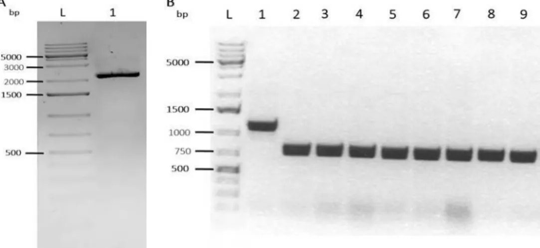
Figure 11. PGK Puro PA fragment cut out from p3E PGK Puro PA BP and digestion of pDONR221 AttB PhiC31 and PCR to confirm PGK Puro PA insertion in pDONR221 AttB PhiC31 plasmid in a 1% agarose gel electrophoresis. . 25
Figure 12. GTS Cherry H2B eGFP fragment cut out from pUC57-Amp_VEcad_GTS.mCherry-2A-H2B.eGFPpA_FRT.Neo.FRTpA and PCR to confirm GTS Cherry H2B eGFP cassette insertion in pDONR221 AttB PhiC31 PGK Puro PA plasmid in a 1% agarose gel electrophoresis. ... 26
Figure 13. Real‐time PCR plot of the cDNA of U2OS cells transfected and not transfected with the targeting vector. Gene‐specific primers were designed to the hairpin of the PhiC31 mRNA precursors ... 27
Figure 14. PhiC31 gene expression in Ct values relative to housekeeping gene obtained by real-time PCR. ... 28
Figure 15. Microscopy images of the control cells and cells transfected with the targeting vector, both transfected with the expression vector. ... 29
xiv
Figure 17. U2OS cells transfected with a construct comprising the GTS (Golgi-specific targeting sequence) fused to mCherry, a 2A self-cleavage peptide, and histone H2B fused to eGFP. ... 33
xv
LIST OF ABBREVIATIONS
Amp Ampicillin
att Attachment
CAG Chicken beta actin
Cam Chloramphenicol
cDNA Complementary DNA
CIP Calf Intestinal Alkaline Phosphatase CMV Cytomegalovirus
CO2 Carbon Dioxide
CRISPR Clustered Regularly Interspaced Short Palindromic Repeats
crRNA CRISPR RNA
Ct Cycle Threshold
DABCO 1,4-Diazabicyclo[2.2.2]Octane DAPI 4',6-Diamidino-2-Phenylindole dCas9 Nuclease-deficient Cas9 dH2O Distilled Water
Dll4 Delta-like Ligand 4
DMEM Dulbecco's Modified Eagle Medium DNA Deoxyribonucleic Acid
dNTPs Deoxyribonucleoside Triphosphates E. coli Escherichia coli
EB Embryoid Body
EDTA Ethylenediaminetetraacetic Acid
ES Embryonic Stem
EtOH Ethanol
G418 Geneticin
GAPDH Glyceraldehyde-3-Phosphate Dehydrogenase GOI Gene of Interest
H2O Water
Kan Kanamycin
LB Luria Broth
mRNA Messenger RNA
ON Overnight
PBS Phosphate Buffer Solution PCR Polymerase Chain Reaction
PFA Paraformaldehyde
RNA Ribonucleic Acid RNAi RNA Interference
RT Room Temperature
xvi SSRs Site-specific Recombinases
TALENs Transcription-activator Like Effector Nucleases TBE Tris/Borate/EDTA
Tm Melting Temperature tracrRNA Trans-activating crRNA
Tris Tris(hydroxylmethyl)aminomethane UTR Untranslated Regions
UV Ultra-violet
VEGF Vascular Endothelial Growth Factor
VEGFR Vascular Endothelial Growth Factor Receptor
WT Wild Type
ZFNs Zinc-finger Nucleases
LIST OF UNITS
% Percent
°C Degree Celsius
µg/µL Microgram per Microliter
µL Microliter µM Micromolar bp Base Pair h Hour kb Kilobase Pair L Liter M Molar min Minute mL Milliliter
ng/µL Nanogram per Microliter rpm Rotations per Minute
sec Second
V Volt
1
1. INTRODUCTION
Blood vessels have being studied for many centuries and even though we now know some of the molecular mechanism controlling the aspects that make the vascular system a very complex, stereotyped and hierarchical network, there are still a lot of unanswered questions behind the blood vessel formation. Importantly, the formation and maintenance of a functional vascular network is fundamental for growth and development of multicellular organisms as an incorrect vascular assembly is linked to several pathological states such as solid tumors, cerebral cavernous malformations, hemangiomas, retinopathies and arteriovenous malformations. The first vessels in the developing embryo form through vasculogenesis, after which angiogenesis, the physiological process through which new blood vessels form from pre-existing ones, is responsible for most, if not all, blood vessel growth during development and in disease. During sprouting angiogenesis, there are two distinct types of cells, the tip and stalk cells, whose phenotype is known to be regulated by a VEGF/Notch signaling.
In the past years, we have witnessed a significant increase in both the development and use of transgenic technologies that have been used to aid our fundamental understanding of biologic mechanisms, but that have also facilitated the development of a range of disease models that have a huge impact upon our approach to human disease. Embryonic stem cells are derived from totipotent cells of the embryo and therefore, can serve as a putative source of numerous types of differentiated cells, thus generating every cell type in the body. Two distinctive properties distinguish embryonic stem cells, their pluripotency and their capacity for self-renewal under defined conditions. Embryonic stem cells can be genetically modified in vitro and thus be used to study many different mechanisms in different assays, particularly, they are used in sprouting assays by creating embryoid bodies. Embryoid bodies are the result of the in vitro aggregation of embryonic stem cells and are utilized to test the differential potential of these cells. The major advantage of this system for our laboratory is that it can be manipulated by using different embryonic stem cells with different modifications or distinct genetic background, creating a chimera (a single organism composed of genetically distinct cells) to visualize the dynamics of endothelial cells and study the efficiency of sprouting and migration during angiogenesis.
The current technologies used to insert a gene into a living cell are limited by the random nature of the insertion of the new sequence into the genome. The new gene is positioned arbitrarily, and may inactivate or disturb the functioning of other genes or even cause severe unwanted effects. Furthermore, these technologies offer no degree of reproducibility, as there is no guarantee that the new sequence will be inserted at the same place in two different cells. The precise genetic modification can be achieved in embryonic stem cells by taking advantage of homologous recombination to target single-copy transgenes to specific sites or to modify existing
2
genes in situ, but this technique is very time-consuming because it is necessary to target and select the positive clones.
Site-specific recombinases recognize short DNA sequences (typically between 30 and 40bp) and mediate the recombination between these elements resulting in excision, integration, inversion, or exchange of DNA fragments. The PhiC31 integrase is a sequence-specific recombinase that by homologous recombination and using attachment (att) sites, can integrate a plasmid of any size in an irreversible way. Taking advantage of these properties and in order to overcome the difficulties inherent to the existing methods for cell genome manipulation, we developed a way to easily manipulate embryonic stem cells to test embryoid bodies, increasing the efficiency and speed of the clone selection process.
We created two vectors, one with the target genome and the other with the protein that we want to express. Each of the vectors has a PhiC31 attachment site and, through the endogenous expression of PhiC31 integrase, induced by the first vector, the expression plasmid is inserted in the genome of the cell, in a specific and efficient way. The major advantages of this system is that it enables a specific area of the DNA to be modified, thereby increasing the precision of the correction or insertion, preventing any cell toxicity and offering perfect reproducibility. At the moment, our strategy revealed to have potential as the insertion of the targeting vector in U2OS cells in culture worked well and cells are expressing PhiC31 integrase but, we were still not able to create an expression vector able to confirm the efficiency of our technique. The next step is to test the constructs in embryonic stem cells to validate it so it can, posteriorly, be applied to different systems of other fields of biology.
This study took place at the Vascular Morphogenesis Laboratory, in Instituto de Medicina Molecular, Lisbon, and was performed under the supervision of Dr. Cláudio Areias Franco, group leader of the laboratory that is mainly focused on the understanding of the molecular mechanisms regulating coordinated endothelial cell behavior during sprouting and remodeling phases of the angiogenic process. Improving the knowledge on the molecular regulation of vascular morphogenesis will certainly create new possibilities for medical prevention and treatment of various human conditions.
3
2. BACKGROUND
The correct development of blood vessels is essential for all aspects of tissue growth and physiology in vertebrates as they supply oxygen and nutrients to the organism. In the adult, vessels are normally quiescent; however, structural or functional vessel abnormalities may occur. An inadequate vessel maintenance or growth causes ischemia in diseases such as myocardial infarction, stroke, and neurodegenerative or obesity-associated disorders, whereas excessive vascular growth or abnormal remodeling promotes many ailments including cancer, inflammatory disorders, and eye disease [1][2]. The formation of an elaborate hierarchically branched vascular network, through either vasculogenesis or angiogenesis, relies on a series of coordinated morphogenic events that involves complex and highly dynamic interactions between endothelial cells and their environment. In the embryo, new vessels form de novo via the assembly of mesoderm-derived endothelial precursors, angioblasts, that differentiate into a primitive vascular network, in a process called vasculogenesis [3]. The subsequent sprouting from the pre-existing vessels, angiogenesis, leads to the stabilization of that network remodeling it into arteries, veins and capillaries [4] thus creating the complex, hierarchical and functional vascular network and we know, allowing the correct blood supply to all tissues.
2.1.
S
PROUTINGA
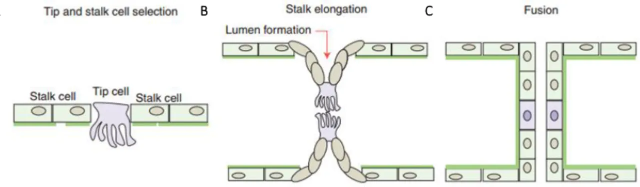
NGIOGENESISIn order to constantly respond to metabolic and growth demands, the vascular system is highly dynamic and endothelial cells are able to sense and respond to the environmental signals, initiating the angiogenic process. Biological signals such as hypoxia, ischemia and/or blood vessel damage, promote the upregulation of pro-angiogenic factors and, attracted by these factors, endothelial cells become motile and invasive by protruding filopodia that guide the new blood vessel in a certain direction (see Figure 1A). These so-called tip cells lead new sprouts and probe the environment for guidance cues. Following tip cells, stalk cells are able to proliferate to support sprout elongation and establish the vessel. These two cell types constitute the so-called vascular sprout, which remains connected to the original vessels while migrating into the avascular tissue. Tip cells anastomose with cells from neighboring sprouts to build vessel loops and the posterior initiation of blood flow, establishment of a basement membrane, and recruitment of mural cells stabilize the new connections (see Figure 1B and C). The iteration of the sprouting process continues until pro-angiogenic signals cease, and quiescence is re-established.
4
Figure 1. Schematic model of sprout initiation, vessel branching, and maturation. (A) Angiogenesis is activated in response to local growth factors, that (re)activates the quiescent endothelial cells differentiates them into tip and stalk cells. (B) Endothelial cells proliferate and collectively invade the tissue while remaining connected to the original vascular network and the tip cells contact other sprouts. (C) The new connection between different sprouts occurs through tip cell fusion (anastomosis) and the formation of the vascular lumen initiates blood flow. As a functional vessel loop is formed, there is a reduction of the release of endothelial growth factors, supporting the establishment of quiescence. Image adapted from [5].
2.1.1. VEGF AND NOTCH/DLL4 SIGNALING PATHWAYS
The specification of endothelial cells into tip and stalk cells within the activated endothelium is determined by the feedback loop between VEGF and Notch1/delta-like ligand 4 (Dll4) signaling pathways [6][7]. The resulting cell-fate specification mechanism is based on a Notch-mediated lateral inhibition process, in which a cell becomes tip cell and prevents its immediate neighbors from acquiring the same phenotype. This cell adopts this phenotype due to stochastic differences in local vascular endothelial growth factor (VEGF) concentrations, in filopodia elongation (and thus VEGF exposure) or in transcription rate that leads to small imbalances where one cell will express slightly higher Dll4 levels and, thus, dominate its neighbors by activating more Notch signaling [8][9]. The cell with more Dll4, and less Notch activity, will be selected as the tip cell. The activation of Notch inhibits VEGF receptor 2 (VEGFR2), indirectly inhibiting Dll4 expression levels, thereby reinforcing the dominance of the selected tip cell and limiting the number of tip cells induced by VEGF (see Figure 2) [10][11]. Besides downregulating VEGFR2, Notch signaling also affects VEGFR1 and VEGFR3 expression. Notch activation leads to increased levels of VEGFR1. This reduces the angiogenic sprouting response to VEGF, as both receptor variants act as a decoy for the VEGF ligand and limit VEGFR2 activation [12][13]. VEGFR3 is most strongly expressed in the leading tip cells and is downregulated by Notch signaling in the stalk cells [14]. Collectively, these molecular regulatory processes lead to increased responsiveness of the tip cell to VEGF and decreased sensitivity of stalk cells to this factor.
A B v d C v d
5 Recent observations challenge the idea of stable tip and stalk cell selection as their phenotype revealed to be dynamic and transient, relying on continued competition between the cells within blood vessel sprouts [15]. As a consequence, tip cells are overtaken and exchanged by neighboring stalk cells.
2.1.2. COMPETITION BETWEEN TIP AND STALK CELLS
Recent time-lapse imaging studies have shown the dynamic shuffling between tip and stalk cells at the leading front of growing sprouts, both in vitro (in embryoid bodies sprouting assays) and in vivo (in mice and zebrafish). The positional exchange suggests that the cells have constantly to reevaluate the VEGF/Notch signaling loop when they contact new neighboring cells, competing for the tip cell position based on their relative levels of VEGFR1 and VEGFR2. These receptors’ activities mediate the expression level of the Dll4 ligand: lower expression levels of VEGFR1 or higher levels of VEGFR2 result in higher levels of Dll4 expression and, hence, an increased ability of a cell to suppress its neighboring cells from becoming tip cells [15]. This proposes that the tip cell is constantly challenged by cells within the stalk cell region of the sprout to demonstrate its dominance in terms of VEGFR levels, ensuring that the cell with the best guiding capacities leads the sprout.
Figure 2. Tip to stalk cell lateral inhibition during sprouting angiogenesis. VEGF interacts with VEGFR2, expressed at the surface of the endothelial cells of the quiescent vessels. The VEGF signaling output is modulated, enhancing the binding activity and signaling of VEGF through VEGFR2. Under VEGF stimulation, Dll4 expression is up-regulated in the tip cells. In turn, Dll4 ligand activates Notch signaling in the stalk, consequently suppressing the tip cell phenotype. Notch signaling activation reduces VEGFR2 expression and increases VEGFR1/sVEGFR1 as well as the expression of different Notch target genes. In contrast, the tip cell receives low Notch signaling, allowing high expression of VEGFR2 but low VEGFR1. Image adapted from [5].
6
2.1.3. EMBRYOID BODY SPROUTING ASSAY AS A TOOL TO MEASURE CELL COMPETITION
Embryonic stem (ES) cells are pluripotent cells derived from blastocyst-stage early mammalian embryos that are capable of differentiation into all three germ layers and, therefore, have the potential to develop any type of tissue. Because of this particular characteristic, the maintenance and differentiation of embryonic stem cells contributed greatly to significant discoveries in developmental biology. When cultured in suspension without anti-differentiation factors, embryonic stem cells spontaneously differentiate and form three-dimensional multi-cellular pluripotent aggregates called embryoid bodies (EBs) [16]. This structure facilitates multimulti-cellular interactions, in which cell-cell contact exists, and consists of a mix of cell populations with ectodermal, mesodermal, and endodermal origins. Embryoid body’s aggregates recapitulate many aspects of cell differentiation during early mammalian embryogenesis offering the opportunity to mechanistically study these differentiation events of tridimensional assemblies of pluripotent cells with the great advantage that genetic manipulation of endothelial stem cells can be studied for gene mutations or knockouts that prove to be lethal during normal embryonic development in vivo [17]. As differentiation continues, a wide range of cell types are developed within the embryoid body’s environment and, consequently, this technique has been widely utilized as a trigger during the process of embryonic stem cells differentiation in vitro. The embryoid bodies’ potential covers different research fields and several gene knockout studies have being made using this technique. For example, embryoid bodies were used as an alternative for studying hematopoiesis [18][19], for complementary studies of cardiomyocytes differentiation [20] and for accessing endothelial cells behavior [21][22]. Currently, there are several methods with unique peculiarities used to form embryoid bodies from embryonic stem cells that allow for different objectives to be attained [23][24].
2.1.3.1. ASSESSING SPROUTING CAPACITY,DYNAMICS AND COMPETITION OF CELLS
The embryoid body model is valuable to easily access vessel development, by manipulation and visualization at a high resolution as the endothelial cell conventional cultures do not provide a proper microenvironment for it. Three dimensional models allow studying interactions between endothelial cells, adjacent non-endothelial cells and matrix, which are known to be essential in the regulation of vascular processes, as well as the metabolism and sprouting efficiency of these cells.
The sprouting capacity of endothelial cells during sprouting angiogenesis can be visualized by using embryoid bodies by designing a mosaic sprouting assays using embryonic stem cells. These cells, as said before, are able to form embryoid bodies that, when cultured in a solidified collagen matrix and treated with VEGF, exhibit robust differentiation into the endothelial cell lineage and an angiogenic behavior that resembles the major steps of sprouting angiogenesis in vivo [25][26].
Previous work described that chimeric cultures of embryonic stem cell, originated from 1:1 mixtures of two wild-type embryonic stem cell lines, revealed mosaic vascular sprouts. Deeper analysis of the genotypic origin of
7 the leading tip cells have shown an equal participation of each cell population to the leading tip cells, proving that wild-type cells of different genetic background have equal potential to acquire the tip cell phenotype (see Figure 3A and B)[15].
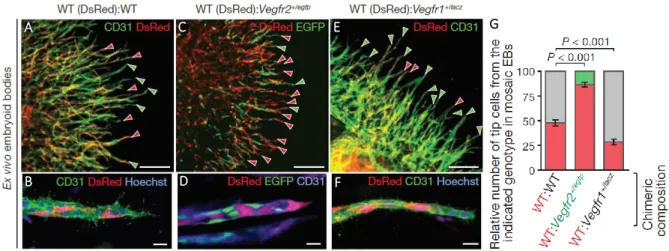
To investigate the interactions between cells in the selection process and the role of VEGFR levels in it, the same group studied different combinations of endothelial cells with half the amount of VEGFR1 or VEGFR2 competing with wild-type neighboring cells for the leading position [15]. It was shown that endothelial cells derived from embryonic stem cells heterozygous for the Vegfr2 allele (Vegfr2+/egfp, that exhibited approximately half the VEGFR2 levels, comparing to wild-type cells), when mixed with wild-type cells in a 1:1 ratio in mosaic cultures, contributed to only around 13% of the tip cells, showing that reduced Vegfr2 levels selectively impair the ability of cells to acquire the tip phenotype. When varying Vegfr1 levels using chimeric cultures of wildtype and Vegfr1+/lacz embryonic stem cells (that exhibited approximately half the VEGFR1 levels in comparison to wild-type cells), 70% of the tip cells were derived from the Vegfr1 heterozygote population. This demonstrates that a cell with lower Vegfr1 expression has a higher probability of acquiring the leading position, whereas a cell with lower Vegfr2 expression has reduced ability to take the lead, when competing with a wild-type neighboring cell, suggesting that the balance of VEGFR2 and VEGFR1 expression in individual endothelial cells affects their potential to become tip cells during sprouting angiogenesis (see Figure 3).
A C E F v d G v d
Figure 3. VEGFR levels in mosaic-tip-cell selection in vitro. (A, B) Sprouting vasculature (green) of a chimeric embryoid body composed of two different wild-type strains, immunolabelled with antibodies specific to CD31. One strain has ubiquitous expression of the fluorophore DsRed-MST (tip cells indicated by red arrowheads) and the other one is a non-fluorescent embryonic stem cell line (R1) (tip cells indicated by green arrowheads). (C, D) Chimeric embryoid body of wild-type (red) and Vegfr2 heterozygote cells (Vegfr2+/egfp, green) mixed in a 1:1 ratio. Wild-type tip cells are indicated by red arrowheads, Vegfr2+/egfp tip cells are indicated by green arrowheads. (E, F) Chimeric embryoid body of wild-type cells (red; tip cells are indicated by red arrowheads) and Vegfr1 heterozygote cells (tip cells are indicated by green arrrowheads), mixed in a 1:1 ratio. (G) Quantification of tip cells with the indicated genotype in chimeric embryoid bodies. Number of counted tip cells per condition: 630–708 (n embryoid bodies per condition = 6, WT:WT; 11, WT:vegfr2+/egfp; 9, WT:vegfr1+/lacz). Values represent means ± s.e.m. Scale bars: a, c, e, 200μm; b, d, f, 20μm. Image adapted from [15].
B v d D v d
8
Despite embryonic stem cells and embryoid bodies assays’ great potential and usefulness for this kind of studies, there is a limited number of genetically modified cell lines available. This results in a gap where knock-downs of genes, expression of fluorescent reporters and overexpression of specific isoforms, including fluorescently-tagged proteins, point mutations forms and dominant-negative/active isoforms are lacking.
2.2.
H
OW TOM
ANIPULATES
TEMC
ELLSEmbryonic stem cells have one great advantage over other cell types that is their accessibility to genetic manipulation. These cells are able to remain pluripotent after genetic modifications and so, they are widely used and there is a range of effective techniques that have been established for gene delivery and manipulation of embryonic stem cells. These methodologies include electroporation, transfection and infection protocols, and also different approaches for inserting, deleting, or changing the expression of genes.
2.2.1. GENETIC ENGINEERING
The strategies used in genetic engineering usually require the permanent modification of the target genome. For this to happen, the insertion of a construct into the genome of the cell needs to be stable but the prevailing approaches for this over the last years have been based in methods that lead to a random integration. Randomly integrating technologies enable users to create stable systems leading to lasting expression which results in a random insertion of selected DNA fragments into the host genome without the use of DNA homology. Besides providing a valuable tool for long-term expression in embryonic stem cells, random insertion leads to inconsistent integration sites and unpredictable expression patterns. In addition, the locus of insertion can result in partial or complete silencing of endogenous genes of embryonic stem cells, which can result in insertional mutagenesis with subsequent genome instability or toxicity [27][28].
Site-specific modification is an ideal method to avoid variable expression patterns and copy number variation as a result of random integration. These tools enable the targeting of specific sites within the chromosome where silencing is minimized. Gene targeting using traditional methods via homologous recombination has been extensively used to specifically alter genes. This method involves the introduction of a targeting construct homologous to the target gene sequence of the embryonic stem cell [29]. Despite offering, in principle, great specificity to the integration process, homologous recombination has an extremely low efficiency and is very laborious. With this in mind, new strategies have been developed to achieve integration at predetermined target sites with high efficiency, based on site-specific recombinases like Cre, FLP and PhiC31 integrase.
2.2.1.1. SITE-SPECIFIC RECOMBINASES
Site-specific recombinases (SSRs) are highly specialized enzymes that perform rearrangements of DNA fragments by recognizing and binding to short DNA sequences, sites. Recombination sites are typically between
9 30 and 200 nucleotides to which the recombinase binds, flanking a central crossover sequence at which the recombination takes place [30][31][32].
FLP and Cre recombinases belong to the integrase family of recombinases (also termed “λ integrase” family) and do not need any accessory factors to mediate recombination. The FLP recombinase (FLP) recognizes FRT sites and mediates recombination between them [33]. The Cre (causes recombination) recombinase recombines loxP (locus of crossover (×) in P1) target sites [34] and shares the common integration mechanism with FLP. A considerable limitation of both the FLP/FRT and the Cre/loxP system exists when used for integration and inversion. In both cases two identical target sites (homotypic sites) are present in close proximity after the recombination event, which then can serve again as substrates for a further recombination event, i.e., for excision or reinversion. In other words, FLP and Cre are “bidirectional” recombinases. Because excision reactions are kinetically favored over integrations, the integrated DNA is highly unstable [35].
The integrase of the Streptomyces phage PhiC31 normally mediates the integration of the phage genome [36] into the bacterial chromosome through heterotypic recombination sites, termed attB (attachment site Bacterium) and attP (attachment site Phage) sites, and like FLP and Cre, it does not need any accessory factors to mediate this integration [37]. The two sequences, though largely different, share a 3bp central region, where the crossover occurs, and this central region is flanked by imperfect repeats. Recombination between the two attachment sites creates the hybrid sites attL and attR (Left and Right) that are no longer substrates for the PhiC31 integrase, thus rendering the recombination irreversible (see Figure 4) [38][39]. This distinguishes it from Cre and FLP, and thus the PhiC31 integrase, as a unidirectional recombinase, is an efficient tool to insert transgenes into a genome [40][41]. This recombination system was successfully applied in bacteria [37], yeast [42], mammalian cell lines [41] [43], Xenopus [44], Drosophila [45][46][47] and plants [48][49] and so, the PhiC31-based transgenesis has become widely used. In this
technique, the attB site-containing vectors are co-injected with PhiC31 integrase encoding mRNA into embryos that harbor a previously introduced transgenic attP site in their genome. The enzyme will then recombine the vector attB with the genomic attP sites, resulting in the integration of the full transgene vector into the genomic
Figure 4. PhiC31 Integrase System. The phiC31 integrase mediates recombination between the two attachment sites. In the presence of PhiC31 integrase, an attB-containing donor plasmid can be unidirectionally integrated into a target genome through recombination at sites with sequence similarity to the native attP site.
10
attP locus with high efficiency, regardless of its size. The well-characterized integration sites eliminate time and effort to map transgene insertions and eliminate the need for several independent lines per transgene, thus effectively reducing the space requirement and workload of generating, analyzing, and maintaining transgenics. Although site-specific recombination systems are diverse, the common property that makes these enzymes so attractive (the ability to specifically and autonomously integrate, excise, or invert defined sequences of DNA) also limits their practical utility. Site-specific recombinases are involved in essential biological functions, what demonstrates their strict specificity toward their natural target. Indeed, the use of these enzymes in mammalian cells requires, either the presence of rare pre-existing recognition sites or the previous introduction of specific target sites within the host genome by homologous recombination [40][50]. Therefore, besides site-specific recombinases potential as transformative tools for targeted genetic engineering, their application is conditioned by technical constraints. In order for this technology to reach its full potential, methods for the enhancement and design of custom recombinases capable of modifying specific DNA sequences are required.
2.2.1.2. CRISPR/CAS9
Recently, a new class of genome engineering tools was reported and it has provided a much simpler and more economic method for gene-targeted modification, the CRISPR/Cas9 system. This follows the previous efforts in genetic manipulation, including homologous recombination [51] and RNA interference (RNAi) [52]. Other recent approaches to targeted genome modification, like zinc-finger nucleases (ZFNs) [53] and transcription-activator like effector nucleases (TALENs) [54], enable researchers to generate permanent mutations by introducing double-stranded breaks to activate repair pathways but are costly and time-consuming approaches, limiting their general use. The clustered regularly interspaced short palindromic repeats (CRISPR) are unique sequences in between DNA repeats that naturally occur and were shown to play an essential role in immunity of selected
bacteria and archaea, enabling the organisms to respond to and eliminate invading genetic material. These
repeats were initially discovered in the 1980s in E. coli [55], but their potential was only confirmed with the demonstration that S. thermophilus can acquire resistance against a bacteriophage by integrating a genome fragment of an infectious virus into its CRISPR locus [56].
There are three different types of CRISPR mechanisms, being type II the most studied. In this case, DNA from viruses or plasmids is cut into small fragments and integrated into a CRISPR locus among a series of short repeats. The loci are transcribed, and transcripts are then processed generating small RNAs (crRNA – CRISPR RNA), which are used to guide effector endonucleases that target invading DNA based on sequence complementarity [57]. This CRISPR mechanism distinguishes from the others as only one Cas protein (Cas9) is required for gene silencing. Cas9 (also known as Csn1), comes from Streptococcus pyogenes and has been shown, through knockdown and rescue experiments, to be a key player in this CRISPR system as it participates in the processing of crRNAs, and is responsible for the destruction of the target DNA. To achieve site-specific DNA
11 recognition and cleavage, Cas9 has to be complemented with a crRNA and a separate trans-activating crRNA (tracrRNA), partially complementary to the crRNA [57][58]. More recently, researchers developed a simplified two-component system that combines tracrRNA and crRNA into a single synthetic single guide RNA (sgRNA). This mechanism was shown to be as effective as the original CRISPR in guiding targeted gene alterations [57]. To date, there are three variants of the Cas9 nuclease that have been implemented in genome-editing protocols. The first is the wild-type Cas9, which can site-specifically generate a DNA double-strand break at the targeted genome locus that activates repair through error-prone nonhomologous end joining or homology-directed repair. In the absence of a template, the nonhomologous end joining is activated, resulting in insertions and/or deletions that disrupt the target loci. In the presence of a donor template with homology to the targeted locus, the homology-directed repair pathway operates, allowing for precise mutations to be made (see Figure 5A) [59][60]. The second variant has increased precision and is a Cas9 mutant form, known as Cas9D10A, with only nickase activity. This means that it cleaves only one DNA strand, and does not activate nonhomologous end joining. Instead, when provided with a homologous repair template, DNA repairs are conducted via the high-fidelity homology-directed repair pathway only, resulting in reduced insertion and/or deletion mutations (see Figure 5B) [57][61]. The last variant is a nuclease-deficient Cas9 (dCas9) with mutations in two specific Cas9 domains that inactivate cleavage activity, but do not prevent DNA binding and therefore, this variant can be used to sequence-specifically target any region of the genome without cleavage [62][63], as a gene silencing or activation tool by fusing with various effector domains [64][65] and as a visualization tool, for instance, the fusion with enhanced green fluorescent protein (EGFP) allowed visualizing repetitive DNA sequences with a single sgRNA or nonrepetitive loci using multiple sgRNAs (see Figure 5C) [66].
Figure 5. CRISPR/Cas9 genome engineering with Cas9 nuclease variants. (A) Wild-type Cas9 nuclease site specifically cleaves double-stranded DNA. Non-homologous end joining can result in insertions/deletions disrupting the target sequence. Alternatively, precise mutations and knock-ins can be made by providing a homologous repair template and exploiting the homology directed repair pathway. (B) Mutated Cas9 makes a site specific single-strand nick. Two sgRNA can be used to introduce a staggered double-stranded break which can then undergo homology directed repair. (C) Nuclease-deficient Cas9 can be fused with various effector domains allowing specific localization. For example, transcriptional activators, repressors, and fluorescent proteins. Image adapted from [83].
12
Besides being a faster alternative for cell genome manipulation compared to traditional gene targeting methods, the CRISPR/Cas9 system has some limitations. First, there is the possibility of off-site effects, when a mutation is introduced at non-specific loci with similar, but not identical, homology to the target sites. These can be difficult to identify and require scanning the genome for mutations at sites with sequence similarity to the sgRNA target sequence. Still, this mechanism has better targeting efficiency comparing with the previously established methods (TALENs or ZFNs) that, in human cells, were only able to achieve efficiencies ranging from 1% to 50% [67][68]. In contrast, the Cas9 system has been reported to have efficiencies up to 70% in zebrafish and plants [69], and ranging from 2–5% in induced pluripotent stem cells [70]. In addition, other studies were able to improve genome targeting up to 78% in one-cell mouse embryos, and to achieve effective germline transmission through the use of dual sgRNAs to simultaneously target an individual gene [71]. Second, mosaicism can occur when nucleases do not cut the DNA at the one cell stage of embryonic development of a mouse line and when that happens, mice with a mutant allele in only some of their cells can be produced. Multiple alleles are another drawback of the technique, due to the fact that the healing of the nuclease cleavage site by non-homologous end joining can produce cohorts of mice with different mutations from the same targeting constructs, requiring genome sequencing to verify the nature and position of the specific mutation. The production of mice with mosaics of multiple mutations is also possible and breeding may be required to segregate and isolate mice that carry single mutations.
Despite the limitations, the CRISPR/Cas9 system has been widely adopted and successfully used to target important genes in many cell lines and organisms, including human [70], bacteria [72], zebrafish [73], C. elegans [74], plants [70], Xenopus tropicalis [75], yeast [76], drosophila [77], monkeys [78], rabbits [79], pigs [74], rats [80] and mice [81]. This technique requires only the redesign of the crRNA to change target specificity, contrasting with other genome editing tools, where redesign of the protein-DNA interface is required. The rapid progress in developing Cas9 into a set of tools for cell and molecular biology research has been remarkable, likely due to the simplicity, high efficiency and versatility of the system. Of the designer nuclease systems currently available for precision genome engineering, the CRISPR/Cas system is by far the most user-friendly and its potential and usefulness seems to be unlimited.
2.3.
P
URPOSE OFT
HIST
HESISTaking into account the difficulties that the previous established methods bring to the genome editing process, in this thesis we propose a new mechanism to insert a gene, of any size, into an embryonic stem cell in a fast and efficient manner. This will allow the easy and versatile manipulation of embryonic stem cells to create embryoid bodies and mouse lines that will posteriorly allow the study of several different mechanisms in transversal fields of biology.
13
3. MATERIALS & METHODS
3.1.
P
OLYMERASEC
HAINR
EACTIONPolymerase chain reaction (PCR) is a cell-free technique, which is used to amplify a specific DNA sequence by in
vitro enzymatic replication assisted by a DNA polymerase. In an exponential manner a small amount of DNA is
amplified into a large amount of DNA in a very short period. The region comprised between the two selected primers is the region of interest to be amplified. The DNA containing the sequence to be amplified is incubated in a test tube with the primers, each complementary to the ends of the targeted DNA, the four deoxynucleotides and a DNA polymerase.
The PCR process consists of a series of about 25-35 subsequent cycles and each cycle consists of three precisely time- and temperature-controlled steps (denaturation, annealing and extension). The first step separates the double stranded DNA into two single strands by use of a high temperature (95°C - 98°C, depending on the manufacturers’ indications). This denaturing step breaks the hydrogen bonds between the two strands. Then the temperature is lowered to the primers’ specific annealing temperature to allow them to base pair to their complementary sequences on the template strands. Further the reaction is heated to 72°C, the optimal temperature for the heat stable DNA polymerase to replicate the single stranded DNA segments. The DNA polymerase uses deoxynucleotides as building blocks of the new strands.
PCRs were set up with either Phusion High-Fidelity DNA Polymerase from Thermo Scientific for DNA extraction or NZYTaq 2× Green Master Mix from NZYTech to identify positive clones using optimal reaction conditions according to the manufacturer’s protocol (see list of primers in the Appendix).
Table 1. Component and cycling instructions for the PCR reactions. The reagents pipetting information (left) and cycling conditions (right) for both Phusion High-Fidelity DNA Polymerase (top) and NZYTaq 2× Green Master Mix (bottom). X°C represents the annealing temperature of the primers used in the reaction.
Components 50µL Reaction Final Concentration Cycle Step Temperature Time Cycles
H2O up to 50µL Initial Denaturation 98°C 30sec 1
5X Phusion HF Buffer 10µL 1X Denaturation 98°C 5-10sec
25-35
10 mM dNTPs 1µL 200µM each Annealing X°C 10-30sec
Forward Primer - 0.5µM Extension 72°C 15-30sec
Reverse Primer - 0.5µM Final Extension 72°C 5-10min 1
Template DNA - 1pg-10ng 4°C hold
Phusion DNA Polymerase 0.5µL 0.02U/µL
Components 20µL Reaction Final Concentration Cycle Step Temperature Time Cycles
H2O up to 20µL Initial Denaturation 95°C 120sec 1
Forward Primer - 0.5µM Denaturation 95°C 30-60sec
25-35
Reverse Primer - 0.5µM Annealing X°C 30-60sec
Template DNA - 0.01-0.5μg Extension 72°C 60sec/kb
NZYTaq 2× Green Master Mix 10µL Final Extension 72°C 5-10min 1
14
3.2.
A
GAROSEG
ELE
LECTROPHORESISAgarose gel electrophoresis was used to confirm each step of the cloning process and to separate PCR products to purify the desired DNA fragments. These fragments are separated by applying an electric field to move the negatively charged molecules through an agarose matrix that are separated by size in the agarose gel matrix. Shorter nucleic acid molecules move faster and migrate further than longer ones due to the fact that these drift more easily through the pores of the gel.
The matrix used was 1% w/v% agarose dissolved in 1xTBE buffer (1M Tris-base, 1M Boric acid, 0.02M EDTA). 1kb DNA Ladder or 1kb Plus DNA Ladder from New England Biolabs was run in parallel to the samples and DNA Gel Loading Dye (6X) from Thermo Scientific was added to all samples before loading them in the gel (except in the case of reactions assembled with NZYTaq 2× Green Master Mix that may be directly loaded onto agarose gels). Electrophoresis was carried out in 1X TBE buffer at 100V for 40-60 minutes. Visualization of DNA was performed by exposure to UV-light using Chemidoc XRS+ (Bio-Rad) and the products of the reactions were confirmed by size comparison with the DNA ladder.
3.3.
DNA
E
XTRACTION FROMA
GAROSEG
EL ANDP
URIFICATIONGel extraction is used to isolate the desired fragment of DNA from the agarose gel following electrophoresis. This technique involves identifying the fragments of interest, isolating the corresponding band, isolating the DNA from it and removing the accompanying salts and stain.
The DNA fragments were visualized on a Dual-Intensity Transilluminator using the low setting of UV-light and the desired band was physically removed by cutting it out from the agarose gel with a clean scalpel. The extraction of the DNA fragments from the gel slice was performed using QIAquick® Gel Extraction Kit from Qiagen following the manufacturer’s protocol. DNA was eluted from the spin column using 30µL of dH2O.
3.4.
E
TOH
P
RECIPITATIONEthanol precipitation is a commonly used technique to purify and/or concentrate nucleic acids (DNA or RNA) preparations in aqueous solution.
For this procedure salt and ethanol are added to the aqueous solution (1/10 volume of sodium acetate (3M, pH 5.2) and 2.5–3.0 X volume (calculated after addition of sodium acetate) of 96% ethanol), followed by a 2 hours incubation at -20°C, which forces the precipitation of nucleic acids out of solution. After precipitation the nucleic acids can then be separated from the rest of the solution by 20 minutes centrifugation at 4°C at 13000rpm. The pellet is washed in 200µL of cold 70% ethanol then after a further 15 minutes centrifugation at 4°C at 13000rpm the ethanol is removed, and the nucleic acid pellet is air dried before being resuspended.
15
3.5.
D
IGESTION OFDNA
WITHR
ESTRICTIONE
NZYMESRestriction enzyme digestion takes advantage of naturally occurring enzymes that recognize specific sequences in DNA and cleave it at these specific sites. Restriction digestion of plasmid constructs provides a fast and cost-efficient method of gaining indirect sequence information, giving insights for the presence or absence of an insert, orientation of the insert, plasmid size, and site-specific sequence data. This technique is also useful to produce a DNA fragment that can be cloned directly into a vector. Plasmid DNA is digested with one or more restriction enzymes selected to give a distinct DNA band pattern that is easily resolved by electrophoresis. DNA digestions were performed using restriction enzymes from New England Biolabs® or Thermo Scientific. Restriction enzyme digestions were performed under optimal conditions described by the manufacturer (see list of all the restriction enzymes in the Appendix).
3.6.
L
IGATION OFDNA
F
RAGMENTSThe ligation of DNA fragments is accomplished by covalently connecting the insert DNA (gene or fragment of interest) into a compatibly digested vector backbone and is mediated by a DNA ligase enzyme that catalysis the formation of covalent phosphodiester linkages, which permanently join the nucleotides together. This reaction involves DNA fragments that have been generated by restriction enzyme digestion. Most restriction enzymes digest DNA asymmetrically across their recognition sequence, resulting in a single stranded overhang on the digested end of the DNA fragment. The overhangs, called sticky-ends, when compatible, meaning that the overhanging base pairs on the vector and insert are complementary, allow the vector and insert to bind to each by the ligation reaction. PCR usually generates blunt-ended products and also the digestion with some restriction enzymes, meaning that there are no overhanging base pairs in the fragment. The ligation of blunt-ended products does not involve base-pairing of the protruding ends, instead, the reaction depends on random collisions between the blunt-ends.
Ligation of DNA fragments was performed using T4 DNA Ligase from New England Biolabs® with supplemented buffer. Ligation took place under optimized reaction conditions depending on the ligation type (see Table 2).
Table 2. Reaction mixtures and conditions that were used for either sticky- or blunt-end ligations in the present study. Sticky-end Ligation Blunt-end Ligation
Components 20µL Reaction Components 20µL Reaction
Vector DNA (4kb) 50ng (0.020pmol) Vector DNA (4kb) 50ng (0.020pmol)
Insert DNA (1kb) 37.5ng (0.060pmol) Insert DNA (1kb) 37.5ng (0.060pmol)
10X T4 DNALigase Buffer 2µL 10X T4 DNALigase Buffer 2µL
Nuclease-free water up to 20µL 50% PEG 4000 solution 2µL
T4 DNA Ligase 1µL Nuclease-free water up to 20µL
Incubate 1H at RT T4 DNA Ligase 1µL
16
3.7.
G
ATEWAY®
T
ECHNOLOGYThe Gateway® Technology is a cloning system based on site-specific recombinases, providing a fast and efficient way to insert one or multiple DNA sequences into vectors. Two recombination reactions constitute the basis of this technology: the BP reaction, that allows recombination of an attB substrate (attB-PCR product or a linearized attB expression clone) with an attP substrate (donor vector) creating an attL-containing entry clone; and the LR reaction, that facilitates recombination of an attL substrate (entry clone) with an attR substrate (destination vector) thus creating an attB-containing expression clone.
To perform these recombination reactions we used Gateway® BP Clonase® II Enzyme mix and Gateway® LR Clonase® II Enzyme mix for the respective reaction and optimized the manufacturer’s protocol for higher efficiency of both reactions (see Table 3).
Table 3. Optimized reaction mixtures and conditions for both BP and LR reactions.
BP Reaction LR Reaction
Components 8µL Reaction Final Concentration Components 8µL Reaction Final Concentration
attB PCR Product or
Linearized attB Clone - 50fmol
5' Entry Clone - 10fmol
Middle Entry Clone - 10fmol
Donor Vector - 150fmol 3' Entry Clone - 10fmol
TE buffer, pH 8.0 up to 8µL Destination Vector - 150fmol
BP Clonase™ II 2µL TE buffer, pH 8.0 up to 8µL
Incubate ON at RT LR Clonase™ II 2µL
Proteinase K Solution 1µL Incubate ON at RT
Incubate 10 minutes at 37°C Proteinase K Solution 1µL
Incubate 10 minutes at 37°C
3.8.
T
RANSFORMATION OFDNA
INTOC
OMPETENTC
ELLSTransformation is the process by which foreign DNA is introduced into host bacteria. The transformation process involves compromising the bacterial membrane permeability (by heat-shock, electric current, etc.), allowing the DNA to be introduced into the cytosol of the bacterium. The outgrowth of bacteria following transformation allows repair of the bacterial surface and selection of recombinant cells if the newly acquired DNA conveys antibiotic resistance to the transformed cells. Transformation of bacteria with plasmids is important for replicating plasmids.
To perform the transformation, we used 25 - 50µL (depending on the strain; 25µL One Shot® ccdB Survival™ 2 T1R Competent Cells from Invitrogen™; 50µL NZY5α Competent Cells from NZYTech) of competent cells were thawed on ice and 2-5μL of plasmid was added to the cells. Incubation of the cells continued on ice for 30 minutes and then the cells were heat pulsed at 42°C for 45 seconds followed by 2 minutes incubation on ice. 300 - 900µL of RT LB medium was added to the cells and following incubation at 37°C for 1 hour. 50/100μL of sample was plated out on agar plates with appropriate antibiotic(s) for selection. Plates were incubated at 37°C
17 ON. The resulting colonies were tested by PCR and the positive clones inoculated in 5 - 6mL of LB media with the correct antibiotic(s) and cultivated at 37°C in a shaker ON. Plasmids were isolated using the QIAprep® Spin Miniprep Kit from Qiage) according to manufacturer’s protocol. The obtained DNA was quantified using a full-spectrum UV-Vis spectrophotometer, Nanodrop 2000, Thermo Scientific.
3.9.
S
EQUENCINGDNA sequencing is the process of determining the precise order of nucleotides within a DNA strand. Sequence data were obtained by LIGHTRUN sequencing, GATC biotech, of the purified plasmid DNA (5µL of DNA 80 - 100ng/µL) with the desired primer (5µL of 5µM primer).
3.10.
T
RANSFECTION OFU2OS
C
ELLSU2OS cells were routinely cultured with Dulbecco's Modified Eagle Medium (DMEM), high glucose, pyruvate, from Gilbco™, and incubated at 37°C and 5% CO2 to ensure a stable environment for optimal cell growth. Cells were transfected with Lipofectamine™ 3000 Reagent kit from Invitrogen® according to the manufacturer’s protocol. In the following day, the medium was changed to eliminate dead cells and toxic reagents.
3.11.
RNA
E
XTRACTIONFor RNA extraction from cells we used the RNeasy® from Qiagen and followed the manufacturer’s protocol for purification of total RNA from animal cells using spin technology. We used confluent cells in a 12-well plate, disrupted in Lysis Buffer complemented with β-mercapthoethanol and homogenized. Ethanol is then added to the lysate, creating conditions that promote selective binding of RNA to the RNeasy membrane when the sample is applied to the RNeasy Mini spin column. The total RNA binds to the membrane, and contaminants are efficiently washed away, which results in high-quality RNA, eluted in 35µL of RNase-free water. All bind, wash, and elution steps are performed by centrifugation in a microcentrifuge. The RNA was then quantified using Nanodrop 2000, Thermo Scientific.
3.12.
R
EAL-T
IMEPCR
To perform PCR using RNA as a starting template, the RNA must first be reverse transcribed into cDNA in a reverse transcription (RT) reaction. We first digested 1µg of the RNA samples with DNase I, RNase-free, from Thermo Scientific, following the manufacturer’s protocol, to digest single- and double-stranded DNA. To inactivate DNase I we added EDTA to a final concentration of 5mM before heating for 10 minutes at 65°C. The cDNA synthesis from RNA was performed using SuperScript® IV First-Strand Synthesis System for RT-PCR from Thermo Scientific and following the manufacturer’s protocol. The cDNA samples were diluted 25 times for the subsequent quantitative real-time PCR reactions.
18
Quantitative real-time PCR was performed using a 7500 Fast Real-Time PCR System (Applied Biosystems®) with Power SYBR® Green PCR Master Mix from Applied Biosystems®, following the standard program. In each well of a MicroAmp® Fast Optical 96-well Reaction Plate from Applied Biosystems®, we added 5µL of cDNA sample and 15µL of qRT-PCR mixture (10µL of Power SYBR® Green PCR Master Mix, 0.5µL of forward and reverse primer’s mix at 4µM and 4.5µL of Milli-Q RNase/DNase free water). The expression levels were normalized to GAPDH (a housekeeping gene).