Proteins of the canine seminal plasma
Proteínas do plasma seminal canino
Annice Aquino-CortezI* Lúcia Daniel Machado da SilvaI Airton Alencar de AraújoII Erika da Silva Bezerra de MenezesIII Arlindo de Alencar Araripe Noronha MouraIV ISSN 1678-4596
ABSTRACT
Studies have been performed to identify the proteins present in canine seminal plasma (SP) and relate them to sperm quality as well as to discover molecular markers of reproductive tract diseases. There is evidence that heparin-binding proteins, zinc-binding proteins, and lactoferrin as well as the matrix metalloproteinase, superoxide dismutase, catalase, and glutathione peroxidase enzymes are associated with canine sperm quality. Other studies indicate that prolactin and enzymes like arginine esterase, acid phosphatase, and alkaline phosphatase could be successfully used as biomarkers of reproductive disorders. Thus, the present literature review aims to address aspects related to
proteins of the canine SP, their influence on fertility, and their
importance as biomarkers of reproductive disorders.
Key words: seminal plasma, dog, proteins.
RESUMO
Pesquisas têm sido realizadas para identificar as
proteínas presentes no plasma seminal canino, com o intuito de relacioná-las com a qualidade espermática, bem como buscar por marcadores moleculares de patologias do trato reprodutivo. Há evidências de que as proteínas ligadoras de heparina, ligadoras de zinco, a lactoferrina, bem como as enzimas matrix metalloproteinase, superoxide dismutase, catalase e a glutationa peroxidase estão relacionadas com a qualidade seminal canina. Outras pesquisas indicam que a prolactina, e as enzimas arginina esterase, fosfatase ácida e fosfatase alcalina poderiam ser utilizadas com sucesso como biomarcadores de doenças reprodutivas. Assim, esta revisão de literatura objetiva abordar aspectos relacionados às proteínas
do plasma seminal canino, suas influências sobre a fertilidade, e sua
importância como biomarcadores de doenças reprodutivas.
Palavras-chave: plasma seminal, cão, proteínas.
INTRODUCTION
Secretions produced by the testicles, epididymis, and accessory sex glands constitute the seminal plasma (SP) (MANN & MANN, 1981; REGO et al., 2014). The prostate is the only accessory sex gland
of dogs, and its fluid constitutes approximately 95%
of the ejaculate (IGUER-OUADA & VERSTEGEN,
2001). It is possible that the prostate fluid has a unique
relationship with the spermatozoa in dogs and affects them differently than in other species.
Various proteins have been identified in
the canine SP, and studies have attempted to show
how these macromolecules can affect sperm quality
and fertility (KIKUCHI et al., 2003; SOUZA et al., 2007; SAENGSOI et al., 2011;
MOGIELNICKA-BRZOZOWSKA et al., 2012). The identification of
SP proteins should provide a better understanding of canine reproductive physiology (SOUZA et al., 2007), assisting in the development of new semen storage protocols (BECCAGLIA et al., 2009).
Prostate disorders have been observed in dogs (SMITH, 2008), and these disorders deserve special attention. Studies have shown that different proteins could be used as biomarkers for the diagnosis of prostate pathologies such as benign prostatic hyperplasia (SAGOLS & NAVARO, 2013). In addition to prostate diseases, other reproductive — REVIEW —
ILaboratório de Reprodução de Carnívoros (LRC), Universidade Estadual do Ceará (UECE), Avenida Dr. Silas Munguba, 1700,
60714-903, Fortaleza, Ceará, Brasil. E-mail: annicevet@gmail.com. *Corresponding autor.
IILaboratório de Nutrição e Produção de Ruminantes (LANUPRUMI), Universidade Estadual do Ceará (UECE), Fortaleza, CE, Brasil. IIIDepartment of Animal and Dairy Sciences, Mississippi State University, Mississippi State, United States of America.
IVLaboratório de Fisiologia Animal, Universidade Federal do Ceará (UFC), Fortaleza, CE, Brasil.
disorders such as azoospermia (SCHAFER-SOMI et al., 2013) and obstruction of the vas deferens or epididymis (GOBELLO et al., 2002) could also be diagnosed using biomarkers.
The identification of proteins in the
canine SP, including molecular markers, can provide
important benefits for human medicine, such as early
diagnosis of diseases of the male reproductive tract, because canines are an excellent model for the study of complex diseases in humans (ROWELL et al., 2011).
Given the above findings, the present
literature review aims to address aspects related
to the proteins of the canine SP, their influence
on fertility, and their importance as biomarkers of reproductive disorders.
Proteins of the canine SP
Experimental evidence from several studies suggests that the seminal proteins aid in maintaining the viability and survival of spermatozoa in the female reproductive tract (SAENGSOI et al., 2011; DRUART et al., 2013), capacitation (MANJUNATH & THÉRIEN, 2002), and spermatozoa binding to the zona pellucida of the oocyte (BECCAGLIA et al., 2009). The proteins of the canine SP also act as signaling molecules for the female immune system, modulating spermatozoa rejection or tolerance in the female genital tract (RODRÍGUEZ-MARTÍNEZ et al., 2011).
In recent decades, the proteomic profiles
of the semen of various species have been studied to elucidate the effects of the different protein groups
present in the SP and how they influence sperm cells
(SOUZA et al., 2007; MOURA et al., 2010; DRUART et al., 2013). Interestingly, ejaculation is fractionated in many species (humans, swine, canines, and
equines), and the interactions between the proteins of
the canine SP and the spermatozoa, secretions, and epithelium of the female genital tract are still unknown (RODRÍGUEZ-MARTÍNEZ et al., 2011). In dogs, a species with fractionated ejaculation, the few existing studies on the subject have sought possible links between the different proteins isolated in the ejaculate and the sperm concentration (KIKUCHI et al., 2003), motility, vigor; percentage of morphologically normal spermatozoa, functionality and integrity of the sperm membrane (SOUZA et al., 2007); and sperm viability (SAENGSOI et al., 2011).
The concentrations of the proteins of the canine SP range from 1.88g dL-1
(AQUINO-CORTEZ, 2003) to 2.3g dL-1 (MOTHEO et al.,
2014). Higher protein concentrations are present in the second fraction of the ejaculate (rich in spermatozoa, 4.15g dL-1) than in the third fraction
or prostatic fluid (2.00g dL-1, BARTLETT, 1962).
In an experiment conducted with German shepherd and Rottweiler dog breeds, the total protein
concentration of prostatic fluid did not differ
between the studied breeds or between the good-
and poor-quality ejaculates (AQUINO-CORTEZ,
2003). When performing a proteomic analysis of the canine SP by one-dimensional electrophoresis on a denatured dodecyl-sulfate-polyacrylamide
gel (12% and 18% SDS-PAGE), 31 protein bands
with molecular weights ranging from 2.71kDa to 139.63kDa were reported; no seasonal variation
in the protein profile was observed (MARTINS, 2005). In a subsequent study, which also used
one-dimensional electrophoresis with different
polyacrylamide concentrations (13% and 22% SDS-PAGE), 37 protein bands were identified. Bands
B4 (67kDa) and B5 (58.6kDa) were positively associated with motility, vigor, morphologically normal spermatozoa, and membrane integrity (SOUZA et al., 2007).
The canine SP may also be involved in the process of sperm capacitation. During ejaculation, several proteins bind to the plasma membrane of spermatozoa, blocking the progesterone receptors located in the acrosome region (SIRIVAIDYAPONG et al., 1999). Coating proteins and glycoproteins of prostatic origin are gradually removed during capacitation, and other proteins can bind to the exposed receptors to induce the acrosome reaction (ROTA et al., 2007).
A group of SP proteins thought to be involved in capacitation and modulation of the acrosome reaction belongs to the heparin-binding protein (HBP) group (MILLER et al., 1990). HBPs bind to the spermatozoa surface after ejaculation and affect fertility due to their modulatory role during the acrosome reaction (MANJUNATH et al., 1993). SOUZA et al. (2006),
using heparin affinity chromatography followed by one-dimensional electrophoresis (12% and 18%
SDS-PAGE), reported that 19 protein bands of the canine SP bound to the heparin column. Of these 19 proteins, a 61.5kDa protein was particularly thought be involved in the canine acrosome reaction (SOUZA et al., 2006). The most studied HBP in several species is osteopontin (OPN), which has been found in high concentrations in canine SP (SOUZA et al., 2009). Although OPN has
been associated with fertility in equines (BRANDON
et al., 1999) and bovines (MOURA et al., 2006), this relationship has not yet been well established in canines (SOUZA et al., 2009); however, it is believed to exert a similar function in canines as in other species.
the fertilization process. The ZnBPs of the canine SP comprise 13 protein bands (11.6 to 152.3kDa) (MOGIELNICKA-BRZOZOWSKA et al., 2012). Zinc is a cofactor for more than 80 metalloenzymes involved in DNA transcription and protein synthesis. These two pathways are the major components of germ cell maturity; therefore, zinc is believed to be important for reproduction (HADWAN et al., 2013).
Enzymes with antioxidative action are present in the semen and are involved in the removal of reactive oxygen species (ROS). ROS are active molecules produced during oxygen reduction, and some evidence suggests that the spermatozoa of mammals are capable of generating ROS, such as the peroxide anion (O2-) and hydrogen peroxide (H
2O2),
during the processes of maturation and storage in the epididymis (AITKEN, 2002). ROS, when produced in high concentrations, lead to many deleterious effects on the function and viability of spermatozoa (CASSANI et al., 2005). ROS can induce oxidative damage to DNA, lipids, and proteins of the cellular systems and inhibit sperm movement (AITKEN, 2002). The production of these highly reactive metabolites is of major importance in the signal transduction pathways that control capacitation and determine how long these cells can remain in a functional and viable state as they migrate and are stored in the epididymis (AITKEN, 2002).
Therefore, spermatozoa are extremely dependent upon the antioxidant protection provided by the reproductive tract environment (ANGRIMANI et al., 2014a). The most important antioxidant enzyme of the mammalian semen is superoxide dismutase (SOD, CASSANI et al., 2005). Other antioxidant enzymes such as catalase (CAT) and glutathione peroxidase
(GPx) have also been identified in the canine SP
(CASSANI et al., 2005; KAWAKAMI et al., 2007; STRZEZEK et al., 2009; NEAGU et al., 2011).
SOD catalyzes the dismutation of O2- into
H2O2, and in mammalian spermatozoa, this enzymatic antioxidant system protects against oxidative stress during ejaculation and the passage through the female genital tract (CASSANI et al., 2005). A higher activity of SOD was observed in oligozoospermic dogs than in normozoospermic dogs (CASSANI et al., 2005). KAWAKAMI et al. (2007) reported a lower activity of SOD in the semen of asthenospermic dogs than in dogs with normal sperm motility. These studies show the importance of SOD in controlling oxidative stress,
which significantly affects the sperm quality of dogs.
CASSANI et al. (2005) reported the SOD activity in the three fractions of canine semen and suggested that this enzyme has a protective effect against the oxidative
stress in the canine spermatozoa. Subsequently, SOD
activity was also demonstrated in the corpus, caput,
and cauda of the canine epididymis, with no significant
difference in the enzyme concentration among the three ejaculated fractions and the three epididymis segments (ANGRIMANI et al., 2014b).
NEAGU et al. (2011) studied the relationship between cellular damage caused by freezing and thawing semen and the activity of antioxidant enzymes in the canine SP. In this study, fresh semen was added to TRIS-glucose extender and centrifuged at 700g for eight minutes. The spermatozoa pellet was frozen, and the SP was frozen/thawed for further evaluation of the activity of the antioxidant enzymes. The authors reported a positive correlation between the activity of SOD of canine SP and sperm velocities post thaw (n=20, r=0.604, P<0.01) (NEAGU et al., 2011). KOBAYASHI et al. (2014) evaluated the effect of SOD on in vitro spermatozoa by adding 30 unit mL-1 of SOD (Wako Pure Chemical Industries
Co., Ltd., Osaka, Japan) to spermatozoa in minimal essential medium (MEM). After 24h of incubation at 38ºC, the percentage of sperm viability, motility, and hyperactivation in SOD-MEM were higher than those in MEM only, showing that SOD can improve the
sperm quality and/or male fertility through protection
from oxidative stress (KOBAYASHI et al., 2014). CAT is an antioxidant enzyme with catalytic activity that converts hydrogen peroxide
into oxygen and water. CAT was identified in the
sperm fraction of normal dogs and in asthenospermic dogs at lower concentrations (KAWAKAMI et al., 2007). CAT was not detected in the canine SP in a study performed in 2009 (STRZEZEK et al., 2009) However, recently, AGRIMANI et al. (2014b) reported CAT in the three ejaculated fractions but not in the segments of the canine epididymis (caput, corpus, and cauda) and suggested a primary contribution of the male accessory sex glands to the content of the seminal CAT of dogs. An in vitro study showed that the addition of 150UI ml-1 of CAT to
commercial TRIS-lecithin extender during chilling
(5ºC for four days) did not improve the semen quality
but did increase the number of spermatozoa bound to the zona pellucida of the oocyte (BECCAGLIA et al., 2009), demonstrating that CAT plays an important role in canine fertilization.
2010), some evidence suggests that GPx is present in canine epididymal secretions (BEIGLBOCK et al., 1998), and a greater activity of this enzyme was reported in samples collected from the cauda of the epididymis than in samples collected from the other segments (caput and corpus) and in the three ejaculated fractions. Therefore, this result indicates that the epididymis may be the main source of GPx in canine semen (ANGRIMANI et al., 2014b). The presence of GPx in the three segments of the canine epididymis might suggest that this enzyme plays an important role in the overall process of sperm maturation (ANGRIMANI et al., 2014a). GPx has an enzymatic activity in the canine SP of 3.98±0.29U ml-1 (STRZEZEK et al., 2009); it is negatively
correlated with the percentage of spermatozoa with linear motility (n=20, r=-0.679, P<0.05) (NEAGU et al., 2011) and positively correlated with percentage of spermatozoa with an intact acrosome membrane (r=0.49, P=0.02) (ANGRIMANI et al., 2014a).
Other proteins strongly bind to the spermatozoa plasma membrane such as lactoferrin, an iron-binding protein (ROBERTS & BOURSNELL, 1975). This protein has a molecular weight of
75.2kDa and was identified and purified in the SP of
healthy dogs. Its average concentration was 77±59µg mL-1, and it showed a significant positive correlation
with the sperm concentration (r=0.7025, P<0.01) (KIKUCHI et al., 2003). The origin of lactoferrin in canine SP has not been determined.
The latent and active forms of the matrix metalloproteinase enzymes (MMP-2 and MMP-9)
have also been identified in canine SP (SAENGSOI
et al., 2011). These proteolytic enzymes may be associated to the cleavage of protein components of the extracellular matrix or the cytoplasm of spermatozoa, in addition to contribute to the remodeling of the basal membrane during the development of seminiferous
tubules and subsequent release of differentiated stem
cells. According to SAENGSOI et al. (2011), MMPs may be involved in the process of sperm apoptosis. The enzymes pro-MMP-9, MMP-9, pro-MMP-2, and MMP-2 exhibit molecular weights of 92, 80, 72, and 62kDa, respectively. In a study by SAENGSOI et al. (2011), the activity of latent and active
MMP-9 was higher in poor-quality semen, suggesting
that active pro-MMP-9 and MMP-9 contribute to semen alterations, because high levels of pro-MMP-9 and pro-MMP-9 may be derived from abnormal spermatogenesis. The enzymes pro-MMP-2 and MMP-2 were negatively and positively correlated
with sperm quality, respectively. Considering these
results, the authors suggest that these enzymes can
be used as potential indicators of sperm functionality (SAENGSOI et al., 2011).
Many proteins and enzymes are unbound in the SP; however, these molecules may be present in small extracellular membrane-bound vesicles produced by epithelial cells lining the prostatic acini called prostasomes (BURDEN et al., 2006).
These vesicles were identified in the canine SP
by transmission electron microscopy (ZELLI et al., 2013), have a spherical shape with different sizes and a mean diameter of 117.6±86.9nm, and are surrounded by single-, double-, or multiple layered laminar membranes. According to ZELLI et al. (2013), the enzymes adenosine-deaminase, 5’ nucleotidase, ADPase, ATPase, dipeptidyl peptidase IV, alkaline phosphatase, and acid phosphatase are responsible for the enzymatic activity of the canine prostasomes SP. However, more studies are needed to determine the exact location where the prostasomes are produced in the genital tract of male dogs and to determine their functions in canine reproductive processes (ZELLI et al., 2013).
Proteins of the canine SP as biomarkers of reproductive tract diseases
Despite the anatomical and histological differences between the prostates of dogs and humans (LOWSETH et al., 1990), dogs are considered the experimental model of choice for humans and endangered species (KIRCHHOFF, 2002; THOMASSEN & FARSTAD, 2009) for several reasons including their easy accessibility (ROWELL et al., 2011) and large phenotypic diversity (STARKEY et al., 2005).
Studies to determine the protein composition of canine SP can provide a better understanding of some aspects of prostate diseases as well as assist in producing treatments that are more effective. The use of molecular biomarkers has been studied to assist the early diagnosis of prostate pathologies in dogs, but these methods are not yet routinely available (MUSSEL et al., 2010). Furthermore, studies of canine reproduction contribute to the physiological knowledge of other species, which can enable the development of new biotechnologies and facilitate the treatment of diseases related to sperm maturation (MA et al., 2013).
prostatic hyperplasia (SAGOLS & NAVARO, 2013).
Despite the strict specificity of CPSE for trypsin and
its preference for synthetic substrates containing arginine, the in vivo significance of its proteolytic
activity in dogs is still unknown.
The role of CPSE in the canine SP has not been fully elucidated. One study suggests that CPSE can bind to the canine spermatozoa and catalyze the cleavage of proteins of the plasma membrane surface (ISAACS & COFFEY, 1984). It is also possible that CPSE is responsible for the hydrolysis of cervical mucus and participates in the regulation of sperm motility in the uterus (DUBÉ, 1994).
Although CPSE and prostate-specific
antigen (PSA), a marker of prostate tumors in humans, belong to the same protein family, studies have demonstrated that the CPSE concentration is higher in the serum of dogs with benign prostatic hyperplasia and could be used as a promising tool for the diagnosis of non-neoplastic canine prostatic disorders (GOBELLO et al., 2002).
Alkaline phosphatase is secreted in the epididymis and not in the prostate, as previously reported (FRENETTE et al., 1986). KUTZLER (2005)
identified the activity of seminal alkaline phosphatase
in the epithelial cells of the caput, corpus, and cauda of the epididymis as well as in the seminiferous tubules. Alkaline phosphatase is a dephosphorylation enzyme present in various organs, including the liver. Although the activity of alkaline phosphatase in the semen has was not been fully established yet (KUTZLER, 2005), it is believed that this enzyme is involved in the sperm glycolytic pathway and fructose formation (MANN, 1964). In fact, alkaline phosphatase, in alkaline pH, acts by catalyzing the hydrolysis of monophosphate esters
such as fructose 1-phosphate, fructose 6-phosphate, and fructose 1,6-diphosphate (TESKE et al., 1986; KUTZLER, 2005). Measurement of the enzymatic activity of alkaline phosphatase in the canine SP has been used for the diagnosis of incomplete ejaculation or azoospermia in dogs (SCHAFER-SOMI et al., 2013), and a reduced concentration of alkaline phosphatase in the SP suggests bilateral obstruction of the vas deferens or epididymis (GOBELLO et al., 2002).
While the role of acid phosphatase in the SP has not been fully elucidated, the phosphorylcholine
degradation and subsequent release of phosphorous
may be the main physiological activity of this enzyme (MANN, 1964). Although prostatic acid phosphatase and PSA are routinely used as prostatic markers for monitoring prostate carcinoma recurrence in humans, studies of these markers in dogs are still controversial (GOBELLO et al., 2002).
Prolactin, a hormone synthesized and secreted by lactotroph cells in the anterior pituitary
gland, was also identified in the prostatic fluid
and can be related to the pathogenesis of benign prostatic hyperplasia in dogs because it can induce abnormal prostate growth. A slight increase in the prolactin concentration was detected in the prostatic
fluid of older dogs diagnosed with benign prostatic
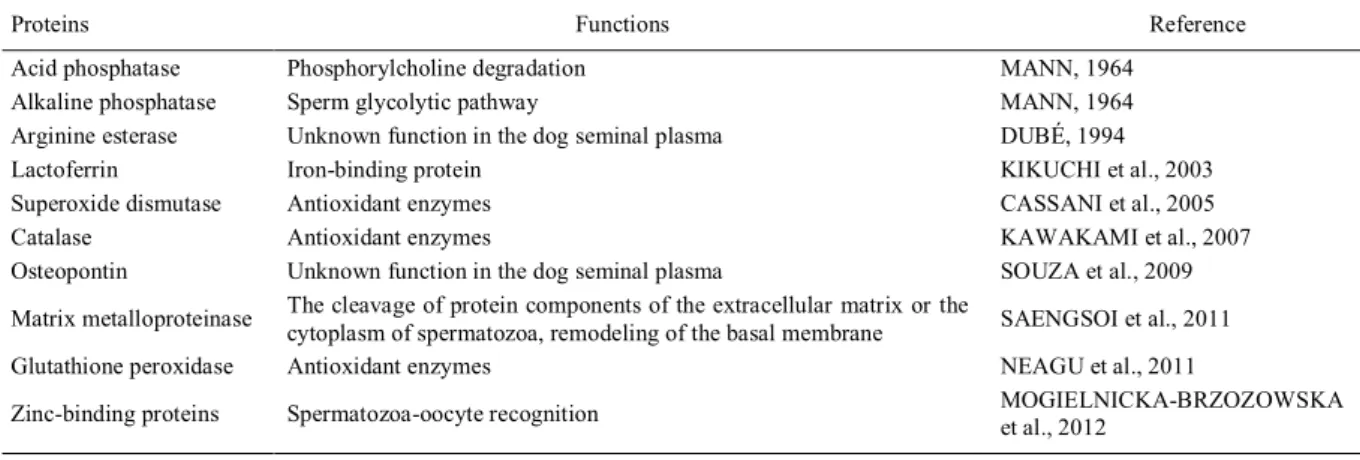
hyperplasia (WOLF et al., 2012). An overview about the proteins of the canine SP and their functions can be seen on table 1.
CONCLUSION
The SP proteins have been the subject of numerous studies in several species of domestic
animals. There is sufficient experimental evidence
Table 1 - Proteins of the canine seminal plasma and their functions.
Proteins Functions Reference
Acid phosphatase Phosphorylcholine degradation MANN, 1964
Alkaline phosphatase Sperm glycolytic pathway MANN, 1964
Arginine esterase Unknown function in the dog seminal plasma DUBÉ, 1994
Lactoferrin Iron-binding protein KIKUCHI et al., 2003
Superoxide dismutase Antioxidant enzymes CASSANI et al., 2005
Catalase Antioxidant enzymes KAWAKAMI et al., 2007
Osteopontin Unknown function in the dog seminal plasma SOUZA et al., 2009
Matrix metalloproteinase The cleavage of protein components of the extracellular matrix or the
cytoplasm of spermatozoa, remodeling of the basal membrane SAENGSOI et al., 2011
Glutathione peroxidase Antioxidant enzymes NEAGU et al., 2011
Zinc-binding proteins Spermatozoa-oocyte recognition MOGIELNICKA-BRZOZOWSKA
of their function regarding sperm capacitation, the acrosome reaction, oxidative processes, reactions of the female immune system, interaction with the epithelium of the uterine tube, and fertilization.
Dogs are the best experimental model for comparative studies with humans due to the similarity of their respective accessory sex glands and prostate growth. In addition, studies on canine SP components provide information that enables the understanding of sperm function and the reproductive tract of wild species of the same family, such as wolves, coyotes, and others. At all of these levels, detailed studies on the composition of canine SP and
its specific biological function are still needed. Such
molecules, in both animals and humans, are potential candidates for fertility markers as well as markers of pathophysiological processes.
REFERENCES
AITKEN, R.J. Epididymal cell types and their functions. In: ROBAIRE, B.; HINTON, B.T. (Eds.). Active oxygen in spermatozoa during epididymal transit. New York: Kluwer Academic/Plenum Publishers, 2002. 558p, p.435-447.
ANGRIMANI, D.S.R. et al. Role of residual cytoplasm on oxidative status during sperm maturation in dogs. Animal Reproduction Science, v.151, p.256-261, 2014a. Available from: <http://www. sciencedirect.com/science/article/pii/S037843201400339X>. Accessed: Aug. 21, 2015. doi: 10.1016/j.anireprosci.2014.10.023.
ANGRIMANI, D.S.R. et al. Sperm maturation in dogs: sperm profile
and enzymatic antioxidant status in ejaculated and epididymal spermatozoa. Andrologia, v.46, p.814-819, 2014b. Available from: <http://onlinelibrary.wiley.com/doi/10.1111/and.12154/epdf>. Accessed: Aug. 21, 2015. doi: 10.1111/and.12154.
AQUINO-CORTEZ, A. Avaliação dos componentes bioquímicos do líquido prostático e suas correlações com a qualidade espermática canina. 2003. 79f. Dissertação (Mestrado em Ciências Veterinárias) – Programa de Pós-Graduação em Ciências Veterinárias, Universidade Estadual do Ceará, CE.
BARTLETT, D.J. Studies on dog semen. II. Biochemical characteristics. Journal of Reproduction and Fertility, v.3, p.190-205, 1962. Available from: <http://www.reproduction-online.org/content/3/2/173.short>. Accessed: Jun. 09, 2015. doi: 10.1530/jrf.0.0030173.
BECCAGLIA, M. et al. Tris-Lecithin extender supplemented with antioxidant catalase for chilling of canine semen. Reproduction in Domestic Animal, v.44, p.345-349, 2009. Available from: <http:// onlinelibrary.wiley.com/doi/10.1111/j.1439-0531.2009.01410.x/ abstract;jsessionid=4667E0DE3CB9718AD63BF46252D6A 2DE.f01t02>. Accessed: Jun. 09, 2015. doi: 10.1111/j.1439-0531.2009.01410.x.
BEIGLBOCK, A. et al. Dog epididymis-specific mRNA
encoding secretory glutathione peroxidase-like protein.
Journal of Reproduction and Fertility, v.112, p.357-367, 1998. Available from: <http://www.reproduction-online.
org/content/112/2/357.short>. Accessed: Jun. 09, 2015. doi: 10.1530/jrf.0.1120357.
BRANDON, C.I. et al. Two-dimensional polyacrylamlde gel
electrophoresis of equine seminal plasma proteins and their
correlation with fertility. Theriogenology, v.52, p.863-873, 1999. Available from: <http://www.sciencedirect.com/science/article/pii/ S0093691X99001788>. Accessed: Jun. 09, 2015. doi: 10.1016/ S0093-691X(99)00178-8.
BURDEN, H.P. et al. Prostasomes - their effects on human male reproduction and fertility. Human Reproduction Update, v.12, n.3, p.283-292, 2006. Available from: <http://humupd. oxfordjournals.org/content/12/3/283.long>. Accessed: Jun. 09, 2015. doi: 10.1093/humupd/dmi052.
CASSANI, P. et al. Relationship between total superoxide dismutase activity with lipid peroxidation, dynamics and morphological parameters in canine semen. Animal Reproduction Science, v.86, p.163-173, 2005. Available from: <http://www.sciencedirect. com/science/article/pii/S0378432004001629>. Accessed: Jun. 09, 2015. doi: 10.1016/j.anireprosci.2004.06.006.
CHAPDELAINE, P. et al. Identification of arginine esterase as
the major androgen-dependent protein secreted by dog prostate and preliminary molecular characterization in seminal plasma.
Journal of Andrology, v.5, p.206-210, 1984. Available from: <http://onlinelibrary.wiley.com/doi/10.1002/j.1939-4640.1984. tb02395.x/abstract>. Accessed: Jun. 17, 2015. doi: 10.1002/j.1939-4640.1984.tb02395.x.
DRUART, X. et al. Proteomic characterization and cross species comparison of mammalian seminal plasma. Journal of Proteomics, v.91, 13-22, 2013. Available from: <http://www. sciencedirect.com/science/article/pii/S1874391913003059>. Accessed: Jun. 09, 2015. doi: 10.1016/j.jprot.2013.05.029. DUBÉ, J.Y. Prostatic kallikreins: biochemistry and physiology.
Comparative Biochemistry and Physiology Part C: Pharmacology, Toxicology and Endocrinology, v.107, n.1, p.13-20, 1994. Available from: <http://www.sciencedirect.com/science/ article/pii/1367828094900043?np=y>. Accessed: Jun. 09, 2015. doi: 10.1016/1367-8280(94)90004-3.
FRENETTE, G. et al. Proteolytic activity of arginine esterase from dog seminal plasma towards actin and other structural proteins. Comparison with trypsin and kallikrein. International Journal of Biochemistry, v.18, n.8, p.697-703, 1986. Available from: <http:// www.sciencedirect.com/science/article/pii/0020711X86903927>. Accessed: Jun. 09, 2015. doi: 10.1016/0020-711X(86)90392-7. GOBELLO, C. et al. Serum and seminal markers in the diagnosis of disorders of the genital tract of the dog: a mini-review.
Theriogenology, v.57, p.1285-1291, 2002. Available from: <http:// www.sciencedirect.com/science/article/pii/S0093691X02006283>. Accessed: Jun. 12, 2015. doi: 10.1016/S0093-691X(02)00628-3. HADWAN, M.H. et al. The key role of zinc in enhancement of total antioxidant levels in spermatozoa of patients with asthenozoospermia. American Journal of Molecular and Cellular Biology, v.2, p.52-61, 2013. Available from: <http://
IGUER-OUADA, M.; VERSTEGEN, J.P. Long-term preservation of chilled canine semen: effect of commercial and laboratory prepared extenders. Theriogenology, v.55, p.671-684, 2001. Available from: <http://link.periodicos.capes.gov.br.ez76. periodicos.capes.gov.br/sfxlcl41?url_ver=Z39.88-2004&url_ ctx_fmt=fi/fmt:kev:mtx:ctx&ctx_enc=info:ofi/enc:UTF-8&ctx_ver=Z39.88-2004&rfr_id=info:sid/sfxit.com:azlist&sfx. ignore_date_threshold=1&rft.object_id=954925464194&svc. fulltext=yes>. Accessed: Jun. 12, 2015. doi: 10.1016/S0093-691X(01)00435-6.
ISAACS, W.B.; COFFEY, D.S. The predominant protein of canine seminal plasma is an enzyme. Journal of Biological Chemistry, v.259, n.18, p.11520-11526, 1984. Available from: <http://www. jbc.org/content/259/18/11520.long>. Accessed: Jun. 12, 2015. KAWAKAMI, E. et al. Superoxide dismutase and catalase activities in the seminal plasma of normozoospermic and asthenozoospermic beagles. Journal of Veterinary Medical Science, v.69, p.133-136, 2007. Available from: <https://www.jstage.jst.go.jp/article/ jvms/69/2/69_2_133/_article>. Accessed: Jun. 12, 2015. doi: 10.1292/jvms.69.133.
KIKUCHI, M. et al. Seminal plasma lactoferrin but not transferrin
reflects gonadal function in dogs. Journal of Veterinary Medical Science, v.65, p.679-684, 2003. Available from: <https://www. jstage.jst.go.jp/article/jvms/65/6/65_6_679/_article>. Accessed: Jun. 12, 2015. doi: 10.1292/jvms.65.679.
KIRCHHOFF, C. The dog as a model to study human epi- didymal function at a molecular level. Molecular Human Reproduction, v.8, p.695-701, 2002. Available from: <http:// molehr.oxfordjournals.org/content/8/8/695.full.pdf+html>. Accessed: Aug. 22, 2015. doi: 10.1093/molehr/8.8.695.
KOBAYASHI, M. et al. Superoxide dismutase activity in the
oviductal and uterine fluid of the bitch and the effects of the
enzyme on viability, motility and hyperactivation of canine sperm in vitro. Journal of Veterinary Medical Science, v.76, p.741-743, 2014. Available from: <http://www.ncbi.nlm.nih. gov/pmc/articles/PMC4073345/>. Accessed: Jun. 12, 2015. doi: 10.1292/jvms.13-0545.
KUTZLER, M.A. Semen collection in the dog. Theriogenology, v.64, p.747-754, 2005. Available from: <http://www.sciencedirect. com/science/article/pii/S0093691X0500186X>. Accessed: Jun. 12, 2015. doi: 10.1016/j.theriogenology.2005.05.023.
LOWSETH, L.A. et al. Age-related changes in the prostate and testes of the Beagle dog. Veterinary Pathology, v.27, p.347-353, 1990. Available from: <http://vet.sagepub.com/ content/27/5/347.full.pdf+html>. Accessed: Jun. 17, 2015. doi: 10.1177/030098589002700507.
MA, L. et al. Spink13, an epididymis-specific gene of the
Kazal-type serine pro- tease inhibitor (SPINK) family, is essential for the acrosomal integrity and male fertility. Journal of Biological Chemistry, v.288, p.10154-10165, 2013. Available from: <http://www.jbc.org/content/288/14/10154.full.pdf+html>. Accessed: Aug. 22, 2015. doi: 10.1074/jbc.M112.445866. MANJUNATH, P. et al. Major proteins of bovine seminal vesicles bind to spermatozoa. Biology of Reproduction, v.49, p.27-37, 1993. Available from: <http://www.biolreprod.org/ content/50/1/27.full.pdf+html>. Accessed: Aug. 23, 2015. doi: 10.1095/biolreprod50.1.27.
MANJUNATH, P.; THÉRIEN, I. Role of seminal plasma
phospholipid-binding proteins in sperm membrane lipid modification
that occurs during capacitation. Journal of Reproductive Immunology, v.53, p.109-119, 2002. Available from: <http:// www.sciencedirect.com/science/article/pii/S0165037801000985>. Accessed: Jun. 16, 2015. doi: 10.1016/S0165-0378(01)00098-5. MANN, T. Biochemistry of semen and of the male reproductive tract. London: Methuen, 1964. 493p, p.329-390.
MANN, T.; L-MANN, C. Biochemistry of seminal plasma and
male acessory fluids: applications to andrological problems. In:
_____. Male reproductive function and semen. Themes and trends in phisiology, biochemistry and investigative andrology. Berlim: Springer Verlag, 1981. 495p, p.269-336.
MARTINS, M.I.M. Efeito da sazonalidade sobre a função testicular de cães. 2005. 123f. Tese (Doutorado em Reprodução Animal) - Botucatu, Faculdade de Medicina Veterinária e Zootecnia da Universidade Estadual Paulista – Campus de Botucatu, SP. MILLER, D.J. et al. Heparin-binding proteins from seminal plasma bind to bovine spermatozoa and modulate capacitation by heparin.
Biology of Reproduction, v.42, p.899-915, 1990. Available from: <http://www.biolreprod.org/content/42/5-6/899.full.pdf>. Accessed: Aug. 18, 2015. doi: 10.1095/biolreprod42.6.899.
MOGIELNICKA-BRZOZOWSKA, M. et al. Isolation and characterization of zinc-binding proteins of canine seminal plasma. Polish Journal of Veterinary Sciences, v.15, p.493-498, 2012. Available from: <http://link.periodicos.capes.gov.br.ez76. periodicos.capes.gov.br/sfxlcl41?url_ver=Z39.88-2004&url_
ctx_fmt=fi/fmt:kev:mtx:ctx&ctx_enc=info:ofi/enc:UTF-8&ctx_
ver=Z39.88-2004&rfr_id=info:sid/sfxit.com:azlist&sfx.ignore_ date_threshold=1&rft.object_id=991042754052872&svc. fulltext=yes>. Accessed: Jun. 12, 2015. doi: 10.2478/v10181-012-0076-5.
MOTHEO, T.F. et al. Semen parameters and seminal plasma protein
and biochemical profiles of dogs with benign prostatic hyperplasia
after botulinum toxin type A intraprostatic injection. Ciência Rural, v.44, n.6, p.1113-1118, 2014. Available from: <http://www.scielo. br/scielo.php?pid=S0103-84782014000600026&script=sci_ arttext&tlng=es>. Accessed: Jun. 12, 2015. doi: 10.1590/S0103-84782014000600026.
MOURA, A.A. et al. Identification of proteins in the accessory sex gland fluid associated with fertility indexes of dairy bulls:
a proteomic approach. Journal of Andrology, v.27, p.201-211, 2006. Available from: <http://onlinelibrary.wiley.com/ doi/10.2164/jandrol.05089/full>. Accessed: Jun. 17, 2015. doi: 10.2164/jandrol.05089.
MOURA, A.A. et al. Proteomics of cauda epididymal fluid from
mature Holstein bulls. Journal of Proteomics, v.73, p.2006-2020, 2010. Available from: <http://www.sciencedirect.com/science/ article/pii/S1874391910001831>. Accessed: Aug. 23, 2015. doi: 10.1016/j.jprot.2010.06.005.
MUSSEL, C. et al. Canine prostatic diseases: diagnosis methods.
and its relation with sperm quality and lipid peroxidation post
thaw. Theriogenology, v.75, p.10-16, 2011. Available from: < h t t p : / / w w w. s c i e n c e d i r e c t . c o m / s c i e n c e / a r t i c l e / p i i / S0093691X10003614>. Accessed: Jun. 12, 2015. doi: 10.1016/j. theriogenology.2010.07.004.
REGO, J.P.A. et al. Seminal plasma proteome of electroejaculated
Bos indicus bulls. Animal Reproduction Science, v.148, p.1-17, 2014. Available from: <http://www.sciencedirect.com/science/ article/pii/S0378432014001353>. Accessed: Jun. 12, 2015. doi: 10.1016/j.anireprosci.2014.04.016.
ROBERTS, T.K.; BOURSNELL, J.C. The isolation and characterization of lactoferrin from sow milk and boar seminal plasma. Journal Reproduction and Fertility, v.42, p.579-582, 1975. Available from: <http://www.reproduction-online.org/ content/42/3/579.full.pdf+html>. Accessed: Jun. 17, 2015. doi: 10.1530/jrf.0.0420579.
RODRÍGUEZ-MARTÍNEZ, H. et al. Seminal plasma proteins: what role do they play? American Journal of Reproductive Immunology, v.66, p.11-22, 2011. Available from: <http:// onlinelibrary.wiley.com/doi/10.1111/j.1600-0897.2011.01033.x/ epdf>. Accessed: Jun. 12, 2015. doi: 10.1111/j.1600-0897.2011.01033.x.
ROWELL, J.L. et al. Dog models of naturally occurring cancer.
Trends in Molecular Medicine, v.17, p.380-388, 2011. Available from: < h t t p : / / w w w. s c i e n c e d i r e c t . c o m / s c i e n c e / a r t i c l e / p i i / S 1 4 7 1 4 9 1 4 11 0 0 0 4 3 8 > . Accessed: Jun. 16, 2015. doi: 10.1016/j.molmed.2011.02.004.
ROTA, A.A. et al. Effect of post-thaw dilution with autologous
prostatic fluid on dog semen motility and sperm acrosome status. Theriogenology, v.67, p.520-525, 2007. Available from: <http:// www.sciencedirect.com/science/article/pii/S0093691X06004869>. Accessed: Jun. 12, 2015. doi: 10.1016/j.theriogenology.2006.09.001. SCHAFER-SOMI, S. et al. Measurement of alkaline phosphatase in canine seminal plasma – an update. Reproduction in Domestic Animals, v.48, p. e10-12, 2013. Available from: <http:// onlinelibrary.wiley.com/doi/10.1111/j.1439-0531.2012.02025.x/ epdf>. Accessed: Jun. 12, 2015. doi: 10.1111/j.1439-0531.2012.02025.x.
SAENGSOI, W. et al. Detection of matrix metalloproteinase (MMP)-2 and MMP-9 in canine seminal plasma. Animal Reproduction Science, v.127, p.114-119, 2011. Available from: <http://www. sciencedirect.com/science/article/pii/S0378432011001795>. Accessed: Jun. 12, 2015. doi: 10.1016/j.anireprosci.2011.07.004. SAGOLS, E.; NAVARRO, C. Follow up of CPSE concentrations after Osaterone treatment in dogs. In: EVSSA CONGRESS. Reproduction and Pediatrics in Dogs, Cats and Exotics, 16., 2013, Toulouse, France. Proceedings… Tolouse: EVSSA, 2013. 184p, p.169.
SIRIVAIDYAPONG, S. et al. Acrosome reaction in dog sperm is induced by a membrane localized progesterone receptor. Journal of Andrology, v.20, p.537-544, 1999. Available from: <http:// onlinelibrary.wiley.com/doi/10.1002/j.1939-4640.1999.tb02553.x/ epdf>. Accessed: Jun. 12, 2015. doi: 10.1002/j.1939-4640.1999. tb02553.x.
SMITH, J. Canine prostatic disease: a review of anatomy, pathology, diagnosis, and treatment. Theriogenology, v.70, p.375-383, 2008. Available from: <http://www.theriojournal.com/article/ S0093-691X(08)00226-4/pdf>. Accessed: Jun. 16, 2015. doi: 10.1016/j.theriogenology.2008.04.039.
SOUZA, F.F. et al. Herapin-binding proteins of canine seminal plasma. Theriogenology, v.66, p.1606-1609, 2006. Available from: < h t t p : / / w w w. s c i e n c e d i r e c t . c o m / s c i e n c e / a r t i c l e / p i i / S 0 0 9 3 6 9 1 X 0 6 0 0 11 2 9 > . Accessed: Aug. 18, 2015. doi: 10.1016/j.theriogenology.2006.02.016.
SOUZA, F.F. et al. Characteristics of seminal plasma proteins and their correlation with canine semen analysis. Theriogenology, v.68, p.100-106, 2007. Available from: <http://www.sciencedirect. com/science/article/pii/S0093691X07001641>. Accessed: Jun. 12, 2015. doi: 10.1016/j.theriogenology.2006.11.016.
SOUZA, F.F. et al. Osteopontin in seminal plasma and sperm membrane of dogs. Reproduction in Domestic Animals, v.44, p.283-286, 2009. Available from: <http://onlinelibrary.wiley.com/ doi/10.1111/j.1439-0531.2009.01447.x/epdf>. Accessed: Jun. 12, 2015. doi: 10.1111/j.1439-0531.2009.01447.x,
STARKEY, M.P. et al. Dogs really are man’s best friend - Canine genomics has applications in veterinary and human medicine!
Briefings in Functional Genomics and Proteomics, v.4,
p.112-128, 2005. Available from: <http://bfg.oxfordjournals.org/ content/4/2/112.full.pdf+html>. Accessed: Aug. 22, 2015. doi: 10.1093/bfgp/4.2.112.
STRZEZEK, R. et al. Characteristics of antioxidant system in dog semen. Polish Journal of Veterinary Sciences, v.12,
p.55-60, 2009. Available from: <http://www.researchgate.net/profile/
Rafal_Strzezek2/publication/24443607Characteristicsofantioxi dantsystemindogsemen/links/02bfe51374c504fe88000000.pdf>. Accessed: Jun. 12, 2015.
TESKE, E. et al. Separation and heat stability of the corticosteroid-induced and hepatic alkaline phosphatase isoenzymes in canine plasma. Journal of Cromatography, v.369, p.349-356, 1986. Available from: <http://www.sciencedirect.com/science/article/ pii/S0021967300901419?np=y>. Accessed: Jun. 12, 2015. doi: 10.1016/S0021-9673(00)90141-9.
THOMASSEN, R.; FARSTAD, W. Artificial insemination in
canids: a useful tool in breeding and conservation. Theriogenology, v.71, p.190-199, 2009. Available from: <http://www.sciencedirect. com/science/article/pii/S0093691X08006481>. Accessed: Aug. 22, 2015. doi: 10.1016/j.theriogenology.2008.09.007.
WOLF, K. et al. Testicular steroids, prolactin, relaxin and prostate gland markers in peripheral blood and seminal plasma of normal dogs and dogs with prostatic hyperplasia. Reproduction in Domestic Animals, v.47, p.243-246, 2012. Available from: <http:// onlinelibrary.wiley.com/doi/10.1111/rda.12083/epdf>. Accessed: Jun. 12, 2015. doi: 10.1111/rda.12083.
ZELLI, R. et al. Ultrastructural and enzymatic activity of membranous vesicles isolated from canine seminal plasma.