UNIVERSIDADE FEDERAL DE SÃO CARLOS
PROGRAMA DE PÓS-GRADUAÇÃO EM BIOTECNOLOGIA
EFEITO IN VITRO E IN VIVO DA TERAPIA FOTODINÂMICA ANTIMICROBIAL SOBRE STREPTOCOCCUS MUTANS PRESENTES EM ACESSÓRIOS
METÁLICOS ORTODÔNTICOS
Vitor Hugo Panhóca
Vitor Hugo Panhóca
EFEITO IN VITRO E IN VIVO DA TERAPIA FOTODINÂMICA ANTIMICROBIAL SOBRE STREPTOCOCCUS MUTANS PRESENTES EM ACESSÓRIOS
METÁLICOS ORTODÔNTICOS
Tese apresentada ao Programa de Pós-Graduação em Biotecnologia da Universidade Federal de São Carlos, como parte dos requisitos para obtenção do título de doutor em biotecnologia.
Orientadores: Prof. Dr. Vanderlei Salvador Bagnato Prof. Dr. Clóvis Wesley Oliveira de Souza
Ficha catalográfica elaborada pelo DePT da Biblioteca Comunitária/UFSCar
P191ev
Panhóca, Vitor Hugo.
Efeito in vitro e in vivo da terapia fotodinâmica antimicrobial sobre streptococcus mutans presentes em acessórios metálicos ortodônticos / Vitor Hugo Panhóca. -- São Carlos : UFSCar, 2015.
98 f.
Tese (Doutorado) -- Universidade Federal de São Carlos, 2015.
1. Terapia fotodinâmica. 2. Streptococcus mutans. 3. Curcumina. 4. Surfactante. 5. Aparelhos ortodônticos. I. Título.
Aos meus pais, Heitor e Neide, pela minha educação; algo que me torna uma pessoa forte para a luta diária e melhor para compreender a vida.
A Maria Eneida, minha mulher, pelo incentivo e compreensão da
minha dedicação na vida acadêmica e profissional tornando possível
que eu pudesse obter meu título de Doutor.
Ao meu filho, Lorenzo, pelos momentos de felicidade e convivência,
que me estimulam na luta para superar as dificuldades diárias.
AGRADECIMENTOS
A Deus, por minha criação e formação, que permitiu a graça e a capacidade de poder evoluir como pessoa e profissionalmente.
Aos meus pais, meu filho Lorenzo, minha enteada Bruna, familiares e, em especial, a minha mulher, pelas palavras de incentivo, carinho, que me proporcionam alicerce firme para construir e desenvolver meu trabalho.
A todos aqueles que, direta ou indiretamente – entre eles: professores, doutorandos, professores convidados, alunos, secretarias, funcionários –, colaboraram com a realização deste trabalho.
Meu agradecimento ao Prof. Dr. Vanderlei S. Bagnato, por ter acreditado na minha capacidade e ter aberto as portas da Universidade de São Paulo (USP) e de seu laboratório, para que eu pudesse aprimorar meus conhecimentos científicos e evoluir também profissionalmente. Sua postura sempre atenciosa com minhas perguntas e meus anseios sempre me motivaram a seguir adiante em meus estudos.
A Prof. Dr. Clóvis Wesley Oliveira de Souza, pela transmissão de conhecimentos sobre cultivo microbiológico em seu laboratório do Departamento de Morfologia e Patologia da UFSCar, que possibilitaram a realização dos meus experimentos com terapia fotodinâmica. E também pelos seus ensinamentos e orientação em microbiologia, contribuindo significativamente para a completa realização deste trabalho.
Aos companheiros do laboratório de Biofotônica do IFSC que me ajudaram direta e indiretamente em meu trabalho. Tenho que agradecer muito a vocês: Profª Cristina, Natália, Lili, Sebastião, Dirceu, Clóvis, Thaila, Mariana, Bruno Pereira, Patrícia, Ila, Dyleis, Ana Paula, Hilde, Gustavo, Carol, Mirian, Cintia, Ramon, Marcelinho, Fernanda Carbinato, Larissa, Toninho, Marcela, Marciana, Alessandra, Hérica e Ana Elisa.
A Posdoc Drª Priscila Menezes e ao pessoal do LAT que forneceu apoio em eletrônica para os aparelhos usados em minha pesquisa e, portanto, me auxiliaram na realização deste trabalho e na obtenção da patente do aparelho de LED intra-bucal. Obrigado, a toda a equipe!
Ao amigo e cirurgião-dentista Doutor Fernando L. E. Florez, sempre atencioso para me auxiliar na escrita dos artigos, o que me auxiliou na obtenção do título de Doutor.
A Doutora Fernanda Paolillo pela amizade e ajuda em realizar os gráficos e a análise estatística deste trabalho.
Ao pessoal da secretaria do Grupo de Óptica, por estarem sempre dispostos a ajudar. Muito obrigado, Isabel, Bene, Adriane e Cristiane!
A secretária da PPGBIOTEC UFSCar, Claudia Pastega, meus agradecimentos pela atenção dada durante todo o meu doutoramento.
Ao bibliotecário Lucas Pessota, por ter formatado e corrigido este texto, permitindo melhor entendimento para os que lerem este trabalho.
Aos colegas de turma, pela convivência agradável e pelos conhecimentos e experiências compartilhadas.
“A melhor coisa que você pode fazer
para
cultivar a verdadeira sabedoria é praticar a
consciência
de que o mundo é um sonho.”
RESUMO
33.54 mW/cm2 e 212 mW/cm2). A avaliação da redução microbiana, e da fração de sobrevivência em cada um dos níveis estudados, foi realizada utilizando-se o teste ANOVA Kruskal-Wallis. A análise estatística mostrou que apenas os grupos 6 e 7 apresentaram resultados de inativação bacteriana que apresentavam diferenças estatisticamente significantes (p <0,05) em relação aos resultados observados no grupo 1. Estes resultados indicam que a TFDA associada com o surfactante SDS pode ser utilizado como agente auxiliar conveniente para promover a descontaminação bucal.
ABSTRACT
Demineralization and caries are often present in patients with orthodontic braces due to the increased accumulation of dental biofilm containing Streptococcus mutans, which produces acids that attack the tooth surface. The Antimicrobial Photodynamic Therapy (APDT) has been shown as an alternative in microbial inactivation in dentistry. This work, carried out both in
vitro and in vivo, aims to evaluate the antimicrobial effect of APDT on Streptococcus mutans present in the dental biofilm accumulated on the metal surface of orthodontic appliances and in patients undergoing orthodontic treatment. The in vitro study evaluated the susceptibility of biofilm formed by S. mutans on the metal surface under the application of orthodontic appliances APDT induced Curcumin diluted surfactant (SDS) and blue light-emitting diode (LED). The samples were treated with the photosensitizer (PS) at a concentration of 1 g/L, 0.1% surfactant and exposed to LED light (455 ± 10 nm, 46mW/cm²), fluence of 30 J/cm². The experimental groups studied were: control group (P-L-S-); light (P-L+S-), surfactant (P-L-S+) and photosensitizer (P+L-S-) groups; surfactant-light (P-L+S+), photosensitizer-surfactant (P+L-S+) and PDT (P+L+S-) groups; and the PDT-surfactant (P+L+S+) group. Each group was named accordingly to the addiction or not of the photosensitizer (P+ or P-), surfactant (S+ or S-) and application or not of light (L+ or L-), respectively. The colonies grown on plates of
fraction in each of the studied levels, was performed using the Kruskal-Wallis ANOVA test. Statistical analysis showed that only G6 and G7 presented results of bacterial inactivation that showed statistically significant differences (p<0.05) compared to those observed in G1. These results indicated that APDT, when combined with SDS surfactant, may be used as an adjunct agent for convenient oral decontamination promoter in vivo.
LISTA DE FIGURAS
LISTA DE TABELAS
LISTA DE ABREVIATURAS
BHI - Brain Heart Infusion
CTAC - Cloreto de Cetiltrimetilamônio EGM - Estreptococus do Grupo Mutans ERO - Espécies Reativas de Oxigênio DMSO - Sulfóxido de Dimetilo
FS - Fotossensibilizador
GTFS - Amilase e Enzimas Glucosiltransferase HpD - Hematoporfirina
HPS - N-dimetil-3-amônio-1-propanosulfonato IFSP - Instituto de Física de São Carlos
LED - Diodo Emissor de Luz MB - Azul de Metileno PDT - Photodynamic Therapy
PEC - Polissacarídeos Extra-celulares Insolúveis PIC - Polissacarídeos Intra-celulares
TFD - Terapia Fotodinâmica
TFDA - Terapia Fotodinâmica Antimicrobial USP - Universidade de São Paulo
SUMÁRIO
1 INTRODUÇÃO ... 17
2 Streptococcus mutans ... 20
3 TERAPIA FOTODINÂMICA ... 22
3.1 Curcumina ... 23
3.2 Luz ... 26
4 SURFACTANTE ... 30
5 CLOREXIDINA ... 32
6 OBJETIVO ... 34
7 OBJETIVOS ESPECÍFICOS ... 35
8 ARTIGO EXPERIMENTO IN VITRO ... 36
9 ARTIGO EXPERIMENTO IN VIVO ... 53
10 DISCUSSÃO ... 68
11 CONCLUSÃO ... 74
REFERÊNCIAS ... 75
APÊNDICE A ... 82
APÊNDICE B ... 90
1 INTRODUÇÃO
O Streptococcus mutans é o agente etiológico responsável pela descalcificação dental, formação da cárie e desenvolvimento de doenças periodontais nos seres humanos. Por esse motivo tem sido estudado a sua inativação em odontologia por vários pesquisadores1,2. Os métodos tradicionais mais utilizados para descontaminação bucal são limpeza dental profilática feita com instrumental profissional em odontologia, escovação dental, uso de fio dental e bochechos com solução antimicrobiana. Dentre estes métodos, a escovação dental é o método mais utilizado para a higiene bucal e reconhecida na literatura como terapia para o controle do biofilme dental3. O biofilme dental é uma comunidade multi-espécies de micro-organismos em uma matriz de polissacarídeos que se adere a superfície dental, essa estrutura do biofilme confere uma característica protetora aos S mutans da ação de antimicrobianos. Os agentes antimicrobianos para bochechos são utilizados como procedimentos complementares a escovação mecânica para maior controle da higiene bucal dos indivíduos. No entanto, essas soluções antimicrobianas que apresentam efeito na descontaminação bucal apresentam substâncias ativas que provocam efeitos colaterais e devido ao seu mecanismo de ação podem gerar cepas microbianas resistentes4.
A terapia fotodinâmica (TFD) é um conhecido tratamento oxidativo utilizado para o tratamento oncológico e também pode ser usado em terapia antimicrobiana. A utilização da TFD com ação antimicrobiana surgiu em 1900, quando o médico alemão Oscar Raab combinou a aplicação de luz solar e corantes como a acridina para inativar micro-organismos unicelulares
– Paramecium caudatum5. A TFD antimicrobial (TFDA) consiste em aplicar uma droga conhecida como fotossensibilizador (FS) e luz sobre um micro-organismo que na presença de oxigênio orgânico produz espécies reativas de oxigênio (ERO) e por reação de oxi-redução provocam danos aos componentes estruturais do micro-organismo levando a sua inativação.
substâncias naturais como a hipericina, bacteroclorinas e clorinas, tal como a Photodithazine® (russa). Na odontologia os FSs mais usados como antimicrobianos são o azul de metileno e o azul de toluídina, da família das fenotiazinas, que são de origem sintética. Outras substâncias sintéticas de segunda geração são as naftalocianinas e ftalocianinas. A Curcumina surgiu recentemente como um FS alternativo na TFDA, é de origem natural e foi escolhido para ser utilizado neste estudo, por ser de baixo custo e biodegradável na natureza. A Curcumina é um corante obtido a partir da raiz do açafrão-da-Índia (Curcuma longa) muito utilizado como aditivo alimentar e é excitada na TFDA com luz azul.
Várias fontes de luz podem ser utilizadas na TFDA; a literatura descreve o emprego de diferentes sistemas de LASER com intensidade de baixa potência, porém, novos estudos a partir do desenvolvimento dos diodos emissores de luz (LEDs) tem mostrado ser uma eficiente fonte de luz aplicada na TFDA6,8.O LED como fonte de luz na TFDA quando comparados aos sistemas de LASERs disponíveis apresentam vantagens como irradiar maior área de tecido, ter menor consumo de energia e ainda possuem um tempo útil de vida maior7,8. Por esses motivos, o uso do LED na TFDA tornou-se mais atraente, facilitando sua implementação em terapias fotônicas aplicada à saúde.
A preocupação com a estética facial tem origem pré-histórica relatado em estudos de Paleontologia9 e nesse assunto se insere o tratamento ortodôntico que diariamente inclui maior quantidade de indivíduos que buscam não apenas melhor função mastigatória, mas também harmonia facial através de correção dentária com o uso de aparatos ortodônticos. Os aparelhos ortodônticos são compostos de bandas, braquetes e outros dispositivos intra-bucais que alteram a microbiota bucal aumentando o acúmulo de S. mutans.10,11 Diante do maior acúmulo de micro-organismos cariogênicos durante o tratamento ortodôntico terapias coadjuvantes no controle do biofilme dental tem sido estudadas para controle da microbiota bucal no sentido de se obter uma terapia que seja considerada a ideal.
Os surfactantes são moléculas anfifílicas que tem mostrado serem agentes ativos capazes de aumentar a permeabilidade da parede celular e agirem como desagregador do biofilme por sua atividade detergente. A TFDA utilizando azul de metileno (MB) combinado com surfactante foi utilizada para aumentar a permeabilidade em células de C. albicans diminuindo a formação de biofilme, sua habilidade de adesão e sua resistência às outras drogas.13 O surfactante pode ser considerado aditivo interessante para potencializar o efeito antimicrobiano na TFDA pois aumenta a solubilidade da Curcumina e promove alteração da membrana celular microbiana e, em sinergismo com os FS, aumenta o efeito antimicrobiano da TFDA, que pode promover melhor controle microbiológico do biofilme dental em indivíduos portadores de aparatos ortodônticos.14,16
2 Streptococcus mutans
Os Estreptococus do Grupo mutans (EGM) são micro-organismos frequentes nos dentes e cavidade bucal. Pertencem a este grupo de Estreptococcus: Streptococcus mutans, S. sobrinus, S. cricetus, S. rattus, S. ferus, S. macacae e S. downei. Os EGM fazem parte também da flora da faringe, sistema gastro-intestinal, vias áreas superiores e sistema urinário. Os S mutans são anaeróbios facultativos que se coram com Gram e apresentam morfologia de cocos. Os S mutans além de adesividade a superfície dental podem também atingir no sistema circulatório e por via endovenosa atingir às válvulas cardíacas podendo causar endocardite sub-aguda. O S sobrinus está envolvido no processo inicial de adesão e formação do biofilme dental contribuindo para a proliferação do S mutans e dando início ao processo de cárie dental.17 Os biofilmes dentais ou também chamadas placas dentais são comunidades de micro-organismos aglomerados e contidos em uma matriz autoproduzida que se adere a diferentes superfícies, assim como o dente, acessórios metálicos ortodônticos e implantes dentais metálicos.
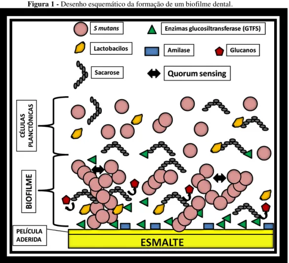
O S mutans é um micro-organismo acidófilo e acidogênico, ou seja, capaz de metabolizar açúcares e produzir ácidos sendo resistente aos mesmos. Uma de suas principais características é não crescer em caldo contendo altas concentrações de sal. Devido as suas características acidogênicas são responsáveis por seu potencial em produzir desmineralizações do esmalte e cárie dental. A produção de ácidos para o desenvolvimento da cárie não ocorre apenas por S mutans, outros micro-organismos acidogênicos como os lactobacilos também estão envolvidos neste processo.18 Os S mutans a partir de açúcares (sacarose, glicose, frutose e maltose) encontrados no meio bucal são capazes de produzir por ação enzimática (amilase e enzimas glucosiltransferase - GTFS), glucanos (polissacarídeos extra-celulares insolúveis - PECs), que são responsáveis por formar a estrutura e dar adesão do biofilme dental. (Figura 01) Os PECs produzidos pelos S mutans, além da adesão do biofilme apresenta outra função fisiológica de formarem uma matriz extra-celular porosa contendo pequenos canais que permitem que os ácidos produzidos pelos S mutans se difundam em direção a superfície dental levando a desmineralização dental. Para se ligar aos glucanos os S mutans usam proteínas de superfícies chamadas proteínas ligantes de glucano, que podem se ligar de maneira covalente e não covalente a parede celular microbiana.
matriz de polissacarídeos proporcionam permeabilidade ao biofilme permitindo que penetrem nutrientes, metabólitos, oxigênio e drogas com atividades antimicrobianas. Outro fator de proteção e sobrevivência que os micro-organismos em biofilme apresentam é o quorum sensing, que é a capacidade que estas células apresentam em se comunicar de maneira coordenada enviando sinais para a expressão gênica de fatores de virulência, adaptação à escassez de nutrientes e de defesa a substâncias tóxicas.19 (Figura 01) Os S mutans também produzem polissacarídeos intra-celulares (PICs) a partir de carboidratos encontrados no meio bucal que servem de reserva de nutrientes para serem metabolizados quando as fontes de açúcares estão ausentes.
Figura 1 - Desenho esquemático da formação de um biofilme dental.
3 TERAPIA FOTODINÂMICA
A Terapia fotodinâmica (TFD) foi introduzida na terapia médica hodierna em 1904, por Oscar Raab, na inativação de células, microrganismos e moléculas. Essa modalidade de tratamento denominada de TFD tem sua origem no termo em inglês Photodynamic Therapy (PDT).20 Essa técnica tem como base a utilização combinada de luz visível com comprimento de onda específico, com certos tipos de drogas que são denominadas de fotossensibilizadores (FS), que, quando utilizados em conjunto e na presença de oxigênio, produzem uma reação de oxidação das células cancerígenas que provocará a lise das células-alvo e, por conseqüência, a indução de morte da célula tumoral. A TFD tem sido mostrada em vários estudos como uma terapia viável e alternativa com potente efeito antimicrobiano.1,6,7 Na TFD a reação de oxi-redução de células tumorais e micro-organismos ocorre por fotoativação dos FSs que se transformam em um estado eletrônico super-excitado gerando energia que reage com o oxigênio orgânico livre produzindo espécies reativas de oxigênio (ERO). Esses radicais livres não possuem especificidade por sítio de ação, reagindo com vários substratos biomoleculares das células vivas levando as células tumorais a apoptose e inativação microbiana. Figura 02.
Figura 2 - Esquema representativo da reação de oxi-redução de células tumorais e micro-organismo promovida pela TFD
A inativação de microrganismos pela TFDA pode apresentar diversas vantagens em relação à utilização de agentes antimicrobianos tradicionais. Primeiramente, a morte bacteriana é rápida, diminuindo a necessidade da manutenção de altas concentrações de substâncias químicas por longos períodos, como no caso do uso de antibióticos e anti-sépticos21 e, em segundo lugar, como a morte bacteriana não está ligada à mediação de radicais químicos agindo sobre alvos específicos, o desenvolvimento de cepas resistentes seria improvável.22,23 Finalmente, como nem o fotossensibilizador, nem a luz de baixa intensidade empregada são bactericidas quando utilizados isoladamente, a destruição das bactérias pode ser controlada restringindo-se a região irradiada, evitando a destruição da microbióta normal em outros locais. O efeito da TFDA como um antimicrobiano é influenciado não apenas pelo tipo de micro-organismo tratado, mas também por outros fatores como o tipo de FS, a concentração do FS, a sua localização durante a irradiação, o período de pré-incubação do FS, o tipo de fonte de luz utilizado, a dosimetria de luz aplicada e a concentração de oxigênio orgânico livre presente no local a ser tratado.
3.1 Curcumina
Figura 3 - Estrutura química da Curcumina
Fonte: disponível em < http://www.drvictorsorrentino.com.br/o-poder-da-curcumina-na-prevencao-e-tratamento-de-doencas/ >.
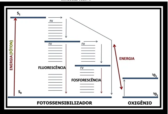
Na TFDA, no mecanismo de fotoativação são importantes as características fotofísicas do FS empregado para que ocorra alto rendimento quântico por parte do FS. O rendimento quântico é a quantidade de moléculas de FS que absorvem fótons e produz energia, o qual é dependente do oxigênio molecular disponível no local irradiado para que ocorra uma eficaz TFDA. O ideal é que um FS além de alto rendimento quântico com o comprimento de onda específico utilizado, ele apresente outras características, como: alta produção de oxigênio singleto; eliminação rápida pelas células que não devem ser atacadas; não serem tóxicas no escuro; fácil diluição em água; alta estabilidade física cinética e termodinâmica; síntese fácil, rápida e barata; curto período de pré-incubação para absorção pelas células-alvo e não provocarem mutação nos tecidos tratados.
oxigênio singleto (1O
2) apresenta maior efeito oxidante que o próprio oxigênio molecular (3O2) e por este motivo tem ação tóxica capaz de inativar os micro-organismos (Figura 04).
Figura 4 - Diagrama de Jablonski adaptado mostrando transferência de energia para produção de oxigênio molecular reativo
Fonte: elaborado pelo autor (Panhóca, 2015).
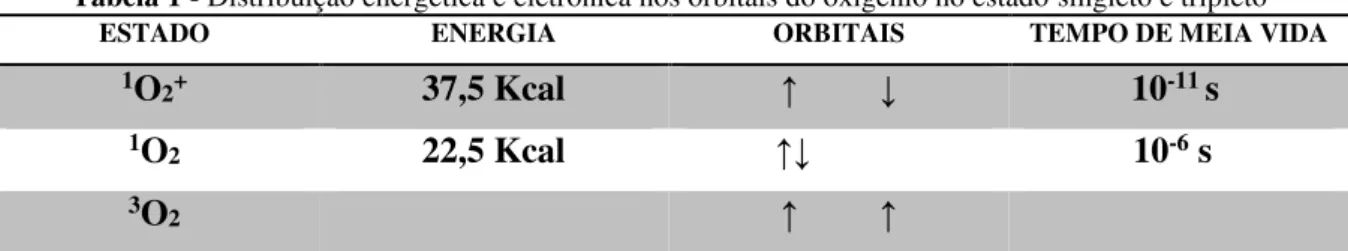
O oxigênio singleto pode se apresentar em dois estados: 1O
2 seria o estado excitado que tem dois elétrons com spin opostos no mesmo orbital, energia de 22,5 Kcal acima do estado fundamental e tempo de meia vida curto em solvente aquoso de 10-6 s; e 1O
Tabela 1 - Distribuição energética e eletrônica nos orbitais do oxigênio no estado singleto e tripleto
ESTADO ENERGIA ORBITAIS TEMPO DE MEIA VIDA
1O2+ 37,5 Kcal ↑ ↓ 10-11 s
1O2 22,5 Kcal ↑↓ 10-6 s
3O2 ↑ ↑
Fonte: adaptado de Martinez, Medeiros e Di Mascio.63
A Curcumina apresenta espectro máximo de absorção da luz na região do violeta e azul (300-500nm)24 (Figura 05). Na presente pesquisa, a solução de Curcumina utilizada para a TFD dos grupos experimentais analisados foi formulada com sais de Curcumina produzidos pela indústria PDT Pharma, Cravinhos, SP, Brasil.
Figura 5 - Gráfico mostrando o espectro de absorbância da Curcumina
Fonte: adaptado de Alzate Ceballos et al.61
3.2 Luz
excitar FSs e na sequência produzir oxigênio citotóxico e obter uma taxa de redução microbial eficiente nesta terapia.
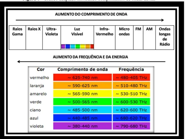
A luz azul apresenta comprimento de onda por volta de 440 a 485nm que é menor em relação ao vermelho com comprimento de onda por volta de 625 a 740nm(Figura 06). Considerando essa diferença de comprimentos de ondas, pode-se dizer que ocorre menor penetração da luz azul nos tecidos do que a luz vermelha. No entanto, a luz azul produz fótons, pacotes de energia, com maior frequência e carga energética capaz de promover de maneira mais eficaz a excitação eletrônica da Curcumina para inativação microbial fotodinâmica. Embora a luz azul seja mais interessante no mecanismo fotodinâmico quando se usa Curcumina como FS e se deseja entregar maior quantidade de energia em uma superfície, cuidado deve ser tomado quando se irradia com luz azul os tecidos vivos, por ser mais absorvida superficialmente e apresentar maior energia acumulada nesta região pode ocorrer aumento de temperatura superficial levando a dano térmico destes tecidos em função da energia cinética gerada no local irradiado que altera a termodinâmica tecidual.
Figura 6 - Gráfico adaptado mostrando comprimentos de ondas da luz
Na TFDA é necessário que o FSs, moléculas fotoabsorvedoras, sejam fotoativadas com um comprimento de onda específico resultando em um estado superexcitado destas moléculas, tornando-as eficazes no mecanismo de oxidação e inativação microbiana. A fonte de luz mais utilizada em TFD tem sido o LASER de baixa potência, porém atualmente vários trabalhos mostram a mesma eficácia quando se usa o LED como fonte de luz8,29. Os aparelhos com fonte luz LED apresentam como principais diferenças do LASER de baixa potência, a falta de coerência entre as ondas eletromagnéticas e não terem uma banda estreita monocromática de luz. Estas características não alteram a capacidade do LED em produzir EROs que vão oxidar e inativar os micro-organismos. Os fótons gerados pelos aparelhos de LASER de baixa potência ou LED, independente da coerência das ondas eletromagnéticas, quando entram em contato com moléculas FSs são capazes de excitá-las e por transferência de energia para o oxigênio orgânico produzir substâncias tóxicas (EROs) responsáveis pelo mecanismo de oxi-redução microbiano. Os aparelhos feitos com LED apresentam vantagens em relação ao LASER por apresentarem maior área de irradiação, consumirem menor energia, ter maior durabilidade e menor custo de produção.30
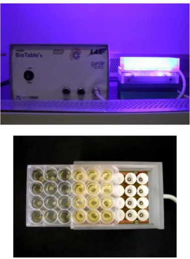
Neste estudo foi utilizado para o experimento in vitro sistema de irradiação, denominado Biotable, desenvolvido pelo Laboratório de Apoio Tecnológico – IFSC - USP, São Carlos, Brasil. A Biotable contém 24 LEDs dispostos de maneira a irradiar de forma homogênea as placas acrílicas com 24 poços contendo as amostras experimentais. A Biotable apresenta potência de 46mW/cm2 e produz luz na cor azul com comprimento de onda igual a 450 ±10 nm, ressonante com a coloração amarela da Curcumina.
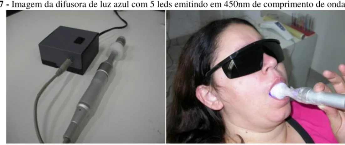
operador dos aparelhos de luz para descontaminação bucal utilizaram óculos de proteção ocular evitando danos que os comprimentos de ondas nesta terapia possam causar aos olhos.
Figura 7 - Imagem da difusora de luz azul com 5 leds emitindo em 450nm de comprimento de onda.
Fonte: elaborado pelo autor (Panhóca, 2015).
Figura 8 - Imagem do Bright Max Evolution – MMOptics com LED azul de 450nm de comprimento de onda.
4 SURFACTANTE
Surfactantes são agentes ativos de superfície. Os surfactantes são muito utilizados na indústria alimentar com o objetivo de inibir a formação de biofilmes em superfícies de equipamentos industriais.31 Sua principal característica é a capacidade de ser absorvido em superfícies e áreas de interface entre diferentes materiais. Outra característica dos surfactantes é apresentar capacidade de redução de tensão superficial para fluídos aquosos, o que permite que tenham ação detergente, humidificante e emulsionante. Por este motivo são muito utilizados pela indústria para produtos de limpeza e cosmética. No preparo da solução para bochecho o surfactante aumenta a solubilização das moléculas de Curcumina diminuindo sua agregação e potencializando o seu efeito quântico antimicrobiano na TFDA.
Figura 9 - Estrutura molecular do surfactante
Fonte: elaborado pelo autor (Panhóca, 2015).
O surfactante pode ser considerado aditivo interessante para potencializar o efeito antimicrobiano na TFDA, pois promovem alteração da membrana celular microbiana tornando-a mtornando-ais permeável, permitindo mtornando-aior penetrtornando-ação de FS, tornando-aumenttornando-ando o seu efeito tornando-antimicrobitornando-ano, o que pode promover melhor controle microbiológico do biofilme dental em indivíduos portadores de aparatos ortodônticos.14,16 Na literatura encontramos estudo mostrando a eficácia antimicrobial quando se aplica TFDA utilizando azul de metileno (MB) combinado com vários tipos de surfactantes, SDS (aniônico), CTAC (catiônico), HPS (zwiteriônicos) e Triton X-100 (não-ionico); os surfactantes na referida pesquisa foram utilizados para aumentar a permeabilidade em células de C. albicans, diminuir sua adesividade por superfícies, diminuir a formação de biofilme e diminuir sua resistência às outras drogas.14 Na presente pesquisa o
5 CLOREXIDINA
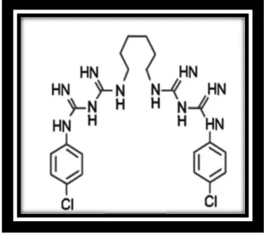
No controle do biofilme dental para prevenir doenças bucais crônicas, tais como: cárie, gengivite e periodontite; tem sido utilizado vários tipos de solução enxaguatória, porém a clorexidina a 0,12% é considerada a mais eficiente.32 A clorexidina foi sintetizada nos anos 40 e apresentava sua estrutura molecular como pode ser visto na figura 10. Após isso, Rose e Swain formularam a clorexidina em 1956 e foi comercializada como anti-séptico para ferimentos de pele.33 Desde a década de 70, estudos tem mostrado o efeito de inibição completa do biofilme dental e prevenção de gengivite quando se aplica diariamente a clorexidina, desde que o agente seja administrado de tal forma que ele entre em contato com todas as superfícies dos dentes.34 A clorexidina é um detergente catiônico, da classe das bisbiguanidas, disponíveis nas formas de acetato, hidrocloreto e digluconato, sendo esta última a forma mais comunmente utilizada como antimicrobial em odontologia.
Figura 10 - Estrutura química da clorexidina (1,6-di(4-clorofenildiguanida)hexano
Fonte: disponível em <
http://pt.wikipedia.org/wiki/Gluconato_de_clorexidina#mediaviewer/File:Chlorhexidine.png >.
protetora osmótica, a partir disto se inicia os eventos subsequentes diretamente dependente da concentração de clorexidina no meio celular. Em doses mais altas causa coagulação e precipitação das proteínas citoplasmáticas e a morte microbiana.35 A clorexidina por ser uma molécula dicatiônica apresenta capacidade química de ligar a película dental adquirida e membrana bacteriana ao mesmo tempo. Isso resulta em uma substantividade, que é a capacidade de se manter ativa no meio bucal, durante aproximadamente 8 a 12 horas, inativando os micro-organismos que buscam colonizar a superfície dental. Outro fator que influência a substantividade da clorexidina é o pH bucal, que sendo ácido aumenta sua eficácia.36
6 OBJETIVO
7 OBJETIVOS ESPECÍFICOS
Avaliação in vitro do efeito da TFDA aplicando LED azul (450±10nm) com Curcumina solubilizada em surfactante (SDS) sobre biofilme formado por S mutans em superfície metálica de acessórios utilizados no tratamento ortodôntico.
Avaliação in vivo do efeito da TFDA aplicando LED azul (450±10nm) com Curcumina solubilizada em surfactante (SDS) sobre S mutans na cavidade bucal de indivíduos portadores de aparelhos ortodônticos fixos, utilizando um novo dispositivo de LED intra-bucal com potência média de luz.
8 ARTIGO EXPERIMENTO IN VITRO
O artigo a seguir foi publicado originalmente em Journal Physical Science and Application, no volume 4 de 2014. ISSN: 2159-5348
ENHANCEMENT OF THE PHOTODYNAMIC THERAPY EFFECT ON
STREPTOCOCCUS MUTANS BIOFILM
a,bV. H.Panhóca*, bM. C. Geralde, bT. Q Corrêa, aM. T. Carvalho, bC. W. O. Souza, aV. S.
Bagnato
aUniversity of São Paulo – USP, Physics Institute of São Carlos – IFSC, Optical Group,
Biophotonics Laboratory, São Carlos, SP, Brazil
bFederal University of São Carlos - UFSCar, Biotechnology Pos-Graduate Program, São
Carlos, SP, Brazil
*Corresponding Author: Department of Physics and Material Science, Institute of Physics of
São Carlos, University of São Paulo, São Carlos, SP, 13560-970, Brazil
E-mail: vhpanhoca@msn.com
ABSTRACT
Biofilm is a community of bacteria, less susceptible to traditions treatments. Although
photodynamic therapy (PDT) is a very effective way to microorganism inactivation, in biofilm
it is not as efficient as it is in planktonic bacteria cultures. Our objective is to increase the
efficiency of PDT by adding one element to the treatment. Therefore, this in vitro study
evaluates the susceptibility of a biofilm formed by Streptococcus mutans on metallic surface of
orthodontic accessories under the application of PDT with a surfactant. Samples obtained from
blades of orthodontic bands (NiCr), where used as adhesion surface for the biofilm. They were
of light (455nm). Eight experimental groups were studied, including the positive and negative
controls. The results show that the group with PDT and surfactant had a significant decrease
(p<0.001) in viability. In this case, the reduction observed was of 5.6 log10 (CFU/ml) in
comparison to the control group. We have shown that, even though the biofilm is very tough
and complex structure, we are able to promote almost the complete inactivation of S. mutans in
systems similar to an orthodontic treated patient’s mouth.
Keywords: Photodynamic Therapy, Light-induced damage, Oral Biofilm, Streptococcus
mutans, Curcumin, Surfactant.
INTRODUCTION
There are two forms for bacteria to survive in their natural environment. They may be
free-floating, as a planktonic cell, or attached to a surface, as in a biofilm. The biofilm is a
complex community in which most of bacteria live. It is a well-organized community that
adheres to surfaces and is embedded in an extracellular adherent layer. Bacterial resistance
increases when they are organized as biofilm. Also, once in a biofilm, the bacteria display
different characteristics from those that they had as a free-floating organism. Biofilms can be
found in many places, and the human mouth has perfect conditions for the bacteria to grow.
For that reason, biofilms are precursor for most common oral diseases, such as caries
and periodontal disease. Prior to the formation of the biofilm, a thin microorganism layer
adheres on the surface of the tooth or gum. These microorganisms use salivary proteins as
substrate for adhesion. Streptococcus mutans produces adhesins that together with these
produce allow the accession of other types of microorganism to form the mature biofilm, and
finally the dental plaque.
The dental biofilm can be categorized into sub- and supra-gingival. It is formed
subsequent to an increase of saccharolytic and acidogenic microorganisms, such as
Streptococcus mutans and lactobacillus, which occur in most people who eat sucrose-rich food.
Hence, these biofilms produce acids that cause tooth demineralization and caries. Four
components are needed to create the carious lesion (1, 2): a host, high-carbohydrate diet,
microorganisms and time.
The Streptococcus mutans, that may be one of the most common bacteria in the oral
cavity, produces a polyhedral matrix to structure the biofilm. The microorganisms existent in a
biofilm become sheltered by this polyhedral matrix, reducing the action of antimicrobial agents.
Compared to planktonic cells, the biofilm structure protects the bacteria, and other
microorganisms. The thickness of this structure prevents contact between antimicrobial agents
and microorganisms, keeping the chemicals away from the deeper layers of the biofilm. As a
result, the biofilm boosts the growth rate of the microorganisms (2-4). Consequently,
microorganisms embedded in biofilms are 10 to 1000 times more resistant to antimicrobial
agents (5, 6).
One cause that enhances the biofilm formation in the mouth is the installation of
orthodontic accessories (7). The usage of orthodontic accessories creates new retention surfaces
for microorganisms in the oral cavity, increasing their retention. Therefore, it increases biofilm
formation and the amount of bacteria in the mouth (7). The high level of S. mutans in the oral
cavity of patients with orthodontic accessories, compared to non-orthodontic treatment patients,
is cause both by the presence of the accessories and also by the poor hygiene control of it by
Photodynamic therapy (PDT) is a well-known medical therapy for cell inactivation and
microorganism control (8, 9). This technique is based on the combined use of light and some
types of drugs, called photosensitizers (PS), which produce an oxidation reaction. This
oxidative reaction can them cause the death or inactivation of selected cells. PDT can also be
used as a therapy with a potent antimicrobial effect (10). Inactivation of microorganisms by
PDT can ensure several advantages over the traditional use of antimicrobials. First, bacterial
killing is rapid, reducing the need to maintain high concentrations of chemicals for long periods,
as in the case of antibiotics and antiseptics usage (11). Secondly, bacterial killing is not linked
to the intervention of chemicals, so, development of resistance is improbable to happen (12).
Finally, since neither the PS, nor the light, are bactericides by themselves, the destruction of
bacteria can be controlled by restricting the irradiated region, preventing the destruction of the
normal micro-biota elsewhere.
PDT is an alternative therapy for preventing and treating dental caries and periodontal
diseases. It is an innovative way to control the formation of the bacterial biofilm, controlling
the incidence of these pathogens. Most studies present high efficiency of PDT to planktonic
bacteria. On the other hand, since the biofilm protects its microorganisms, the results over
clinical trials are not as efficient (13). Therefore, researches over systems that emulate the
patients’ mouth are so important to find the “perfect therapy”. Thus, the purpose of this paper
is to evaluate the effect of the antimicrobial activity of PDT on biofilms formed on orthodontic
accessories. This in vitro study evaluates the use of PDT and a surfactant on metal surfaces,
observing the susceptibility of biofilms formed by S. mutans. Our objective is to increase the
performance of PDT, adding one element to the treatment, in a system that emulates the mouth
real environment.
For this study 24 metal samples measuring 5x5mm were used, obtained from
orthodontic band blades (NiCr). On the metal surface, S. mutans biofilms were growth and
threated with PDT and a surfactant. The PDT was induced by curcumin and light emitting diode
(LED) in the blue range, and the surfactant used was the sodium dodecyl sulfate (SDS). There
were tested eight experimental groups, each test repeated three times and each solution
measured twice.
Biofilm growth and CFU preparation
For Streptococcus mutans biofilm, an inoculum from stock culture (ATCC 25175) was
grown in Brain Heart Infusion (BHI) broth (Difco, Detroit, USA) and incubated at 37°C for 24
hours. The suspensions were centrifuged, 3000 rpm per 15 minutes (Excelsa II centrifuge,
model 206-BL, FANEM), and the bacterial pellet was dispersed into BHI broth with 20%
sucrose. To archive the desired population density, the culture was adjusted to obtain
standardized suspension containing 106 cells/ml, the optical density of the final suspensions
was verified using a digital spectrophotometer (FEMTO 600).
The biofilm was grown in orthodontic band metal blades (orthodontic appliance -
Tecnident, São Carlos, Brazil), cut into 5x5 mm squares, and sterilized in autoclave. Each
sample was placed inside individuals wells of a 24-well microtiter plate. Then, aliquots of 1ml
of the S. mutans were transferred to the plates. The biofilms were incubated at 37°C up to 7
days, with the growth medium changed every two days. All the samples were washed in PBS
solution and placed in a new well into the 24 microtiter plate to be prepared for experimental
treatment. They were separated between eighth groups and treated accordingly.
After treatment, the samples and theirs solutions were transferred to eppendorfs, 1ml of
PBS was added, and they were homogenized, to break and loosen the biofilm into the solution,
removed. The solutions were diluted up to 10-5 and uniformly spread to petri dishes containing
culture medium BHI AGAR (Difco, Detroit, USA) plus 20% sucrose. They were aerobically
cultured at 37°C for 48 hours prior to the colony-forming unit (CFU) count.
Experimental treatment procedure
The experimental groups included: control group (P-L-S-); light (P-L+S-), surfactant
(P-L-S+) and photosensitizer (P+L-S-) groups; surfactant-light (P-L+S+),
photosensitizer-surfactant (P+L-S+) and PDT (P+L+S-) groups; and the PDT-photosensitizer-surfactant (P+L+S+) group. Each
group was named accordingly to the addiction of the photosensitizer (P+ or P-), surfactant (S+
or S-) and application of light (L+ or L-).
All the samples were placed in a new well into the 24 microtiter plate and received the
correct solution necessary for treatment. To the control (P-L-S-) and light (P-L+S-) groups,
there was added 1 ml of a solution of PBS with 5% of DMSO. There was added 1 ml of
surfactant solution to the surfactant (P-L-S+) and surfactant-light (P-L+S+) groups. The
surfactant solution was made in PSB with 0.1% of SDS and 5% of DMSO. To the
photosensitizer (P+L-S-) and PDT (P+L+S-) groups it was added 1 ml of a photosensitizer
solution. This solution had 1 mg/ml of curcumin, and was made in PBS with 5% of DMSO.
And to the photosensitizer-surfactant (P+L-S+) and PDT-surfactant (P+L+S+) groups, there
was added 1 ml of a photosensitizer-surfactant solution, which had 1 g/mL of curcumin, 0.1%
of surfactant, and 5% of DMSO in PBS.
All the experimental groups were kept in the dark for 5 minutes, incubation of the PS,
before the application of light in the designated groups. The experimental groups with light
(L+) were then irradiated in a home-made blue LED device (high power royal blue LEDs,
355mW, centered at 455 nm with 20 nm bandwidth), for 10 minutes and 54 seconds, ensuring
was made to guarantee uniform distribution of light (46 mW/cm² intensity) in the plane were
the samples were placed. The experimental groups (L-) where no light was applied were kept
in the dark at room temperature, for the same amount of time.
Figure 1 – Photography of the 24-well blue Biotable and details of the metal plate samples, placed in a 24-well microtiter plate, being put in place on the LED device.
Data analysis
The effect of the photodynamic therapy and surfactant on the biofilms was evaluated by
counting colony-forming units. The results were statistically evaluated by ANOVA, differences
were considered when p <0.05. The difference between treated groups with the control group
RESULTS AND DISCUSSION
To ensure biofilm formation, selected metallic samples were imaged with confocal
microscopy (Figure 2). The images showed the formation of S. mutans biofilm after 7 days of
cultivation on the metallic samples of orthodontic appliance (NiCr). The photodynamic
inactivation was also observed, by means of a live-dead biological marker, imaging plate before
and after PDT treatment. The confocal reflection images show the metal surface, indicating the
presence of colonies of S. mutans. The confocal fluorescence images show the fluorescence of
the propidium iodide (PI) dye and curcumin in the bacteria cells. The colonies can be
highlighted when both images are superposed, showing the correspondence between the
colonies seen by the reflection and fluorescence images.
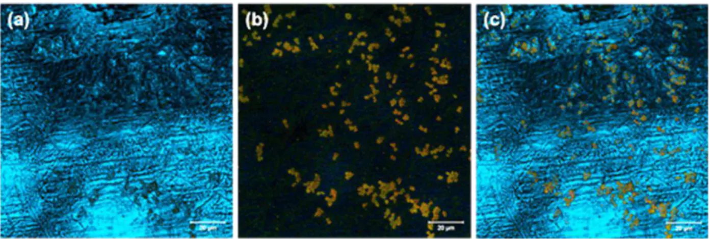
Figure 2 - Biofilms of S. mutans on metallic sample with 7days, after PDT treatment: (a)
Confocal reflection imaging, the image show the appearance of the metal surface, indicating the presence of S. mutans colonies. (b) Confocal fluorescence image, showing
the fluorescence of the propidium iodide (PI) dye and curcumin. The colonies appear as bright spots in the image, red indicates dead cells and yellowish-green show the living microorganisms. (c) Reflection and fluorescence images superposed (a+b). The image
The confocal reflectance images present the surface of the metal sample. It was expected
that the orthodontic appliance squares show some roughness, and appears in the image as a
random pattern. It is also noticed a pattern similar to a bunch of grapes, and the superposition
of the reflectance and fluorescence images confirm that those patterns correspond to the biofilm.
Meanwhile, the fluorescence image show red and yellow-green dots. The yellow-green dots are
the fluorescence of the curcumin within the bacteria cells. The red dots are the fluorescence if
the propidium iodide (PI) dye, which appear in the dead cells.
Figure 3 - Reduction of CFU for S. mutans biofilms. Individual difference made by
ANOVA One Way - post hoc Turkey. Groups with the same letter are not significantly different (p>0,005).
The bacterial effects of PDT and the surfactant were evaluated by the cell viability
(CFU/ml). The efficacy of the treatment differs depending on the association of curcumin and
surfactant (Figure 3). They showed significant reductions in the viability of the S. mutans (over
highest reduction in the cell viability of the biofilms was equivalent to 5.6 log10 (CFU/ml) and
correspond to the association of PDT and surfactant.
The results show remarkable behavior of the bacteria population depending on the
association of curcumin, surfactant and the application of light (Table 1). In some cases the
illumination can promote biostimulation, but none of the studied cases presented a relatively
relevant increase on the bacterial population. Applying just PDT (P+S-L+) or surfactant
(P-S+L-), it was observed small decreases on the S. mutans population. The same occur when
light was applied mutually with surfactant (P-S+L+), but in this case the antibacterial effect was
somehow smaller. The test group where only the photosensitizer was used (P+S-L-) was
statistically similar to the control group, confirming that curcumin, by itself, is not capable to a
significant bacteria inactivation. On the other hand, the association of curcumin and SDS
(P+S+L-) upheld an even smaller decrease in the population. However, when the three where
used together (P+S+L+), it was observed a huge antibacterial effect (-5.6 log10). In this case,
only one in a thousand part of the population survived.
Table 1- Expression of cell viability (CFU/ml), related to the control group data, for all experimental groups. Groups with the same letter are not significantly different
(p>0,005).
Experimental
groups CFU Percentage difference Expression (log10)
(P-S-L-) a 0
(P-S-L+) a +29.17 +0.1
(P-S+L-) b -99.22 -2.1
(P-S+L+) b c -97.84 -1.6
(P+S-L-) a d -42.91 -0.2
(P+S-L+) b e -97.46 -1.5
(P+S+L-) c d e -93.37 -1.1
The oral microbiota is composed of more than 500 types of microorganisms, even
though S. mutans is the etiologic agent of caries and demineralization. It is also responsible for
the adhesion, colonization and dental biofilm formation (14). This study evaluated particularly
the susceptibility of S. mutans biofilm on the metal surface (NiCr) of orthodontic blade band
by means of photodynamic therapy with curcumin (1mg/ml), 0.1% of SDS and LED
illumination. As expected, we have shown that photodynamic therapy reduces the number of
living cells of S. mutans in a biofilm model created on metallic surface (15-17). The presence
of photosensitizer, without irradiation, was not able to achieve an effective antibacterial
outcome. This confirms the essential rule of light to obtain an anti-microbial effect (18, 19) by
means of PDT.
The most significant finding was the lack of viability of S. mutans on the surfaces of the
samples treated with curcumin combining PDT with surfactant. The result for this group
(P+S+L+) is above the minimal bactericidal concentration, which is the minimum concentration
able to reduce 4 log10 on the survival of a microorganism (20). Significant decrease on the
viability of microorganisms was also observed when biofilms were exposed to the surfactant
alone (P-S+L-), surfactant with light (P-S+L+), photosensitizer with light (P+S-L+), or the
combined use of surfactant and curcumim without light (P+S+L-), compared to the control
group (P-S-L-). For these groups, the reduction on the bacteria viability was above 90%.
Depending on the application, 90% of reduction on a microorganism population might be
sufficient. Although the results show that the bacterial inactivation can be almost complete
when the surfactant was used together with PDT.
The surfactants are amphipathic molecules composed of a hydrophobic portion and a
hydrophilic portion. The first is often a nonpolar hydrocarbon chain, while the second can be
characteristics, are highly potent and act as emulsifiers, reducing interfacial and superficial
tension (21). They are responsible for modifications on the ion channels of the bacterial
membrane via trans-membranes proteins, similar to the porins. This allows the passage of
solutes through the membrane, increasing the permeability of these membranes (21-24).
Surfactants also work as: emulsifiers, dispersing in water hydrocarbons or other insoluble
compounds; reducing the adhesion and releasing surfaces cell; and presenting antibiotic
activity.
The mechanism of action of PDT is not connected to the mediation of chemical radicals
acting in a single target, which is the case of the antibacterial products. Photosensitizes act
producing reactive oxygen species (ROS) which have no specific site of action. This avoids the
formation of strains resistant to antibiotics (20, 21, 25, 26). Photodynamic therapy applied as
antimicrobial therapy can be also advantageous over mechanical removal methods, since it can
reach places such as recesses and protrusions of the orthodontic accessories (27). It may also
enable safe treatment of patients with special needs or difficulty in oral hygiene during
orthodontic treatment, avoiding oral infections, such as dental caries, gingivitis and periodontal
pockets (13, 27).
Several hypotheses could explain the synergism of the PDT action with surfactant as
antimicrobial therapy in S. mutans biofilm. The surfactant could be increasing the number of
working cycles of PS, protecting it from oxidation, and increasing the efficacy of PDT. It could
also be acting increasing the permeability of the curcumin through the cell membrane,
increasing the concentration of PS molecules in the bacteria. One other hypothesis is that the
surfactant is breaking the biofilm structure, and this allows the PDT to be more effective.
Although the surfactant, SDS, can be used as antimicrobial by itself, the combined effects of it
larger than one could expect from using both treatments separately. Therefore more studies
must be done in order to thoroughly understand this mechanism.
CONCLUSIONS
The result of this study shows higher reduction in the S. mutans population, compared
to previous investigations (15, 19). We demonstrate significant removal of S. mutans biofilm
after application of PDT with SDS as antimicrobial therapy. PDT associated to surfactants
promoted a high inactivation of S. mutans biofilm (5.6 log10) on the surface of metallic
orthodontic accessories. We have shown that, even though the biofilm is very tough and
complex structure, we are able to promote almost the complete inactivation of S. mutans in a
system similar to an orthodontic treated patient’s mouth. And this inactivation was achieved
with a protocol that does not promote bacterial resistance that applied PDT and SDS.
Given the findings, our results suggest that the surfactant acts on the microorganisms’
membrane making them permeable to solutes, and breaking the biofilm structure. The surfactant
leads to higher the input of oxygen molecules and curcumin from the extracellular medium into
the microorganism. Therefore, the synergistic action of the SDS and PS produces higher
amounts of reactive oxygen species (ROS) upon irradiation, optimizing the antimicrobial effect
of PDT. At the same time, protecting the PS molecules from oxidation and increasing its
lifetime of the PDT cycle. These hypotheses should be put to test in supplementary
investigations.
AKNOWLEDGMENT
The authors would like to acknowledge the support of the Brazilian agencies FAPESP,
CNPQ and CAPES, through founding and scholarships. V. H. Panhóca would like to
acknowledge scientific contributions and helpful advice from Clóvis Grecco and Fernanda
REFERENCES
1. P. H. Keyes, "The infectious and transmissible nature of experimental dental caries.
Findings and implications," Arch Oral Biol 1(304-320 (1960)
2. E. Newbrum, Cariology, Baltimore (1978).
3. T. Fusayama and S. Terachima, "Differentiation of two layers of carious dentin by
staining," J Dent Res 51(3), 866 (1972)
4. T. Fusayama and S. Terashima, "Differentiation of two layers of carious dentin by
staining," Bull Tokyo Med Dent Univ 19(1), 83-92 (1972)
5. M. J. Sedlacek and C. Walker, "Antibiotic resistance in an in vitro subgingival biofilm
model," Oral Microbiol Immunol 22(5), 333-339 (2007)
6. C. R. Fontana, A. D. Abernethy, S. Som, K. Ruggiero, S. Doucette, R. C. Marcantonio,
C. I. Boussios, R. Kent, J. M. Goodson, A. C. Tanner and N. S. Soukos, "The antibacterial
effect of photodynamic therapy in dental plaque-derived biofilms," J Periodontal Res 44(6),
751-759 (2009)
7. J. A. Corbett, L. R. Brown, H. J. Keene and I. M. Horton, "Comparison of Streptococcus
mutans concentrations in non-banded and banded orthodontic patients," J Dent Res 60(12),
1936-1942 (1981)
8. S. George, A. Kishen, “Photophysical, photochemical, and photobiological
characterization of methylene blue formulations for light-activated root canal disinfection,”
Journal of Biomedical Optics 12(3), 034029 (2007)
9. R. Darlenski, J. W. Fluhr, “Photodynamic therapy in dermatology: past, present, and
future,” Journal of Biomedical Optics 18(6), 061208 (2012)
10. M. Wainwright, "Photodynamic antimicrobial chemotherapy (PACT)," Journal of
11. H. Greenwell and N. Bissada, "Emerging concepts in periodontal therapy," Drugs
62(18), 2581-2587 (2002)
12. P. Bidault, F. Chandad and D. Grenier, "Risk of bacterial resistance associated with
systemic antibiotic therapy in periodontology," Journal of the Canadian Dental Association
73(8), 721-725 (2007)
13. N. Araujo, C. Fontana, M. Gerbi and V. Bagnato, "Overall-Mouth Disinfection by
Photodynamic Therapy Using Curcumin," Photomedicine and Laser Surgery 30(2), 96-101
(2012)
14. J. Kreth, E. Hagerman, K. Tam, J. Merritt, D. T. Wong, B. M. Wu, N. V. Myung, W.
Shi and F. Qi, "Quantitative analyses of Streptococcus mutans biofilms with quartz crystal
microbalance, microjet impingement and confocal microscopy," Biofilms 1(4), 277-284 (2004)
15. Z. Zou, P. Gao, H. Yin and Y. Li, "Investigation of photodynamic therapy on
streptococcus mutans of oral biofilm," Chinese Optics Letters 6(12), 947-949 (2008)
16. J. Giusti, L. Santos-Pinto, A. Pizzolito, K. Helmerson, E. Carvalho-Filho, C. Kurachi
and V. Bagnato, "Antimicrobial photodynamic action on dentin using a light-emitting diode
light source," Photomedicine and Laser Surgery 26(4), 281-287 (2008)
17. I. C. Zanin, M. M. Lobo, L. K. Rodrigues, L. A. Pimenta, J. F. Höfling and R. B.
Gonçalves, "Photosensitization of in vitro biofilms by toluidine blue O combined with a
light-emitting diode," Eur J Oral Sci 114(1), 64-69 (2006)
18. J. O'Neill, C. Hope and M. Wilson, "Oral bacteria in multi-species biofilms can be killed
by red light in the presence of toluidine blue," Lasers in Surgery and Medicine 31(2), 86-90
19. M. Schneider, G. Kirfel, M. Berthold, M. Frentzen, F. Krause and A. Braun, "The impact
of antimicrobial photodynamic therapy in an artificial biofilm model," Lasers in Medical
Science 27(3), 615-620 (2012)
20. G. Jori, C. Fabris, M. Soncin, S. Ferro, O. Coppellotti, D. Dei, L. Fantetti, G. Chiti and
G. Roncucci, "Photodynamic therapy in the treatment of microbial infections: basic principles
and perspective applications," Lasers Surg Med 38(5), 468-481 (2006)
21. P. A. V. Fernandes, I. R. d. Arruda, A. F. A. B. d. Santos, A. A. d. Araújo, A. M. S.
Maior and E. A. Ximenes, "Antimicrobial activity of surfactants produced by Bacillus subtilis
R14 against multidrug-resistant bacteria," Brazilian Journal of Microbiology 38(704-709
(2007)
22. F. Peypoux, J. M. Bonmatin and J. Wallach, "Recent trends in the biochemistry of
surfactin," Appl Microbiol Biotechnol 51(5), 553-563 (1999)
23. M. Nitschke and G. M. Pastore, "Biossurfactantes: propriedades e aplicações," Química
Nova 25(772-776 (2002)
24. J. P. Lyon, R. R. Rezende, M. P. Rabelo, C. J. de Lima and L. M. Moreira, "Synergic
effect of photodynamic therapy with methylene blue and surfactants in the inhibition of Candida
albicans," Mycopathologia 175(1-2), 159-164 (2013)
25. F. Lauro, P. Pretto, L. Covolo, G. Jori and G. Bertoloni, "Photoinactivation of bacterial
strains involved in periodontal diseases sensitized by porphycene-polylysine conjugates,"
Photochemical & Photobiological Sciences 1(7), 468-470 (2002)
26. A. Tavares, C. M. Carvalho, M. A. Faustino, M. G. Neves, J. P. Tomé, A. C. Tomé, J.
A. Cavaleiro, A. Cunha, N. C. Gomes, E. Alves and A. Almeida, "Antimicrobial photodynamic
therapy: study of bacterial recovery viability and potential development of resistance after
27. T. S. Mang, D. P. Tayal and R. Baier, "Photodynamic therapy as an alternative treatment
9 ARTIGO EXPERIMENTO IN VIVO
O artigo a seguir foi submetido em periódico internacional.
ORAL DECONTAMINATION USING ANTIMICROBIAL PHOTODYNAMIC THERAPY APPLIED IN ORTHODONTIC PATIENTS.
Panhóca, V.H.1,2,a, Florez, F.L.E.3,b, Corrêa, T.Q1,c, Souza, C.W.O.1,d, Bagnato,V.S2,d. 1 Biotechnology Graduate program, Federal University of São Carlos.
2 São Carlos Institute of Physics, Group of Optics, University of São Paulo.
3 College of Dentistry, Department of Dental Materials, University of Oklahoma Health
Sciences Center.
a Clinical Researcher and Ph.D. Student. b Postdoctoral Fellow.
c Ph.D. Student. d Full Professor.
All authors have completed and submitted the ICMJE Form for Disclosure of Potential Conflicts of Interest, and none were reported.
Address correspondence to: Fernando Luiz Esteban Florez, D.D.S., M.S, Ph.D.
Department of Dental Materials, College of Dentistry, The University of Oklahoma Health Sciences Center.
1201 North Stonewall Avenue. Oklahoma City, Oklahoma. 73117
E-mail: fernando-esteban-florez@ouhsc.edu
Abstract
were randomly distributed within seven experimental groups: G1 - Negative control, G2 - Curcumin mouthwash, G3 - Curcumin mouthwash+SDS, G4 - light irradiation, G5 - APDT, G6 - APDT+SDS and G7 - Positive Control (Chlorhexidine mouthwash). Non-stimulated saliva samples were collected from each one of the patients in three steps (S), as follows: S1 - Initial condition, S2 - Treatment with mouthwashes (water, Chlorhexidine or Curcumin) and S3 - After APDT treatment. The efficacy of the proposed treatment protocols was assessed in function of the survival fraction observed in each group. The obtained results were statistically analyzed using variance analysis ANOVA Kruskal-Wallis test. The statistical analysis demonstrated that only the groups 6 (APDT+SDS) and 7 (Chlorhexidine 0.12%) has presented bacterial inactivation results that were statistically significant differences (p<0.05) from the results observed in the group 1 (negative control). These results indicate that the APDT when associated with the SDS surfactant can be used as a coadjutant and convenient agent to promote the oral decontamination in vivo.
Introduction
The dental specialty that is dedicated to the study, prevention and treatment of dentofacial alterations is known as Orthodontics. Its main objective is to attain a more balanced relation of position between teeth, facial arches and facial bones through the use of orthodontic appliances. Dental brackets, wires and metallic bands are some of the components that commonly are used during this kind of treatment1.
Once these components had been adapted over tooth surfaces (buccal or lingual), the patient experiences a drastic decrease in his ability to properly perform their routine oral hygiene and, at the same time, the installation of these devices, will also promote a significant increase on the amount of available surfaces for bacterial aggregation. This problem is exacerbated even further, because the microorganisms present in the oral microflora preferentially aggregate on dental materials surfaces, such as, restorative materials (amalgam and composite resins) and orthodontic appliances2,3.
dislodgement. Published studies on the matter have shown that, microorganisms in biofilms have 10 to 1000 times higher resistance levels to antimicrobial agents, when compared with the antibacterial resistance observed for the same microorganisms in its planktonic phase5,6. Other studies had also shown that patients under orthodontic treatment have higher Streptococcus mutans loads when compared with patients without the use of orthodontic appliances.
The biofilm growth during the evolution of the orthodontic treatment, will promote the establishment of a degradation cycle that might result initially in the appearance of white lesions (ICDAS - 0, 1 and 2) around the brackets' attachment sites and active carious lesions (ICDAS - 3 to 6) around the metallic bands7-9.
Preventive measures to avoid the installation and progression of caries diseases include oral hygiene instructions (brushing techniques, flossing techniques, use of mouthwashes, etc.), orientation about the impact of dietary habits on patient's overall oral health, prescription of
fluoride toothpastes (≥1000 ppm) and mouthwashes. In this direction, and taking into
consideration, the concepts of prevention and minimum intervention currently recommended by modern Dentistry, it becomes of critical importance, the development of techniques, products and devices that can aid the maintenance of daily oral health9.
One alternative currently available to control the microbial load in the oral cavity is the Antimicrobial Photodynamic Therapy (APDT)10-12. Its mechanism of action is based on the organic molecules oxidization through the generation of reactive oxygen species (ROS) mediated by light13,14. Studies published in the literature had demonstrated the obtainment of varying degrees of success for both Gram-positive and Gram negative microorganisms15. In addition, these studies had also emphasized that, in function of the non-specific nature of the oxidative reaction, it is unlikely that the repeated use of the APDT will promote the development of resistant bacterial strains10,13,16,17.
Modern strategies combine some antimicrobial agents with a variety of surfactants. The molecular structure of these compounds are composed by a hydrophobic (non-polar) and a hydrophilic portion capable of exerting tensioactive properties of great importance and great economic value for pharmaceutical, medical and hygiene product industries22,23. Studies in the literature suggest that the mechanism of action of such components may increase the bacterial cells' extracellular permeability by promoting an electrical charge unbalance that will lead into a higher penetration of the antimicrobial agent inside of the cells which, consequently, will lead into the attainment of higher levels of bacterial inactivation20,21.
Hence, the present clinical and randomized study investigated the synergistic effect of the surfactant Sodium Dodecyl Sulfate (SDS) on the levels of inactivation of Streptococcus mutans promoted by the use of the APDT using Curcumin as photosensitizer. The null hypothesis tested was, that the attainment of higher inactivation levels does not depends on the addition of SDS to the Curcumin photosensitizer.
Material and Methods:
The Ethics Committee in Human Beings of the São Carlos Federal University (UFSCAR) approved the execution of the present clinical and randomized study under protocol
258.461 (2013) and title “Optimization of the antimicrobial effect of Photodynamic Therapy on Streptococcus mutans in patients with orthodontic appliances". The study was registered with NIH ClinicalTrials ( NCT02337192 ).
Inclusion and Exclusion criteria
Experimental groups and Conditions
After the screening process and selection, patients were randomly distributed in each one of the experimental groups described in Table 1 through a simple draw, in order to investigate the effect of the use of the SDS on the attainment of higher levels of bacterial inactivation with the use of the APDT as an oral decontamination agent.
Table 1. Description of experimental groups and conditions.
Saliva collection
The collection of non-stimulated saliva (n=3; 3mL/collection) were performed at the following steps: i) immediately before the swishing (Curcumin, Chlorhexidine or water), ii) after swishing and iii) after performing the APDT treatments. To that end, 15 mL sterile polypropylene tubes were used (Falcon, BD Science, Durham, NC). After each saliva collection, the saliva samples were identified and stored in a dark and refrigerated environment. The samples of each patient in each experimental step were used to determine the total bacterial load.
GROUPS
N=24 TREATAMENTS
APLICATION PROTOCOLS Swishing time (s) Irradiation Time (s) Power Output (mW/cm2)
1 DMSO water solution at 5% (Negative control)
120 s 0 0
2 Swish with Curcumin 120 s 0 0
3 Swish with Curcumin + SDS 120 s 0 0
4 Irradiation with blue light 120 s 2x 180 212 & 34.5
5 APDT 120 s 2x 180 212 & 34.5
6 APDT + SDS 120 s 2x 180 212 & 34.5