UNIVERSIDADE FEDERAL DA PARAÍBA
CENTRO DE CIÊNCIAS DA SAÚDE
PROGRAMA DE PÓS-GRADUAÇÃO EM ODONTOLOGIA
ATIVIDADE ANTIFÚNGICA, MECANISMO DE
AÇÃO, CITOTOXICIDADE E AÇÃO ANTIBIOFILME
DA CLORAMINA T SOBRE Candida spp.
Gabriela Lacet Silva Ferreira
GABRIELA LACET SILVA FERREIRA
ATIVIDADE ANTIFÚNGICA, MECANISMO DE AÇÃO,
CITOTOXICIDADE E AÇÃO ANTIBIOFILME DA CLORAMINA T
SOBRE
Candida
spp.
Dissertação apresentada ao Programa de
Pós-Graduação em Odontologia, da
Universidade Federal da Paraíba, como
parte dos requisitos para obtenção do
título de Mestre em Odontologia
–
Área de
Concentração
em
Ciências
Odontológicas.
Orientador: Prof. Dr. Ricardo Dias de
Castro
João Pessoa
F383a Ferreira, Gabriela Lacet Silva.
Atividade antifúngica, mecanismo de ação, citotoxidade e ação antibiofilme da cloramina T sobre Candida spp. / Gabriela
Lacet Silva Ferreira.- João Pessoa, 2015. 56f. : il.
Orientador: Ricardo Dias de Castro Dissertação (Mestrado) - UFPB/CCS
1. Odontologia. 2. Estomatite sob prótese. 3. Higienizadores de dentadura. 4. Cloraminas. 5. Candidíase bucal. 6. Biofilmes.
iii
DEDICATÓRIA
iv
AGRADECIMENTOS
À Deus, que ilumina meus caminhos a cada dia e é minha fortaleza,
meu amparo e meu guia maior! Nas vitórias, louvo-O acima de tudo e nos
momentos difíceis, busco aconchego nos braços d`Ele.
Aos meus pais, Jamacy e Elda, que são responsáveis pela pessoa que
sou hoje. Foram exemplo de amor e dedicação, renunciaram muitas vezes a vida
deles para cuidar da minha, sonharam os meus sonhos, comemoraram comigo as
vitórias e me deram força nos momentos mais difíceis! Acima de tudo, me
ensinaram o amor e o sentido de sermos uma família!
À minha família, especialmente meus irmãos Felipe e Júlia, que
dividiram comigo momentos inesquecíveis, e aos meus avós maternos, Expedito
(
In memorian
) e Osmarina (
In memorian
), aos quais não deixarei jamais de ser
grata pelo amor e incentivo em todas as fases da minha vida.
Ao meu marido Inalmar Segundo, que sempre me incentivou e
acompanhou de perto os caminhos da minha vida acadêmica, foi meu
companheiro de noites em claro, meu incentivo quando o cansaço era maior que
tudo e a cada dia faz minha vida mais feliz.
À Ricardo Castro, que além de orientador sempre foi amigo e
compreensivo. Devo à ele todas as minhas vitórias deste caminho na vida
acadêmica, pois sempre com paciência preocupou-se em ensinar ao invés de
punir, aconselhar ao invés de impor, e hoje tem todo o meu respeito e admiração.
Nunca terei como retribuir tanto conhecimento e tanta atenção que recebi.
À Larissa Peixoto, que em tão pouco tempo tornou-se mais que uma
colega de mestrado e fez dos nossos momentos no laboratório muito mais que
uma rotina, mas instantes de amizade e companheirismo, e partilha de todo o
mérito deste trabalho.
v
À todos os colegas e professores do PPGO/UFPB que, cada um da
sua maneira, contribuiu para os momentos de aprendizado e alegria durante todo
o tempo de curso do mestrado. Em especial, agradeço aos professores Lúcio
Roberto Cançado Castellano, Fabíola Galbiatti de Carvalho Carlo e Edeltrudes de
Oliveira Lima, por terem dispensado tempo, cedido seus espaços e contribuído
com seus conhecimentos para a concretização deste trabalho.
vi
RESUMO
Introdução:
Diante das limitações para o uso do hipoclorito de sódio na
desinfecção de próteses dentárias e da necessidade do controle da proliferação
fúngica nestes sítios, faz-se necessário o estudo de novas substâncias para este
fim. Assim como o hipoclorito de sódio, a cloramina T (CAT) é um composto de
cloro com ação antibacteriana e alto poder de cloração e oxidação, e sua ação
antifúngica merece ser estudada.
Objetivos:
Avaliar a atividade antifúngica,
antibiofilme, citotoxicidade e mecanismo de ação da cloramina T trihidrato sobre
Candida
spp.
Materiais e Métodos:
Foram determinadas as concentrações
inibitória mínima (CIM) e fungicida mínima (CFM) da CAT sobre 7 cepas de
Candida albicans
,
Candida tropicalis
,
Candida krusei
e
Candida glabrata.
Foi
avaliada a cinética de inibição do crescimento de
C. albicans
em diferentes
tempos e concentrações e realizado microcultivo de
C. albicans
em ágar fubá
para avaliação da possível alteração da micromorfologia frente a diferentes
concentrações da substância. O provável mecanismo de ação sobre parede e
membrana celular fúngica foi verificado através da determinação da CIM na
presença, respectivamente, de sorbitol e ergosterol. A inibição da aderência inicial
de células fúngicas, formação e redução do biofilme de
C. albicans
foram
avaliados após contato curto (1 min) e prolongado (8 h) com a substância e a
formação do biofilme foi mensurada através de leitor de absorbância. Nistatina e
hipoclorito de sódio foram utilizados como controles positivos. A citotoxicidade foi
avaliada pelo método da hemólise. Foi realizada análise estatística descritiva e
inferencial, considerando α=5%.
Resultados:
A CIM
75%encontrada para a CAT foi
vii
(48 h) (p
≤
0,05). No teste de citotoxicidade, a CAT provocou hemólise entre 61 e
67,7%.
Conclusão:
A CAT apresenta atividade antifúngica sobre
Candida
spp. e
apresenta ação fungicida sobre a maioria das cepas testadas. Sua ação é
imediata e prolongada na inibição do crescimento de
C. albicans
e provavelmente
ocorre tanto em parede quanto em membrana celular. A CAT causa alterações na
micromorfologia de
C. albicans
e possui atividade antibiofilme, sendo efetiva na
inibição da aderência inicial das células fúngicas, bem como na formação e
redução do biofilme.
viii
ABSTRACT
Introduction:
Given the limitations to the use of sodium hypochlorite in the
disinfection of dental prostheses, and the need for control of fungal proliferation in
these sites, it is necessary the study of new substances for this purpose. Even as
sodium hypochlorite, chloramine T (CAT) is a chloro compound with antibacterial
action and high power of chlorination and oxidation, and their antifungal action
needs to be studied.
Objectives:
To evaluate the antifungal activity, mechanism of
action, cytotoxicity and anti-biofilm action of chloramine T trihydrate on
Candida
spp.
Materials and Methods:
It was determined the minimum inhibitory
concentration (MIC) and minimum fungicidal concentration (MFC) of CAT on
seven strains of
Candida albicans
,
Candida tropicalis
,
Candida krusei
and
Candida glabrata.
It was evaluated the growth kinetics of
C. albicans
by counting
colony forming units (CFU) method in different time
s and substance’
concentrations and was performed microculture of
C. albicans
in cornmeal agar
plus tween 80 to assess the possible change of its micromorphology with different
concentrations of the substance. The possible mechanism of action on fungal cell
wall and membrane has been verified by determination of the MIC in the presence,
respectively, of sorbitol and ergosterol. The inhibition of initial fungal cell
adherence and formation and the reduction in
C. albicans
biofilm were evaluated
after short (1 min) and extended (8 h) contact with the substance and biofilme
formation was by absorbance reader. Nystatin and sodium hypochlorite were used
as positive controls. Cytotoxicity was assessed through the hemolysis method. It
was conducted descriptive and inferential statistical analysis, considering α = 5%.
Results:
The MIC
75%found for CAT was 781.3 µg/mL, and the MFC/MIC ratio
ix
h) and MIC x 4 (48h) concentrations (p ≤0.05).
In the cytotoxicity assay, CAT led
to hemolysis between 61 and 67.7%.
Conclusions:
CAT has antifungal activity
against Candida spp. and has fungicidal action on most of the strains tested. Its
action is immediate and prolonged in inhibition of
C. albicans
growth and probably
occurs both in the wall and in the cell membrane. CAT causes changes in the
micromorphology of
C. albicans
and has antibiofilm activity, being effective in
inhibiting the initial adherence of the yeast cells as well as in the formation and
reduction of biofilm.
x
LISTA DE ABREVIATURAS E SIGLAS
ASD
–
Ágar Sabouraud Dextrose
ATCC -
American Type Culture Collection
CAT
–
Cloramina T Trihidrato
C. albicans
–
Candida albicans
C. glabrata
–
Candida glabrata
C. krusei
–
Candida krusei
C. tropicalis
–
Candida tropicalis
CBS
–
Central Bureau voor Schimmelcultures,
Coleção Holandesa de Cepas
Fúngicas.
CIM (MIC)
–
Concentração Inibitória Mínima (Minimum Inhibitory Concentration).
CFM (MFC)
–
Concentração Fungicida Mínima (Minimum Fungicidal
Concentration).
CSD
–
Caldo Sabouraud Dextrose
DMSO
–
Dimetilsulfóxido
IZ-
Instituto Zimotécnico
–
Universidade de São Paulo, Piracicaba, Brazil.
PBS
- Phosphate buffered saline
xi
SUMÁRIO
1 INTRODUÇÃO
12
2 CAPÍTULO I
ANTIFUNGAL ACTIVITY, MECHANISM OF ACTION AND
ANTI-BIOFILM ACTION OF CHLORAMINE T ON
Candida
spp.
15
3 CONSIDERAÇÕES GERAIS
40
4 CONCLUSÃO
44
REFERÊNCIAS
45
APÊNDICE 01
TERMO DE CONSENTIMENTO LIVRE E ESCLARECIDO (TCLE)
48
ANEXO 01
CERTIDÃO DE APROVAÇÃO DO COMITÊ DE ÉTICA
49
ANEXO 02
NORMAS PARA SUBMISSÃO DO ARTIGO AO PERIÓDICO
“CLINICAL ORAL INVESTIGATIONS”
12
1. INTRODUÇÃO
A estomatite protética é uma doença bucal que está associada ao
uso de próteses totais, na maioria dos casos, ou parciais removíveis, e
caracterizada por inflamação da mucosa subjacente a prótese. Embora seja,
em grande parte dos casos, assintomática, alguns indivíduos relatam ardência
bucal, desconforto ou gosto ruim diante da ocorrência da doença. O uso
contínuo da prótese e a qualidade de sua adaptação, fluxo salivar, hábito de
fumar, condições imunológicas, fatores dietéticos e a microbiota oral têm sido
mencionados como fatores envolvidos no surgimento e determinação da
severidade da doença (GENDREAU; LOEWY, 2011; NAVABI et al., 2013;
MARINOSKI; BOKOR-BRATIC; CANKOVIC, 2014; MARTORI et al., 2014).
Estudos têm mostrado que aproximadamente 90% dos casos de
estomatite protética são causados por leveduras, principalmente
Candida
albicans
, mas têm sido isoladas outras espécies do gênero
Candida
tanto nas
próteses quanto nas mucosas de pacientes, como
Candida parapsilosis
,
Candida tropicalis
,
Candida glabrata
,
Candida krusei
,
Candida guilliermondii
e
Candida dubliniensi
s, as quais também podem estar associadas ao surgimento
da doença (FIGUEIRAL et al., 2007; KABAWAT et al., 2014; KILIC et al., 2012).
A presença de leveduras na cavidade bucal de pacientes sadios é
esperada e tem sido mencionada cada vez mais na literatura, principalmente
em coletas microbiológicas das superfícies das próteses dentárias (FIGUEIRAL
et al., 2007). No entanto, sabe-se que a candidíase está bastante relacionada a
queda imunológica no hospedeiro e é frequentemente citada em portadores da
Síndrome da Imunodeficiência Adquirida (SIDA) e sujeitos submetidos à terapia
imunossupressora (BOSCO et al., 2003; GOLECKA et al., 2006).
13
É preconizado na odontologia que o controle do biofilme formado
nestas superfícies seja realizado através da remoção mecânica, representada
pela escovação das superfícies das próteses dentárias, aliada à desinfecção
química, representada pela utilização de um agente antimicrobiano na forma de
creme dental ou solução higienizadora (PANZERI et al., 2009; HUH et al.,
2014). O agente químico mais indicado tem sido o hipoclorito de sódio (Figura
01), no entanto o mesmo apresenta algumas características indesejáveis, como
corrosão de metais e alteração de cor da resina acrílica das próteses e odor e
sabor residual desagradáveis (FELTON et al., 2011; AMIN et al., 2014;
NEPPELENBROEK et al., 2015).
A cloramina T (CAT
–
Figura 01) é um composto de cloro que possui
comprovada ação antibacteriana (FUURSTED; HJORT; KNUDSEN; 1997;
ARNITZ; NAGL; GOTTARDI, 2009). De acordo com a literatura, possui alto
poder de cloração e oxidação (GOTTARDI; NAGL, 2005). Na odontologia, a
CAT tem sido utilizada como bactericida e desinfetante na composição do gel
de Papacárie
®, proposto para remoção químico-mecânica de cárie dentária
(MIYAGI et al., 2006). Como bochecho, na concentração de 0,25%, a CAT
apresentou efeito prolongado na redução na quantidade de micro-organismos
salivares (PITTEN; KRAMER, 1999). Também podem ser encontrados na
literatura alguns estudos voltados ao desenvolvimento de cremes dentais
compostos de CAT indicados para higienização de próteses dentárias
(PANZERI et al., 2009; ANDRADE et al., 2012).
14
A realização deste trabalho justifica-se pelo fato de que ainda que a
CAT tenha sido avaliada na odontologia quanto a suas propriedades
antimicrobianas, os estudos podem ser considerados pontuais e voltados para
sua atividade antibacteriana. Poucas informações são encontradas sobre a
atividade antifúngica da substância (PANZERI et al., 2009; ANDRADE et al.,
2012) e não foram encontrados estudos relevantes acerca da atividade
antibiofilme da mesma. Sendo assim, existe a necessidade da realização de
estudos a respeito da sua atividade antifúngica frente a uma variedade de
cepas consideradas patogênicas e ação antibiofilme, visto que o mesmo
representa uma forma de organização e proteção dos micro-organismos e,
ainda, no caso da estomatite protética, tem papel essencial no sucesso do
tratamento e possibilidades de recidiva da doença (CHANDRA et al., 2001;
SALERNO et al., 2011).
Este estudo propôs agregar novos conhecimentos nesta área,
através da verificação da atividade antifúngica da CAT por meio da
determinação das concentrações inibitória mínima (CIM) e fungicida mínima
(CFM) sobre diferentes espécies do gênero
Candida
, estudo da cinética de
inibição do crescimento fúngico e efeito sobre a micromorfologia de
C. albicans
,
bem como determinação do possível mecanismo de ação da mesma. Em
adição a isto, almejando a utilização da solução da substância como agente na
desinfecção de próteses, foi realizado o teste de atividade antibiofilme sobre a
aderência inicial, formação e redução do biofilme maduro.
A hipótese alternativa prevê que a CAT possui efeito antifúngico e
apresente atividade sobre o biofilme de
C. albicans
.
Este trabalho se apresenta na forma de um capítulo e compreende o
artigo intitulado “Atividade antifúngica, mecanismo de ação e ação antibiofilme
da cloramina T sobre
Candida
spp.
”, que será submetido para apreciação no
15
2. CAPÍTULO 1
O manuscrito a seguir foi submetido para publicação no periódico
“Clinical Oral Investigations”, cuja classificação QUALI
S/CAPES na área
Odontologia é A1 (2015).
Antifungal and anti-biofilm activity, mechanism of action and cytotoxicity of chloramine T on Candida spp.
Abstract
Objectives: Given the scarcity of denture disinfection resources, this study aimed to evaluate
the antifungal and anti-biofilm activity, mechanism of action and cytotoxicity of chloramine T trihydrate (CAT) on seven strains of Candida genus. Materials and Methods: The minimum
inhibitory (MIC) and minimum fungicidal concentrations (MFC) of CAT were determined.
Changes in the growth kinetics of C. albicans and possible changes in its micromorphology
were evaluated. The possible mechanism of CAT action on the fungal cell wall and cell membrane was verified. The inhibition of initial fungal cell adherence and formation and the reduction in C. albicans mature biofilm were evaluated. Cytotoxicity was assessed by the
hemolysis method. Nystatin and sodium hypochlorite were used as positive controls. It was conducted descriptive and inferential statistical analysis, considering α = 5%. Results: The
MIC75% found for CAT was 781.3 µg/mL. CAT demonstrated an immediate and sustained action
in the kinetic test, likely acting on the cell wall and cell membrane simultaneously, and caused changes to the micromorphology of C. albicans. The biofilm test produced results similar to
those of sodium hypochlorite for inhibition of mature biofilm formation. CAT was more effective in reducing the mature biofilm after successive contacts of 1 min with a concentration of MIC x 2 (24 h) and MIC x 4 (48 h) (p≤0.05). In the cytotoxicity assay, CAT led to hemolysis between 61 and 67.7%. Conclusions and Clinical Relevance: CAT shows antifungal and anti-biofilm activity
and has good potential for application as a denture disinfectant agent.
16
Acknowledgement
This study was carried out at in the Laboratory of Oral Microbiology - Tropical Medicine Center (NUMETROP); Laboratory for Cell Culture and Analysis (LACEC); Integrated Laboratory of Biomaterials (LABIO) and Laboratory of Mycology, all of these belonging to the Center for Health Sciences, Federal University of Paraiba, Paraiba, Brazil. The strains were generously provided by the Microbiology Division, Center for Chemical, Biological and Agricultural research of the State University of Campinas, Piracicaba, Sao Paulo, Brazil.
1 Introduction
Denture stomatitis is an inflammation of the oral mucosa that is associated with the use of removable dentures, with the majority of cases relating to the use of complete upper dentures. This disease is strongly associated with the presence of the Candida genus but also
has the following etiological and/or aggravating factors: frequent trauma, bad denture adaptation, salivary flow deficiency, smoking, poor hygiene, continuous denture use and immune deficiency [1-4].
As oral candidiasis has been associated with the use of dentures, many studies have investigated the most prevalent species of Candida, with Candida albicans the most cited.
However, other species, such as Candida tropicalis, Candida krusei and Candida glabrata, have
also been isolated from patients' mucosa and denture bases and are thought to be important in the disease's etiology [3,5,6]. Fungal isolation does not necessarily indicate disease, but dentures can be a favorable site for the proliferation and adherence of microorganisms that are likely to promote the emergence of disease under the adverse conditions mentioned above.
Given the substantial number of denture users, there is a need to control sites that may encourage the presence and maintenance of fungal species in the oral cavity. Mechanical denture cleaning is the most commonly used method, but the scientific community recognizes the need to use chemicals adjunctively in denture hygiene, and studies have been developed in this regard [7-9].
Sodium hypochlorite has been indicated as a denture cleaning solution [7,10] but has undesirable characteristics, such as an unpleasant residual odor and flavor, risk of mucosal burns, metal corrosion and acrylic resin color change [7,11,12]. These limitations support the need to study compounds that effectively inhibit and reduce denture biofilm but that have fewer side effects.
Chloramine T (CAT) is an active chlorine compound with the chemical formula C7H7ClNNaO2S. This synthetic compound has recognized antimicrobial activity, which is cited
17
lack of investigations focusing on the antifungal activity and mechanism of action of CAT, it is important to conduct studies with a greater diversity of Candida species and that evaluate the
mode of action of the compound and its action on biofilm.
Given the antimicrobial potential offered by CAT and the need to study chemicals with antifungal abilities for application as aids in mechanical denture cleaning, this study aimed to investigate the antifungal activity, mechanism of action and anti-biofilm action of CAT on
Candida genus strains.
2 Materials and Methods
2.1 Microorganisms
The strains of Candida spp. used for the microbiological tests were obtained from
American Type Culture Collection (ATCC): C. albicans ATCC 60193, C. tropicalis ATCC 750
and C. krusei ATCC 3413; from the Dutch collection Central Bureau voor Schimmelcultures
(CBS): C. albicans CBS 562, C. tropicalis CBS 94 and C. krusei CBS 73; and from the IZ
collection (Instituto Zimotécnico ESALQ - USP): C. glabrata IZ 07.
2.2 Determination of minimum inhibitory concentration (MIC)
The MIC was determined using the microdilution technique in Sabouraud’s dextrose broth (SDB, KASVI, Curitiba, Brazil) according to the protocol proposed by the Clinical and Laboratory Standards Institute (CLSI) [22]. The antifungal activity of chloramine T trihydrate (CAS 7080-50-4, INLAB, São Paulo, Brazil) was evaluated to an initial concentration of 25,000 µg/mL against the fungal strains of the Candida genus aforementioned.
An initial volume of 100 µL of SDB was initially plated in each well of a microtiter plate. A volume of 100 µL of CAT solution was then placed into the first well of each column and serially diluted by withdrawing an aliquot of 100 µL from the most concentrated well and placing it into the next well. At the end of each column, the last aliquot withdrawn was discarded.
A volume of 100 µL of fungal strain inoculum prepared in accordance with CLSI standards [22] in SDB at a final concentration equivalent to 2.5 x 10³ CFU/mL was then added to each well. The plates were incubated for 24 h at 37°C, and then 50 µL of TCT dye (2,3,5-triphenyl tetrazolium chloride), to confirm the presence of viable microorganisms, was added to each plate well and then incubated again for 24 h. For the reading, wells with red staining were considered to contain viable microorganisms. The MIC was taken to be the lowest concentration of the compound that visibly inhibited fungal growth.
18
culture medium and a DMSO control (1%, dimethyl sulfoxide, Sigma-Aldrich, São Paulo, Brazil), which was used for the preparation of the nystatin solution, were performed at the same time as the test.
2.3 Determination of minimum fungicidal concentration (MFC)
Based on the results obtained for the MIC, 50 µL of the contents of the wells at the MIC and the two concentrations immediately higher (MIC x 2 and MIC x 4) was seeded in Petri dishes containing Sabouraud’s dextrose agar (SDA, KASVI, Curitiba, Brazil). The plates were incubated at 37°C for 24 h. The reading was performed by visual observation of fungal growth in the culture medium. The MFC was taken to be the lowest concentration capable of inhibiting visible growth in the subculture [22].
The MFC/MIC ratio was calculated to determine whether the compound had fungistatic (MFC/MIC ≥ 4) or fungicidal (MFC/MIC < 4) activity [23].
2.4 Evaluation of fungal growth kinetics
The study of the effect of each compound on the growth and multiplication of C.
albicans ATCC 60193 fungal cells was performed using the colony forming units (CFU) counting
method [24-25]. The evaluation times set for this test corresponded to T0 (initial), T1 (1 hour after the initiation), T2 (2 h), T4 (4 h), T6 (6 h), T8 (8 h), T12 (12 h) and T24 (24 h after the start of the test).
The test was performed in a 96-well plate using the protocol proposed in the microdilution technique [22] and CAT was added in concentrations of the MIC, MIC x 2 and MIC x 4. Nystatin and sodium hypochlorite were used as positive controls. Growth control of the tested strain and sterility control of the culture medium were performed concurrently.
For the evaluation of fungal growth kinetics, after homogenization, 10 µL of the contents of the wells was seeded at predefined time intervals into Petri dishes containing SDA, and these were incubated at 37°C for 24 h for a subsequent CFU count. After incubation, the count was performed, and the values were transformed into log CFU/mL and presented in the form of a fungal cell death curve.
19
2.5 Verification of the possible mechanism of action of the compounds on the fungal cell wall - sorbitol test
The microdilution technique [22] was performed in the presence of sorbitol (INLAB, São Paulo, Brazil), an osmotic protector, to verify the possible mechanism of action of CAT on the cell wall of C. albicans (ATCC 60193 and CBS 562). The technique used was similar to that
performed when determining the MIC; however, the inoculum used was prepared with sorbitol to a final concentration of 0.8 M. The plates were incubated at 37°C, and readings were performed after 24 and 48 h of incubation.
The positive controls used in this test were caspofungin diacetate (Sigma-Aldrich, São Paulo, Brazil), with an initial concentration of 5 µg/mL [26,27], and sodium hypochlorite, initially at 2,500 µg/mL. The MIC in the presence of sorbitol was taken as the lowest concentration of the compound that visibly inhibited fungal growth [25,28,29].
2.6 Verification of the possible mechanism of action of the compounds on the fungal cell wall - ergosterol test
The test in the presence of exogenous ergosterol (Sigma-Aldrich, São Paulo, Brazil) at concentrations of 100, 200 and 400 µg/mL was performed using the microdilution technique [22]. The inocula of the C. albicans strains (ATCC 60193 and CBS 562) were
prepared separately by means of SDB culture plus ergosterol at the three tested concentrations. Each concentration was tested in triplicate. The plates were incubated at 37°C, and readings were taken after 48 h [29].
The positive controls used in this test were nystatin [30,31] and sodium hypochlorite, with respective initial concentrations of 12.5 and 2,500 µg/mL. A control was also prepared with 96% ethanol and Tween 80, which were used in the preparation of ergosterol solutions. The MIC in the presence of ergosterol was taken as the lowest concentration of the compound that visibly inhibited fungal growth.
2.7 Evaluation of the effect of the compound on fungal micromorphology
Microculture in cornmeal agar (CA) plus Tween 80 [32,33] was used to evaluate possible changes in the fungal micromorphology of C. albicans CBS 562 caused by CAT at
CIM75% and higher concentrations.
20
formed by sterile Petri dishes and filter paper soaked in sterile distilled water to maintain system moisture and prevent drying of the medium. The system was incubated at 37°C for a period of 48 h.
Evaluation of fungal micromorphology was performed using light microscopy at 40x magnification for observation of the formation of characteristic structures such as blastoconidia, pseudohyphae and chlamydospores. Nystatin and sodium hypochlorite were used as positive controls in the test.
2.8 Evaluation of the antifungal activity of CAT on C. albicans biofilm
The tests evaluated CAT's anti-biofilm activity at concentrations of the MIC, MIC x 2 and MIC x 4 at three different time points: the initial adherence of fungal cells and the formation and reduction of C. albicans mature biofilm. To this end, groups were named
according to the time of addition of the compound, contact time with the fungal cells and time for reading of biofilm formation (Table 1).
The tests were performed on C. albicans ATCC 60193 monospecies biofilm, and
the compound's contact times with the fungal cells were selected to simulate two forms of clinical application: a short contact period of 1 min every 8 h, simulating immersion of the denture into the compound during the time devoted to daily oral hygiene, and prolonged contact for 8 consecutive hours, simulating soaking dentures in the compound overnight.
The tests were performed in triplicate. Sodium hypochlorite was used as a positive control at concentrations of the MIC, MIC x 2 and MIC x 4 in all groups. Sterility and growth controls were performed following the same methodology for each group.
2.8.1 Evaluation of the compound's anti-biofilm activity on initial adherence to fungal cells
In a 96-well flat-bottom microtiter plate, 100 µL of inoculum prepared in SDB and containing 2.5 x 10⁵ CFU/mL was transferred to each well of the plate with the aid of a pipettor. A volume of 100 µL of the compound at concentrations corresponding to the MIC, MIC x 2 and MIC x 4, as determined in advance, was then added to the wells corresponding to group 1. The plate was incubated for 2 h at 37°C, which allowed the yeast to stay adhered to the bottom of the wells.
21
Table 1 Characterization of groups formed to evaluate the anti-biofilm activity of the compound according to the time point of addition of the compound, contact time with the fungal cells and elapsed time for biofilm measurement
Group Description
Group 1 - Evaluation of the compound's anti-biofilm activity on initial adherence tofungal
cells
Compound added before initial adherence. Contact time: 2 consecutive hours. Time
reading: 48 h. Group 2 - Evaluation of compound's
anti-biofilm activity on the formation of C. albicans
mature biofilm
Compound added after initial adherence (2 h). Contact time: 1 min. Time reading: 48 h.
Group 3 - Evaluation of compound's anti-biofilm activity on the formation of C. albicans
mature biofilm
Compound added after initial adherence (2 h). Contact time: 8 consecutive hours. Time
reading: 48 h. Group 4 - Evaluation of compound's
anti-biofilm activity on the reduction of C. albicans
mature biofilm
Compound added after formation of mature biofilm (48 h). Contact time: 3 x 1 min. Time
reading: 24 h. Group 5 - Evaluation of compound's
anti-biofilm activity on the reduction of C. albicans
mature biofilm
Compound added after formation of mature biofilm (48 h). Contact time: 6 x 1 min. Time
reading: 48 h. Group 6 - Evaluation of compound's
anti-biofilm activity on the reduction of C. albicans
mature biofilm
Compound added after formation of mature biofilm (48 h). Contact time: 8 h. Time
reading: 24 h. Group 7 - Evaluation of compound's
anti-biofilm activity on the reduction of C. albicans
mature biofilm
Compound added after formation of mature biofilm (48 h). Contact time: 2 x 8 h. Time
reading: 48 h.
To perform the reading and quantification of the formed biofilm, after incubation, the wells were washed twice with 200 µL of PBS and air dried for 45 min. A volume of 100 µL of 0.4% crystal violet aqueous solution was added to each well and remained in contact with the biofilm for 45 min. After incorporation of the dye, the wells were washed three times with 200 µL of sterile distilled water and immediately decolorized with 200 µL of 95% ethanol. Forty-five minutes after this last procedure, 100 µL of the decolorized solution was transferred to a well in a new plate, and the amount of crystal violet was measured at 600 nm in an absorbance reader (GloMax-Multi, PROMEGA) [34].
22
% inhibition ≤ 25%, a score of 2 indicated 25% < % inhibition ≤ 50%, a score of 3 indicated 50% < % inhibition ≤ 75%, and a score of 4 indicated 75% < % inhibition ≤ 100%.
The data were statistically analyzed using the Mann-Whitney statistical test (p ≤ 0.05), and the scores for each compound in a given group were compared for each concentration. The Kruskal-Wallis statistical test (p ≤ 0.05) was performed for comparisons between concentrations of the same compound in each group.
2.8.2 Evaluation of compound's anti-biofilm activity on the formation of C. albicans mature
biofilm
For initial adherence of the fungal cells, 100 µL of inoculum prepared in SDB and containing 2.5 x 10⁵ CFU/mL was transferred to each well of a 96-well flat-bottom microtiter plate with the aid of a pipettor. The plate was then incubated for 2 h at 37°C in an incubator.
After the adherence stage, the fungal strain suspension was aspirated from each well, and the plate was washed twice with 200 µL of PBS to remove the non-adhered cells. After washing, 100 µL of SDB medium was transferred into each well. A total of 100 µL of each compound, at concentrations corresponding to the MIC, MIC x 2 and MIC x 4, was then added to the group 2 and 3 wells.
For group 2 wells, compounds remained in the wells for 1 min. The wells were then washed again twice with 200 µL of PBS, and finally, 100 µL of SDB was added to each well, and the plate was incubated for 48 h at 37°C for later reading.
For group 3 wells, the compounds remained in the wells for 8 consecutive hours. The wells were then again washed twice with 200 µL of PBS, and finally, 100 µL of SDB was added to each well, and the plate was incubated for 48 h at 37°C for later reading.
After incubation, the steps for reading and quantification of the formed biofilm were followed as described in the previous section. Percentage inhibition was calculated based on the values of the experimental groups and the growth control values and categorized and analyzed as described above.
2. 8. 3 Evaluation of the compound's anti-biofilm activity on the reduction of C. albicans mature
biofilm
For this test, the initial fungal cell adherence procedures were the same as described in the previous section.
23
After 48 h from the start of mature biofilm formation, the biofilm reduction test was performed with the addition of the compound at concentrations of the MIC, MIC x 2 and MIC x 4. Initially, the culture medium was aspirated from the wells to remove planktonic cells. The wells were then washed twice in 200 µL of PBS. After washing, 100 µL of SDB medium was transferred into each well.
A volume of 100 µL of the compound at the tested concentrations was then added to the wells corresponding to groups 4, 5, 6 and 7.
For groups 4 and 5, the compounds remained in the wells for 1 min. The wells were then again washed twice with 200 µL of PBS, and finally, 100 µL of SDB was added to each well, and the plate was incubated at 37°C. The compounds were added to the wells every 8 h according to the aforementioned procedure.
For group 4, the reading was taken 24 h after the first addition of the compound, totaling 3 applications of 1 min. For group 5, the reading was taken after 48 h, and there were 6 applications in total.
For groups 6 and 7, the compound remained in the wells for 8 consecutive hours. After this period, the wells were washed twice with 200 µL of PBS, and finally, 100 µL of SDB was added to each well, and the plate was returned to the incubator.
For group 6, the reading was performed 24 h after the addition of the compound, representing a total contact time of the test compounds with the yeast of 8 h.
For Group 7, 100 µL of the compound was again placed in the wells 24 h after the start of the experiment and remained in contact with the biofilm for 8 h, following the same washing procedure and addition of the culture medium after completion of the contact time. The plate was returned to the incubator until 48 h from the start of the test period, and the reading was performed after 16 h of exposure of the biofilm to the compound for two alternating periods. The controls and biofilm quantification procedures were performed in the same manner as described for the aforementioned test.
2.9 Evaluation of cytotoxicity through the hemolysis method
Peripheral blood samples were collected by venipuncture into heparinized tubes from five healthy individuals aged between 18 and 40 years, without hemoglobin or erythrocyte disorders and with no history of these diseases among first-degree relatives. Following total blood collection, samples were centrifuged at 1200-1500 rpm for 15 minutes, and plasma, platelets and white blood cells were separated [35]. The erythrocytes were washed three times in 15 ml of phosphate buffered saline (PBS) and then diluted to a 2% solution in PBS. Initially, 50 µl of different concentrations of CAT (ranging from 25,000 to 781.25 µg/ml based on its previouslydetermined MIC75%) were dispensed into the wells of a 96-well U-bottom microdilution
24
each well were transferred to a 96-well flat-bottom microdilution plate to measure the hemolytic activity based on absorbance (Multi-GloMax) using a 560 nm wavelength filter. The assay was performed in duplicate, and the positive and negative controls consisted of distilled water and saline solution, respectively [36]. Sodium hypochlorite at 0.5% was also tested as a positive control. The percent of hemolysis was calculated using the formula: % hemolysis = (AS - AN / AP - AN) x 100 where, AS, AN, and AP correspond to the absorbance of the test substance, negative control and positive control, respectively. The research proposal has been reviewed and approved by the human research ethics committee from the Center of Health Sciences of the Federal University of Paraiba (CAAE: 43914615.0.0000.5188).
3 Results
3.1 Determination of the MIC and MFC
Table 2 shows the data describing the antifungal activity of CAT, sodium hypochlorite and nystatin on the C. albicans, C. tropicalis, C. krusei and C. glabrata strains
tested. The MIC values found varied between 195.3 µg/mL for C. albicans CBS 562 and 1,562.5
µg/mL for C. tropicalis CBS 94 and C. glabrata IZ 07. The concentration of 781.3 µg/mL can be
defined as the concentration able to inhibit 75% of the tested strains (MIC75%). For the MFC, the
concentrations ranged between 390.6 and 3,125 µg/mL, which were observed for the two tested species C. krusei and C. glabrata, respectively.
The solvent (DMSO) that was used in the preparation of the nystatin solution showed no antifungal activity. The MFC/MIC ratio indicates the fungicidal activity for all concentrations of the compounds, except for the activity of CAT against C. albicans CBS 562,
25
Table 2 Antifungal activity of chloramine T (CAT) and the positive controls sodium hypochlorite (NaOCl) and nystatin on the Candida genus strains
Strain Compound (µg/mL) MIC (µg/mL) MFC MFC/MIC Antifungal activity
Candida albicans
ATCC 60193
CAT 781.3 1,562.5 2 Fungicidal
NaOCl 625.0 625.0 1 Fungicidal
Nystatin 0.391 0.781 2 Fungicidal
Candida albicans
CBS 562
CAT 195.3 781.3 4 Fungistatic
NaOCl 312.5 625.0 2 Fungicidal
Nystatin 0.391 0.781 2 Fungicidal
Candida tropicalis
ATCC 750
CAT 781.3 781.3 1 Fungicidal
NaOCl 625.0 625.0 1 Fungicidal
Nystatin 0.391 0.391 1 Fungicidal
Candida tropicalis
CBS 94
CAT 1,562.5 1,562.5 1 Fungicidal
NaOCl 1,250.0 1,250.0 1 Fungicidal
Nystatin 0.391 0.391 1 Fungicidal
Candida krusei
ATCC 3413
CAT 390.6 390.6 1 Fungicidal
NaOCl 312.5 312.5 1 Fungicidal
Nystatin 1.563 1.563 1 Fungicidal
Candida krusei
CBS 73
CAT 390.6 390.6 1 Fungicidal
NaOCl 312.5 312.5 1 Fungicidal
Nystatin 0.781 0.781 1 Fungicidal
Candida glabrata
IZ 07
CAT 1,562.5 3,125.0 2 Fungicidal
NaOCl 1,250.0 1,250.0 1 Fungicidal
Nystatin 0.391 0.781 1 Fungicidal
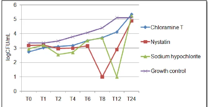
3.2 C. albicans growth kinetics
The kinetics test identified differences in the behavior of C. albicans from the first
26
with no statistical difference between them (p > 0.05). At the MIC x 4 concentration (Fig 3), all compounds were effective at reducing the number of CFU/mL, but CAT and sodium hypochlorite showed greater inhibition than nystatin (p ≤ 0.05).
After 1 h, there was no inhibition by the compounds at the MIC concentration, and after 2 h of contact, no compound at any tested concentration was able to significantly inhibit fungal cells. In the period between 4 and 8 h of contact of the compounds with fungal cells, all compounds were equally capable of reducing fungal growth (p ≤ 0.05) at all tested concentrations. At the time corresponding to 12 h, CAT at the MIC concentration was less effective than nystatin and sodium hypochlorite, although CAT did significantly inhibit fungal growth. After 24 h, none of the compounds at the MIC concentrations was able to inhibit fungal cell growth.
The CAT concentrations of MIC x 2 and MIC x 4 caused effective inhibition at all studied times (p ≤ 0.05) except after 2 h.
Figures 01, 02 and 03 present fungal behavior in response to the action of CAT, nystatin and sodium hypochlorite at MIC, MIC x 2 and MIC x 4 concentrations, respectively.
Fig 1 Candida albicans ATCC 60193 fungal growth kinetics for chloramine T, sodium
27
Fig 2 Candida albicans ATCC 60193 fungal growth kinetics for chloramine T, sodium
hypochlorite and nystatin at MIC x 2 concentrations (chloramine T: 1,562.2 µg/mL; nystatin: 0.78 µg/mL; sodium hypochlorite: 1,250 µg/mL)
Fig 3 Candida albicans ATCC 60193 fungal growth kinetics for chloramine T, sodium
28
3.3 CAT mechanism of action
The presence of sorbitol and exogenous ergosterol resulted in an increase in the MIC of CAT on the C. albicans strains studied. This result indicates that CAT likely acts on both
the fungal cell wall and the cell membrane. The same is presumable to sodium hypochlorite, which also showed an increase of CIM in the presence of sorbitol and ergosterol. Table 3 shows the results obtained in the presence of sorbitol, and Figure 04 illustrates the behavior of the compounds in the presence of exogenous ergosterol at different concentrations.
Table 3 Effect of chloramine T (CAT), sodium hypochlorite (NaOCl) and caspofungin on the fungal cell wall of Candida albicans in the presence and absence of 0.8 M sorbitol
Strain Compound (µg/mL) MIC without sorbitol
MIC (µg/mL) with sorbitol
Candida albicans
ATCC 60193
CAT 781.3 6,250.0
NaOCl 625.0 2,500.0
Caspofungin < 0.039 > 5
Candida albicans
CBS 562
CAT 195.3 3,125
NaOCl 312.5 2,500.0
Caspofungin < 0.039 5.0
3.4 Effect of CAT on C. albicans micromorphology
Microscopic evaluation of C. albicans CBS 562 microculture identified the presence
29
Fig 4 Effect of chloramine T, sodium hypochlorite and nystatin on Candida albicans (ATCC
60193 and CBS 562) fungal cell membrane in the absence and presence of exogenous ergosterol at concentrations of 100, 200 and 400 µg/mL. Values (x MIC) are presented as a function of the MIC of each compound in the absence of exogenous ergosterol
3.5 Effect of CAT on the adherence, formation and reduction of C. albicans mature biofilm
In the anti-biofilm activity tests, sodium hypochlorite was better at reducing the C.
albicans adherence at MIC x 2 (p≤0.05), but CAT demonstrated activity similar (p>0.05) to that
of sodium hypochlorite at all concentrations for the groups evaluating the inhibition of the formation of C. albicans biofilm (Table 04). However, CAT was more effective (p≤0.05) at
reducing the C. albicans mature biofilm than sodium hypochlorite at MIC x 2 in group 4 and at
MIC x 4 in group 5 (Table 05). In the groups with long contact times of the compound with mature biofilm (8 h), there was no difference between CAT and sodium hypochlorite at the concentrations tested. There was also no statistically significant difference between the concentrations of each compound in the groups studied.
30
3.6 Cytotoxicity of CAT on human erythrocytes
All tested concentrations of CAT in the range 25,000 to 762.25 µg/ml demonstrated hemolytic activity between 61 and 67.7%. Hypochlorite, used as a control, caused complete destruction of the erythrocytes immediately after being added to the wells, which was considered as parameter of 100% hemolytic activity. Saline (0.9% NaCl), used as a negative control, did not cause hemolysis as expected.
Fig 5 Candida albicans (CBS 562) micromorphology (40x magnification)in the absence (growth
31
Table 4 Categories of percentage values for inhibition of initial fungal cell adherence and the formation of Candida albicans ATCC 60193 mature biofilm in the
groups, as characterized by different contact times, after 48 h of incubation
Different uppercase letters on the lines represent a significant difference between the compounds in each group. Equal lowercase letters in each column represent similarity between the concentrations of each compound.
Inhibition of initial adherence Inhibition of mature biofilm formation
Group 1
(Contact time: 2 h)
Group 2
(Contact time: 1 min)
Group 3
(Contact time: 8 h)
Concentration Chloramine T Sodium
hypochlorite Chloramine T
Sodium
hypochlorite Chloramine T
Sodium hypochlorite
MIC 25% < % inhibition ≤ 50%Aa
25% < % inhibition ≤
50%Aa
25% < % inhibition ≤
50%Aa
25% < % inhibition ≤
50%Aa
% inhibition ≤ 25%Aa
25% < % inhibition ≤
50%Aa
MIC x 2 % inhibition ≤ 25%Aa 50% < % inhibition
≤ 75%Ba 25% < % inhibition ≤ 50%Aa 25% < % inhibition ≤ 50%Aa 25% < % inhibition ≤ 50%Aa 25% < % inhibition ≤ 50%Aa
MIC x 4 25% < % inhibition ≤ 50%Aa
25% < % inhibition
32
Table 5 Categories of percentage values for the reduction of Candida albicans ATCC 60193 mature biofilm in the groups, as characterized by different contact
times.
Different uppercase letters on the lines represent a significant difference between the compounds in each group. Equal lowercase letters in each column represent similarity between the concentrations of each compound.
Group 4
(Contact time: 3 x 1 min)
Group 5
(Contact time: 6 x 1 min)
Group 6
(Contact time: 8 h)
Group 7
(Contact time: 2 x 8 h).
Concentration Chloramine T Sodium
hypochlorite Chloramine T
Sodium
hypochlorite Chloramine T
Sodium
hypochlorite Chloramine T
Sodium hypochlorite
MIC
50% < % inhibition ≤
75%Aa
% inhibition ≤ 25%Aa
% inhibition ≤ 25%Aa
% inhibition ≤ 25%Aa
50% < % inhibition ≤
75%Aa
50% < % inhibition ≤ 75%Aa
25% < % inhibition ≤
50%Aa
25% < % inhibition ≤
50%Aa
MIC x 2
50% < % inhibition ≤
75%Aa
% inhibition ≤ 25% Ba
% inhibition ≤ 25%Aa
% inhibition ≤ 25%Aa
50% < % inhibition ≤
75%Aa
25% < % inhibition ≤ 50%Aa
25% < % inhibition ≤
50%Aa
25% < % inhibition ≤
50%Aa
MIC x 4
50% < % inhibition ≤
75%Aa
25% < % inhibition ≤
50%Aa
25% < % inhibition ≤
50%Aa
% inhibition ≤ 25%Ba
50% < % inhibition ≤
75%Aa
25% < % inhibition ≤ 50%Aa
25% < % inhibition ≤
50%Aa
25% < % inhibition ≤
33
4. Discussion
Although CAT has been studied for many years and it has been approached in several ways, there is still a lack of studies regarding its use for dentistry and directed toward its antimicrobial activity and cytotoxicity [20,21]. This is the first study to evaluate the antifungal activity of CAT on C. albicans, C. tropicalis, C. krusei and C. glabrata and the effect on C. albicans growth
kinetics and micromorphology. In addition, the mechanism of action and anti-biofilm activity of CAT were ascertained.
The MIC75% of CAT for the tested strains was 781.3 µg/mL standing out the value
195.3 µg/mL found for the strain of C. albicans CBS 562, and the MFC/MIC ratio reflects the
fungicidal activity of the compound for most of the strains, except for C. albicans CBS 562. The
variation in MIC can be explained by the different genetic profile of the strains and possible acquired resistance mechanisms. Regarding the difference between fungistatic and fungicidal activity, the literature cites this as concentration dependent in most cases, with the same compound able to act in both manners [25,37].
There were not found in the literature studies to compare the MIC results front of genus Candida strains. However, a study carried out to assess the fungicidal activity of CAT at a
concentration of 100 µg/ml on a clinical strain of Aspergillus fumigatus, did not find positive results
for the substance [38]. This result does not mean that CAT has no action on the tested strain and cannot be compared to those obtained in the present study due to the methodological difference to determine the antifungal activity and the discrepancy between the concentrations tested, whereas 100 µg/mL is below the MIC values obtained for the strains of Candida spp.
When tested against bacterial strains (Escherichia coli, Enterococcus faecalis,
Staphylococcus aureus, Pseudomonas aeruginosa, Klebsiella pneumoniae, Proteus mirabilis,
Enterobacter cloacae, Staphylococcus epidermidis, and Serratia marcescens), the literature shows
CAT MIC values ranging between 500 and 2.000 µg/mL, values found approximate to this study [39].
A previous study of C. albicans death kinetics in response to CAT noted that a concentration of 1,000 µg/mL caused a 2 log10 reduction in CFU/mL after 30 min of contact, and a
concentration of 100 µg/mL achieved this reduction in 1 h [13]. In the present study, the lowest concentration tested, 781.3 µg/mL, was able to significantly reduce (p ≤ 0.05) the number of fungal cells immediately after addition of CAT (T0), but this concentration was not able to reduce the log10
level over time. The concentration of 1,562.5 µg/mL (MIC x 2) decreased the CFU/mL after 6 h of contact, and MIC x 4 (3.125 µg/mL) resulted in complete removal of fungal cells after 1 h.
Some chemical characteristics of CAT, such as its efficient chlorination and oxidation of microorganisms [40] and its high stability and ability to maintain chlorine levels for a prolonged time [15], explain, respectively, the rapid antifungal effect at T0 and the prolonged action of CAT that caused significant inhibition of fungal growth up to 12 h at all concentrations tested.
34
However, a moderate chlorine layer is not able to decrease cell viability, and therefore, penetration of the compound into the cell is important, as it is followed by destruction of vital components by oxidation [40].
Studies claim that penetration ability is related to molecular size and polarity. However, it was found that although CAT is considerably larger than other active chlorine compounds and it presents as an anion [13], CAT has highly reactive compounds and a high capacity for chlorine layer formation that has an immediate destructive impact on the microbial surface [40]. The results of the present study are supported by these findings because the MIC of CAT increased in the presence of both sorbitol and exogenous ergosterol, suggesting a simultaneous action on the cell membrane and cell wall.
The study of C. albicans micromorphology in the presence of antifungal agents is
important because fungal form is one of several factors that affect its virulence and pathogenicity. The formation of pseudohyphae and hyphae causes tissue invasion by enabling the cell to exert a mechanical force favorable for tissue penetration. Chlamydospores are considered to be resistance structures [41,42]. The CA culture medium used for C. albicans microculture in the present study
facilitates the expression of virulence forms and allows us to evaluate possible changes caused by the presence of the compounds.
At all concentrations tested, CAT reduced the formation of pseudohyphae and led to an absence of chlamydospores. Sodium hypochlorite at concentrations of 1,250 and 2,500 µg/mL completely inhibited the presence of filaments and chlamydospores. This result is important because it complements the others presented in the present study and because it may be inferred that CAT, at the concentrations tested, promotes the general maintenance of fungal cells in the form of blastoconidia, which are considered less virulent.
Taking into account the adherence and penetration capacity of C. albicans on the
acrylic resin of dentures and the fact that hyphae lead to the formation of a thicker biofilm more resistant to removal [43], the results presented here are even more important for the indication of CAT as a solution for denture disinfection, underscoring the potential of this compound in this area. Whereas biofilms are the most common organization of microorganisms, as they provide greater protection and resistance, testing on C. albicans biofilm is essential to corroborate
the information obtained in terms of planktonic cells and to approximate the real world as much as possible. In denture stomatitis, the biofilm formed on the denture merits attention because disease can recur after treatment of the mucosa when there is maintenance of the biofilm adhering to the material [43,45].
In the present study, it was decided to conduct tests on a C. albicans mono-species
35
methodology was suggested, which can be used in future studies for the purpose of standardization for presentation and analysis of this type of data.
The results obtained showed that CAT has similar anti-biofilm activity to sodium hypochlorite regarding inhibition of the initial adherence of the fungal cells and the formation of mature biofilm. The tested concentrations did not differ significantly. In the groups testing its ability to reduce C. albicans mature biofilm, CAT was more effective than the hypochlorite at the MIC x 2
concentration in group 4, simulating 3 x 1 min contacts over 24 h, and at MIC x 4 concentration for groups 5 and 6, simulating 6 x 1 min contacts over 48 h. In both cases, CAT decreased the biofilm by 50 to 75%. In the groups with contact for 8 consecutive hours, there were no significant differences between the compounds.
The results of Panzeri et al. [20] differ from those found in the present study because in their study, a CAT-based dentifrice at a concentration of 10,000 µg/mL was not able to significantly reduce the number of CFU/mL of C. albicans present in denture biofilms after 2 min of
daily brushing for 21 days. In the same way, a clinical trial with dentifrices containing the CAT 2.000 and 10,000 µg/mL [21], used three times daily for 7 days, found that they were not able to significantly remove the biofilm of dental prosthesis. The differences in the formulation and the dosage may have affected the activity of the active compound and may thus explain the difference between these results.
Substances indicated for denture disinfection can be harmful and thus should not be in contact with the patient’s mucosal tissues, that is why it is recommended to thoroughly wash the denture after disinfection. However, it is critical to study the potential cytotoxic effects of a given substance before indicating it for clinical use. With this purpose, we evaluated the cytotoxicity of CAT using the hemolysis method, which can be considered a preliminary, although meaningful, assay to evaluate the characteristics of test substances.
All CAT concentrations tested, either higher or lower than its MIC, showed hemolytic activity between 61 and 67.7%, suggesting that such effect is not concentration dependent. Sodium hypochlorite caused complete hemolysis immediately after contact with the erythrocytes, confirming its high cytotoxicity. According to the literature [46], CAT did not cause visible cellular effects on a human cell line of squamous cell carcinoma at concentrations between 1 and 10 µg/ml after 30 min exposure. At higher concentrations (100, 1,000 and 10,000 µg/ml), it led to changes in cell morphology after 30 min and complete fragmentation after 24 h.
36
The results demonstrate the good potential for the use of CAT for denture disinfection. It is suggested, therefore, that further tests be performed toward the evaluation of cytotoxicity, the verification of anti-biofilm activity using a multi-species model and the investigation of the effect of CAT on the properties of denture components.
5 Conclusion
CAT offers antifungal activity against C. albicans, C. tropicalis, C. krusei and C.
glabrata and has a fungicidal action on most of the tested strains; It is able to significantly reduce
the growth of C. albicans immediately after initial contact and has prolonged inhibitory activity, likely
acting on the cell wall and cell membrane simultaneously. CAT changes C. albicans
micromorphology, causing reductions in the filamentous form and the inhibition of chlamydospores and also has C. albicans anti-biofilm activity and is effective at inhibiting the initial adherence of
fungal cells and biofilm formation and at reducing mature biofilm. It has high hemolytic activity, however lower than that of sodium hypochlorite.
Ethical approval
All procedures performed in studies involving human participants were in accordance with the ethical standards of the institutional and national research committee and with the 1964 Helsinki declaration and its later amendments or comparable ethical standards.
The research proposal has been reviewed and approved by the human research ethics committee from the Center of Health Sciences of the Federal University of Paraiba (CAAE: 43914615.0.0000.5188).
Conflict of Interest
The authors declare that they have no conflict of interest.
References
[1] Gendreau L, Loewy ZG (2011) Epidemiology and etiology of denture stomatitis. J Prosthodont 20: 251–260
[2] Navabi N, Gholamhoseinian A, Baghaei B et al (2013) Risk factors associated with denture stomatitis in healthy subjects attending a dental school in southeast Iran. Sultan Qaboos University Med J 13: 574-580
[3] Marinoski J, Bokor-Bratić M, Čanković M (2014) Is denture stomatitis always related with candida infection? A case control study. Med Glas (Zenica) 11: 379-384
37
[5] Kilic K, Koc AN, Tekinsen FF et al (2012) Assessment of Candida species colonization and
denture-related stomatitis in bar- and locator- retained overdentures. J Oral Implantol. doi: 10.1563/AAID-JOI-D-12-00048.1
[6] Kabawat M, de Souza RF, Badaró MM et al (2014) Phase 1 clinical trial on the effect of palatal brushing on denture stomatitis. Int J Prosthodont 27:311-319
[7] Felton D, Cooper L, Duqum I et al (2011) Evidence-Based Guidelines for the Care and Maintenance of Complete Dentures: A Publication of the American College of Prosthodontists. J Prosthodont 20: S1–S12
[8] Gornitsky M, ParadisI I, Landaverde G et al (2002) A clinical and microbiological evaluation of denture cleansers for geriatric patients in long-term care institutions. J Can Dent Assoc 68: 39-45
[9] Yildirim-Bicer AZ, Peker I, Akca G et al (2014) In vitro antifungal evaluation of seven different
disinfectants on acrylic resins. Biomed Res Int. doi: 10.1155/2014/519098
[10] Lima EM, Moura JS, Del Bel Cury AA et al (2006) Effect of enzymatic and NaOCl treatments on acrylic roughness and on biofilm accumulation. J Oral Rehabil 33: 356-362.
[11] Amin F, Iqbal S, Azizuddin S et al (2014) Effect of denture cleansers on the color stability of heat cure acrylic resin. J Coll Physicians Surg Pak 24: 787-790
[12] Neppelenbroek KH, Kurokawa LA, Procópio ALF et al (2015) Hardness and surface roughness of enamel and base layers of resin denture teeth after long-term repeated chemical disinfection. J Contemp Dent Pract 16): 54-60
[13] Arnitz R, Nagl M, Gottardi W (2009) Microbicidal activity of monochloramine and chloramine T compared. J Hosp Infect 73:164-170
[14] Gottardi W, Debabov D, Nagl M (2013) N-chloramines, a promising class of well-tolerated topical anti-infectives. Antimicrob Agents Chemother 57:1107-1114
[15] Austin JH, Taylor HD (1918) Behavior of hypochlorite and of chloramine-t solutions in contact with necrotic and normal tissues in vivo. J Exp Med 27: 627-633
[16] Dakin HD, Cohen JB, Kenyon J(1916) Studies in antiseptics (II): on chloramine: its preparation, properties and use. Br Med J 1: 160-162
[17] Sweet JB, Macynski AA (1985) Effect of antimicrobial mouth rinses on the incidence of localized alveolitis and infection following mandibular third molar oral surgery. Oral Surg Oral Med Oral Pathol 59: 24-26
[18] Miyagi SPH, Mello I, Bussadori SK et al (2006) Response of cultured pulpal fibroblasts to Papacárie® gel. Rev. Odontol. Univ. São Paulo 18: 245-249
[19] Rolland SL, Carrick TE, Walls AW et al (2007) Dentin decontamination using chloramine T prior to experiments involving bacteria. Dent Mater 23:1468-1472
[20] Panzeri H, Lara EH, Paranhos HF et al (2009) In vitro and clinical evaluation of specific
dentifrices for complete denture hygiene. Gerodontology 26: 26-33
[21] Andrade IM, Silva-Lovato CH, Souza RF et al (2012) Trial of experimental toothpastes regarding quality for cleaning dentures. Int J Prosthodont 25:157-159
[22] Clinical and Laboratory Standards Institute (CLSI) (2002) Protocol M27-A2. Reference method for broth dilution antifungal susceptibility testing of yeasts. NCCLS: Pennsylvania