INTERDISCIPLINARY APPROACH BETWEEN DENTISTRY
AND SPEECH-LANGUAGE PATHOLOGY IN TREATMENT
OF CHILDREN WITH EARLY CHILDHOOD CARIES
Atuação interdisciplinar odontologia/fonoaudiologia
no tratamento de paciente com cárie precoce da infância
Luciana Tiemi Inagaki (1), Daniela Galvão de Almeida Prado (1),
Alexsandra Shizue Iwamoto (1), João Sarmento Pereira Neto (2), Maria Beatriz Duarte Gavião (2),
Regina Maria Puppin-Rontani (2), Fernanda Miori Pascon (2)
(1) Programa de Pós-Graduação em Odontologia pela
Facul-dade de Odontologia de Piracicaba – UniversiFacul-dade Esta-dual de Campinas, Piracicaba – SP – Brasil.
(2) Departamento de Odontologia Infantil da Faculdade de
Odontologia de Piracicaba – Universidade Estadual de Campinas, Piracicaba – SP – Brasil.
Conlict of interest: non-existent
caregivers may be provided with proper instructions about oral hygiene and good dietary habits, in order to preserve the integrity of children’s oral health1. In
addition, periodic visits to the dentist are important for monitoring the child’s dentition and craniofacial growth1,2. In Brazil, only a small percentage of
children receive dental care at the recommended age, as the demand for these services is related to socioeconomic and cultural factors; thus, the higher these levels are, the greater will be the concerns about the oral health2,3. Consequently, children from
families with a low socioeconomic level, and who have many dental problems, tend to seek dental
INTRODUCTION
It is recommended that children’s irst dentist
appointment should be scheduled before the eruption of primary dentition, so that their parents and/or
ABSTRACT
The interdisciplinary integration between dentistry and speech therapy can provide adequate treatment of dental and myofunctional alterations. This case report presented the dental treatment of children
with three years old with early childhood caries, premature loss of the primary maxillary incisors due
trauma, esthetic and functional rehabilitation and speech therapy. The dental care proceeding was oral hygienic instructions, dietary recommendations and dental restoration with resin composite due to caries lesions in teeth 64, 84, 85, 74 and 75 (occlusal surface) and 51, 61, 52 and 62 (smooth
surface). After one year of follow-up, the teeth 51 and 61 were extracted (dental trauma history in irst appointment), because the teeth presented extensive external reabsorption. Subsequently, esthetic
and functional space maintainer was placed in the upper anterior region. Speech evaluation was performed using the MBGR protocol (orofacial myofunctional), where was analyzed the orofacial functions, mobility and muscular tone. The scores were attributed for each item available in the
protocol. Thus, the dificult of lips and tongue were conirmed, the cheek tonus were reduced and
alterations in the speech. The speech therapy was established during three month once a weekly and
this improved of all aspects changed with conirmation of adequate scores of MBRG protocol. In the
dentistry treatment was observed satisfactory clinical results for children and parents. It was concluded that interdisciplinary approach between dentistry and speech therapy provided adequate treatment for oral conditions presented by children providing oral health and favorable prognosis.
11. Furthermore, chewing is the initial stage of the
digestive process, and involves development of the
craniofacial complex, central nervous system and
dental occlusion12.
Moreover, chewing is considered a learned and adaptable function that depends on numerous factors, among them the increase in intraoral space by means of craniofacial growth. The development of this function is considered gradual, because in the
beginning, it is shown by mandibular and maxillary
movements of coming together and moving apart. After eruption of the molars it becomes more effective, with contraction of the cheeks and mandibular rotation. This evolution of chewing is facilitated by variety of food offered during the craniofacial maturation process, which ends up
demanding increasingly complex patterns13.
Speech is another important function of stomato-gnathic system affected in cases of premature loss of primary teeth. It is known that verbal language
begins from the birth and becomes more complex as
the child develops. At the age of 2 to 4 years further
expansion occurs in the phonological system14.
Systematic simpliications of the phonological rules
that affect the class or sequence of sounds, are noted as the main features during phonological development, and these disappear over the course of time15.
Dificulty with organizing the phonological rules
is deined as phonological disorder. This is charac
-terized by substitutions, omissions and distortions that child performs during speech acquisition. These alterations may be related to cognitive linguistic
dificulty, associated with sound perception and/or
production16.
This aim of this clinical case report was to present the interdisciplinary therapy provided by Dentistry and Speech Language-Pathology in the treatment of a preschool-age child with mild early childhood caries, involving premature loss of the
maxillary central incisors due to pathological root
resorption resulting from trauma and subsequence rehabilitation of aesthetics and function.
CASE REPORT
Dental approach
This case report was approved by the Research Ethics Committee of the Piracicaba Dental School, University of Campinas (protocol number 087/2014).
The caregivers of patient L.S.G., a 3-year-old girl, sought attention at the pediatric dentistry clinic of the Piracicaba Dental School – FOP/UNICAMP, because of the presence of dental caries. In the interview, the mother reported that her daughter had never received dental care, and the reason care services only in emergencies for restorative
treatment and extractions3.
Early childhood caries is deined as the presence
of caries lesions in primary dentition in children under
six years of age1, with the stages of lesions classiied
as mild, when caries is present at least one of
maxillary incisors and/or on the maxillary irst molars;
moderate, when caries is present on the vestibular
surface of one or both irst mandibular molars; and
severe, if multiple dental surfaces are involved4. The
consequences of severe early childhood caries and premature loss of primary dentition can compromise the child’s quality life, as they affect aesthetics, nutrition, speech development, dental arch integrity, development and eruption of permanent successor teeth, and contribute to the establishment of delete-rious oral habits 4-6.
Apart from early childhood caries, the early
loss of maxillary primary incisors can also cause
sequelae. In cases of premature loss by trauma, the
consequences could be: avulsion; extraction after
injury due trauma complications and poor prognosis, the traumatized primary tooth could cause risk to the proper development of permanent tooth, early
exfoliation due to pathological root resorption, and inlammatory complications, including adjacent
tissues around the traumatized tooth6,7. Among
primary teeth, maxillary central incisors are those
most frequently involved in injuries compared with
other teeth, followed by maxillary lateral incisors and
mandibular central incisors6,8.
When considering the stomatognathic system,
deined as a morphofunctional unit located in oral
cavity, with functions such as breathing, sucking, chewing and phonoarticulation9, and the relationship
of this system with the integrity of dental arch, the early loss of incisors may lead to the development of harmful oral habits and change the development of speech6. The dental arch, one of the static
struc-tures forming part of the stomatognathic system, is morphologically and functionally considered ideal when there is harmony between all the structures and functions of system. That is to say, when there is a favorable relationship between bony bases, and perfect adaptation between occlusal surfaces and teeth in the intercuspal position10. Therefore,
changes in phonoarticulatory structures can compromise the functions of the stomatognathic system.
With respect to chewing, the literature does not emphasize the importance of primary incisors in the preparation of food processing for digestion6.
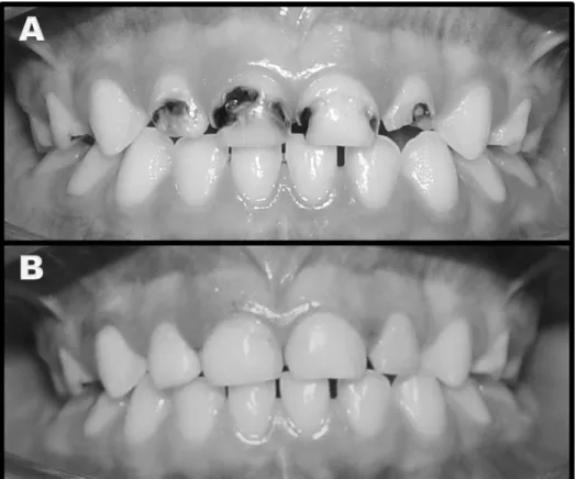
and vestibular incisal surface of teeth 51, 61 (with a history of previous trauma), 52 and 62 (Figure 1A).
The radiographic examination showed integrity of
alveolar bone, with absence of periapical lesion in the teeth affected by caries (Figure 2A).
for seeking the dental clinic was because of the presence of dark spots in the child’s anterior
maxillary teeth. The clinical examination showed
that patient was at the stage of primary dentition, with presence of caries lesions on the occlusal surface of teeth 64, 84, 85, 74, 75 and middle thirds
arches, as well as bite registration in wax, in order to
fabricate the space maintainer, with stock teeth for the region of the missing teeth.
After extraction of teeth 51 and 61 (Figure 2C)
the space maintainer was immediately placed, and monthly appointments were scheduled for
the patient in the irst three months, with intention
of evaluating how she adapted to the new device.
During this period, dificulties in pronunciation of
some words and changes in speech were noted in the patient, who was referred to a speech therapist for assessment.
Myofunctional THERAPY approach
The speech evaluation was performed by a
speech therapist expert in Orofacial Myology.
Orofacial functions, tone and mobility of the phono-articulatory organs were observed according to the Orofacial Myofunctional Protocol (MBGR)17.
The option was to use the MBGR protocol to assess orofacial functions because it has been well reported in the literature, and is used in the area of orofacial myology. This protocol enabled measurement of the changes found, complete evaluation and establishment of a prognosis, however, in this study, only the results found with Treatment planning was accomplished in a
preventive approach, with instructions given to child and caregivers about oral hygiene and dietary habits. As regards curative treatment, composite resin direct restorations were performed in all the teeth affected by caries, under local anesthesia
with absolute isolation of the operative ield with
a rubber dam. Carious tissue was removed with spherical diamond-coated and carbide burs. The dental restorative materials used were the one-step adhesive system Adper Easy One (3M, ESPE, St. Paul, USA) and composite resin Filtek Z350 (3M, ESPE, St. Paul, USA) shade B1 for enamel and A1 for dentin. The materials were light polymerized in accordance with the manufacturer’s instructions.
After this, inishing of the restorations was performed using speciic diamond points, and one week later, they were polished by means of the SofLex system
(3M, ESPE, St. Paul, USA) (Figure 1B).
After follow-up of one year, a new radiographic
examination of teeth 51 and 61 revealed external
root reabsorption and periapical lesion in both teeth, although the child had shown no pain
symptom-atology (Figure 2B). Thus, extraction of these teeth,
and placement of a removable, aesthetic-functional space retainer were indicated. Accordingly,
impres-sions were taken of the maxillary and mandibular
were very important to encourage the child and her caregivers to take care of their oral health. The choice of a simple approach, avoiding the use of technical terms (with emphasis on popular and local terms such as “tooth dirt”, “worm that eats the tooth”, “brush to clean the tooth”, etc.) was convenient to improve the patient’s family’s understanding about correct tooth brushing habits, how to use dental
loss, how to readjust dietary habits and the impor
-tance of preventing new caries lesions.
The patient was receptive to the invasive proce-dures of restorative treatment. During the sessions, behavioral changes were observed in the child, who had been shy and ashamed in the beginning of treatment. After restoration of the anterior teeth, the child began to smile and to communicate more frequently. At this time it was noted that she needed myofunctional therapy assistance. Fortunately,
the early loss of maxillary central incisors did not
change the patient’s self-esteem, and she accepted placement of the space maintainer.
Myofunctional therapy approach
in the evaluation, diminished cheek tone, tongue
with increased height, and dificulty with mobility
were observed, so that the patient was unable to perform lateralized lip protrusion to the right and left, and tongue vibration. As regards oral functions, when chewing the patient presented unsystematic lip closure, increased speed and presence of atypical muscle contractions. During swallowing, no changes were observed and the patient presented a predominantly nasal breathing mode.
With reference to speech, the patient
demon-strated simpliications when speaking the consonant clusters (/tr/, /gl/, /th/, /l/), in addition to the family
history of speech disorders. Thus, the character-ization of a phonological disorder was evident.
In view of the results found in the evaluation, the strategies applied in myofunctional therapy were
established, such as isotonic exercises for mobility of the tongue and cheek, isometric exercises to
tone cheek, and awareness of the correct chewing pattern. The work of speech involved improvement in the articulation point, with words that were part of the phonetic environment of the child being used, in addition to the use of synesthetic and auditory cues. The patient was treated for three months with weekly sessions lasting for 30 minutes, totaling 12 sessions.
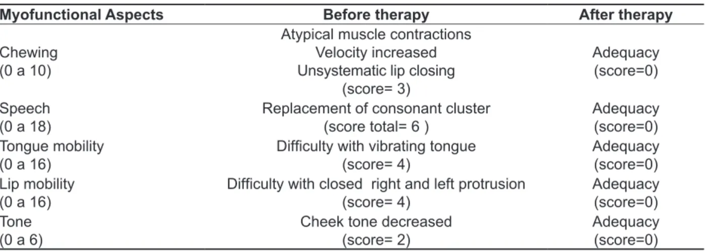
Table 1 shows the evaluated myofunctional aspects and results with scores obtained in the MBGR protocol, before and after the completion of therapy.
regard to mobility, tone and orofacial functions were addressed.
According to the proposed protocol, the assessment of orofacial functions was recorded by means of images of chewing, swallowing and speech tests. In the chewing test, a wafer type biscuit was used to analyze the chewing pattern and atypical muscle contractions. To evaluate the direct swallowing of liquid, lip sealing, position of the tongue, containment of food, presence of contraction of the orbicularis and mentalis muscles, head movement and coordination of swallowing were analyzed. With regard to speech, samples of naming pictures, counting numbers from zero to 20 and spontaneous speech were obtained.
The respiratory type was evaluated by
obser-vation, and was classiied during the clinical examination as middle/lower or middle/upper. The
breathing mode was observed considering the position of the lips, jaw and tongue, in addition to observing whether closure of the mouth occurred at any time, thus classifying the mode as nasal, mouth- or oronasal breathing.
As regards mobility of the lips and tongue, the patient was requested to perform motor tasks for the lips: protrusion, retraction and alternating the tasks with the lips opened and closed; lateralized protrusion to the right and left, popping in protrusion and retraction and also alternated. For the tongue: protrusion, alternating between protrusion, and
retraction, elevating the apex onto the incisive
papilla, alternating between raising and lowering on the papilla, raising the tongue onto the upper lip, raising and lowering alternately touching the lips, touching the right and left commissure, alter-nating these movements, touching the left and right cheek internally and alternating the movements,
pop the apex, body popping, sucking against the palate and vibrating. Each movement was classiied
as: performed properly, approximately or as not performed.
With regard to tone, the upper and lower lip, chin, tongue, right and left cheek were evaluated, and
classiied as normal, decreased or increased.
In the analyses, scores were assigned to each item investigated. Thus, the sum of individual scores was considered to obtain the total score of each function. When appropriate, a zero value was assigned; higher values when changed, and the higher the score was, the greater was the change.
RESULTS
Dental approach
Table 1 - Results of myofunctional therapy
Myofunctional Aspects Before therapy After therapy
Chewing (0 a 10)
Atypical muscle contractions Velocity increased Unsystematic lip closing
(score= 3)
Adequacy (score=0)
Speech (0 a 18)
Replacement of consonant cluster (score total= 6 )
Adequacy (score=0) Tongue mobility
(0 a 16)
Dificulty with vibrating tongue
(score= 4)
Adequacy (score=0) Lip mobility
(0 a 16)
Dificulty with closed right and left protrusion
(score= 4)
Adequacy (score=0) Tone
(0 a 6)
Cheek tone decreased (score= 2)
Adequacy (score=0)
DISCUSSION
This case report showed the importance of interdisciplinary interaction/cooperation between Pediatric Dentistry and Speech Language-Pathology in the treatment of a preschool child with mild early childhood caries and consequent
premature loss of maxillary central primary incisors
as a result of trauma. The oral conditions of the patient when assistance was sought at FOP/
UNICAMP Pediatric Dentistry Clinic, relect the
challenges of public oral health programs with respect to early childhood caries. According to the Brazilian Oral Health Survey18 the Brazilian mean
index for decayed, missing and illed teeth (dmft)
for 5-year-old children is 2.43, accounting for over
80% of the index. Nevertheless, when the results
of each Brazilian region are compared separately, the interior of the southeastern region, in which the patient’s family is inserted, shows a mean dmft of 74.9%. Furthermore, in Brazil, there is no program with the main focus on evaluation of oral problems
affecting children under ive years of age, despite
the attention given to epidemiological oral health issues5.
The absence of oral health promotes negative impacts on the quality life of children, because it compromises the development of chewing function and speech, and can interfere in psychological factors that impair the child’s performance at school5. In a cross-sectional study, early childhood
caries was shown to cause a negative relection of
oral health on the quality of life of children aged 2 to 5 and their families19. The authors showed that
the greatest impact factors related by children were
toothache, dificulty to eat and frustration. For family
members, the feelings of guilt followed by discomfort
about children’s oral health were the most signiicant
factors expressed. In another study, the association
between parents’ guilt feeling and oral health of their children showed that when early childhood caries was present, parents became increasingly worried because of the complaints of pain20. Studies in
different cultures also conirm the negative impact
that oral health disorders can affect the life quality of preschool children and their families21-23.
Apart from early childhood caries, another important factor that impairs the quality of life and development of the stomatognathic system is the premature loss of primary teeth, caused by caries lesions and/or dental trauma. There is a consensus in the literature about the anterior teeth being the most affected by trauma in comparison with the other teeth in the dental arch, with the central incisors being more vulnerable, especially between the ages of two to four8,24,25.
In an evaluation of 620 cases of traumatized
primary teeth, 86% were maxillary central incisors
and the most frequent types of trauma found were
subluxation (32.7%), intrusion (29.3%) and avulsion
(20.1%)8. Other authors have found that lateral
luxation was the most common injury (33.3%), and
among the treatment plans, regular reviews (39.4%)
and extraction (29.3%) were the most common24.
In this case report, the patient’s family were unable to specify the type of trauma and time of injury,
therefore, the prognosis of the maxillary anterior
teeth was based on the initial radiograph taken
during the child’s irst appointment. In this radio
-graph, the alveolar ridge and periodontal space presented without pathologies, which let to opting for clinical and radiographic follow up procedures.
The external root reabsorption of maxillary central incisors, which was observed after approximately
but phonological aspects, featuring a linguistic change. The therapy provided improvement in the
patient’s speech, in which the phonological simplii
-cations were eliminated, as shown in Table 1. With respect to chewing, the study showed that
there was no change in mastication after extraction
of the incisors, because children’s growth continued satisfactorily, since the diet was adapted to dental changes29. However, other authors have reported
that this occurrence cannot be extrapolated to
cases of trauma, since trauma causes sudden tooth
loss and is associated with infections, pain and difi
-culty with incision even before tooth extraction6. In
this case, the extraction can even ease the pain and
discomfort, improving the ability to eat. However the incision of food must be considered because it is an important step, and the space maintainer together with myofunctional therapy helped the patient to maintain adequate chewing function. In addition, there was reduction in atypical muscle contractions, improvement in speed, and lip closure became systematic.
Moreover, the fact that the patient had decreased
tone of the cheeks can be explained because
sagging occurs when they are not used to move food from the vestibules to the occlusal surfaces during mastication30. Thus, hypotonia of the patient’s
cheeks may have been related to the changes in chewing presented.
The interdisciplinary work between dentistry and myofunctional therapy in this clinical case provided a suitable and favorable treatment prognosis. Thus, the importance of this joint work between the two areas allowed more precise diagnosis of the dental and myofunctional disorders.
CONCLUSION
In this case report, the interdisciplinary inter-action between Dentistry and Speech Language-Pathology was important for obtaining successful clinic treatment, which involved both preventive and curative approaches, which comprised the preventive and curative treatment of early childhood caries, functional and aesthetic reestablishment required
because of the premature loss of maxillary incisors,
and phonetic and myofunctional adaptation. Thus, the patient was gained considerable improvement in oral health, and in the development of speech, chewing and self-esteem.
the previous history of trauma. Dental trauma may promote damage to the stomatognathic system because of triggering reabsorption processes in mineralized tissues such as the teeth and bone; and root reabsorption is considered an irreversible complication that causes loss of the tooth7.
In a study with children between 9 months and 5 years of age, who had a history of trauma to the
maxillary incisors, 70% of root reabsorption occurred during the irst year after the trauma, with this period
ranging between 1 to 45 months7. In addition,
in cases of recurrent trauma, there are greater chances of developing pathological root resorption, irrespective of age7. In this case report, due to the
presence of root resorption in the maxillary central incisors with an unfavorable prognosis, extraction
was indicated with subsequent placement of an aesthetic-functional space maintainer. In this period, speech alterations were noted in the patient and myofunctional therapy treatment was recommended. In childhood, anatomic and functional alterations can promote disturbances in the stomatognathic system, and premature tooth loss can interfere in the development of this system, and consequently, in orofacial functions. These disorders require phonoaudiological assistance, and thus the impor-tance of emphasizing the dentist knowledge about the oral alterations, in order to refer the patient to a speech therapist.
In the reported case, the placement of a space maintainer enabled suitable posture of the tongue, because in the absence of anterior teeth, tongue could become interposed between the arches, causing impairments in speech, chewing and swallowing26. When the tongue is in a different
position in the oral cavity, it is unable to fulill the role
of modeling of dental arches27. Thus, the presence
of a space maintainer in the maxillary anterior
region allowed the correct positioning of the tongue, prevented postural changes and provided balance of the entire stomatognathic system.
As regards the association of changes in speech and early loss of deciduous teeth, the literature has shown that in children with premature loss of incisors resulting from previous trauma, changes in speech such as frontal lisp, probably do not occur. If it does happen, the changes are transient and resolved with the eruption of the permanent teeth successors28. However, in the case of the patient in
this report dificulty was not phonetic, in other words
REFERENCES
1. American Academy on Pediatric Dentistry; American Academy of Pediatrics. Policy on
early childhood caries (ECC): classiications,
consequences, and preventive strategies. Pediatr Dent. 2008-2009;30(7 Suppl):40-3.
2. Goettems ML, Ardenghi TM, Demarco FF, Romano AR, Torriani DD. Children’s use of dental
services: inluence of maternal dental anxiety,
attendance pattern, and perception of children’s quality of life. Community Dent Oral Epidemiol. 2012;40(5):451-8.
3. Ardenghi TM, Vargas-Ferreira F, Piovesan C,
Mendes FM. Age of irst dental visit and predictors
for oral healthcare utilisation in preschool children. Oral Health Prev Dent. 2012;10(1):17-27.
4. Feitosa S, Colares V, Pinkham J. The psychosocial effects of severe caries in 4-year-old children in Recife, Pernambuco, Brazil. Cad Saude Publica. 2005;21(5):1550-6.
5. Bönecker M, Abanto J, Tello G, Oliveira LB. Impact of dental caries on preschool children’s quality of life: an update. Braz Oral Res. 2012;26 Suppl 1:103-7.
6. Holan G, Needleman HL. Premature loss of primary anterior teeth due to trauma – potential
short- and long-term sequelae. Dent Traumatol. 2013 Oct 20. doi:10.1111/edt.12081. [Epub ahead of print]
7. Cardoso M, Rocha MJ. Identiication of factors
associated with pathological root resorption in traumatized primary teeth. Dent Traumatol. 2008;24(3):343-9.
8. Da Silva Assunção LR, Ferelle A, Iwakura ML, Cunha RF. Effects on permanent teeth after
luxation injuries to the primary predecessors: a
study in children assisted at an emergency service. DentTraumatol. 2009;25(2):165-70.
9. Ribas MO, Reis LFG, França BHS, Lima AAS. Cirurgia ortognática: orientações legais aos ortodontistas e cirurgiões bucofaciais. Rev Dent Press Ortodon Ortop Facial. 2005;10(6):75-83. 10. Almeida MEC de, Vedovello Filho M, Vedovello SAS, Lucatto A, Torrezan AT. Prevalência da má oclusão em escolares da rede estadual do município de Manaus, AM – Brasil. RGO – Revista Gaúcha de Odontologia. 2007;55(4):389-94.
11. Tagliaro ML, Calvi CL, Chiapetta ALM. A Fase de incisão no processo da mastigação: enfoque clínico. Rev CEFAC. 2004;6(1):24-8.
12. Douglas CR. Fisiologia Aplicada a Fonoaudiologia. 2th ed. Ganabara Koogan; 2006. RESUMO
A integração interdisciplinar entre odontologia e fonoaudiologia pode proporcionar tratamento ade-quado das alterações dentárias e miofuncionais. Este relato de caso clínico apresenta o tratamento odontológico em criança com três anos de idade com cárie precoce da infância leve, com conse-quente perda dos incisivos centrais superiores devido a trauma, a reabilitação estética e funcional e tratamento fonoaudiológico. Os procedimentos clínicos odontológicos foram instrução de higiene bucal, aconselhamento dietético e realização das restaurações com resina composta devido ao aco-metimento por lesão cariosa dos dentes 64, 84, 85, 74, 75 (oclusal); e dentes 51, 61, 52 e 62 (face
vestibular). Após um ano de preservação foi realizada a exodontia dos dentes 51 e 61 (com história
de trauma anterior à primeira consulta), devido a reabsorção externa avançada. Em seguida, mante
-nedor de espaço estético-funcional removível foi colocado na região anterior superior. Avaliação
fono-audiológica foi realizada utilizando o protocolo Miofuncional Orofacial (MBGR), sendo veriicadas as
funções orofaciais, mobilidade e tônus muscular. Foram atribuídos escores para cada item avaliado
no protocolo. Conirmou-se diiculdade de mobilidade dos lábios e língua com diminuição do tônus
da bochecha e alterações na fala. A terapia fonoaudiológica foi estabelecida durante três meses com
periodicidade semanal, havendo melhora em todos os aspectos alterados, conirmados pela ade
-quação dos escores do Protocolo MBGR. No tratamento odontológico foram observados resultados clinicamente satisfatórios para a criança e responsáveis. Concluiu-se que o trabalho interdisciplinar entre a Odontologia e Fonoaudiologia proporcionou tratamento adequado para as condições bucais apresentadas pela criança, proporcionando saúde bucal e prognóstico favorável.
22. Krisdapong S, Somkotra T, Kueakulpipat W. Disparities in Early Childhood Caries and Its Impact on Oral Health-Related Quality of Life of Preschool Children. Asia Pac J Public Health. 2012 Mar 16. [Epub ahead of print]
23. Kramer PF, Feldens CA, Ferreira SH, Bervian
J, Rodrigues PH, Peres MA. Exploring the impact
of oral diseases and disorders on quality of life of preschool children. Community Dent Oral Epidemiol. 2013;41(4):327-35.
24. Arikan V, Sari S, Sonmez H. The prevalence and treatment outcomes of primary tooth injuries. Eur J Dent. 2010t;4(4):447-53.
25. Piovesan C, Guedes RS, Casagrande L, Ardenghi TM. Socioeconomic and clinical factors associated with traumatic dental injuries in Brazilian preschool children. Braz Oral Res. 2012;26(5):464-70.
26. Dixit UB, Shetty RM. Comparison of soft-tissue,
dental, and skeletal characteristics in children with and without tongue thrusting habit. Contemp Clin Dent. 2013;4(1):2-6.
27. Andrade FV, Andrade DV, Araújo AS, Ribeiro
ACC, Deccax LDG, Nemr K. Alterações estruturais
de órgãos fonoarticulatórios e más oclusões dentárias em respiradores orais de 6 a 10 anos. Rev CEFAC. 2005:7(3):318-25.
28. Gable TO, Kummer AW, Lee L, Creaghead HÁ,
Moore LJ. Preamature loss of the maxillary primary
incisors: effect on speech production. ASDC J Dent Child 1995;62:173-9.
29. Christensen JR, Fields HW. Space Maintenance in the primary dentition. In: Casamassimo SP, Fields HW, Mc Tigue DJ, Nowak AJ, editors. Pediatric Dentistry – Infancy Through adolescence. 5th ed. St Louise, MO: Elsevier Inc.; 2013. P. 379.
30. Felício CM. Fonoaudiologia aplicada a casos odontológicos: motricidade oral e audiologia. 1ª edição. São Paulo: Pancast; 1999.
13. Gomes LM, Bianchini EMG. Caracterização da função mastigatória em crianças com dentição decídua e dentição mista. Rev CEFAC. 2009;11(Suppl3):324-33.
14. Wertzner HF, Pagan LO, Galea DES; Papp ACCS. Características fonológicas de crianças com transtorno fonológico com e sem histórico de otite. Rev Soc Bras Fonoaudiol. 2007;12(1):41-7.
15. Wertzner HF, Alves RR, Ramos ACO. Análise do desenvolvimento das habilidades diadococinéticas orais em crianças normais e com transtorno fonológico. Rev Soc Bras Fonoaudiol. 2008;13(2):136-42.
16. American Psychiatric Association. Diagnostic and statistical manual of mental disorders DSM-IV. 4th ed. Washington DC: APA; 1994.
17. Genaro KF, Berretin-Felix G, Rehder MIBC,
Marchesan IQ. Avaliação miofuncional orofacial – protocolo MBGR. Rev CEFAC. 2009;11(2):237-55. 18. Brasil. Ministério da Saúde. SB Brasil 2010 – Pesquisa Nacional de Saúde Bucal Resultados principais. Brasília (DF): Ministério da Saúde; 2012 [visitado:2013Dec15]. Available from: http://bvsms. saude.gov.br/bvs/publicacoes/pesquisa_nacional_ saude_bucal.pdf
19. Martins-Júnior PA, Vieira-Andrade RG, Corrêa-Faria P, Oliveira-Ferreira F, Marques LS, Ramos-Jorge ML. Impact of early childhood caries on the oral health-related quality of life of preschool children and their parents. Caries Res. 2013;47(3):211-8. 20. Carvalho TS, Abanto J, Mendes FM, Raggio DP, Bönecker M. Association between parental guilt and oral health problems in preschool children. Braz Oral Res. 2012;26(6):557-63.
21. Pani SC, Badea L, Mirza S, Elbaage N. Differences in perceptions of early childhood oral health-related quality of life between fathers and mothers in Saudi Arabia. Int J Paediatr Dent. 2012;22(4):244-9.
Received on: May 24, 2014 Accepted on: September 01, 2014
Mailing address: Fernanda Miori Pascon
Departamento de Odontologia Infantil – Área de Odontopediatria
Faculdade de Odontologia de Piracicaba – Universidade Estadual de Campinas Av. Limeira, 901
Piracicaba – SP – Brasil CEP: 13414-903