EVALUATION OF THE EFFICACY OF LOW-LEVEL LASER
THERAPY (LLLT) IN THE TREATMENT OF TEMPOROMANDIBULAR
DISORDERS: A RANDOMIZED CLINICAL TRIAL
Avaliação da eicácia do laser de baixa intensidade no tratamento das
disfunções têmporo-mandibular: estudo clínico randomizado
Maria Helena Chaves de Vasconcelos Catão(1), Polyana Sarmento de Oliveira(2),
Roniery de Oliveira Costa(3), Vanda Sanderana Macêdo Carneiro(4)
(1) State University of Paraiba – UEPB, Campina Grande, PB, Brazil.
(2) State University of Paraiba – UEPB, Campina Grande, PB,
Brazil.
(3) State University of Paraiba – UEPB, Campina Grande, PB,
Brazil.
(4) State University of Paraiba – UEPB, Campina Grande, PB,
Brazil.
Conlict of interest: non-existent
with dental and facial factors, which relate to the stomatognathic system2,3.
Signs and symptoms of TMD are present in 86% of the population, being more frequent among
women, also related to dental occlusion and
emotional stress4. Are considered signs: limited mouth opening, joint sounds and deviation of the
mandible to one side during opening and closing5,
muscle spasm, pain relex, impaired joint motion, crepitus, headache and hearing disorders6. Otologic
symptoms are represented by decreased hearing,
vertigo and tinnitus7, 8, which can be related to the
ontogenetic and anatomical relationship between the middle ear and masticatory structures2.
The “Research Diagnostic Criteria for
Temporomandibular Disorders” (RDC / TMD)9
provides the best classiication for TMD, as it includes not only methods for the physical diagnostic classiication of TMD, present on its axis I, but at the
INTRODUCTION
The temporomandibular disorders (TMD) are
diseases that consist of a series of clinical signs and symptoms, which involves temporomandibular joint (TMJ) and / or masticatory musculature. It rarely occurs affecting only joint or muscle, but mostly cases have complex symptoms1. The TMD
has multifactorial etiology and may be associated
ABSTRACT
Purpose: the effectiveness of lasertherapy in the treatment o temporomandibular of pain with temporomandibular disorders.Method: it consisted of a randomized clinical trial divided into two
groups: Group 1: AsGaAl laser; Group 2: InGaAIP laser, 20 patients between 19 and 35 years old, diagnosed with signs and symptoms of TMD. Patients had the range of motion for maximum mouth
opening and laterality registered at the beginning and at the end of the laser treatment. Laser was
applied in four pre-auricular points three times a week during a month, in a total of 12 sessions to each patient. The patients’ pain was noted based on the use of visual analogue scale (VAS) and also by physical examination of the pain points. Results: there was a signiicant reduction (p<0.028) of
the level of pain in both treatment groups, but the G1 had higher signiicance. The evolution of the threshold of muscle sensivity showed a statistically signiicant difference for G1 and G2. Laser therapy
in Group 1 improved the mouth opening 4.643 mm on average, while in Group 2, the average was 3.71 mm per patient. Conclusion: there was effectiveness in both lasers in the pain control and mouth opening of patients.
same time it alos provides methods to assess the
intensity and severity of chronic pain and levels of depressive symptoms present in axis II.
The TMD, as it is a complex condition, requires a treatment based on a correct diagnosis, established based on informations about possible etiological factors, through the survey of signs and symptoms for each patient10. Among the different treatments we
can use thermotherapy, electrotherapy, ultrasound, iontophoresis, some analgesics, and low-intensity
laser therapy 11, the last one usually indicated
when there are presence of pain, restriction of mandibular movements, tissue inlammation and
joint instability12.
The low-intensity laser comes in many cases as
an alternative therapy for the treatment to disorders
of the maxillo-facial region and joint pain, neuralgia
and paresthesia13. The main justiication for the use
of low intensity laser therapy (LILT) on TMD is due to its analgesic effects (seen in most studies in the
literature14,15) anti-inlammatory properties and its
tissue repairing effect with modulation of cellular activity16-18. LILT inluences changes with metabolic
energetic and functional character, as it promotes increasing of cells strength and vitality, leading them to their normal functioning quickly 19.
The LILT has demonstrated an ability to assist in the symptomatic treatment of pain, providing a considerable degree of comfort to the patient after application10. The great advantage of laser applica -tions in the treatment of TMD is that it is a nonin-vasive therapy20, with low cost, and currently has
being widely used in dental practice, reducing the
demand related to the surgery or the use of drugs to treat relief pain and tissue regeneration21.
The application of laser therapy in TMD patients
has demonstrated an ability to relieve pain in few
minutes after application, promoting well-being signiicant. However, LILT is an adjuvant treatment in pain relief by its analgesic action, which allows the patient’s return to daily chores, providing confort
and better quality of life.
Thus, the objective of this study was to evaluate the effectiveness of low intensity laser in the
treatment of pain in patients with TMD treated with
twelve sessions by visual analogue scale for pain (VAS) and measurement range of mouth opening.
METHOD
This study consisted of a descriptive and a quantitative survey. The descriptive part of the study was obtained by analyzing the clinical physical examination of patients before and after application low-intensity laser therapy three times per week in
patients with painful symptoms of TMD.
For the muscle tension evaluation, it was
performed palpation of the masseter, temporal,
frontal and extensors of the cervical spine, using the classiication of Jensen et al.22, which described the
following scores: 0 – no pain or discomfort, 1 – slight discomfort, 2 – moderate pain and 3-severe pain.
To evaluate pain in a quantitative way we used
the visual analogue scale (VAS), initially clariied
and then applied to patients at initial evaluation and
then weekly after the session.
The patients were submitted to a questionnaire
about symptoms of TMJ, consisting of 10 questions designed to evaluate the TMD – anamnestic index DMF (Fonseca et al.23). After illing the clinical
form and the performance of clinical examination,
we evaluated the pain intensity by visual analog
scale (VAS) before application of low-intensity laser therapy and then a weekly review was made of pain
evaluation.
For the study, we selected 20 subjects, 18
females and two males, aged between 19 and
58 years referred to the pain clinic already with a diagnosis of temporomandibular dysfunction, based on Fonseca23 clinical examination. The sample was randomly divided by lot into two equal groups: the
irst, called G1 (n = 10) was treated with infrared laser (GaAlAs) according to the protocol. The second group, called G2 (n = 10), was submitted to the red laser treatment (InGaAlP).
The applications of low-intensity laser were performed in the following points: ive points around
the joints with pain – in the joint posterior portion,
with open mouth (auriculo temporal nerve region and bilaminar zone) – the anterior part of articulation in sigmoidea notch, with the mouth at rest position (non-occluding teeth). After localization of the muscle in question by means of palpation, the LILT was applied on the most painful points (including trigger points) with an equidistance between those of 1 cm2.
The therapeutic dental laser Biowave (Kondortech) was applied emitting infrared radiation
with a wavelength of 830 nm, power 40 mW, the
beam delivery system through direct contact with skin, with a focusing area of 0.20 cm2. The 4J/cm²
dose was applied by point at the time of 1’40seg, punctually. The red laser group received red irradi -ation, with a wavelength of 660 nm, power 30 mW,
the beam delivery system through direct contact with the skin, with focusing area of 0.20 cm2, and
amplitude movements of maximum mouth opening,
lateral movement to left and right.
The participants were patients who agreed to sign an Informed Consent Form (ICF). The devel -opment of this study followed the requirements of
Resolution 196/96 of the National Health Council
/ Brazilian Ministry of Health, with approval by the
Ethics Committee in Research of our institution with
No. 0149.0.133.000-09.
Data were analyzed using descriptive statistics, inferential and comparative analyzes. We used the Wilcoxon test and t test for paired samples.
Data were entered and analyzed using the SPSS
(Statistical Package for Social Sciences) version 13.0. The margin of error of the statistical tests
was 5%.
RESULTS
Of the 20 patients evaluated, 18 patients were female (90%) and two males (10%) with mean age
of 28.2 years to 39.1 years. It was observed that,
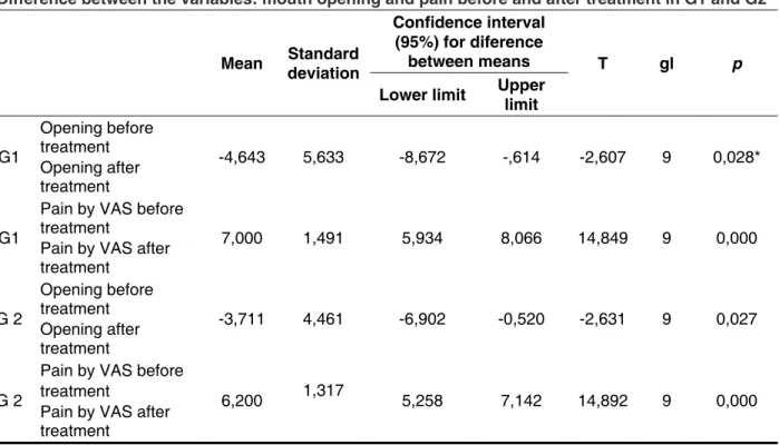
before treatment with infrared laser (GaAlAs), the opening mouth in group 1 averaged 41.35 mm, and
after the treatment, the mean aperture increased to 46.16 mm. According to the t test, there is suficient evidence that the mouth opening after treatment with the infrared beam was statistically
higher than before treatment (p <0.028). Similarly occurred with the visual analogue scale (VAS) pain,
as the average of the group before the laser was
8.4, and after treatment this number decreased to 1.4, showing signiicant difference (p <0.00). In group 2, treated with red laser (InGaAlP), it could
be observed that before treatment mouth opening average was 46.34 mm, and after it the mouth
opening increased to 50.05 mm. By applying the t test the result showed that there is suficient evidence that the mouth opening after treatment with the red laser is statistically higher than before the treatment at 5% signiicance (p <0.00). Similarly occurred with the visual analogue scale (VAS) of
pain, where before treatment, the pain average was
8.1 and after treatment decreased to 1.9, with statis
-tical signiicance (p <0.027). Observed changes in
mouth opening between the groups treated with
infrared and red lasers, and visual analog scale of pain by VAS among patients treated with infrared and red lasers, it was veriied a statistically signif
-icant difference at 5 % signiicance (Table 1 and
Table 2).
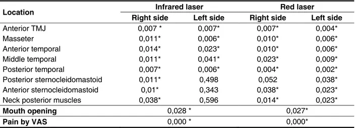
Table 3 shows nociceptive points during movement of the patients after the application of the laser. In both groups, about the auscultation of joint sounds, the crack was the most prevalent
Difference between the variables: mouth opening and pain before and after treatment in G1 and G2
Mean Standard deviation
Confidence interval (95%) for diference
between means T gl p
Lower limit Upper limit
G1
Opening before treatment Opening after treatment
-4,643 5,633 -8,672 -,614 -2,607 9 0,028*
G1
Pain by VAS before treatment
Pain by VAS after treatment
7,000 1,491 5,934 8,066 14,849 9 0,000
G 2
Opening before treatment Opening after treatment
-3,711 4,461 -6,902 -0,520 -2,631 9 0,027
G 2
Pain by VAS before treatment
Pain by VAS after treatment
6,200 1,317 5,258 7,142 14,892 9 0,000
Table 1 – Mouth opening and pain in patients suffering from Temporomandibular Dysfunction (TMD) before and after infrared (Group 1) and red (Group 2) laser treatments
* Statistical signiicance for comparison, using the T test. Source: Research conducted in Campina Grande, PB, Brazil.
Mean N Standard deviation
Red Group 1
Initial opening 41,73 10 10,003
Final opening 46,37* 10 7,618
Initial pain 8,40 10 1,430
Final pain 1,40 10 1,506
Infrared Group 2
Initial opening 46,34 10 8,852
Final opening 50,05* 10 6,521
Initial pain 8,10 10 1,197
Final pain 1,90 10 1,197
Table 2 – Mean of initial/ inal mouth opening and initial/ inal pain by VAS of patients suffering from
temporomandibular dysfunction (TMD) before and after treatment with red (Group 1) and infrared (Group 2) laser
DISCUSSION
In the present study the highest concentration
of individuals with temporomandibular disorder is among women aged between 21 and 30 years
in accordance with Okeson researchs 4,24-27. One
possible explanation for the higher prevalence of pain in women, according to Salvador et al.26 lies
in the fact that women have lower levels of muscle
strength under fatigue than men, and plasma
concentrations of the main anabolic hormones (testosterone GH and IGF-1), at rest or after intense exercise, that are very different for men and women.
The most prevalent joint sounds of this research were the crackling, followed by the jump and crepi -tation, appearing in 80% of patients with TMD. These
data corroborate Lopez28, which said the joint noise
is the irst sign of TMD to manifest in more than 70% of cases. Moresca and Urias29 found that the higher
frequency of joint noises in women demonstrates
a greater female predisposition to develop TMJ
problems, corroborating what was observed in this
study.
According to Dworkin and Leresche30, the
restriction of mandibular mobility and pain are considered the main clinical signs of temporoman
-dibular disorders. In adults, the maximum opening of
the jaw average is between 53 and 58 mm, ranging from 40 to 60 mm. The mandibular opening is usually
smaller in women than in men and decreases with
age as it were seen by Friedman31. However, most
of the patients in this study had no restriction of the maximum voluntary opening when was used 40 mm as the normal reference to measure opening. In the
present study it was found that the initial average
maximum mouth opening in Group 1 was 41.73
mm and the average end, after the twelve sessions
of laser therapy, was 46.37 mm. The restriction of mouth opening was not the main complaint of the sample, but the improvement in this feature can be understood as a secondary effect of pain reduction. These results are consistent with Kogawa32, who reported to found an average mouth opening before therapy of 44.65 mm, and after treatment with laser,
48.5 mm. Studies indicate that pain presents a several incidence in TMD, besides a large amount of associated signs and symptoms such as headaches and neck region pain 33,34.
The use of low-intensity laser therapy is a
nonin-vasive treatment modality with low cost, which has been widely used to control several diseases, among which, the muscular-joint diseases, for the relief of pain and tissue regeneration. This technique has been certiied as beneicial in the treatment of TMD. Treatment of TMD is based on a correct diagnosis,
Location Infrared laser Red laser
Right side Left side Right side Left side
Anterior TMJ 0,007 * 0,007* 0,007* 0,004*
Masseter 0,011* 0,006* 0,010* 0,006*
Anterior temporal 0,014* 0,023* 0,010* 0,006*
Middle temporal 0,011* 0,041* 0,023* 0,009*
Posterior temporal 0,007* 0,006* 0,004* 0,002*
Posterior sternocleidomastoid 0,011* 0,498 0,052 0,038*
Anterior sternocleidomastoid 0,01* 0,343 0,038* 0,023*
Neck posterior muscles 0,038* 0,596 0,014* 0,023*
Mouth opening 0,028 * 0,027*
Pain by VAS 0,000 * 0,000*
Table 3 – Algic locations of patients suffering from temporomandibular dysfunction (TMD) before and after treatment with red (Group 1) and infrared (Group 2) laser
* Statistical signiicance for comparison, using the T test. Source: Research conducted in Campina Grande, PB, Brazil.
Types %
Estalido Salto Crepitação
NDN
35 25 20 20
Total 100%
Table 4 – Noise joint present in patients with temporomandibular dysfunction (TMD) during clinical examination prior to treatment with laser therapy
established based on information of possible
etiological factors, through the survey of signs and symptoms for each patient35. By means of the VAS
(Visual Analogue Scale), we observed the evolution
of painful symptoms in both treatment groups
in this study. It was found that in both treatment groups was statistically signiicant reduction in pain, conirming Frare Nicolau36 about lasers eficiency
in controlling inlammation, what shows its effect in reducing pain through the absorption of exudates and by the allogeneic substances removing. The low-intensity laser therapy presents an analgesic local mechanism, working directly to reduce inlam
-mation, which favors the elimination of allogeneic substances, stimulates a relex action and leads to the production of substances such as endorphins, blocking the pain, as it improves local microcir
-culation and blood supply in areas of muscular
tension37. Therefore, the laser acts as a stabilizer of
the rest membrane potential factor by acting directly
on nerve endings and maintaining longer analgesia,
which makes the transmission of painful stimulus38.
The results of this study indicate a signiicant
improvement in mouth opening in both groups, and
these results corroborate those found by McNeely
et al 39,40 that in their studies demonstrated that laser
therapy yielded satisfactory effects on the param -eters used. However, the pain relief the present
study showed signiicant results in both groups with regard to reduction of pain of the table, different from those found by McNeely39.
With regard to the type of laser used in accor
-dance with the data obtained in this study, the reduction of pain after treatment was signiicant
for both lasers, the infrared and the red laser, with
an average scale reduction of 7 and 6.2 , respec -tively. There are not studies in the literature about
the red laser application for temporomandibular dysfunction, and studies in this situation usually are with the infrared laser and control group (placebo), without comparative study comparing both, red and infrared lasers. The results of this study indicate the complexity of temporomandibular dysfunction, since it is inluenced by different factors, among which we can mention the psychoemotional and the activities
performed by the individual in their day to day4. However, it may be questioned why some patients
do not respond to such treatment and also why in some cases the patients reported exacerbation of
symptoms10,19, which becomes necessary adjust -ments in dose or the sessions interval for the laser
application, being possible that some TMJ conditions
may not respond in the same model than others.
Factors such as stress, time to disease progression and severe loss of vertical dimension negatively inluence the analgesic eficacy of low intensity
laser19. For professionals dealing with patients with
TMD, laser therapy has become an invaluable aid
for this type of treatment, often by eliminating the
use of analgesics, anti-inlammatories and muscle relaxants. This type of therapy is very effective, due to it also promotes biomodulation, an important factor
in the treatment of degenerative disorders, being the
correct dosage essential for successful treatment. Maybe that’s the reason that some patients in the reviewed studies remained symptomatic after the LILT application sessions. Furthermore, it is still needed more studies that evaluate the effectiveness of protocols for low-intensity laser administration.
Laser therapy has demonstrated an ability to assist
in the symptomatic treatment of pain, providing a considerable degree of comfort to the patient, moments after its application, and its improvement in mouth opening reached with range of motion of the temporomandibular joint can also be achieved, according to the assessment tools used.
CONCLUSIONS
Laser therapy induced a reduction in symptoms after application and increased the patient’s mouth opening. The evolution of the muscle pain through the irst to the last session in the clinical evaluation on the threshold of muscle tenderness demonstrated difference between the infrared laser and red laser. It has been found effective in application of both
laser emission, infrared and Red, in the treatment of
TMD patients pain and recovery of mouth opening. The laser is a supportive therapy effective in treating
patients with temporomandibular disorder, relieve
pain symptoms without changes in the etiology or cause of the disorder, but etiologic factors should
therefore be viewed and disposed so that the
REFERENCES
1. McNeill C. Management of temporomandibular disorders: concepts and controversies. J Prosthet
Dent. 1997;77(5):510-22.
2. Ash MM, Ramford SP, Schmioseroer J. Oclusão.
2.ed. São Paulo: Santos, 2001. P.272.
3. Valetic´-Peruzovic´M, Alajbeg I, Prpic´-Mehicic´G, Juros V, Illes D, Pelivan I. Acta Medica Croatica.
2008;62(2):179-87.
4. Bove SRV,Guimarães AS, Smith RL.Caracterização dos pacientes de um ambulatório de disfunção temporomandibular e dor orofacial.
Rev Latino Enferm 2005;13(5):686-91.
5. Dworkin SF, Leresche L. Research diagnostic criteria for temporomandibular disorders: review, criteria, examinations and speciications, critique. J
Craniomandib Disord. 1992;6:301-55.
6. Detamore MS, Athanasiou KA. Structure and function of the temporomandibular joint disc: implications for tissue engineering. J Oral Maxillofac
Surg. 2003;61(4):494-506.
7. Ramínez LM, Ballesterol LE, Sandoval GP.
Otological symptoms among patients with temporimandibular joint disorders. Revista Médica
de Chile. 2007;135(12):1582-90.
8. Felício CM, Melchior MDEO, Ferreira CL, Da Silva MA. Otologic symptoms of temporomandibular disorder and effect of orofacial myofunctional disorder and effect of orofacial myofunctional
therapy. Cranio. 2008;26(2):118-25.
9.Dworkin SF, Leresche L. Research diagnostic criteria for temporomandibular disorders: review, criteria, examinations and speciications
critique. J. Craniomandib. Disord. Facial Oral
Pain.1992;6(4):300-55.
10.Venancio RA, Camparis CM, Lizarelli
RFZ. Laser no Tratamento de Desordens
Temporomandibulares. J. Bras. Oclusão, ATM, Dor Orofac. 2002;7:229-34.
11. Carlsson GE. Epidemiology and treatment need
for temporomandibular disorders. J. Orofac. Pain, Carol Scream. 1999;3(4):232-7.
12. Kalamir A, Pollard H, Vitello AL, Bonello R. Manual
Therapy for temporomandibular disorders: a review of literature.J Bodyw Mov Ther. 2007;11:84-90.
13. Beckerman H, Bie RA, Bouter LM, Cuyper HJ, Oostendorp RA. The eficacy of laser therapy for musculoskeletal and skin disorders: a criteria-based meta-analysis of randomized clinical trials. Phys
Ther. 1992;72(7):483-91.
14. Hansson TL. Infrared laser in the treatment of craniomandibular disorders arthrogenous pain. J
Prosth Dent. 1989;61(5):614-7.
15. Kreisler MB, Haj HA, Noroozi N, Willershausen
B. Eficacy of low level laser therapy in reducing postoperative pain after endodontic surgery – A randomized double blind clinical study. Int J Oral Ma¬xillofac Surg. 2004;33(1):38-41.
16. Brugnera JRA. Biomodulatory effect of lasertherapy-clinical indications. Dentistry Braz
Dent J. 2004;15(Suppl):60.
17. Catão MHCV. Os benefícios do laser de baixa intensidade na clínica odontológica na estomatologia.
Rev Bras Patol Oral. 2004;3(4):214-8.
18. Netto BP, Maior BSS, Oliveira RG, Teixeira ML,Miranda ME. Laserterapia de baixa intensidade
no tratamento de desordens temporomandibulares.
R. Fac. Odontol. 2007;48 (1/3): 88-91.
RESUMO
Objetivo: avaliar a eicácia do laser de baixa intensidade no tratamento da dor em pacientes com desordens temporomandibulares. Método: consistiu de um ensaio clínico randomizado divididos em
dois grupos: Grupo 1: laser AsGaAl, Grupo 2: laser InGaAlP, do qual participaram 20 pacientes entre 19 e 35 anos de idade, com diagnóstico de sinais e sintomas de DTM. Os pacientes tinham a ampli
-tude de movimento para abertura máxima da boca e lateralidade registados no início e no inal do tratamento a laser. O Laser foi aplicado em quatro pontos pré-auriculares, totalizando 12 sessões três vezes por semana, durante um mês. Dor dos pacientes foi registrado com base na utilização da
escala analógica visual (EAV) e também por exame físico dos pontos álgicos. Resultados:
observou--se redução signiicante (p<0,028) do nível de dor em ambos os grupos, porém no G1 a signiicância foi maior. A evolução do limiar de sensibilidade muscular evidenciou diferença estatisticamente sig
-niicante (p<0,05) para G1 e G2. A laserterapia no Grupo 1 melhorou a abertura bucal em média de 4,643 mm, enquanto no Grupo 2, a média foi de 3,71 mm por paciente. Conclusão: houve eicácia
em ambos os lasers no controle da dor e abertura bucal dos pacientes.
19. Pinheiro ALB, Cavalcanti ET, Rego T, Pinheiro
M, Manzi CTA. Low power laser therapy in the
management of disorders of the maxilofacial region.
J.Clin.Laser Med.Surg. 1997;15(4):181-3.
20. Kato MT, Kogawa EM, Santos CN, Conti PCR. Tens and low level laser therapy in the management
of temporomandibular disorders. J Appl Oral Sci.
2006;14(2):130-5.
21. Fikackova H, Dostalova L, Vosicka R, Peterova V, Navratil L, Lesak J. Arthralgia of
the temporomandibular joint and low-lewel laser therapy. Photomed Laser Surg. 2006;21(1):522-7. 22. Jensen R, Rasmussen BK, Pedersen B, Olesen
J. Cephalic muscle tenderness and pressure
pain threshold in a general population. Pain. 1992;48(2):197-203.
23. Fonseca DM, Bonfante G, Valle AL, Freitas SFT. Diagnóstico pela anamnese da disfunção craniomandibular. RGO.1994;42(1):23-8.
24. Okeson JP. Tratado das desordem temporomandibular e oclusão. 4ªed. São Paulo: Artes Médicas. 2000;123-5.
25. Macfarlane TV. et al. Orofacial pain: Just another chronic pain? Results from a population-based
survey. Pain, Amsterdam, 2002;99(3):453-8.
26. Salvador EP, Cyrino ES, Gurjão ALD, Dias
RMR, Nakamura FY, Oliveira AR. Comparação
entre o desempenho motor de homens e mulheres
em séries múltiplas de exercícios com pesos. Rev
Bras Med Esporte. 2005;11(5):257-61.
27. Nekora-Azak A. Temporomandibular disorders in relation to female reproductive hormones: a
literature review. J Prosthet Dent. 2004;91(5):492-3.
28.Lopez VJ.El laser em tratamiento de las disfunciones de ATM. Rev Actual Estomatol Madrid.
1986;46(355):35-40.
29. Moresca R, Urias D. Estudo epidemiológico dos
ruídos da ATM em adultos jovens sulbrasileiros.
Jornal Brasileiro de Oclusão, ATM & Dor Orofacial.
2001;1(2):121-9.
30. Dworkin SF, LeRresche L. Research diagnostic criteria for temporomandibular disorders: review, criteria, examinations and speciications, critique. J
Craniomandib Disord.1992;6:301-5.
31 Friedman MH. Closed lock. A survey of 400 cases.
Oral Surg Oral Med Oral Pathol.1993;75(4):422-7. 32. Kogawa EM, Kato MT, Santos CN, Conti PCR.
Evaluation of the eficacy of low-level laser therapy (LLLT) and the microelectric neurostimulation (MENS) in the treatment of myogenic temporomandibular disorders: a randomized clinical trial. J Appl Oral Sci. 2005;13(3):280-5.
33.Kato MT, Kogawa EM, Santos CN, Conti PCR. Tens and low-level laser therapy in the management
of temporomandibular disorders. J Appl Oral Sci.
2006;14(2):130-5.
34.Mcneely ML, Olivo SA, Magee DJ. A systematic review of the effectiveness of physical therapy
interventions for temporomandibular disorders. Phys Ther. 2006; 86(5):710-25.
35. Venancio AR, Camparis CM, Zanirato FLR.
Low intensity laser therapy in the treatment of temporomandibular disorders: a double-blind study. J Oral Rehabil. 2005;32(11):800-7.
36. Frare JC, Nicolau RA. Clinical analysis of the effect of laser photobiomodulation (GaAs – 904 nm) on temporomandibular joint dysfunction. Rev Bras
Fisioter . 2008; 12(1):37-42.
37. Simunovic Z. Low Level laser therapy with trigger points technique: a clinical study on 243 patients. J Clinical. 1996;11:163-7.
38. Fikackova H, Dostalova L, Vosicka R, peterova V, Navratil L, Lesak J. Arthralgia of the
temporomandibular joint and low level laser therapy. Photomed Laser Surg. 2006;21(1):522-7.
39. Mcneely ML, Olivo SA, Magee DJ. A systematic review of the effectiveness of physical therapy
interventions for temporomandibular disorders. Phys Ther. 2006; 86(5):710-25.
40. Certiner S, Kahraman SA, Yucetas S. Evaluation
of low-level laser therapy in the treatment of temporomandibular disorders. Photomed Laser Surg. 2006;24(5):637-4.
Received on: April 10, 2012 Accepted on: July 12, 2012
Mailing Address:
Maria Helena Chaves de Vasconcelos Catão
Universidade Estadual da Paraíba, Centro de
Ciências Biológicas e da Saúde, Departamento de Odontologia. Correspondência para: Catão MHCV. Rua Juvêncio Arruda s/n, Bodocongó
Campina Grande – PB CEP: 58100-000