CASE REPORT
Nystagmus in a newborn: a manifestation of Joubert
syndrome in the neonatal period
Inês Salva,
1Carolina Albuquerque,
2Ana Moreira,
3Catarina Dâmaso
41Department of Pediatrics, Hospital de Dona Estefânia, Lisbon, Portugal 2Department of Pediatrics, Hospital Vila Franca de Xira, Lisbon, Portugal
3Hospital de Dona Estefânia, Lisbon, Portugal
4Hospital Vila Franca de Xira, Lisbon, Portugal
Correspondence to Dr Inês Salva, [email protected]
Accepted 25 December 2015
To cite:Salva I, Albuquerque C, Moreira A, et al.BMJ Case Rep Published online: [please includeDay Month Year] doi:10.1136/bcr-2015-213127
SUMMARY
Joubert syndrome is a rare disorder, usually autosomal recessive, with a prevalence of 1:80 000 to 1:100 000. This disease presents most commonly as breathing irregularities, although the two major clinical criteria are hypotonia and developmental delay, sometimes associated with ocular movement abnormalities. The severity of the presentation varies, ranging from mild cases with normal intelligence to severe developmental delays associated with early death. We report a case of a newborn who presented to the emergency department for absent ocularfixation and torsional nystagmus without other neurological abnormalities. Her cranial MR showed cerebellar vermis agenesis and a molar tooth sign. Her laboratory evaluation, and renal and abdominal ultrasound were normal. An electroretinogram showed mixed retinal dystrophy and an AHI1 homozygous missense c.1981T>C mutation was identified ( parents are carriers). Throughout infancy, she has shown mild developmental delay and hypotonia, but no respiratory abnormalities. Owing to variable expressivity, a high level of suspicion is required.
BACKGROUND
Joubert syndrome is an autosomal recessive disease (in rare cases X linked) characterised by altered development of the cerebellum and brainstem.1 2
The estimated prevalence is between 1:80 000 and 1:100 000 and it is probably underdiagnosed. The average age for diagnosis is 33 months but there are reports of diagnosis as late as 25 years of age, and many patients die before the diagnosis is ever made.2–7 Despite the fact that clinical
characteristics are often present in the neonatal period, there is a significant delay in their recogni-tion.8 This disease affects both genders and all races equally.2
While the underlying physiopathology is not clear, it may be related to a failure in the develop-ment of rhombomeres, and the V and X cranial nerves from the ectoderm, leading to defective cerebellar development.2 The cerebellum controls balance and motor coordination and the vermis is responsible for posture and rhythmic modulation that originate stereotyped movements such as walking.2 9 10
The most common presentation is intermittent effortless tachypnoea that may or may not be asso-ciated with apnoea, with no bradycardia and no cyanosis.2 11This disease may also present as hypo-tonia, ataxia, oculomotor apraxia, seizures, mental retardation, autism and delayed psychomotor development.3 7 12–14 IQs vary widely, ranging
from profound cognitive delay to normal intelligence.12
Cranial MR classically shows underdevelopment of the cerebellar vermis, along with the molar tooth sign, which is essential for the diagnosis, although not specific.2This sign derives from four anomalies: increased depth and length of the inter-peduncular fossa with a narrow isthmus, increased thickness of the cerebral superior pedunculae, dilated and anteriorly deviated fourth ventricle with ‘bat wing’ appearance and cerebellar vermis hypoplasia or dysplasia.2 11 15
Other syndromes that are also associated with the molar tooth sign include Debakan-Arima drome, COACH syndrome, Senior-Loken syn-drome, Varadi-Papp synsyn-drome, Cogan oculomotor apraxia syndrome, Bardet-Biedl syndrome and nephronophtisis. These syndromes are included in the same group of diseases designated Joubert syn-drome and related diseases ( JSRD).3 The respira-tory pattern, including intermittent tachypnoea and/or apnoea, is characteristic of Joubert syndrome.2
Joubert syndrome may be classified into six groups according to organ involvement: pure, with ocular, renal or hepatic involvement, with ocular and renal involvement, and with orofacial or digital defects.16 Alternative classification systems have been suggested, such as dividing cases according to the presence or absence of retinal dystrophy.17
Although cardiac screening is not currently recommended in JSRD, there are descriptions of association with aortic stenosis, bicuspid aortic valve and atrial septal defect that may be caused by an overlap with other ciliopathies, such as Bardet-Biedl syndrome.18
The first genetic locus ever associated with Joubert syndrome was in the long arm of chromo-some 9. It is currently known that the genes asso-ciated with this disease are responsible for proteins expressed in cilia or centrosomes, leading to inclu-sion in the group of ciliopathies.3 6 Despite all efforts to find specific genetic loci, genetic defects are found in only 50% of cases. Currently, there are 19 known causative genes, including the AHI1 (chromosome 6) and CEP290 (chromo-some 13).19 20
CASE PRESENTATION
A previously healthy 24-day-old newborn girl pre-sented to the emergency department for absent ocularfixation.
She was born at 41 weeks—by vaginal delivery after an uneventful pregnancy—with a birth weight
Salva I,et al.BMJ Case Rep2016. doi:10.1136/bcr-2015-213127 1
of 3550 kg, the second child of two healthy 24-year-old parents with no history of consanguinity. She was discharged from the nursery after 48 h.
Her family history was remarkable for an uncle with trisomy 21 caused by a de novo mutation. She had no other relevant family or personal history.
She had been observed in another paediatric emergency department 2 days before, where the family was told that her absent fixation could be a part of her normal visual development.
On physical examination, she had conjugated erratic ocular movements suggestive of congenital torsional nystagmus, with a positive blink reflex, associated with tremor of the upper limbs. The ocular media, fundus and optic disk were normal. Her pupils were reactive to light symmetrically. The neurological examination revealed rapid, jerky and irregular conjugated eye movements; the patient was alert and had normal flexor and extensor tone, head response to traction and response to ventral suspension; her tendon and primitive reflexes were normal, as well as her posture, and she had a positive auditory response. Cranial nerve, motor and sensory function testing was normal.
INVESTIGATIONS
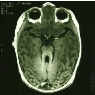
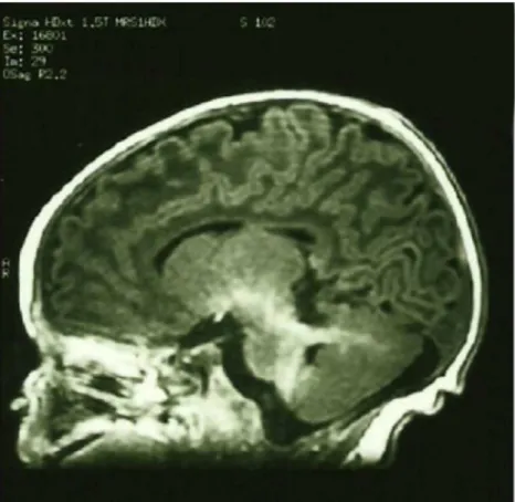
Cranial MR revealed cerebellar vermis agenesis with a mesen-cephalic molar tooth appearance (figures 1and2), as well as a thickened cerebral superior pedunculae and a narrow isthmus (figure 3).
Laboratory examinations revealed haemoglobin 13.1 g/dL, 12 000/μL leucocytes, 44.6% neutrophils, 297 000/μL platelets,
aspartate aminotransferase 42 U/L, alanine aminotransferase 29 U/L, creatinine 0.4 mg/dL and urea 34 mg/dL. Abdominal and renal ultrasound was normal.
An electroretinogram showed mixed retinal dystrophy com-prising attenuated amplitude of cone and rod potentials with preserved macular function.
Gene sequencing was positive for an AHI1 missense c.1981T>C homozygous mutation. The patient’s parents are carriers of the same genetic mutation.
OUTCOME AND FOLLOW-UP
The patient is now 24 months old and has developmental delay, as she does not stand or walk. She sits with support, has a social
smile, babbles and responds to simple commands. She maintains torsional nystagmus and acquires a tilted head position when trying to fixate objects, although she no longer manifests tremor. There is no report of respiratory irregularities.
She has been on a physical therapy and rehabilitation pro-gramme since she was 6 months old, and will maintain follow-up with Neurology, Ophthalmology and Child Development.
DISCUSSION
Criteria for diagnosis of Joubert syndrome include the presence of hypotonia in infancy, developmental delay/mental retardation and at least one of two features including breathing irregularities and abnormal eye movements.17
This case was diagnosed in the neonatal period fulfilling one clinical criterion, abnormal eye movements. The presence of the molar tooth sign in the cranial MR confirmed the diagnosis. The most common manifestation in this age group is respiratory abnormalities, which our patient never developed.2
This case illustrates how important it is to listen to parents’
concerns. Newborns mayfixate on a face and have‘doll’s eye’
movements on turning, but nystagmus and complete absence of ocularfixation are never normal.21
Joubert syndrome is associated with several ocular abnormal-ities, including nystagmus (horizontal, vertical, torsional, pendu-lar or see-saw pattern) and oculomotor apraxia, which are usually present at birth and may improve with age. Strabismus, ocular coloboma, visual loss, ptosis, pigmentary abnormalities of the fundus and decreased vestibulo-ocular reflexes are also manifestations.22
Retinal dystrophy is also associated with polycystic renal disease,23 for which our patient has tested negative thus far.
AHI1 gene mutations are associated with retinal abnormalities and renal progressive disease.24 25 AHI1 missense c.1981T>C
is considered possibly pathogenic according to Mutation Predictor.
Joubert syndrome can evolve variably in three major groups of severity: early death, severe developmental delay with mul-tiple visual and motor deficits, or mild cognitive delay (even normal intelligence).5
Figure 1 Horizontal MRI T2 section of the brain illustrative of the molar tooth sign.
Figure 2 Horizontal MRI T1 section of the brain illustrative of the molar tooth sign.
2 Salva I,et al.BMJ Case Rep2016. doi:10.1136/bcr-2015-213127
There are characteristics to this disease that should be considered, namely the progressive nature of retinal and renal dysfunction, which require frequent follow-up and repeat oph-thalmological examinations, as well as renal ultrasounds.15
Patients may have adverse reactions to anaesthetics based on opiates and nitrous oxide and these should thus be avoided.26
The early diagnosis in this case allowed us to initiate rehabilitation at an early age. We are hoping that this will potentiate the patient’s psychomotor development and minim-ise her deficits.
An early diagnosis is also important to ensure family genetic counselling, allowing decisions regarding future pregnancies.27
Learning points
▸ Joubert syndrome is a rare disease that is caused by hypoplasia or dysplasia of the cerebellar vermis.
▸ Breathing irregularities, hypotonia and developmental delay are the most common signs, although early on, patients may not fulfil all the clinical criteria.
▸ This disease is phenotypically heterogeneous and a high level of suspicion is required to successfully diagnose mild cases. ▸ Mutations in the AHI1 gene are associated mainly with
retinal abnormalities.
▸ Early diagnosis is essential to ensure timely initiation of rehabilitation measures and genetic counselling.
Contributors IS contributed to data collection, analysis and interpretation, and manuscript drafting. CA contributed to data analysis and manuscript drafting. AM and CD contributed to manuscript drafting and critical revision.
Competing interests None declared.
Patient consent Obtained.
Provenance and peer reviewNot commissioned; externally peer reviewed.
REFERENCES
1 Romani M, Micalizzi A, Valente EM. Joubert syndrome: congenital cerebellar ataxia with the molar tooth.Lancet Neurol2013;12:894–905.
2 Merritt L. Recognition of the clinical signs and symptoms of Joubert syndrome. Adv Neonatal Care2003;3:178–86.
3 Brancati F, Dallapiccola B, Valente EM. Joubert syndrome and related disorders. Orphanet J Rare Dis2010;5:20.
4 Choc SA, Choh NA, Bhat SA. MRIfindings in Joubert syndrome.Indian J Ped 2009;76:231–5.
5 Steinlin M, Schmid M, Landua K,et al. Follow-up in children with Joubert syndrome.Neuropediatrics1997;28:204–11.
6 Maria BL, Boltshauser E, Palmer SC,et al. Clinical features and revised diagnostic criteria in Joubert syndrome.J Child Neurol1999;14:583–91.
7 Wolfe L, Lakadamyali H, Mutlu GM. Joubert syndrome associated with severe central sleep apnea.J Clin Sleep Med2010;6:384–8.
8 Akcacus M, Gunes T, Kumandas S,et al. Joubert syndrome: report of a neonatal case.Paediatr Child Health2003;8:499–502.
9 Pellegrino JE, Lensch MW, Muenke M,et al. Clinical and molecular analysis in Joubert syndrome.J Child Neurol1999;14:583–91.
10 Yachnis AT, Rorke LB. Cerebellar and brainstem development: an overview in relation to Joubert syndrome.J Child Neurol1999;14:570–3.
11 Fennel EB, Gitten JC, Dede DE,et al. Cognition, behaviour and development in Joubert syndrome.J Child Neurol1999;14:592–6.
12 Khan AO, Oystreck DT, Seidamehmed MZ,et al. Ophtalmic features of Joubert syndrome.Ophtalmology2008;115:2286–9.
13 Parisi M, Glass I. Joubert syndrome and related disorders. In: Pagon RA, Adam MP, Ardinger HH,et al., eds.GeneReviews [Internet]. Seattle: WA, University of Washington, Seattle, 2003:1993–2015.
14 Singh J, Gathwala G, Agarwal S,et al. Joubert syndrome: a case report.J Indian Med Assoc2011;109:348–9.
15 Singh P, Goraya JS, Saggar K,et al. A report of Joubert syndrome in an infant, with literature review.J Pediatr Neurol2011;6:44–7.
16 Kenner C. Human genetics and implications for neonatal care. In: Kenner C, Lott JW, eds.Comprehensive neonatal nursing: a physiological perspective. 3rd edn. Philadelphia, PA: Saunders, 2003:132–50.
17 Paprocka J, Jamroz E. Joubert syndrome and related disorders.Neurol Neurochir Pol 2012;46:379–83.
Figure 3 Vertical MRI section of the brain and cerebellum illustrative of thickened superior cerebral superior pedunculae and a narrow isthmus.
Salva I,et al.BMJ Case Rep2016. doi:10.1136/bcr-2015-213127 3
18 Karp N, Grosse-Wortmann L, Bowdin S. Severe aortic stenosis, bicuspid aortic valve and atrial septal defect in a child with Joubert syndrome and related disorders ( JSRD)—a case report and review of congenital heart defects reported in the human ciliopathies.Eur J Med Genet2012;55:605–10.
19 Valente EM, Dallapiccola B, Bertini E. Joubert syndrome and related disorders. Handb Clin Neurol2013;113:1879–88.
20 Cheng Y, Eley L, Hynes AM,et al. Investigating embryonic expression patterns and evolution of AHI1 and CEP290 genes, implicated in Joubert syndrome.PLoS ONE 2012;7:e44975.
21 Feigelman S. Thefirst year. In: Kliegman RM, Stanton BMD, Geme JS,et al., eds.Nelson textbook of pediatrics. 19th edn. Philadelphia, PA: Saunders, 2011. Chap 8.
22 Incecik F, Herguner MO, Altunbasak S,et al. Joubert syndrome: report of 11 cases. Turk J Pediatr2012;54:605–11.
23 Saraiva JM, Baraitser M. Joubert syndrome: a review.Am J Med Genet 1992;43:726–31.
24 Valente EM, Brancati F, Silhavy JL,et al. AHI1 gene mutations cause specific forms of Joubert syndrome-related disorders.Ann Neurol2006;59:527–34.
25 Parisi MA, Doherty D, Eckert ML,et al. AHI1 mutations cause both retinal dystrophy and renal cystic renal disease in Joubert syndrome.J Med Genet2006;43:334–9. 26 Habre W, Sims C, D’Souza M. Anaesthetic management of children with Joubert
syndrome.Paediatr Anaesth1997;7:251–3.
27 Angemi JA, Zuccotti JC. Joubert syndrome: report of four adult siblings affected. Rev Neurol2012;54:609–12.
Copyright 2016 BMJ Publishing Group. All rights reserved. For permission to reuse any of this content visit http://group.bmj.com/group/rights-licensing/permissions.
BMJ Case Report Fellows may re-use this article for personal use and teaching without any further permission.
Become a Fellow of BMJ Case Reports today and you can: ▸ Submit as many cases as you like
▸ Enjoy fast sympathetic peer review and rapid publication of accepted articles ▸ Access all the published articles
▸ Re-use any of the published material for personal use and teaching without further permission
For information on Institutional Fellowships contact [email protected]
Visit casereports.bmj.com for more articles like this and to become a Fellow
4 Salva I,et al.BMJ Case Rep2016. doi:10.1136/bcr-2015-213127