Aging-Like Skin Atrophy via Antioxidant Activity in Mice
Shuichi Shibuya1, Yusuke Ozawa1, Kenji Watanabe1, Naotaka Izuo1, Toshihiko Toda1, Koutaro Yokote2, Takahiko Shimizu1,3*
1Department of Advanced Aging Medicine, Chiba University Graduate School of Medicine, Chiba, Japan,2Department of Clinical Cell Biology and Medicine, Chiba University Graduate School of Medicine, Chiba, Japan,3Molecular Gerontology, Tokyo Metropolitan Institute of Gerontology, Itabashi, Tokyo, Japan
Abstract
Cu-Zn superoxide dismutase (Sod1) loss causes a redox imbalance as it leads to excess superoxide generation, which results in the appearance of various aging-related phenotypes, including skin atrophy. Noble metal nanoparticles, such as palladium (Pd) and platinum (Pt) nanoparticles, are considered to function as antioxidants due to their strong catalytic activity. In Japan, a mixture of Pd and Pt nanoparticles called PAPLAL has been used to treat chronic diseases over the past 60 years. In the present study, we investigated the protective effects of PAPLAL against aging-related skin pathologies in mice. Transdermal PAPLAL treatment reversed skin thinning associated with increased lipid peroxidation inSod12/2mice. Furthermore, PAPLAL normalized the gene expression levels ofCol1a1,Mmp2,Has2,Tnf-a,Il-6, andp53in the skin of the Sod12/2 mice. Pt nanoparticles exhibited marked SOD and catalase activity, while Pd nanoparticles only displayed weak SOD and catalase activityin vitro. Although the SOD and catalase activity of the Pt nanoparticles significantly declined after they had been oxidized in air, a mixture of Pd and Pt nanoparticles continued to exhibit SOD and catalase activity after oxidation. Importantly, a mixture of Pd and Pt nanoparticles with a molar ratio of 3 or 4 to 1 continued to exhibit SOD and catalase activity after oxidation, indicating that Pd nanoparticles prevent the oxidative deterioration of Pt nanoparticles. These findings indicate that PAPLAL stably suppresses intrinsic superoxide generation bothin vivoandin vitrovia SOD and catalase activity. PAPLAL is a potentially powerful tool for the treatment of aging-related skin diseases caused by oxidative damage.
Citation:Shibuya S, Ozawa Y, Watanabe K, Izuo N, Toda T, et al. (2014) Palladium and Platinum Nanoparticles Attenuate Aging-Like Skin Atrophy via Antioxidant Activity in Mice. PLoS ONE 9(10): e109288. doi:10.1371/journal.pone.0109288
Editor:Hemachandra Reddy, Oregon Health & Science University, United States of America ReceivedApril 16, 2014;AcceptedAugust 30, 2014;PublishedOctober 15, 2014
Copyright:ß2014 Shibuya et al. This is an open-access article distributed under the terms of the Creative Commons Attribution License, which permits unrestricted use, distribution, and reproduction in any medium, provided the original author and source are credited.
Data Availability:The authors confirm that all data underlying the findings are fully available without restriction. All relevant data are within the paper and its Supporting Information files.
Funding:This research was supported by funds from Musashino Pharmaceutical Co. (http://www.musashino-p.co.jp/). The PAPLAL and the Pd and Pt nanoparticles were provided from Musashino Pharmaceutical and Toyo Kosei pharmaceutical (http://toyokosei.co.jp/). The funders had no role in study design, data collection and analysis, decision to publish, or preparation of the manuscript.
Competing Interests:Musashino Pharmaceutical Company made a grant donation to Department of Advanced Aging Medicine, Chiba University Graduate School of Medicine, for the conduct of the animal study and for the purchase of reagents. Musashino Pharmaceutical was not involved with the scientific content of the research projects run by the Department of Advanced Aging Medicine, Chiba University Graduate School of Medicine. The authors were not paid employees and did not receive any kind of fees from Musashino Pharmaceutical Company. This does not alter the authors’ adherence to all the PLOS ONE policies on sharing data and materials.
* Email: [email protected]
Introduction
Skin aging induced by chronological or intrinsic factors leads to skin atrophy [1]. The amounts of skin collagen components fall in an age-dependent manner in both males and females, resulting in age-related skin thinning in older individuals [2,3]. Evidence suggests that oxidatively modified proteins, DNA, and lipids in the skin and other organs progressively accumulate during aging [4], indicating that reactive oxygen species (ROS) are strongly associated with skin aging. Complex organisms possess multiple antioxidative and repair systems for mitigating oxidative damage. Superoxide dismutase (SOD) plays a central role in antioxidative systems due to its ability to catalyze the conversion of cellular superoxide (O22) to hydrogen peroxide (H2O2). H2O2is further
degraded to O2and H2O by catalase, glutathione peroxidases and
peroxiredoxins. Copper/zinc superoxide dismutase (SOD1) is localized to react with intracellular O2 in the cytoplasm. Our
previous studies have demonstrated thatSod1-deficient (Sod12/2)
mice exhibit increased intracellular O22 concentrations and
various aging-related organ phenotypes, such as age-related macular degeneration [5], fatty deposits in the liver [6,7], skin atrophy [8–11], bone loss and fragility [12,13], progression of Alzheimer’s disease [14], infertility [15], dry eye [16,17], and rotator cuff degeneration [18]. These findings suggest that cytoplasmic O22-mediated oxidative damage is the primary cause
of aging-related changes in various tissues. In particular, Sod1 insufficiency results in both epidermal and dermal atrophy, which is associated with the downregulation of extracellular matrix related-genes, including Col1a1 and Has2 [11]. Therefore, Sod12/2mice constitute a suitable model for studying skin aging in elderly people.
atoms located on their surfaces [19–21]. Such noble metal nanoparticle catalysts are considered to function as antioxidants and are potentially useful in material science and engineering as well as in medical science and clinical therapy [22]. A number of studies have reported that Pt nanoparticles exhibit strong antioxidant activity [22–24]. Recently, Elhusseiny and Hassan reported that a complex of Pd and Pt nanoparticles demonstrated highly potent antitumor and antimicrobial activity [25]. In 1915, Dr. Hideyo Noguchi (Rockefeller University) and Dr. Saburo Ishizuka (a dental surgeon at Tokyo Dental College) formulated a plan for creating a solution of Pd and Pt nanoparticles for clinical use. Twenty-one years later, Dr. Ishizuka successfully prepared a Pd and Pt nanoparticle solution called PAPLAL (Toyokose Pharmaceuticals, Japan) [26]. Since then, PAPLAL has been used to treat Japanese patients with burns, frostbite, hives, lung inflammation, gastric ulcers, and rheumatoid arthritis. PAPLAL has been shown to have various beneficial effects on chronic diseases [26]. In addition, PAPLAL has been approved as a treatment for acute gastric inflammation and chronic gastrin catarrh in Japan under the Pharmaceutical Affairs Law, and it has also been patented as an antioxidant with the Japan Patent Office (Patent No. 3411195, 2003). In vitro studies have reported that PAPLAL exhibits antioxidant activity against superoxide anions and hydroxyl radicals [27,28]. However, no previous studies have investigated the effects of PAPLAL or other metal nanoparticles on skin aging.
In the present study, we investigated the protective effects of PAPLAL against age-related skin pathologies in model mice. We also analyzed the expression profiles of skin-related genes, including those involved in matrix biosynthesis, inflammation, and aging, in order to clarify the underlying mechanisms of the in vivo effects of PAPLAL. In addition,in vitroexperiments were conducted to evaluate the antioxidant activity of PAPLAL.
Materials and Methods
Nanoparticles
Pd and Pt nanoparticles and PAPLAL were provided by Toyokose Pharmaceutical Co. (Tokyo, Japan) through Musashino Pharmaceutical Co. (Tokyo Japan). PAPLAL is composed of a mixture of 0.3 mg/mL (2.82 mM) of Pd nanoparticles and 0.2 mg/mL (1.03 mM) of Pt nanoparticles.
Mice
Sod12/2 mice were purchased from the Jackson Laboratory (Bar Harbor, ME, USA). Genotyping of theSod12/2 allele was performed via genomic PCR using genomic DNA isolated from the tail tip, as reported previously [8]. The animals were housed under a 12-hour light/dark cycle and fedad libitum. In addition, they were maintained and studied according to protocols approved by the animal care committee of the Tokyo Metropolitan Institute of Gerontology and Chiba University.
Transdermal administration inSod12/2mice TheSod1+/+
and Sod12/2 mice were transdermally adminis-tered PAPLAL for four weeks, beginning at four months of age. On the first day of each week, the hair on the backs of the mice was shaved off and then PAPLAL (200mL/mouse) or MilliQ water (200mL/mouse) was applied to the exposed skin once a day. MilliQ water was used as a placebo control for PAPLAL.
Histology
To assess the histological morphology of the treated skin, skin specimens were dissected from the back tissue of the mice, fixed
overnight in a 20% formalin neutral buffer solution (Wako, Osaka, Japan), embedded in paraffin, and sectioned on a microtome at a thickness of 4mm according to standard techniques. Hematoxylin and eosin staining was performed as described previously [6,29]. The thickness of the skin tissue was measured using the Leica QWin V3 imaging software (Leica, Germany).
8-isoprostane content
The skin tissue specimens were homogenized with 0.1 M phosphate (pH 7.4) containing 1 mM of ethylenediaminetetraace-tic acid (Dojindo Laboratories, Kumamoto, Japan) and 50mg/mL
(w/w) of dibutylhydroxytoluene (Wako). The homogenate was centrifuged at 8,0006g for 10 minutes at 4uC, and the total supernatant was used for the assay. The 8-isoprostane concentra-tion of the homogenate was measured using an 8-isoprostane enzyme immunoassay kit (Cayman Chemical Company, MI, USA) according to the manufacturer’s instructions. The protein concentration of the supernatant was assessed using the DC protein assay kit (BioRad, Hercules, CA, USA), and the 8-isoprostane level was normalized to the protein level.
Outgrowth assay
Mouse back skin samples were sterilized with 70% ethanol and rinsed with phosphate-buffered saline (Takara Bio Inc., Shiga, Japan), and then discs measuring 5 mm in diameter were punched out using a dermal punch (Nipro, Tokyo, Japan). The punched skin discs were placed into a 24-well culture plate (Falcon BD, Franklin Lakes, NJ) and cultured with or without PAPLAL ina -minimum essential medium (a-MEM; Life Technologies Corpo-ration, Carlsbad, CA, USA) containing 20% fetal bovine serum (FBS; Life Technologies Corporation), 100 units/mL of penicillin, and 0.1 mg/mL of streptomycin (Sigma-Aldrich, MO, USA) at 37uC in a humidified incubator under 5% CO2and 20% O2. The
number of outgrowing fibroblasts originating from the mouse skin discs was directly counted after 96 h culturing.
Lactate dehydrogenase (LDH) activity
Skin specimens were cultured according to the method described above, and the culture medium was collected after 96 h. The collected medium was centrifuged at 4006gfor 5 min at 4uC, and the total supernatant was used for the subsequent assay. The LDH level was measured using the LDH cytotoxicity assay kit (Cayman Chemical Company) according to the manufacturer’s instructions.
Quantitative PCR
Total RNA was extracted from the back skin using Trizol reagent (Life Technologies Corporation) according to the manu-facturer’s instructions. cDNA was synthesized from 1mg of total
RNA using reverse transcriptase (ReverTra Ace qPCR RT Master MIX, TOYOBO, Osaka, Japan). Real-time PCR was performed on a Mini OpticonTM(Bio-Rad) with SYBR GREEN PCR master mix (Bio-Rad), according to the manufacturer’s instructions. All expression data were normalized to the expression level of the housekeeping gene glyceraldehyde-3-phosphate dehydrogenase (Gapdh). The primer sequences are listed in Table S1.
Antioxidant activity
according to the manufacturer’s instructions. SOD derived from bovine erythrocytes (Sigma-Aldrich, lot number 080M76901V) and catalase derived from bovine liver tissue (Sigma-Aldrich, lot number 1232075) were used as positive controls for SOD and catalase activity, respectively. In order to assess the stability of the antioxidant activity of the different nanoparticles, Pd and Pt nanoparticle solutions were incubated at room temperature for four weeks in tubes in which the air had been replaced with N2. In
a further experiment, Pd and Pt nanoparticle solutions were exposed to air by rotating them for 24 h at room temperature. The SOD and catalase activity levels of each solution were then measured as described above.
Statistics
Statistical analyses were performed using the Student’st-test for comparisons between two groups or Tukey’s test for comparisons between three groups. Differences were considered significant at p-values of less than 0.05. All data are expressed as mean6standard deviation (SD) values.
Results
PAPLAL accelerated wound healing in the aged mice In order to evaluate the protective effects of PAPLAL on the skin, we first investigated the ability of PAPLAL to promote wound healing in aged murine skin (17 months of age). Although the areas of the wounds treated with and without PAPLAL did not differ at two days after wounding, the wounds treated with PAPLAL were significantly smaller than those treated with vehicle at four and six days after wounding (Figure S1A–1B). In addition, PAPLAL treatment was found to have promoted wound healing at 12 days after wounding (Figure S1B). These results demonstrate that the transdermal application of PAPLAL assists in wound healing in aged mice.
PAPLAL attenuated skin atrophy in theSod1-deficient mice
SOD1, an antioxidant enzyme, plays a pivotal role in cellular antioxidative systems. Therefore, in order to investigate the beneficial effects of PAPLAL on the skin symptoms seen in Sod12/2 mice, PAPLAL was transdermally administered to the skin on the backs ofSod12/2and wild-type (Sod1+/+) mice daily
for four weeks, beginning at four months of age. As shown in Figure 1, the skin of theSod12/2mice was significantly thinner than that of theSod1+/+mice, confirming that skin atrophy had
occurred in the Sod12/2 mice. The back skin of theSod12/2 mice that were treated with a high concentration of PAPLAL was significantly thicker (epidermis and dermis: by 46.2% and 19.2%, respectively) than that of the Sod12/2 mice treated with the vehicle. The administration of a low concentration of PAPLAL also improved the skin atrophy observed in theSod12/2mice (the thickness of the epidermis and dermis were increased by 42.0% and 21.1%, respectively) compared with that seen in theSod12/2 mice treated with vehicle. In order to examine the degree of oxidative damage in the skin of theSod12/2mice, we measured the concentration of 8-isoprostane as a representative of lipid peroxidation products. The 8-isoprostane content of theSod12/2 skin was increased by 65.4% compared with that of theSod1+/+
mice, which indicated that lipid peroxidation products had accumulated in it (Figure 1D). Meanwhile, treatment with a high concentration of PAPLAL significantly decreased the 8-isopros-tane content of theSod12/2mouse skin compared with that of the Sod12/2mouse skin treated with the vehicle (Figure 1D).
Next, we investigated the protective effects of PAPLAL against skin damage. Organ culture experiments using skin discs demonstrated that theSod12/2fibroblasts had a markedly lower outgrowth capacity than theSod1+/+
fibroblasts, which indicated that the migration and proliferation of the Sod12/2 fibroblasts were impaired (Figure 2A). In order to analyze the protective effects of PAPLAL, we added PAPLAL to cultures of Sod12/2 skin discs. Treatment with PAPLAL significantly promoted fibroblast outgrowth from the mutant skin discs (Figure 2A). We also measured the LDH activity, which is a marker of skin damage, in the culture medium. The Sod12/2 skin exhibited significantly increased LDH activity by 5.1-fold compared with the Sod1+/+ fibroblasts, which was indicative of skin damage
(Figure 2B). Meanwhile, PAPLAL treatment significantly sup-pressed the LDH activity in the Sod12/2 skin disc culture compared with vehicle treatment (Figure 2B).
In order to investigate the adverse effects of PAPLAL, we transdermally administered PAPLAL into the skin on the backs of Sod1+/+mice. As shown in Figure 3, no significant differences in
skin thickness were observed among theSod1+/+
mice treated with or without PAPLAL. In addition, no abnormalities, such as cell infiltration or PAPLAL deposition, were detected in the skin of the Sod1+/+
mice, suggesting that PAPLAL does not have any adverse effects on the skin, at least in the short term. In the organ culture analysis, PAPLAL treatment ofSod1+/+skin did not induce any
significant change in fibroblast outgrowth capacity or LDH activity detected in skin disc cultures (Figure 2A, 2B). These findings demonstrate that the transdermal application of PAPLAL ameliorates skin atrophy in Sod12/2 mice by suppressing oxidative damage.
PAPLAL normalized the transcriptional profiles of skin-related genes in theSod12/2skin
In order to investigate the mechanisms by which PAPLAL treatment counters skin atrophy inSod12/2 mice, we analyzed the expression patterns of extracellular matrix-related genes in the skin. In theSod12/2mice, the skin mRNA expression level of the type I collagen gene (Col1a1) was significantly reduced, while that of the matrix metalloproteinase 2 gene (Mmp2) were significantly increased, compared with those observed in the Sod1+/+
mice, indicating that collagen biosynthesis was reduced in theSod12/2 mice (Figure 4). Moreover, the mRNA expression of the hyaluronan synthase 2 gene (Has2) was significantly downregu-lated in the skin of theSod12/2mice (Figure 4). In contrast, the mRNA expression levels of Decorin and Ki67 did not differ between theSod1+/+
andSod12/2mice (Figure 4). These results suggest that Sod1 deficiency causes skin thinning due to dysregulation of the extracellular matrix. Among the genes that exhibited altered expression levels, PAPLAL treatment signifi-cantly normalized the mRNA levels ofCol1a1,Mmp2, andHas2, suggesting that PAPLAL treatment increases skin thickness by increasing the concentrations of extracellular matrix components, such as collagen and hyaluronic acid (Figure 4).
The skin of theSod12/2mice also exhibited significantly higher expression levels of inflammatory cytokines, includingTnf-aand Il-6, compared with the skin from the Sod1+/+mice (Figure 4).
Figure 1. Protective effects of PAPLAL against skin atrophy in theSod1-deficient mice.(A) Hematoxylin and eosin staining of the skin on the backs of theSod1+/+andSod12/2mice (five months of age). E, epidermis; D, dermis. The scale bars represent 20mm (top) or 100mm (bottom).
The thickness of the (B) epidermal and (C) dermal layers of the skin on the backs ofSod1+/+
andSod12/2mice treated with PAPLAL (n = 6–8). (D)
8-isoprostane content of the skin on the backs ofSod1+/+andSod12/2mice treated with PAPLAL (n = 5–8). 0.01
6and 16PAPLAL indicate 0.0 or
1-fold concentrations of PAPLAL, respectively. Data are shown as the mean6SD; *p,0.05, **p,0.01. doi:10.1371/journal.pone.0109288.g001
Figure 2. PAPLAL attenuates cellular damage in skin.(A) Relative number of outgrowing fibroblasts in culturedSod1+/+andSod12/2skin
specimens. (B) LDH activity in the medium used to culture theSod1+/+
andSod12/2skin specimens. Data are shown as the mean6SD; *p,0.05,
**p,0.01.
mice. PAPLAL treatment significantly normalized the mRNA expression level ofp53of theSod12/2mice (Figure 4), suggesting that PAPLAL delays skin aging by inhibiting p53 upregulation in Sod12/2mice.
Pd nanoparticles prevented the oxidative deterioration of Pt nanoparticles with respect to their SOD and catalase activity
Since Pt nanoparticles display strong antioxidant activity [22– 24], we assessed the SOD and catalase activity of PAPLAL and its componentsin vitro. The Pd nanoparticles displayed weak SOD Figure 3. PAPLAL is non-toxic in wild-type mice.(A) Hematoxylin and eosin staining of the skin on the backs ofSod1+/+
mice (17–20 weeks of age). E, epidermis; D, dermis. The scale bars represent 20mm (top) or 100mm (bottom). The thickness of the (B) epidermal and (C) dermal layers of the skin on the backs ofSod1+/+
mice treated with PAPLAL (n = 5). doi:10.1371/journal.pone.0109288.g003
Figure 4. PAPLAL improved the transcriptional profiles of skin-related genes in the skin ofSod12/2mice. (A) The relative mRNA
expression levels ofCol1a1,Has2,Mmp2,Decorin,Ki67,Tnf-a,Il-6,p53, andMdm2. Each mRNA expression level was determined using qRT-PCR. Data
are shown as the mean6SD; *p,0.05 vs.Sod1+/+
, **p,0.01 vs.Sod1+/+ ,#p
,0.05 vs.Sod12/2.
and catalase activity, while the Pt nanoparticles exhibited strong SOD and catalase activity (Figure 5A, 5B). PAPLAL, a mixture of Pd and Pt nanoparticles, demonstrated SOD and catalase activity levels that were equivalent to the sum of those exhibited by the Pd and Pt nanoparticles, although no synergistic effects were observed (Figure 5A, 5B).
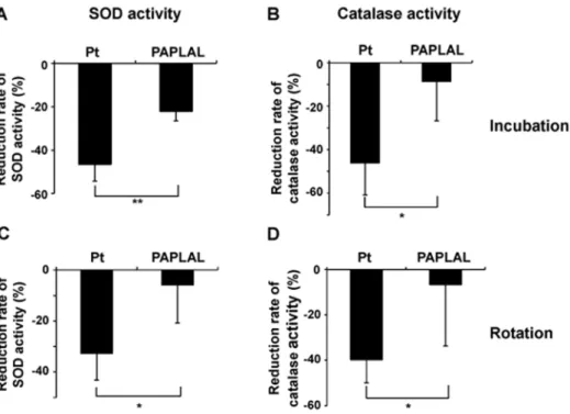
In order to evaluate the stability of the SOD and catalase activity of the tested nanoparticles, Pd and Pt nanoparticles and PAPLAL were stored at room temperature for four weeks. The SOD and catalase activity levels of the stored Pt nanoparticles were dramatically reduced to 46.5% and 46.2%, respectively, of those exhibited by the freshly prepared Pt nanoparticles (Figure 6A, 6B). In contrast, the SOD and catalase activity levels of stored PAPLAL were only reduced by 22.1% and 8.7%, respectively, compared with those of the freshly prepared PAPAL (Figure 6A, 6B). In order to confirm that the reductions in the SOD and catalase activity of the Pt nanoparticles were due to oxidative deterioration during storage, Pt nanoparticles and PAPAL were exposed to air by rotating them for 24 hours. The SOD and catalase activity levels of the Pt nanoparticles that were exposed to air were significantly decreased compared with those of the freshly prepared nanoparticles (SOD and catalase: 33.0% and 39.7%, respectively; Figure 6C, 6D). In contrast, the PAPLAL exposed to air retained its SOD and catalase activity, indicating that the oxidative deterioration of the Pt nanoparticles within it had been prevented (Figure 6C, 6D).
Furthermore, Pd nanoparticles were mixed with Pt nanoparti-cles at various concentration ratios and then stored at room temperature for four weeks. When Pd nanoparticles were added to Pt nanoparticles at a molar ratio of 1 or 2 to 1, the SOD and catalase activity levels of the mixture were markedly reduced after their storage at room temperature (Figure 7). However, when Pd nanoparticles were added to Pt nanoparticles at a molar ratio of 3 or 4 to 1, the SOD and catalase activity levels of the mixture were sustained (Figure 7).
We previously reported that Sod1 loss significantly enhanced intracellular O22 generation in fibroblasts [9,32]. Therefore, we
assessed the antioxidant effects of Pd and Pt nanoparticles on Sod12/2 fibroblasts. Sod12/2 fibroblasts were treated with Pd and/or Pt nanoparticles for 16 hours, and then the levels of intracellular O22were assessed. O22generation was significantly
decreased (by 23.3%) in the Sod12/2 fibroblasts treated with 10mM of Pt nanoparticles, but not in those treated with Pd nanoparticles (Figure S2). Notably, treatment with pre-incubated Pt nanoparticles did not decrease intracellular O22generation in
theSod12/2fibroblasts (Figure S2). These results suggest that Pt nanoparticles possess strong antioxidant effects, such as SOD and catalase activity, and that the Pd in PALAL inhibits the oxidative deterioration of Pt, which enables PALAL to retain strong antioxidant activity over time.
Discussion
PAPLAL attenuates intrinsic skin aging by suppressing oxidative damage
In the present study, we demonstrated that PAPLAL significantly reversed skin thinning by reducing oxidative and cellular damage in Sod12/2 mice (Figures 1 and 2). In addition, PAPLAL and Pt nanoparticles, but not Pd nanoparticles, exhibited SOD and catalase activity (Figure 5). Furthermore, anin vitro experiment found that treatment with Pt nanoparticles, but not Pd nanopar-ticles, significantly reduced O22generation inSod12/2fibroblasts
(Figure S2). These findings suggest that the antioxidant activity of the Pt nanoparticles in PAPLAL contribute to attenuating age-related skin thinning in Sod12/2 mice. In this context, we previously evaluated the ability of several antioxidants to counteract thein vivoage-related changes seen inSod12/2mice and found that the administration of ascorbic acid significantly attenuated bone loss and fragility in Sod12/2 mice [12]. Likewise, the transdermal administration of ascorbic acid derivatives was demonstrated to normalize skin thinning inSod12/2mice [9,10]. Furthermore, Iuchi et al. reported that oral N-acetylcysteine treatment mitigates hemolytic anemia in Sod12/2 mice by suppressing ROS generation in red blood cells [33]. Together, these findings demonstrate that antioxidants, such as PAPLAL, ascorbic acid, and N-acetylcysteine, can improveSod1loss-induced organ pathologies.
PAPLAL normalizes gene expression, including that related to matrix biosynthesis and inflammation, in the skin
As shown in Figure 4, Sod1 loss induced the transcriptional downregulation ofCol1a1andHas2, as well as the upregulation of Mmp2expression, which were indicative of collagen and hyaluronic acid malformation in the atrophic skin of theSod12/2mice.Sod1 loss also upregulated the expression of proinflammatory genes, such asTnf-a and Il-6, in the skin (Figure 4). Tumor necrosis factor (TNF)- a regulates type I collagen expression via the c-Jun N-terminal kinase (JNK) and nuclear factor kappa-light-chain-enhancer of activated B cells (NF-kB) pathways in skin fibroblasts Figure 5. Pt nanoparticles possess SOD and catalase activity.
PAPLAL includes 2.82 mM of Pd nanoparticles and 1.03 mM of Pt nanoparticles. (A) The SOD and (B) catalase activity of 2.82 mM Pd nanoparticles, 1.03 mM Pt nanoparticles, and PAPLAL. Five nM of SOD derived from bovine erythrocytes (A) and 0.2mM of catalase derived from bovine liver tissue (B) were used as positive controls for SOD and catalase, respectively.
[34]. In addition, Galera et al. reported that NF-kB directly suppressesCOL1A1gene transcription in human dermal fibroblasts [35] and accumulates in the nuclei of aged human fibroblasts in association with the downregulation of theCOL1A1gene [36]. An IkB kinase (IKK)-b inhibitor has also been shown to suppress interleukin (IL)-1b-induced collagen degradation by inhibiting the activation of NF-kB and upregulation of matrix metalloproteinases [37]. Taken together, the inflammatory response controls collagen homeostasis via transcriptional mechanisms in fibroblasts. In the present study, PAPLAL treatment normalized the gene expression ofCol1a1andTnf-ain the skin of theSod12/2mice (Figure 4). With respect to other organs, Onizawa et al. reported that the
intranasal administration of Pt nanoparticles reduced NF-kB activity and inhibited pulmonary inflammation in mice exposed to cigarette smoke [38]. Rehman et al. also reported that Pt nanoparticles have anti-inflammatory effects on the lipopolysac-charide-induced inflammatory response by downregulating the expression of IL-1b, TNF-a, and IL-6 in macrophages [39]. Collectively, these findings suggest that PAPLAL and Pt nanopar-ticles suppress the inflammatory response, resulting in improve-ments in the anabolic and catabolic regulation of collagen homeostasis.
Pd nanoparticles stabilize the antioxidant activity of Pt nanoparticles by preventing their oxidative deterioration
Noble metal nanoparticles, such as Pd, Pt, and gold, are considered to function as antioxidants by reducing catalysis [19– 21]. A number of studies have reported that Pt nanoparticles exhibit a strong ability to scavenge ROS, including O22 and H2O2 [22–24]. In a lifespan analysis of C. elegans, Kim et al.
reported that Pt nanoparticles extended the lifespans of wild-type N2 and short-livedmev-1nematodes, in which intracellular ROS accumulated due to respiratory impairment [40]. The present results demonstrated that Pt nanoparticles, but not Pd nanopar-ticles, possess SOD and catalase activity (Figure 5), which is consistent with the above results. Okamotoet al.reported that Pt nanoparticles were oxidized to PtO by oxygen in the air, which resulted in an time-dependent increase in their ability to degrade ascorbic acid [41]. However, the co-incubation of Pd nanoparti-cles with Pt nanopartinanoparti-cles effectively prevented PtO formation via Pt nanoparticles oxidation [41]. Since Pd has a lower oxidation/ reduction potential than Pt, they proposed that Pd reduces Pt2+
to Pt in solution [41]. In order to further investigate the ability of Pd nanoparticles to prevent the oxidative deterioration of Pt, we herein examined the SOD and catalase activity of Pt nanoparticles Figure 6. Pd nanoparticles protected the SOD and catalase activity of Pt nanoparticles against oxidative degradationin vitro.(A) The SOD and (B) catalase activity of 1.03 mM Pt nanoparticles and PAPLAL that had been stored at room temperature for four weeks. (C) The SOD and (D) catalase activity of Pt nanoparticles and PAPLAL that had been rotated for 24 hours in a tube in order to oxidize the nanoparticles. Data are shown as the mean6SD; *p,0.05, **p,0.01.
doi:10.1371/journal.pone.0109288.g006
Figure 7. Pd nanoparticles protected the SOD activity of Pt nanoparticles against oxidative degradation at various molar ratios. Pd nanoparticles were added to Pt nanoparticles at various molar ratios, and the SOD activity of each mixture was measured after four weeks of storage at room temperature.
after storage- or rotation-induced oxidization in the presence of Pd nanoparticles (Figure 5). Predictably, the co-storage of Pt nano-particles with Pd nanonano-particles delayed the oxidative inactivation of the SOD and catalase activity of the former nanoparticles, while in the absence of Pd nanoparticles the Pt nanoparticles were inactivated during incubation or rotation (Figure 6). Indeed, Pt nanoparticles that had been oxidized in air failed to suppress intracellular O22 generation in Sod12/2 cells (Figure S2). In
contrast, even after oxidation in air, PAPLAL, a mixture of Pd and Pt nanoparticles, continued to exhibit SOD and catalase activity, and hence, was able to decrease O22 generation in Sod12/2
fibroblasts (Figure S2).
Notably, when Pd nanoparticles were added to Pt nanoparticles at a molar ratio of 3 or 4 to 1, the SOD and catalase activity levels of the Pt nanoparticles were sustained more effectively than when a molar ratio of 1 or 2 to 1 was employed (Figure 7). In fact, PAPAPL contains Pd and Pt nanoparticles at a molar ratio of 2.74 to 1, suggesting that the excess Pd nanoparticles in PAPLAL effectively protect the Pt nanoparticles from oxidative tion. Since long-term storage accelerates the oxidative deteriora-tion of antioxidants in clinical use, the addideteriora-tion of Pd nanoparticles to such antioxidants might efficiently maintain their bioactivity under oxidative conditions.
PAPLAL is a valuable antioxidant for delaying skin aging In the present study, we directly applied PAPLAL to the skin of Sod12/2mice. PAPLAL treatment effectively improved the skin thinning seen in theSod12/2mice, suggesting that the PAPLAL had been incorporated into epidermal and dermal cells. In a recently electron microscopic study, Okamotoet al.reported that the Pd and Pt nanoparticles in PAPLAL measure 3.5960.56 and 1.9360.34 nm, respectively, in diameter [41]. A previous study found that cells that had been treated with Pt nanoparticles exhibited increased Pt concentrations compared with the control cells [24]. In an inductively coupled plasma mass spectrometry-based study ofC. elegans, Sakaue et al.reported that treatment with Pt nanoparticles increased the internalization of Pt in nematodes [42]. These results suggest that PAPLAL and/or Pt nanoparticles are transdermally taken in by cells, which inhibits the progression of skin pathologies caused by oxidative stress in mice.
In the present study, PAPLAL treatment did not cause any morphological abnormalities, such as cell infiltration or PAPLAL deposition, or cellular damage in mouse skin (Figures 2, 3). Furthermore, PAPLAL has been shown to be free from adverse effects during its clinical use as a treatment for chronic disease in humans [26]. In agreement with our results, several reports have found that treatment with Pt nanoparticles did not induce any alterations in the biological profiles of wild-type mice [38,43]. On the other hand, Newkirk et al. recently reported that the oral administration of a mixture of Pd, Pt, and rhodium had a synergistic toxic effect on eosinophils in rats [44]. Further research on the dynamic state and safety of PAPLAL in the living body is required.
Vitiligo is an acquired depigmentation disorder characterized by H2O2/peroxynitrite-mediated oxidative and nitrative stress in
the skin. Salemet al. reported that treatment with PC-KUS, a UVB-activated pseudocatalase, reduces the levels of epidermal H2O2 and induces repigmentation in vitiligo patients [45,46].
Since PAPLAL possesses catalase activity, it might also be useful as a treatment for H2O2-related skin diseases, including vitiligo.
In conclusion, PAPLAL, which is composed of Pd and Pt nanoparticles at a molar ratio of 2.74 to 1, exhibits potent antioxidant activity (attributed to the effects of Pt nanoparticles) and attenuates aging-related skin pathologies in vivo. The Pd nanoparticles contained in PAPLAL prevent the oxidative deterioration of the Pt nanoparticles by attenuating PtO forma-tion. PAPLAL has been found to have few adverse effects on skin morphology in transdermally treated mice. Consistent with these results, no previous studies have found that PAPLAL induces adverse effects, despite it being used in Japan to treat patients with chronic conditions for over 60 years. Therefore, PAPLAL is considered to be a safe and valuable antioxidant for delaying skin aging in humans.
Supporting Information
Figure S1 PAPLAL improves wound healing in aged mice.(A) Typical pictures of aged C57BL/6 male mice that were treated with or without PAPLAL at six days after wounding. (B) Aged C57BL/6 male mice (17 months of age) were wounded on day 0 and treated with or without PAPLAL for 12 days. Wound size was measured over time. Data are shown as the mean6SD; *p,0.05.
(TIF)
Figure S2 PAPLAL suppresses O22production inSod1
-deficient fibroblasts.Sod12/2dermal fibroblasts were treated with 10mM of Pt nanoparticles, 10mM of Pd nanoparticles, or PAPLAL for 16 hours. Intracellular superoxide generation was detected using a DHE fluorescent probe and calculated as the area of DHE-based fluorescence divided by the number of Hoechst-positive cells. Data are shown as the mean6SD. *p,0.05, **p, 0.01.
(TIF)
Table S1 qRT-PCR primers (DOCX)
Methods S1 Supporting Methods. (DOCX)
Acknowledgments
We thank Dr. Hirofumi Koyama, Dr. Daichi Morikawa, Dr. Keiji Kobayashi, Masato Koike, Isao Masuda, and Ayami Goda (Chiba University) for their valuable technical assistance.
Author Contributions
Conceived and designed the experiments: SS TS. Performed the experiments: SS YO KW NI TT. Analyzed the data: SS TS. Wrote the paper: SS KY TS.
References
1. Poljsak B, Dahmane RG, Godic A (2012) Intrinsic skin aging: the role of oxidative stress. Acta Dermatovenerol Alp Panonica Adriat 21: 33–36. 2. Shuster S, Black MM, McVitie E (1975) The influence of age and sex on skin
thickness, skin collagen and density. Br J Dermatol 93: 639–643.
3. Naylor EC, Watson RE, Sherratt MJ (2011) Molecular aspects of skin ageing. Maturitas 69: 249–256.
4. Finkel T, Holbrook NJ (2000) Oxidants, oxidative stress and the biology of ageing. Nature 408: 239–247.
6. Uchiyama S, Shimizu T, Shirasawa T (2006) CuZn-SOD deficiency causes ApoB degradation and induces hepatic lipid accumulation by impaired lipoprotein secretion in mice. J Biol Chem 281: 31713–31719.
7. Kondo Y, Masutomi H, Noda Y, Ozawa Y, Takahashi K, et al. (2014) Senescence marker protein-30/superoxide dismutase 1 double knockout mice exhibit increased oxidative stress and hepatic steatosis. FEBS Open Bio 4: 522– 532.
8. Murakami K, Inagaki J, Saito M, Ikeda Y, Tsuda C, et al. (2009) Skin atrophy in cytoplasmic SOD-deficient mice and its complete recovery using a vitamin C derivative. Biochem Biophys Res Commun 382: 457–461.
9. Shibuya S, Kinoshita K, Shimizu T (2011) Protective effects of vitamin C derivatives on skin atrophy caused bySod1deficiency. In: Preedy BS, editor. Handbook of diet, nutrition and the skin. The Netherlands: Wageningen Academic. pp. 351–364.
10. Shibuya S, Nojiri H, Morikawa D, Koyama H, Shimizu T (2014) Protective effects of vitamin C on age-related bone and skin phenotypes caused by intracellular reactive oxygen species. In: Preedy BS, editor. Aging: Oxidative stress and dietary antioxidants. New York: Academic Press. pp. 152–159. 11. Shibuya S, Ozawa Y, Toda T, Watanabe K, Tometsuka C, et al. (2014)
Collagen peptide and vitamin C additively attenuate age-related skin atrophy in Sod1-deficient mice. Biosci Biotech Biochem 78: 1212–1220.
12. Nojiri H, Saita Y, Morikawa D, Kobayashi K, Tsuda C, et al. (2011) Cytoplasmic superoxide causes bone fragility owing to low-turnover osteoporosis and impaired collagen cross-linking. J Bone Miner Res 26: 2682–2694. 13. Morikawa D, Nojiri H, Saita Y, Kobayashi K, Watanabe K, et al. (2013)
Cytoplasmic reactive oxygen species and SOD1 regulate bone mass during mechanical unloading. J Bone Miner Res 28: 2368–2380.
14. Murakami K, Murata N, Noda Y, Tahara S, Kaneko T, et al. (2011) SOD1 (copper/zinc superoxide dismutase) deficiency drives amyloid beta protein oligomerization and memory loss in mouse model of Alzheimer disease. J Biol Chem 286: 44557–44568.
15. Noda Y, Ota K, Shirasawa T, Shimizu T (2012) Copper/zinc superoxide dismutase insufficiency impairs progesterone secretion and fertility in female mice. Biol Reprod 86: 1–8.
16. Kojima T, Wakamatsu TH, Dogru M, Ogawa Y, Igarashi A, et al. (2012) Age-related dysfunction of the lacrimal gland and oxidative stress: evidence from the Cu,Zn-superoxide dismutase-1 (Sod1) knockout mice. Am J Pathol 180: 1879– 1896.
17. Ibrahim OM, Dogru M, Matsumoto Y, Igarashi A, Kojima T, et al. (2014) Oxidative stress induced age dependent meibomian gland dysfunction in Cu, Zn-superoxide dismutase-1 (Sod1) knockout mice. PLoS One 9: e99328. 18. Morikawa D, Itoigawa Y, Nojiri H, Sano H, Itoi E, et al. (2014) Contribution of
oxidative stress to the degeneration of rotator cuff entheses. J Shoulder Elbow Surg 23: 628–635.
19. Lewis L, Lewis N (1986) Platinum-Catalyzed Hydrosilylation-Colloid Formation as the Essential Step. J Am Chem Soc 108: 7228–7231.
20. Toshima N, Yonezawa T (1998) Bimetallic nanoparticles-novel mterials for chemical and physical applications. New J Chem 22: 1179–1201.
21. Roucoux A, Schulz J, Patin H (2002) Reduced transition metal colloids: a novel family of reusable catalysts? Chem Rev 102: 3757–3778.
22. Yoshihisa Y, Honda A, Zhao QL, Makino T, Abe R, et al. (2010) Protective effects of platinum nanoparticles against UV-light-induced epidermal inflam-mation. Exp Dermatol 19: 1000–1006.
23. Kajita M, Hikosaka K, Iitsuka M, Kanayama A, Toshima N, et al. (2007) Platinum nanoparticle is a useful scavenger of superoxide anion and hydrogen peroxide. Free Radic Res 41: 615–626.
24. Yoshihisa Y, Zhao QL, Hassan MA, Wei ZL, Furuichi M, et al. (2011) SOD/ catalase mimetic platinum nanoparticles inhibit heat-induced apoptosis in human lymphoma U937 and HH cells. Free Radic Res 45: 326–335. 25. Elhusseiny AF, Hassan HH (2013) Antimicrobial and antitumor activity of
platinum and palladium complexes of novel spherical aramides nanoparticles containing flexibilizing linkages: structure-property relationship. Spectrochim Acta A Mol Biomol Spectrosc 103: 232–245.
26. Ishizuka S (1956) In: Ishizuka S, editor. Generation of PAPLAL. Japan: Juseikai. In Japanese.
27. Tajima K, Watabe R, Kanaori K (2005) Antioxidant activity of PAPLAL a colloidal mixture of Pt and Pd metal to superoxide anion radical as studied by
quantitative spin trapping ESR measurements. Clinical Phamacology and Therapy 15: 635–642. In Japanese.
28. Tajima K, Sakurai Y, Oishi T, Kanaori K, Morimoto K, et al. (2009) Chemical reactivity of Pd-, and Pt-colloid lnvolved in PAPLAL to solvated oxygen and hydroxyl radical. Clinical Phamacology and Therapy 19: 397–404. In Japanese. 29. Nojiri H, Shimizu T, Funakoshi M, Yamaguchi O, Zhou H, et al. (2006) Oxidative stress causes heart failure with impaired mitochondrial respiration. J Biol Chem 281: 33789–33801.
30. Lopez-Otin C, Blasco MA, Partridge L, Serrano M, Kroemer G (2013) The hallmarks of aging. Cell 153: 1194–1217.
31. Tyner SD, Venkatachalam S, Choi J, Jones S, Ghebranious N, et al. (2002) p53 mutant mice that display early ageing-associated phenotypes. Nature 415: 45– 53.
32. Watanabe K, Shibuya S, Koyama H, Ozawa Y, Toda T, et al. (2013)Sod1Loss Induces Intrinsic Superoxide Accumulation Leading to p53-Mediated Growth Arrest and Apoptosis. Int J Mol Sci 14: 10998–11010.
33. Iuchi Y, Okada F, Onuma K, Onoda T, Asao H, et al. (2007) Elevated oxidative stress in erythrocytes due to a SOD1 deficiency causes anaemia and triggers autoantibody production. Biochem J 402: 219–227.
34. Verrecchia F, Mauviel A (2004) TGF-beta and TNF-alpha: antagonistic cytokines controlling type I collagen gene expression. Cell Signal 16: 873–880. 35. Beauchef G, Bigot N, Kypriotou M, Renard E, Poree B, et al. (2012) The p65 subunit of NF-kappaB inhibitsCOL1A1gene transcription in human dermal and scleroderma fibroblasts through its recruitment on promoter by protein interaction with transcriptional activators (c-Krox, Sp1, and Sp3). J Biol Chem 287: 3462–3478.
36. Bigot N, Beauchef G, Hervieu M, Oddos T, Demoor M, et al. (2012) NF-kappaB accumulation associated withCOL1A1transactivators defects during chronological aging represses type I collagen expression through a -112/-61-bp region of theCOL1A1promoter in human skin fibroblasts. J Invest Dermatol 132: 2360–2367.
37. Kondo Y, Fukuda K, Adachi T, Nishida T (2008) Inhibition by a selective IkappaB kinase-2 inhibitor of interleukin-1-induced collagen degradation by corneal fibroblasts in three-dimensional culture. Invest Ophthalmol Vis Sci 49: 4850–4857.
38. Onizawa S, Aoshiba K, Kajita M, Miyamoto Y, Nagai A (2009) Platinum nanoparticle antioxidants inhibit pulmonary inflammation in mice exposed to cigarette smoke. Pulm Pharmacol Ther 22: 340–349.
39. Rehman MU, Yoshihisa Y, Miyamoto Y, Shimizu T (2012) The anti-inflammatory effects of platinum nanoparticles on the lipopolysaccharide-induced inflammatory response in RAW 264.7 macrophages. Inflamm Res 61: 1177–1185.
40. Kim J, Takahashi M, Shimizu T, Shirasawa T, Kajita M, et al. (2008) Effects of a potent antioxidant, platinum nanoparticle, on the lifespan of Caenorhabditis elegans. Mech Ageing Dev 129: 322–331.
41. Okamoto H, Horii K, Fujisawa A, Yamamoto Y (2012) Oxidative deterioration of platinum nanoparticle and its prevention by palladium. Exp Dermatol 21: 5– 7.
42. Sakaue Y, Kim J, Miyamoto Y (2010) Effects of TAT-conjugated platinum nanoparticles on lifespan of mitochondrial electron transport complex I-deficient Caenorhabditis elegans, nuo-1. Int J Nanomedicine 5: 687–695.
43. Takamiya M, Miyamoto Y, Yamashita T, Deguchi K, Ohta Y, et al. (2012) Strong neuroprotection with a novel platinum nanoparticle against ischemic stroke- and tissue plasminogen activator-related brain damages in mice. Neuroscience 221: 47–55.
44. Newkirk CE, Gagnon ZE, Pavel Sizemore IE (2014) Comparative study of hematological responses to platinum group metals, antimony and silver nanoparticles in animal models. J Environ Sci Health 49: 269–280. 45. Salem MM, Shalbaf M, Gibbons NC, Chavan B, Thornton JM, et al. (2009)
Enhanced DNA binding capacity on up-regulated epidermal wild-type p53 in vitiligo by H2O2-mediated oxidation: a possible repair mechanism for DNA
damage. FASEB J 23: 3790–3807.
46. Schallreuter KU, Salem MA, Holtz S, Panske A (2013) Basic evidence for epidermal H2O2/ONOO(2)-mediated oxidation/nitration in segmental vitiligo
is supported by repigmentation of skin and eyelashes after reduction of epidermal H2O2 with topical NB-UVB-activated pseudocatalase PC-KUS.