New report of two patients with mosaic trisomy 9
presenting unusual features and longer survival
Novo relato de dois pacientes com a trissomia do 9 em mosaico apresentando
achados não usuais e uma sobrevida prolongada
Paulo Ricardo Gazzola Zen
I, Rafael Fabiano Machado Rosa
II, Rosana Cardoso Manique Rosa
III, Carla Graziadio
IV, Giorgio
Adriano Paskulin
VUniversidade Federal de Ciências da Saúde de Porto Alegre (UFCSPA) and Complexo Hospitalar Santa Casa de Porto Alegre (CHSCPA),
Porto Alegre, Rio Grande do Sul, Brazil
ABSTRACT
CONTEXT: Mosaic trisomy 9 is considered to be a rare chromosomal abnormality with limited survival. Our objective was to report on two patients with mosaic trisomy 9 presenting unusual indings and prolonged survival.
CASE REPORTS: The irst patient was a boy aged six years and ive months presenting weight of 14.5 kg (< P3), height of 112 cm (P10), head circumference of 49 cm (P2), prominent forehead, triangular and asymmetric face, thin lips, right microtia with overfolded helix, small hands, micropenis (< P10), small testes and hallux valgus. His lymphocyte karyotype was mos 47,XY,+9[4]/46,XY[50]. Additional cytogenetic assessment of the skin showed normal results. The second patient was a two-year-old girl who was initially assessed at ive months of age, when she presented weight of 5.3 kg (< P3), height of 61.5 cm (P2-P10), head circumference of 40.5 cm (P25), sparse hair, micrognathia, right ear with overfolded helix and preau-ricular pit, triphalangeal thumbs and sacral dimple. She also had a history of congenital heart disease, hear-ing loss, hypotonia, delayed neuropsychomotor development and swallowhear-ing disorder. Her lymphocyte karyotype was mos 47,XX,+9[3]/46,XX[69]. Both patients had unusual clinical indings (the irst, hemifacial hypoplasia associated with microtia, with a phenotype of oculo-auriculo-vertebral spectrum, and the sec-ond, triphalangeal thumbs and hearing loss) and survival greater than what is usually described in the literature (< 1 year). Further reports will be critical for delineating the clinical features and determining the evolution of patients with mosaic trisomy 9.
RESUMO
CONTEXTO: A trissomia do cromossomo 9 em mosaico é considerada uma anormalidade cromossômica rara e com limitada sobrevida. Nosso objetivo foi realizar o relato de dois pacientes com trissomia do 9 em mosaico, apresentando achados não usuais e sobrevida prolongada.
RELATO DE CASOS: O primeiro paciente era um menino de seis anos e cinco meses apresentando peso de 14,5 kg (< P3), altura de 112 cm (P10), perímetro cefálico de 49 cm (P2), proeminência frontal, face triangular e assimétrica, lábios inos, microtia à direita com hélix sobredobrado, mãos pequenas, micropênis (< P10), testículos pequenos e hálux valgo. Seu cariótipo em linfócitos foi mos 47,XY,+9[4]/46,XY[50]. O estudo ci-togenético complementar da pele foi normal. A segunda paciente era uma menina de dois anos, avaliada inicialmente aos cinco meses, quando apresentava peso de 5,3 kg (< P3), estatura de 61,5 cm (P2-P10), perí-metro cefálico de 40,5 cm (P25), cabelos esparsos, micrognatia, orelha direita com sobredobramento do hélix e fosseta pré-auricular, polegares trifalangeanos e fosseta sacral. Ela possuía também história de cardiopatia congênita, perda auditiva, hipotonia, atraso do desenvolvimento neuropsicomotor e distúrbio da deglutição. Seu cariótipo de linfócitos foi mos 47,XX,+9[3]/46,XX[69]. Os dois pacientes apresentam achados clínicos não usuais (o primeiro, hipoplasia hemifacial associada à microtia lembrando um fenótipo de espectro óculo-aurículo-vertebral, e o segundo, polegares trifalangeanos e perda auditiva) e uma sobrevida maior àquela usualmente descrita na literatura (< 1 ano). Mais relatos serão fundamentais para delinear o quadro clínico e determinar a evolução de pacientes com trissomia do 9 em mosaico.
IPhD. Adjunct Professor of Clinical Genetics,
Professor of the Postgraduate Pathology Program and Clinical Geneticist, Universidade Federal de Ciências da Saúde de Porto Alegre (UFCSPA), and Complexo Hospitalar Santa Casa de Porto Alegre (CHSCPA), Porto Alegre, Rio Grande do Sul, Brazil.
IIMD. Postgraduate Student and Clinical
Geneticist, Universidade Federal de Ciências da Saúde de Porto Alegre (UFCSPA), and Complexo Hospitalar Santa Casa de Porto Alegre (CHSCPA), Porto Alegre, Rio Grande do Sul, Brazil.
IIIMD. Pediatrician and Postgraduate Student,
Universidade Federal de Ciências da Saúde de Porto Alegre (UFCSPA), Porto Alegre, Rio Grande do Sul, Brazil.
IVMD. Assistant Professor of Clinical Genetics
and Clinical Geneticist, Universidade Federal de Ciências da Saúde de Porto Alegre (UFCSPA), and Complexo Hospitalar Santa Casa de Porto Alegre (CHSCPA), Porto Alegre, Rio Grande do Sul, Brazil.
VPhD. Associated Professor of Clinical Genetics,
Professor of the Postgraduate Pathology Program, Clinical Geneticist and Cytogeneticist, Universidade Federal de Ciências da Saúde de Porto Alegre (UFCSPA), and Complexo Hospitalar Santa Casa de Porto Alegre (CHSCPA), Porto Alegre, Rio Grande do Sul, Brazil.
KEY WORDS:
Mosaicism.
Chromosomes, human, pair 9. Chromosome aberrations. Goldenhar syndrome. Survivorship (Public health).
PALAVRAS-CHAVE:
Mosaicismo.
INTRODUCTION
Trisomy 9 is considered to be a rare chromosomal abnormality. Since the irst descriptions, which were made in 1973, more than 50 patients have been described in the literature, although reports from Brazil are uncommon.1 Trisomy 9 has been reported both
alone and, especially, in mosaic with a normal cell line.2,3
How-ever, the patients usually present similar clinical features, inde-pendent of the presence of mosaicism, characterized by growth retardation, mental deiciency and brain, facial, cardiac, renal and skeletal abnormalities.2
It is interesting to note that normal chromosomal cells are identiied in patients with a previous diagnosis of trisomy 9 alone when a great number of cells are analyzed through cytogenetic molecular techniques such as luorescence in situ hybridization, as demonstrated by Cantú et al.4 Nevertheless, the survival of patients
with mosaicism is longer than that of individuals with trisomy 9 alone. While trisomy 9 patients survive for a mean of 20 days,3 the
follow-up on patients with mosaicism may reach beyond the irst year of life.3,5,6
hus, we report here on two patients with mosaic trisomy 9 presenting uncommon clinical features and a longer follow-up. hese patients were evaluated by medical geneticists at the Clini-cal Genetics Sector of the Universidade Federal de Ciências da Saúde de Porto Alegre (UFCSPA) and Complexo Hospitalar Santa Casa de Porto Alegre (CHSCPA), a service that has existed in the State of Rio Grande do Sul, Brazil, for nearly 35 years.
CASE REPORTS
Patient 1
he patient was a Caucasian boy of six years and ive months of age, the third son of a non-consanguineous couple aged 38 years (mother) and 47 years (father). he family history was unre-markable. he child was born by vaginal delivery, with 30 weeks of gestation, weighing 1,660 g (P50-90) and with an Apgar score of 9 at the ith minute. he mother reported that she had used diazepam (10 mg three times a week), smoked 40 cigarettes per day and consumed alcohol every day throughout her pregnancy. Ater the birth, the child evolved with inadequate weight gain and jaundice, with the need for phototherapy.
he boy underwent let orchidopexy at the age of one year and eight months. His neuropsychomotor development was delayed, since he started to walk without support at the age of two year and six months, spoke his irst words at the age of ive years and, at this age, still did not have sphincter control. Additional evaluation through a brain computed tomography scan showed normal results.
At a physical examination performed at the age of six years and ive months, the patient presented a weight of 14,500 g (< P3), height of 112 cm (P10), head circumference of 49 cm (P2), hand length of 11.5 cm (< P3), middle inger length of 4.5 cm (< P3),
prominent forehead, triangular and asymmetrical face (right hemiface was smaller), smooth philtrum, thin lips, deviation of the labial commissure during speech, microtia of the right ear with overfolded helix, diastasis recti, micropenis [penis length of 4 cm (< P10)], small testicles and hallux valgus (Figure 1). He presented a heart murmur, but echocardiography showed normal results.
Figure 1. Patient 1: six-year-old boy with mosaic trisomy 9.
A
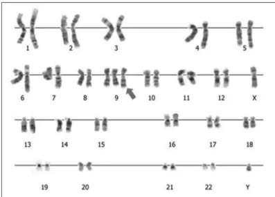
Cytogenetic analysis on GTG-banding karyotypes showed mosaicism between a normal male chromosomal lineage and another with trisomy 9: mos 47,XY,+9[4]/46,XY[50] (Figure 2). he karyotype analysis on ibroblasts was normal: 46,XY[59].
Patient 2
he patient was a two-year-old Caucasian girl, the daughter of parents aged 37 years (mother) and 54 years (father). he gesta-tional and family data were unavailable. An atrial septal defect associated with a ventricular septal defect and patent ductus arte-riosus were diagnosed at birth. She also presented a swallowing disorder and gastroesophageal relux.
Figure 2. Cytogenetic analysis of the patient #1.
*No results were observed using the same search strategies in the Cochrane Library, Lilacs and SciELO databases.
Database* Search strategy Results
PubMed
“Chromosomes, Human, Pair 9” AND “Mosaicism” AND “Trisomy” 42 case reports 11 reviews “Chromosomes, Human, Pair 9” AND “Mosaicism” AND “Trisomy” AND “facial asymmetry” 1 case report “Chromosomes, Human, Pair 9” AND “Mosaicism” AND “Trisomy” AND “thumb” 1 case report
“Chromosomes, Human, Pair 9” AND “Mosaicism” AND “Trisomy” AND “survival” 1 case report 1 review
Scirus
“Chromosomes, Human, Pair 9” AND “Mosaicism” AND “Trisomy” 61 articles “Chromosomes, Human, Pair 9” AND “Mosaicism” AND “Trisomy” AND “facial asymmetry” 1 case report “Chromosomes, Human, Pair 9” AND “Mosaicism” AND “Trisomy” AND “thumb” 1 case report “Chromosomes, Human, Pair 9” AND “Mosaicism” AND “Trisomy” AND “survival” 4 articles
Embase
“Chromosomes, Human, Pair 9” AND “Mosaicism” AND “Trisomy”
20 articles 3 reviews 1 letter 1 conference abstract
1 conference paper “Chromosomes, Human, Pair 9” AND “Mosaicism” AND “Trisomy” AND “facial asymmetry” 0 articles “Chromosomes, Human, Pair 9” AND “Mosaicism” AND “Trisomy” AND “thumb” 0 articles “Chromosomes, Human, Pair 9” AND “Mosaicism” AND “Trisomy” AND “survival” 0 articles Table 1. Results from our review using descriptors for the main features observed in our patients
At a physical examination, at the age of ive months, she pre-sented a height of 61.5 cm (P2-10), weight of 5.3 kg (< P3), head circumference of 40.5 cm (P25), sparse hair, micrognathia, right ear with an overfolded helix and a preauricular pit, long thumbs, sacral dimple and long and tapered toes. Radiographic evaluation of the hands and feet revealed triphalangeal thumbs. he child also had a history of hearing loss (with abnormal auditory evoked potential), signiicant hypotonia and neuropsychomotor delay. Her electroencephalogram was normal, and she did not present seizures. She was using an auditory prosthesis and was being fol-lowed up with physiotherapy and speech therapy.
Cytogenetic evaluation from peripheral blood on GTG-banding karyotypes showed mosaic trisomy 9: mos 47,XX,+9[3]/46,XX[69]. Fibroblast evaluation was not performed.
DISCUSSION
Mosaic trisomy 9 is an infrequent condition associated with limited survival. Our review of the PubMed, Scirus, Embase, Cochrane Library, Lilacs and SciELO databases using speciic descriptors can be seen in Table 1. An additional chromosome 9 in blood tests, which was not detected in other tissues such as the skin,6 as observed with patient 1, or only identiied in ibroblasts,7
has been described in the literature. Schwartz et al., evaluating diferent tissues, such as hepatic, pulmonary and heart cells, demonstrated high variability of the mosaicism found in a single individual with mosaic trisomy 9.8
diferent tissues involved. Moreover, it may be associated with the existence of undetected uniparental disomy in the normal chro-mosomal lineage, which will have occurred during chrochro-mosomal rescue.6 his has only been investigated in a few cases of mosaic
trisomy 9, and in our review, we found only one report with this abnormality.6,7 Additionally, uniparental disomy of chromosome
9 alone has been rarely reported. All the cases were of mater-nal uniparental disomy, and the patients described presented fea-tures of autosomal recessive diseases, such as cartilage-hair hyp-oplasia and Leigh syndrome, for which the genes are located in chromosome 9.9-11
Facial abnormalities are common in cases of mosaic trisomy 9. However, most of the abnormalities observed in our patients difered from those most frequently described in the literature, which include high or narrow forehead, microcephaly, short and upslanting palpebral issures, deep-set eyes, microphthal-mus, epicanthal folds, hypertelorism, broad nasal bridge, bul-bous nose, high arched palate, clet lip or palate, micrognathia and large fontanelles.2 Ear abnormalities, as identiied in our
patients (with the exception of the preauricular pit observed in patient 2), are considered to be very common and have been described in more than 90% of such patients.3 On the other
hand, facial asymmetry caused by hemifacial hypoplasia is an uncommon feature,2,4,6 and this may be related to the
mosa-icism presented by the patients.6 he association of this feature
with ipsilateral microtia observed in patient 1 may also suggest the presence of the phenotype of another condition, the ocu-lo-auriculo-vertebral spectrum (OAVS), also known as hemi-facial microsomia or Goldenhar syndrome. his is considered to be a phenotype characterized by variable clinical and etio-logical features. Although most cases have been sporadic and have not presented any known cause, diferent chromosomal abnormalities have been described in subjects with this pheno-type, and mosaic trisomy 9 is one of them12 (Table 1).
Interest-ingly, the patient described by Willatt et al., with uniparental disomy of chromosome 9 in the normal chromosomal lineage, presented facial asymmetry.6 However, no case of uniparental
disomy of chromosome 9 alone has been reported with these features of OAVS.9-11
Cardiac abnormalities are frequent (around 70% of the cases), and the most common types correspond to those pre-sented by patient 2, i.e. ventricular and atrial septal defects and patent ductus arteriosus. Genitourinary abnormalities afect 73% of the patients, and micropenis and cryptorchidism, as observed in patient 1, are very common among male subjects.3 In
rela-tion to skeletal malformarela-tions, dislocarela-tions and bone absences involving especially the hips, knees, pelvis, ribs, hands and feet are frequent in cases of mosaic trisomy 9.3 Nevertheless, neither
of our patients presented such abnormalities. Our attention was
drawn to the inding of triphalangeal thumbs observed in patient 2, because this was an abnormality that had not previously been described in cases of mosaic trisomy 9 (Table 1) or even in
uni-parental disomy of chromosome 9. In these cases, the impair-ment of the thumbs consisted only of limited abduction. Gastro-intestinal abnormalities are also infrequent, and the swallowing disorder and gastroesophageal relux observed in patient 2 have only been described in a few cases.3
Neuropsychomotor delay and mental retardation are com-mon features acom-mong survivors. However, there are also descrip-tions in the literature of individuals with mosaicism and normal development.3 In our cases, both patients presented
neuropsy-chomotor delay. However, we cannot rule out the possibility that the gestational exposure to tobacco and alcohol and the prema-turity seen in the case of the irst patient may have inluenced his development. Central nervous system abnormalities were not observed in our patients, but abnormalities such as Dandy-Walker malformation have been identiied in trisomy 9 subjects without mosaicism. We did not ind any descriptions of hearing loss in the literature, among patients with mosaic trisomy 9, as observed in patient 2.
Neither of our patients died. However, the mean survival of subjects with mosaic trisomy 9 is still not known, because, as highlighted by other authors,5 a signiicant proportion of the
patients reported with this condition were not dead and were very young at the time of their descriptions (Table 1). Neverthe-less, survival beyond the irst year is considered uncommon.3,5,6 It
seems that patient 1 has good survival prospects, because he does not present major abnormalities such as congenital heart defect or respiratory and gastrointestinal complications. We cannot rule out the possibility that this longer survival may be related to the degree of mosaicism observed or even to the tissue distribution of the trisomy. It is possible that in such cases the trisomic cells may be especially present in “less prime” tissues. In patient 1, for example, the mosaicism was detected only in lymphocytes and not in ibroblasts. Another point to be considered is in relation to the improvement in healthcare for such patients that has come about over the past decades, which may present an inluence on their survival.
CONCLUSION
REFERENCES
1. Moskovitz M, Brener D, Annick RR. Dental management of a child with trisomy 9 mosaicism: a case report. Pediatr Dent. 2006;28(3):265-8. 2. Arnold GL, Kirby RS, Stern TP, Sawyer JR. Trisomy 9: review and report
of two new cases. Am J Med Genet. 1995;56(3):252-7.
3. Wooldridge J, Zunich J. Trisomy 9 syndrome: report of a case with Crohn disease and review of the literature. Am J Med Genet. 1995;56(3):258-64.
4. Cantú ES, Eicher DJ, Pai GS, Donahue CJ, Harley RA. Mosaic vs. nonmosaic trisomy 9: report of a liveborn infant evaluated by luorescence in situ hybridization and review of the literature. Am J Med Genet. 1996;62(4):330-5.
5. Okumura A, Hayakawa F, Kato T, Kuno K, Watanabe K. Two patients with trisomy 9 mosaicism. Pediatr Int. 2000;42(1):89-91.
6. Willatt LR, Davison BC, Goudie D, et al. A male with trisomy 9 mosaicism and maternal uniparental disomy for chromosome 9 in the euploid cell line. J Med Genet. 1992;29(10):742-4.
7. Lindor NM, Michels VV, Jalal S, Shaughnessy W. Trisomy 9 mosaicism in a child with tethered cord. Clin Dysmorphol. 1995;4(2):169-72. 8. Schwartz S, Ashai S, Mejboom EJ, et al. Prenatal detection of trisomy
9 mosaicism. Prenat Diagn. 1989;9(8):549-54.
9. Sulisalo T, Mäkitie O, Sistonen P, et al. Uniparental disomy in cartilage-hair hypoplasia. Eur J Hum Genet. 1997;5(1):35-42.
10. Tiranti V, Lamantea E, Uziel G, et al. Leigh syndrome transmitted by uniparental disomy of chromosome 9. J Med Genet. 1999;36(12):927-8.
11. Slater HR, Ralph A, Daniel A, Worthington S, Roberts C. A case of maternal uniparental disomy of chromosome 9 diagnosed prenatally and the related problem of residual trisomy. Prenat Diagn. 2000;20(11):930-2. 12. Wilson GN, Barr M Jr. Trisomy 9 mosaicism: another etiology for the
manifestations of Goldenhar syndrome. J Craniofac Genet Dev Biol. 1983;3(4):313-6.
Sources of funding: None
Conlict of interest: None
Date of irst submission: October 4, 2010
Last received: December 14, 2010
Accepted: March 10, 2011
Address for correspondence:
Giorgio Adriano Paskulin
Clínica Genética — Universidade Federal de Ciências da Saúde de Porto Alegre e Complexo Hospitalar da Santa Casa de Porto Alegre