23
Revista da Sociedade Brasileira de Medicina Tropical 41(Suplemento II):23-26, 2008
1. Department of Sanitary Dermatology, Eduardo de Menezes Hospital, Hospital Foundation of Minas Gerais State, Belo Horizonte, MG, Brazil. 2. Post-Graduate Program in Health Sciences, Federal University of Minas Gerais, Belo Horizonte, MG, Brazil. 3. State Coordination of Sanitary Dermatology, Minas Gerais State Health Secretariat, Belo Horizonte, MG, Brazil. 4. CEMEPE – Center for Specialized Medical Research and Education, Belo Horizonte, MG, Brazil. 5. KIT Biomedical Research, Royal Tropical Institute, Amsterdam, The Netherlands and Tropical Pathology and Public Health Institute, Federal University of Goiás, Goiânia, Goiás, Brazil.
Address to:Dra.Sandra Lyon. Avenida do Contorno 4852, sala 601, Bairro Funcionários, 30110-032 Belo Horizonte, MG, Brasil.
Phonefax: 55 31 31 3227-0092
e-mail: [email protected]; [email protected]
Association of the ML Flow serological test with slit skin smear
Associação do teste sorológico ML Flow com a baciloscopia
Sandra Lyon
1, Rozana Castorina da Silva
1,2, Ana Cláudia Lyon
1,2,
Maria Aparecida de Faria Grossi
2,3, Sílvia Helena Lyon
1, Maria de Lourdes Azevedo
4,
Samira Bührer-Sékula
5and Manoel Otávio da Costa Rocha
2ABSTRACT
A descriptive, exploratory study was conducted analyzing the association of covariables in the results of the ML Flow serological test and slit skin smear. A total of 60 leprosy cases diagnosed at the state Sanitary Dermatology Referral Center were investigated. Slit skin smear samples were collected from four sites and the results were expressed by the bacillary index. ML Flow was registered in both qualitative and semi-quantitative terms. Cohen’s kappa coefficient was used to study the agreement with Landis and Koch’s observer criteria for interpretation. For statistical analysis, the logistic regression model and Kruskal-Wallis test were used. ML Flow showed a strong association with slit skin smear results, since a gradual increase in BI was accompanied by a semi-quantitative rise in antibody levels measured by ML Flow, with 100% positivity in cases presenting a positive slit skin smear. Given its strong correlation to slit skin smear, the results of this study provide evidence that the ML Flow test could be a valuable auxiliary tool in the classification and treatment of leprosy patients.
Key-words: Leprosy. Serological tests. Slit skin smear. ML Flow.
RESUMO
Realizou-se estudo descritivo e exploratório relacionando as covariáveis aos resultados do teste sorológico ML Flow e baciloscopia. Foram estudados 60 casos novos de hanseníase diagnosticados no Centro de Referência em Dermatologia Sanitária. Para a baciloscopia, foi utilizada a coleta de esfregaço dérmico em quatro sítios, sendo o resultado expresso pelo índice bacilocópico. O ML Flow foi registrado de modo qualitativo e semi-quantitativo. Para o estudo da concordância, foi utilizado o índice de Kappa e, para sua interpretação, os critérios de Landis e Koch. Para análise estatística foram realizados a regressão logística e o teste de Kruskal-Wallis. O ML Flow mostrou forte associação com a baciloscopia, observou-se que o aumento gradativo do índice baciloscópico foi acompanhado pelo aumento semi-quantitativo dos níveis de anticorpos medidos pelo ML Flow, tendo sido positivo em 100% dos casos com baciloscopia positiva. Os resultados deste estudo evidenciaram que o ML Flow, por estar fortemente correlacionado à bacilocopia, poderá tornar-se um valioso instrumento auxiliar na classificação e alocação dos pacientes para fins de tratamento.
Palavras-chaves: Hanseníase. Testes sorológicos. Baciloscopia. ML Flow.
Leprosy (Hansen’s disease) is an infectious disease caused by Mycobacterium leprae and its diagnosis is based on the identification of classic symptoms. It is classified as paucibacillary (PB) or multibacillary (MB) for treatment purposes2.
The slit skin smear is the most important laboratorial test used to identify the causal agent, although it is negative for the PB forms and in some MB cases. In addition, laboratory infrastructure and trained professionals are required for its proper execution and these conditions are often absent in primary healthcare centers2.
The phenolic glycolipid-1 (PGL-1) antigen is Mycobacterium
leprae-specific and leads to the formation of IgG and IgM
antibodies. Correlation exists between IgM titers, leprosy clinical forms and disease activity1. In the lepromatous leprosy, high
levels of anti-PGL-1 have been obtained that tend to decrease with specific treatment3.
ML Flow is an immunochromatographic test that detects IgM antibodies and can assist in the classification of PB and MB leprosy patients by detecting the patient’s bacterial load3 6. It is a
simple, low-cost and easily-conducted test that does not require a laboratory3 and can be used routinely in health centers, including
primary care units6.
For treatment purposes, the classification of leprosy patients
should be based on the number of bacilli present in the body5.
The relation between antibody levels and the bacteriological index (BI) indicates that serology can be used as an alternate technique for patient classification1 7 8.
The combination of clinical and laboratory criteria, more specifically those of serological tests to detect specific antibodies
against the phenolic glycolipids of Mycobacterium leprae, may
be used as an auxiliary test for the correct classification of leprosy patients6. Previous studies have shown that the results of
24
Revista da Sociedade Brasileira de Medicina Tropical 41(Suplemento II):23-26, 2008
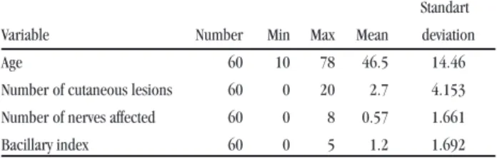
TABLE 1
Descriptive statistics of the 60 new leprosy cases treated at Eduardo de Menezes Hospital. Brazil. 2006.
Standart Variable Number Min Max Mean deviation
Age 60 10 78 46.5 14.46
Number of cutaneous lesions 60 0 20 2.7 4.153 Number of nerves affected 60 0 8 0.57 1.661
Bacillary index 60 0 5 1.2 1.692
TABLE 2
Agreement between slit skin smear and the ML Flow serologic test of the 60 new leprosy cases treated at Eduardo de Menezes Hospital, Brazil, 2006.
Slit skin smear
positive negative total
no % no % no %
ML Flow Positive 24 100.0 18 49.0 42 70.0
Negative - - 18 51.0 18 30.0
Total 24 100.0 36 100.0 60 100.0
kappa coefficient = 0.44, p-value = <0.001
TABLE 4
Distribution of ML Flow results by BI in 60 new leprosy cases treated at Eduardo de Menezes Hospital. 2006.
Bacillary ML Flow test Total
Index 0 +1 +2 +3 +4 no %
0.0 18 9 2 5 2 36 60.0
0.1 – 0.9 0 1 1 0 1 3 5.0
1.0 – 1.9 0 1 2 0 1 4 6.7
2.0 – 2.9 0 0 0 1 3 4 6.7
3.0 – 3.9 0 0 0 1 4 5 8.3
4.0 – 4.9 0 1 0 0 5 6 10.0
5.0 – 5.9 0 0 1 0 1 2 3.3
6.0 0 0 0 0 0 0 0
Total no 18 12 6 7 17
% 30 20 10 11.7 28.3 60 100.0
The aim of this work was to study the relation between antibody levels detected by the ML Flow serological test and the bacterial load, as determined by the bacilloscopic index (BI).
CASUISTIC AND METHODS
The study population consisted of 60 new leprosy cases diagnosed at the dermatology clinic of the Eduardo de Menezes Hospital or referred to this clinic during the period of March to December 2006. The research subjects volunteered to take the ML Flow serological test after signing a free informed consent form, as per the document emitted by the Research Ethics Committee of the Eduardo de Menezes Hospital on 12 April 2006.
The 60 patients were classified according to the World Health Organization (WHO) operational guidelines, considering the number of skin lesions. For the slit skin smear, the BI was determined through the collection of samples taken from four sites: a skin lesion, the opposite-side elbow and earlobes and scored according to Ridley’s logarithmic scale, ranging from zero to six. The ML Flow test was registered qualitatively (positive or negative) and semi-quantitatively (zero, 1+, 2+, 3+
or 4+) according to Bührer-Sékula et al3. Data were collected
on the patient information form for posterior analysis included identification data, number of skin lesions, number of thickened nerves, slit skin smear results and ML Flow results.
Statistical analysis of the data was performed using the ordinal logistic regression model9 and the Kruskal-Wallis test10. For the
agreement analysis, Cohen’s kappa coefficient was used, as well as Landis and Koch’s interpretation criteria.
RESULTS
Patient age varied from 10 to 78 years with an average of 46.5 years. The number of skin lesions varied from 0 to 20 and thickened nerves from 0 to 8. The average BI was 1.2 with a
standard deviation of 1,692 (Table 1).
Seropositivity occurred in 70% of patients, while the slit skin
smear was positive in 40% (Table 2). The agreement observed
between the serological results and slit skin smear was 70%, which is considered moderate (Kappa = 0.44), according to the Landis and Koch criteria. Observation revealed that 18 patients, 50% of the cases with a negative slit skin smear, were seropositive. Among patients with a positive slit skin smear, all were ML Flow-positive.
Table 3 presents the multiple analyses of the factors associated with the seropositivity of the ML Flow test, slit skin smear and the number of cutaneous lesions. In the interpretation of this model, patients presenting a positive slit skin smear had approximately a 19-fold greater chance of testing positive on the ML Flow (OR: 19.38) compared to a patient with a negative slit skin smear result. Patient with 6 or more skin lesions had a 6-fold greater chance (OR: 6.04) of a positive ML Flow compared to patients with 5 lesions or less.
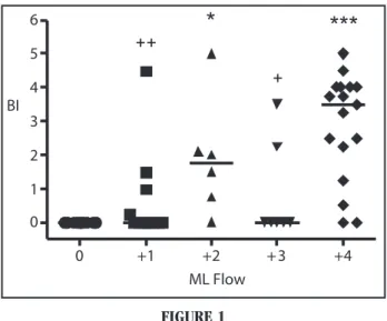
The categorized BI was associated with the serological levels
of the ML Flow, as shown in Table 4 and Figure 1. Among the
patients studied, 60% showed a negative BI and 28.3% presented a BI of 2 or higher, with 18.3% of cases within the BI range of 3.0 to 4.9. Observation also showed that the slit skin smear results were associated with the ML Flow test results.
TABLE 3
Multiple analysis of the factors associated to seropositivity in the ML Flow test of 60 new leprosy cases treated at the Eduardo de Menezes Hospital. Brazil.2006. Variables ML Flow (+) Odds CI (95%) P-value
no % ratio
Slit skin smear
negative 18 42.9 1.0 - 0.007
positive 24 57.1 19.38 (2.28; 164.96) Number of skin lesions
< 5 lesions 23 54.8 1.0
25
Lyon S et al. Association of the ML Flow serological test with slit skin smear0 +1 +2 + 3 +4
0 1 2 3 4 5
6
*
***
++
+
BI
ML Flow
FIGURE 1
Results of the bacillary index according to the semi-quantitative results of the ML Flow test of new leprosy cases treated at Eduardo de Menezes Hospital, Brazil, 2006. 0: N=18; +1: N=12; +2: N=6; +3: N=7; +4: N=17. Statistical analysis was conducted using the Kruskal-Wallis test, followed by Dunn’s post-hoc test for multiple comparisons. *p<0.05; ***p<0.0001 (vs. 0); +p<0.05; ++p<0.01 (vs. +4).
Patients that presented a high BI had a 19-fold chance of presenting a ML Flow serological test in the highest category (4+) compared to those patients with lower BI values (Table 3). Even patients with a low BI can present a high response to the ML Flow test (Table 4 and Figure 1).
Figure 1 shows the association of semi-quantitative results with ML Flow and BI, including the Kruskal-Wallis statistical analysis.
DISCUSSION
Seropositivity in this study was higher (70%) than that witnessed by Lyon (57%)7 8 and Grossi (50.8%)6, and higher than from Nepal
(35.6%)4 and Nigeria (62.9%)4. These studies demonstrate that
the level of antibodies specific to the phenolic glycolipid antigens of
Mycobacterium leprae correlate with the bacterial load of leprosy patients4 6 7 8. The majority of patients classified as MB have high levels
of anti-PGL-1 IgM antibodies, as opposed to those classified as PB, who are generally seronegative. The level of these antibodies is directly correlated to the quantity of Mycobacterium leprae in patients and diminishes throughout the treatment period5. In the current study, a
moderate agreement (kappa: 0.44) was observed between slit skin smear and ML Flow results (Table 2); similar results were obtained by Grossi (Kappa: 0.48)6 and Lyon (Kappa: 0.49)7 8. It should be
emphasized that 18 (50%) of the patients who presented a negative slit skin smear were seropositive, meaning that they would be classified as PB if slit skin smear were used as the only criterion for classification. This fact suggests that a serological test may be more sensitive than the slit skin smear for the detection of true MB cases. In contrast, serology was positive in 100% of patients with a positive slit skin smear (Table 4). In the study by Bührer-Sékula et al3, ML Flow was positive
in 97.4% of MB cases and in 97.8% of patients with a BI ≥ 2. The association between BI and ML Flow indicated that as the BI increases, the results from the serological test also rise, as previously demonstrated by Lyon (2005)7 8.
This study provides evidence of a strong association between the ML Flow serological test and slit skin smear. It also showed that the ML Flow test was capable of detecting seropositivity in half of the patients showing a negative BI, which is of great importance, principally for professionals working in primary healthcare units who would be more confident in classifying leprosy patients, given that their results are closely related to field observations.
CONCLUSION
A statistically significant association was established between the results of the ML Flow serological test and slit skin smear. The study showed that the incorporation of the ML Flow test could be an auxiliary tool in the classification of leprosy patients for treatment purposes in an important number of cases. This may prevent the possibility of insufficient treatment in the case of clinically-classified PB cases with a positive serology and excessive treatment in the case of patients diagnosed as MB who are seronegative.
ACKNOWLEDGEMENTS
The authors would like to thank the State Health Secretariat of Minas Gerais (Belo Horizonte, Brazil) and KIT Biomedical Research Department (Amsterdam, the Netherlands) for their technical support and Netherlands Leprosy Relief (Amsterdam, Netherlands) for financial backing. The NT-P-BSA used in the production of the ML Flow serological test was kindly provided by Dr. Fujiwara of the Institute for Natural Science, Nara University, Nara, Japan.
REFERENCES
1. Barros RPC, Oliveira MLWDR. Detecção de anticorpos específicos para antígeno Glicolipide Fenólico-1 do M. leprae (anti-PGL-1IgM): aplicações e limitações. Anais Brasileiros de Dermatologia 75: 745-753, 2000.
2. Brasil. Ministério da Saúde. Guia para o controle da hanseníase. Cadernos de Atenção Básica, no 10, Brasília, 2002.
3. Bührer-Sékula S, Smits HL, Gussenhoven GC, Leeuwen J, Amador S, Fujiwara T, Klatser PR, Oskam L. Simple and fast lateral Flow Test for classification of leprosy patients and identification of contacts with high risk of developing leprosy. Journal of Clinical Microbiology 41: 1991-1995, 2003.
4. Bührer-Sékula S, Visschedijk J, Grossi MAF, Dhakal KP, Namadi AU, Klaster PR, Oskam L. The ML FLOW test as a point of care test for leprosy control programmes: potential effects on classification of leprosy patients. Leprosy Review 78: 70-79, 2007.
5. Gallo MEN, Ramos Jr LAN, Albuquerque ECA, Nery JAC, Sales AM. Alocação do paciente hanseniano na poliquimioterapia: correlação da classificação baseada no número de lesões cutâneas com os exames baciloscópicos. Anais Brasileiros de Dermatologia 78: 415-424, 2003.
6. Grossi MAF. Estudo das possíveis mudanças na classificação da hanseníase com utilização do Teste ML Flow e suas implicações no tratamento e controle da endemia em Minas Gerais. Tese de Doutorado. Universidade Federal de Minas Gerais, Belo Horizonte, 2005.
26
Revista da Sociedade Brasileira de Medicina Tropical 41(Suplemento II):23-26, 2008
8. Lyon S, Lyon AC, Castorina-Silva R, Grossi MAF, Lyon SH, Bührer-Sékula S, Rocha MOC. A comparison of ML Flow serology and slit skin smears to assess the bacterial load in newly diagnosed leprosy patients in Brazil. Leprosy Review 79: 1-9, 2008.
9. Mccullagh P. Regression models for ordinal data. Journal of the Royal Statistical Society, 42: 109-142, 1980.
10. Zar JH. Bioestatistical analysis. 2nd edition. Englerwood Chiffs: Printice-Hall.