Clinical and histological features of patients with
membra-noproliferative glomerulonephritis classified by
immunoflu-orescence findings
Características clínicas e histológicas de pacientes com
glome-rulonefrite membranoproliferativa classificados por achados de
imunofluorescência
Authors
Cristiane Bitencourt Dias 1 Leonardo Testagrossa 2 Lectícia Jorge 1 Denise Malheiros 2
Viktoria Woronik 1
1 Universidade de
São Paulo, Escola de Medicina, Hospital das Clínicas, Departamento de Nefrologia, São Paulo - SP, Brazil.
2 Universidade de São
Paulo, Escola de Medicina, Hospital das Clínicas, Departamento de Patologia, São Paulo - SP, Brazil.
Submitted on: 01/11/2017. Approved on: 03/29/2017.
Correspondence to: Cristiane Bitencourt Dias. E-mail: cristianebitencourt@ uol.com.br
I
NTRODUCTIONMembranoproliferative glomerulone-phritis (MPGN) diagnosis occurred in 79 (4.2%) patients out of 1849 with glomeru-lonephritis enrolled in Paulista Registry of Glomerulopathies from 1999 to 2005.1 This
glomerulopathy is more often associated with secondary causes, especially infection
DOI: 10.5935/0101-2800.20170078
Background: New classification for
mem-branoproliferative glomerulonephritis has been proposed in the literature. The aim of this study was to compare the clini-cal, biochemiclini-cal, etiology and renal bi-opsy findings of these patients grouped by immunofluorescence as proposed by the new classification. Methods: Patients
with renal biopsy-proven membranopro-liferative glomerulonephritis unrelated to systemic lupus erythematosus, diagnosed between 1999 and 2014. The patients were divided according to immunofluo-rescence: Immunoglobulin positive group, C3 positive only and negative immuno-fluorescence group. Results: We evaluated
92 patients, the majority of which were in the immunoglobulin positive group. Infectious diseases, hepatitis C virus and schistosomiasis, were the most frequent etiology. A negative immunofluorescence group had more vascular involvement in renal biopsy compare with others groups.
Conclusions: The only difference between
the groups was higher vascular involve-ment in renal biopsy in negative immu-nofluorescence group. These new classifi-cation was satisfactory for the finding of etiology in one part of the cases.
ABSTRACT
Keywords: complement system proteins; epidemiology; glomerulonephritis, mem-branoproliferative; microscopy, fluores-cence.
Introdução: Uma nova classificação para
glomerulonefrite membranoproliferativa foi proposta na literatura. O objetivo deste estu-do foi comparar os achaestu-dos clínicos, bioquí-micos, etiológicos e da biópsia renal desses pacientes agrupados por imunofluorescên-cia, conforme proposto pela nova classifi-cação. Métodos: Pacientes com
glomerulo-nefrite membranoproliferativa comprovada por biópsia renal, não relacionada ao lúpus eritematoso sistêmico, diagnosticados entre 1999 e 2014. Os pacientes foram divididos de acordo com a imunofluorescência: grupo positivo por imunoglobulina, grupo positivo por C3 apenas e grupo com imunofluores-cência negativa. Resultados: avaliamos 92
pacientes, a maioria dos quais estava no gru-po de imunoglobulina gru-positiva. Doenças in-fecciosas, o vírus da hepatite C e a esquistos-somose, foram as etiologias mais frequentes. Um grupo com imunofluorescência negativa apresentou maior comprometimento vas-cular na biópsia renal quando comparado com os outros grupos. Conclusões: a única
diferença entre os grupos foi o maior envol-vimento vascular na biópsia renal no grupo de imunofluorescência negativa. Esta nova classificação foi satisfatória para a descober-ta de etiologia em uma parte dos casos.
R
ESUMOPalavras-chave: epidemiologia; glomeru-lonefrite, membranoproliferativa; pala-vra; microscopia, fluorescência; proteínas do sistema complementar.
by hepatitis C virus (HCV) and autoim-mune diseases such as systemic lupus ery-thematosus (SLE) and rheumatoid arthritis.2
However Little et al.3 identified 34% of
Histologically, MPGN is characterized by mesan-gial proliferation, matrix expansion and its interpo-sition between the endothelium and the glomerular basement membrane, giving the capillary loops a dou-ble-contour appearance. In classical studies MPGN was categorized in three types, according to the loca-tion of the electron-dense deposit on electron micros-copy: type I (subendothelial deposits); type II (dense homogeneous intramembranous deposits); type III (a variant of type I, with subepithelial and subendothe-lial deposits). Type I was associated with HCV infec-tion, whereas type II affects younger individuals and was not related to systemic causes.4
Sethi and Fervenza5 proposed a new system of
classifying MPGN based on immunofluorescence (IF), defining two groups: MPGN with immuno-globulin deposition on IF that could be associated to autoimmune diseases, infections, or monoclonal gam-mopathy, whereas MPGN without immunoglobulin deposition but with C3 deposition on IF that is clas-sified as dense deposit disease (DDD) or C3 glomeru-lonephritis after electron microscopy examination. In other study, MPGN with none deposition seen on IF was introduced with other group and could be sec-ondary to membrane reactions, as in thrombotic mi-croangiopathy.6 In this new classification the use of
electron microscopy is mandatory in cases of exclu-sive C3 deposition, in order to differentiate DDD and C3 glomerulonephritis.5
The new classification proposed by Sethi and Fervenza5 emphasizes the various mechanisms
in-volved in the pathogenesis of MPGN, drawing dis-tinctions between MPGN mediated by immune com-plexes, which had immunoglobulin deposition, and MPGN resulting from abnormalities of the alterna-tive complement pathway with C3 deposition only. Some authors believe that idiopathic forms should be very rare emphasizing complement and genetic stud-ies in these patients in order to clear their diagnosis.7
C3 glomerulopathy defined after Sethi and Fervenza5 as a MPGN with exclusive C3 deposition
allows pathological discussion. Pickering et al.8
pro-posed a less restrictive definition with deposition of dominant C3 at least two orders of magnitude more intense than any immunoglobulin. In doing so most cases of immune complex diseases would be excluded.
Because of recent proposed classification, we aimed to conduct a retrospective single center study of MPGN patients grouped by the new IF-based
classification to compare clinical and biochemical characteristics, renal biopsy dates and follow up of patients, as well as the etiology in each group.
M
ETHODSSTUDY DESIGN
This was a retrospective study of patients diagnosed by renal histology with MPGN between 1999 and 2014 in the Nephrology Department of the Hospital das Clínicas of the School of Medicine of University of São Paulo, in São Paulo Brazil. Clinical and bio-chemical characteristics were retrieved from clinical charts on admission and during follow-up.
Patients with SLE based on classical criteria9,10
were excluded, as were those for whom there were in-sufficient data from the renal biopsy or on the patient chart at diagnosis.
BIOCHEMICALANDCLINICALCHARACTERISTICS
At diagnosis were analyzed the following parameters: 24-h proteinuria, measured by the automated colo-rimetric method; urinalysis; serum creatinine, mea-sured by the kinetic colorimetric method; glomerular filtration rate (GFR), calculated by the Modification of Diet in Renal Disease equation;11 C3 and C4
com-plement fractions, measured by immunoturbidimetry, with reference ranges of 90-180 mg/dL and 10-40 mg/ dL, respectively; serology for hepatitis B virus (HBV), HCV, and HIV; antinuclear factor; rheumatoid fac-tor, serum proteins, measured by immunofixation. We assessed those same parameters at the end of fol-low-up, defined as the last visit to the facility.
Hematuria was defined > 10 red blood cells per field in two first morning urine specimens. Hypertension was defined as arterial blood pressure > 140/90 mmHg in two measurements on different days12 or previous use of antihypertensive drugs,
re-gardless of blood pressure levels.
RENALBIOPSY
deposition was defined as a score of 1+ or more for any type of immunoglobulin.
The so-called “full-house” immunofluorescence pattern was defined as concomitant deposition of three immunoglobulins and two complements. In the tubulointerstitial compartment, we assessed fibrosis or atrophy, categorized by the proportional involve-ment of the sample: ≥ 50%; ≤ 10%; or 11-49%. Vascular compartment was evaluated by the presence of any kind of involvement.
STATISTICALANALYSISANDETHICALSTATEMENTS
Continuous variables were expressed as mean ± stan-dard deviation for samples with normal distribution or as median and interquartile range for those with-out, whereas categorical variables were expressed as absolute and relative frequencies. Differences between the three groups for continuous variables were deter-mined by analysis of variance or by the Kruskal-Wallis test, as appropriate. For the analysis of categorical variables, we used the chi-square test. Correlations were obtained by Pearson’s or Spearman’s correla-tion analysis, as appropriate. Values of p < 0.05 were considered statistically significant and all analysis was performed in Prism version 5.0 software.
The study was based exclusively on obtaining the records of patients’ data, so we consider the approval of the Nephrology department as sufficient for its realization.
R
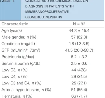
ESULTSAfter excluding patients with lupus nephritis, we ob-tained a sample of 100 patients resting 92 for baseline analysis after exclusion of 3 patients lacking biochem-ical data and 5 lacking IF analysis. Therefore, the final sample comprised 92 patients aged 44.3 ± 15.4 years, 62% male and 38% female. Other characteristics at diagnosis are shown in Table 1. Of note are hematu-ria in 71.7% of cases, hypertension in 55.4%, mean proteinuria of 6.2 ± 3.4 g/day, mean serum albumin of 2.5 ± 0.6 mg/dL, median GFR of 41.5 mL/min/1.73 m2, and serum creatinine of 1.8 mg/dL. After
comput-ing missed data C3 levels in 7 patients and C4 in 6 patients, low levels of C3 were present in 47.8% and for C4 in 31.5%.
ANALYSIS OF GROUPS BY IMMUNOFLUORESCENCE
FINDINGS
Using the new, IF-based MPGN classification
sys-Characteristic N = 92
Age (years) 44.3 ± 15.4
Male gender, n (%) 57 (62.0)
Creatinine (mg/dL) 1.8 (1.3-3.5)
GFR (mL/min/1.73m2) 41.5 (20.0-58.7) Proteinuria (g/day) 6.2 ± 3.2
Serum albumin (g/dL) 2.5 ± 0.6
Low C3, n (%) 44 (47.8)
Low C4, n (%) 29 (31.5) Low C3 and C4, n (%) 25 (27.1)
Arterial hypertension, n (%) 51 (55.4)
Hematuria, n (%) 66 (71.7)
TABLE 1 CLINICALANDBIOCHEMICALDATAON DIAGNOSISINPATIENTSWITH MEMBRANOPROLIFERATIVE GLOMERULONEPHRITIS
The values for continuous variables are expressed as the mean ± SD. or as the median (interquartile range).
GFR, glomerular filtration rate (as estimated with the Modification of Diet in Renal Disease equation).
immunoglobulin positive group, 9 (9.7%) were in the C3 positive group, and 10 (10.8%) were in the IF neg-ative group. Clinical and biochemical data collected at diagnosis showed no differences among the three groups for any of the clinical or biochemical param-eters evaluated (Table 2).
All groups at diagnosis showed low GFR (immu-noglobulin positive 41.0 (20.0-65.0) mL/min/1.73m2 vs. C3 positive 42.0 (15.5-50.0) mL/min/1.73m2vs. IF
negative 39.5 (11.2-53.5) mL/min/1.73m2, p = 0.84)
and nephrotic proteinuria (immunoglobulin positive 6.3 ± 3.4 g/day vs. C3 positive 6.0 ± 3.0 g/day vs.
IF negative 5.5 ± 3.5 g/day, 0.91). Immunoglobulin positive group had more patients with hypertension but with no statistical difference between the other groups and the IF negative group had 100% of cases with hematuria (Table 2). Immunoglobulin positive group showed missed data for C3 levels in 6 patients and for C4 in 6 patients while only one patient in the IF negative group showed missed C3 data.
Table 3 shows associations of MPGN and systemic diseases. Infectious diseases predominate in the three groups with 22 patients in Immunoglobulin positive, 3 in C3 positive and 3 in IF negative.
Parameter
Immunoglobulin
positive C3 positive IF negative p-value
(n = 73) (n = 9) (n =10)
Age (years) 44.9 ± 14.6 44.6 ± 20.0 43.8 ± 15.0 0.44
Male gender, n (%) 45 (61.6) 6 (66.6) 7 (70.0) 0.57
Creatinine (mg/dL) 1.8 (1.1-3.6) 1.8 (1.3-5.4) 2.1 (1.6-5.1) 0.45
GFR (mL/min/1.73m2) 41.0 (20.0-65.0) 42.0 (15.5-50.0) 39.5 (11.2-53.5) 0.84
Proteinuria (g/day) 6.3 ± 3.4 6.0 ± 3.0 5.5 ± 3.5 0.91
Serum albumin (g/dL) 2.5 ± 0.6 2.5 ± 0.3 2.7 ± 0.5 0.22
Low C3, n (%) 35 (52.2) 4 (44.4) 5 (50.0) 0.63
Low C4, n (%) 23 (34.3) 2 (22.2) 4 (40.0) 0.70
Low C3 and C4, n (%) 18 (26.8) 2 (22.2) 3 (30.0) 0.91
Arterial hypertension,
n (%) 41 (56.0) 2 (22.2) 3 (30.0) 0.06
Hematuria, n (%) 51 (70.0) 5 (55.5) 10 (100.0) 0.07
TABLE 2 CLINICALANDBIOCHEMICALPARAMETERSINTHETHREEGROUPSONDIAGNOSIS
The values for continuous variables are expressed as the mean ± SD. or as the median (interquartile range).
ns, not significant; GFR, glomerular filtration rate (as estimated with the Modification of Diet in Renal Disease equation).
Disease
Immunoglobulin
positive C3 positive IF negative p-value
(n = 73) (n = 9) (n = 10)
Infection, n (%) 22 (30.1) 3 (33.3) 3 (30.0) 0.98
Autoimmune disorder,
n (%) 8 (11.0) 0 (0.0) 1 (10.0) 0.57
Dysproteinemia, n (%) 3 (4.1) 1 (11.1) 2 (20.0) 0.14
Other, n (%) 4 (5.5) 1 (11.1) 0 (0.0) 0.56
None, n (%) 36 (49.3) 4 (44.4) 4 (40.0) 0.83
TABLE 3 SYSTEMICDISEASESASSOCIATEDWITHMEMBRANOPROLIFERATIVEGLOMERULONEPHRITISONDIAGNOSIS
present in 8 patients of the immunoglobulin positive group: autoimmune hepatitis (n = 1), Takayasu arte-ritis (n = 1), antiphospholipid syndrome (n = 1) and “lupus like nephritis” defined as MPGN pattern with “full-house” deposits not meeting criteria for lupus was found in 5 patients. Monoclonal gammopathy or dysproteinemia was found in 3 patients of immuno-globulin positive group: lymphoma (n = 1), chronic lymphocytic leukemia (n = 1) and myeloma (n = 1). Furthermore, in 4 patients, there were associations with diseases rarely reported in MPGN: melanoma (n = 1), spherocytosis (n = 1) and alcoholic cirrhosis (n = 2).
In the C3 positive group, there was an association with HCV in 2 patients, HBV in 1, lymphoma in 1, and alcoholic cirrhosis in 1. Among the patients in the IF negative group, there was association with HCV in 2 patients, schistosomiasis in 1, essential cryoglobuli-nemia in 1, myeloma in 1, and Castleman’s syndrome in 1.
The most common associated disease in MPGN patients was HCV infection observed in 14 (15.2%) patients. Excluding those co-infected with HBV (n = 1) and HIV (n = 2), 11 patients with mono infec-tion with HCV were analyzed. Their mean age was 45.8 ± 10.9 years, 63.6% had hypertension, males predominate (63.6%), mean proteinuria was 5.9 ± 2.5 g/day, mean serum albumin was 2.7 ± 0.6 g/dL, median serum creatinine was 2.3 mg/dL (1.1-8.0 mg/ dL), and mean GFR was 33 mL/min/1.73 m2 (6-71
mL/min/1.73 m2). In addition, 81.8% had hematuria,
54.5% had cryoglobulinemia and 80.0% of those tested for C3 and C4 (10 patients) showed low levels of C3 or C4.
the C3 positive group, and 2 were in the IF negative group. After a mean follow-up of 81.6 months, 2 pa-tients had progressed to dialysis.
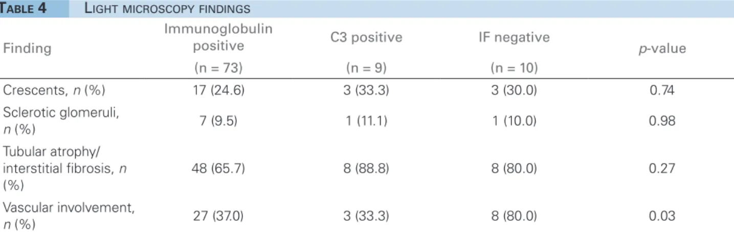
RENALHISTOLOGY
Crescents were observed in 24.6%, 33.3%, and 30.0% of the patients in the immunoglobulin posi-tive, C3 posiposi-tive, and IF negative groups, respec-tively, without statistical difference between them (Table 4). The vast majority showed crescents in less than 10% of the glomeruli, except for two patients, one with HCV who showed crescents in 18% and another with idiopathic MPGN who showed crescents in 44%.
SCLEROTICGLOMERULI
Sclerotic glomeruli were found in 9.5%, 11.1%, and 10.0% of the patients in the immunoglobulin posi-tive, C3 positive and IF negative groups, respectively, without statistical difference between them (Table 4).
TUBULOINTERSTITIALINVOLVEMENT
The vast majority of patients showed some tubu-lar atrophy and interstitial fibrosis, which was ob-served in 65.7%, 88.8%, and 80.0% of the patients in the immunoglobulin positive, C3 positive, and IF negative groups, respectively, without statistical difference between them (Table 4). The proportion of patients showing severe (> 50%) impairment was similar in all three groups: 7.6%, 12.5%, and 10.0%, respectively.
VASCULARINVOLVEMENT
The only findings of vascular involvement were inti-mal fibrosis and hyalinization of the arteriolar wall and they were observed in 37.0%, 33.3%, and 80.0% of the patients in the immunoglobulin positive, C3
Finding
Immunoglobulin
positive C3 positive IF negative p-value
(n = 73) (n = 9) (n = 10)
Crescents, n (%) 17 (24.6) 3 (33.3) 3 (30.0) 0.74
Sclerotic glomeruli,
n (%) 7 (9.5) 1 (11.1) 1 (10.0) 0.98
Tubular atrophy/
interstitial fibrosis, n
(%)
48 (65.7) 8 (88.8) 8 (80.0) 0.27
Vascular involvement,
n (%) 27 (37.0) 3 (33.3) 8 (80.0) 0.03
TABLE 4 LIGHTMICROSCOPYFINDINGS
positive and IF negative groups, respectively, show-ing a statistical difference (p = 0.03) corresponding to more vascular involvement in IF negative group (Table 4).
IMMUNOFLUORESCENCEFINDINGS
Table 5 describes the immunofluorescence findings in the immunoglobulin positive group. IgG was present in 43 patients and was the only immunoglobulin in 6, associated with IgM in 18, associated with IgA in 4, associated with IgM and IgA in 3 and associated with C3 in 12 patients. In 3 patients there was deposition of IgM alone while in 19 IgM deposited with C3 and in one patient IgM deposited with IgA. In 2 patients the only immunoglobulin deposited was IgA together with C3 deposition. The “full-house” pattern was ob-served in 5 patients.
FOLLOW-UP
Twenty four patients from immunoglobulin positive group were lost to follow-up. Among the remaining 49 patients, mean follow-up period was 76.7 ± 52.0 months and 14 patients (28.5%) progressed requir-ing renal replacement therapy while 35 patients ended with mean serum creatinine of 1.5 ± 1.1 mg/dL.
In the C3 positive group, 4 patients were lost to follow-up and the remaining 5 patients had been fol-lowed for an average of 29.2 ± 30.8 months. Four (80%) of those 5 patients required renal replacement therapy, and the remaining patient ended with serum creatinine level of 1.5 mg/dL.
Deposition n = 73
IgG, n (%) 6 (8.2)
IgG + IgM, n (%) 18 (24.7)
IgG + IgA, n (%) 4 (5.5)
IgG + IgM + IgA, n (%) 3 (4.1) IgG + C3, n (%) 12 (16.5)
IgM, n (%) 3 (4.1)
IgM + IgA, n (%) 1 (1.4)
IgM + C3, n (%) 19 (26)
IgA + C3, n (%) 2 (2.7) Full-house pattern, n (%) 5 (6.8)
TABLE 5 IMMUNOGLOBULINANDCOMPLEMENT DEPOSITIONINIMMUNOGLOBULINPOSITIVE GROUP
D
ISCUSSIONOver the 14-year period analyzed, 100 patients were diagnosed with MPGN unrelated to lupus at our facility and 92 fulfilled the criteria for analysis. Immunofluorescence assay revealed that the vast ma-jority (79.3%) of the MPGN patients in our sample showed immunoglobulin deposition, whereas 9.7% showed C3 deposition without immunoglobulin and 10.8% showed no deposition of immunoglobulins or complements. Infection, mainly by HCV, was the most prevalent associated disease in all groups while schistosomiasis was the second most prevalent in im-munoglobulin positive group.
Surprisingly, in spite of the small sample of C3 positive group over immunoglobulin positive, associ-ated infections on diagnosis of MPGN were propor-tionally distributed. We can speculate that in those patients infections could have triggered dysregulation of complement cascade that is known as the basis of pathogenesis of C3 glomerulopathy. Otherwise, as expected, autoimmune disease was absent from C3 positive group. There is scarce literature data on anal-ysis of MPGN patients according to the new classifi-cation. Woo et al.13 described complement mediated
MPGN in 2 patients (4.3%) and immune-mediated MPGN in 39 patients (95.7%) out of 44 diagnosed with MPGN, not distant from our results that shows a predominance of the group with immune complexes.
Considering MPGN associated to HCV, na Italian paper14 showed a 20% association while in our sample
it was 15%. Cryoglobulinemia was present in 54.5% of our HCV positive patients while in literature there are reports 77.7% of incidence.15 Cryoglobulinemia
in HCV negative and without any other systemic dis-ease besides MPGN was found in only one patient in our casuistic.
Considering autoimmune disease we found asso-ciation with MPGN in 8 patients out of 92 evaluated. Seven were in the immunoglobulin positive group and 5 of them showed a lupus-like immunofluores-cence pattern (full house) negatively tested for lupus antibodies and without any clinical manifestations of lupus. Zand et al.16 found an association with
autoim-mune diseases other than lupus in 17 (5.5%) patients out of 308 with MPGN diagnosis, the most common being rheumatoid arthritis and Sjögren’s syndrome.
MPGN relates to multiple myeloma, lymphoma, and leukemia besides other paraproteinemias are rare.17 In our sample, 4 patients had also been
di-agnosed with leukemia, lymphoma or myeloma: 3 in the immunoglobulin positive group and 1 in the C3 positive group. Larsen et al.18 described MPGN
related to dysproteinemia patients with negative monoclonal immunoglobulin deposits that were un-masked only after performing immunofluorescence on formalin-fixed paraffin embedded tissue after pro-tease digestion. Thus, they point to the possibility of misdiagnosis in those cases classifying as MPGN with immunofluorescence without immunoglobulin.
In relation to the clinical and laboratory data there was no difference among the three groups, in-dicating that only the IF is able to differentiate the groups. The data of optical microscopy between the three groups showed only difference in relation to vascular involvement, where the negative IF group had higher incidence than the other. MPGN with negative IF although not reported in the original clas-sification systems, comprises 10.8% of our patients, and it should be seen as a group in which reparative process of thrombotic microangiopathy (TMA) is in-volved besides other cause.6 However, in our patients
we could not see any sign of present TMA although associations with systemic disease (infection, dyspro-teinemia and autoimmune disease) were present in 60% of them.
The idiopathic etiology that could be accepted in im-munoglobulin positive group corresponded to 49.3% of out casuistic, we do not know if electron micros-copy could help to find the etiology in this cases.
Progression to ESRD on follow-up was higher in C3 positive group over immunoglobulin positive or IF negative groups (80%, 28.5% and 44.4%, respectively). Nargund et al.19 characterized 51 subjects with
comple-ment dominant MPGN and 20 immunoglobulin domi-nant MPGN showed ESRD and death in 14 patients (27.4%) complement dominant MPGN and in 6 patients (30%) immunoglobulin dominant MPGN during fol-low up with median of 2 years. Study comparing DDD and C3 glomerulonephritis ESRD occurred in 47% of the DDD and 23% of C3 glomerulonephritis in median follow-up of 28 months.20
C
ONCLUSIONOur data correlate with the proposed new classifica-tion Sethi and Fervenza5 in 83.4% of cases. Only in 11
patients who were diagnosed with infectious disease or autoimmune or gammopathy, they were in no immuno-globulin group. However the primary forms (idiopathic) were present in this study by following the guidelines of the new classification. The only clinical and laboratory differences and histology between groups were the larg-est vascular involvement in negative IF group and worse renal survival in the C3 group.
R
EFERENCES1. Malafronte P, Mastroianni-Kirsztajn G, Betônico GN, Romão Jr JE, Alves MAR, Carvalho MF, et al. Paulista registry of glomerulo-nephritis: 5-year data report. Nephrol Dial Transplant. 2006; 21: 3098-3105. DOI: http://dx.doi.org/10.1093/ndt/gfl237
2. Sethi S. Etiology-Based Diagnostic Approach to Proliferative Glomerulonephritis. Am J Kidney Dis. 2014; 63(4): 561-566. DOI: http://dx.doi.org/10.1053/j.ajkd.2013.11.019
3. Little MA, Dupont P, Campbell E, Dorman A and Walshe JJ. Se-verity of primary MPGN, rather than MPGN type, determines re-nal survival and post-transplantation recurrence risk. Kidney Int. 2006; 69: 504-511. DOI: http://dx.doi.org/10.1038/sj.ki.5000084 4. Schena FP and Alpers CE. Membranoproliferative
glomeru-lonephritis, dense deposit disease, and cryoglobulinemic glo-merulonephritis. In: Jurgen Floege, Richard J. Johnson, John Feehally, editors. Comprehensive clinical nephrology. 3rd ed. Philadelphia, PA: MOSBY; 2010. p. 260-69.
5. Sethi S and Fervenza FC. Membranoproliferative Glomerulone-phritis - A New look at an Old Entity. N Engl J Med. 2012; 366: 1119-1131. DOI: http://dx.doi.org/10.1056/NEJMra1108178 6. Mansani N, Jhaveri KD and Fishbane S. Update on
Membrano-proliferative GN. Clin J Am Soc Nephrol. 2014; 9: 600-608. 7. Fervenza FC, Sethi S and Glassock RJ. Idiopathic
membranoproli-ferativa glomerulonephritis: does it exist? Nephrol Dial Transplant. 2012; 27: 4288-4294. DOI: http://dx.doi.org/10.1093/ndt/gfs288 8. Pickering MC, D’Agati VD, Nester CM, Smith RJ, Haas M, Appel
GB, et al. C3 glomerulopathy: consensus report. Kidney Int. 2013; 84(6):1079-89. DOI: http://dx.doi.org/10.1038/ki.2013.377 9. Tan EM, Cohen AS, Fries JF, Masi AT, McShane DJ, Rothfield
NF, et al. The 1982 revised criteria for the classification of sys-temic lupus erythematosus. Arthritis Rheum. 1982; 25: 1271-1277. DOI: http://dx.doi.org/10.1002/art.1780251101 10. Petri M, Orbai AM, Alarcón GS, Gordon C, Merrill JT, Fortin PR,
et al. Derivation and validation of the Systemic Lupus Internatio-nal Collaborating Clinics classification criteria for systemic lupus erythematosus. Arthritis Rheum. 2012 64: 2677-2686.
11. Levey AS, Bosch JP, Lewis JB, Greene T, Rogers N, Roth D. A more accurate method to estimate glomerular filtration rate from serum creatinine: a new prediction equation. Modifica-tion of Diet in Renal Disease Study Group. Ann Intern Med. 1999; 130(6): 461-470. DOI: http://dx.doi.org/10.7326/0003-4819-130-6-199903160-00002
12. James PA, Oparil S, Carter BL. 2014 Evidence-Based Guideline for the Management of High Blood Pressure in Adults Report From the Panel Members Appointed to the Eighth Joint Natio-nal Committee (JNC 8). JAMA. 2013; 18: E1- E14.
13. Woo SA, Young Ju H, Hyo Kwon S, Lee JH, Jeong Choi S, Cheol Han D, et al. Reanalysis of membranoproliferative glo-merulonephritis patients according to the new classification: a multicenter study. Kidney Res Clin Pract. 2014; 33(4):187-91. DOI: http://dx.doi.org/10.1016/j.krcp.2014.07.006
14. Fabrizi F, Pozzi C, Farina M, Dattolo P, Lunghi G, Badalamenti S, et al. Hepatitis C virus infection and acute or chronic glo-merulonephritis: an epidemiological and clinical appraisal. Ne-phrol Dial Transpl. 1998; 13: 1991-1997.
15. Rikako Hiramatsu R, Hoshino J, Suwabe T, Sumida K, Mise K, Hiramatsu R, et al. Membranoproliferative glomerulonephritis and circulating Cryoglobulins. Clin Exp Nephrol. 2014; 18: 88-94. DOI: http://dx.doi.org/10.1007/s10157-013-0810-z 16. Zand L, Fervenza FC, Nasr SH and Sethi S.
Membranoprolife-rative glomerulonephritis associated with autoimmune diseases. J Nephrol. 2014; 27: 165-171. DOI: http://dx.doi.org/10.1007/ s40620-014-0049-0
17. Cambier JF and Ronco P. Onco-Nephrology: Glomerular Di-seases with Cancer. Clin J Am Soc Nephrol. 2012; 7: 1701-1712. DOI: http://dx.doi.org/10.2215/CJN.03770412 18. Larsen CP, Messias NC, Walker PD, Fidler ME, Cornell LD,
Hernandez LH, et al. Kidney Int. 2015; 88(4): 867-73. 19. Nargund P, Kambham N, Metha K, Lafayette RA.
Clinicopa-thological features of membranoproliferative glomerulonephri-tis under a new classification. Clin Nephrol. 2015; 84: 323-30. DOI: http://dx.doi.org/10.5414/CN108619