Braz Dent J 17(3) 2006
Intraosseous schwannoma 255
Braz Dent J (2006) 17(3): 255-258
Intraosseous Schwannoma of
Mandibular Symphysis: Case Report
Suzie Aparecida de LACERDA1
Luiz Guilherme BRENTEGANI1
Adalberto Luiz ROSA2
Marcelo Vinícius Oliveira VESPÚCIO1
Luiz Antônio SALATA2
1Department of Morphology, Stomatology and Physiology,
School of Dentistry of Ribeirão Preto, University of São Paulo, Ribeirão Preto, SP, Brazil
2Department of Oral and Maxillofacial Surgery and Periodontology,
School of Dentistry of Ribeirão Preto, University of Sao Paulo, Ribeirão Preto, SP, Brazil
Schwannoma (neurilemmoma) is a benign neoplasm originated from the neural sheath and occurring most frequently in the head and neck. Intraosseous schwannomas are rare. The mandible is the most common site of occurrence for these lesions. This article reports the case of an intraosseous schwannoma located in the mandibular symphysis of an 11-year-old boy. The lesion was surgically removed and no radiographic evidence of recurrence was observed after 5 years.
Key Words: intraosseous schwannoma, neurilemmoma, mandibular symphysis.
Correspondence: Profa. Dra. Suzie Aparecida de Lacerda, Departamento de Morfologia, Estomatologia e Fisiologia, Faculdade de Odontologia de Ribeirão Preto, Universidade de São Paulo, 14040-904 Ribeirão Preto, SP, Brasil. Tel: 3602-4041. Fax: +55-16-633-0999. e-mail: [email protected]
ISSN 0103-6440
INTRODUCTION
Schwannoma is a benign neoplasm of neuroectodermal derivation that originates from the Schwann cells (cells that cover peripheral nerves) (1-4). This painless slow-growth lesion may develop at any age and is most frequently located in the soft tissues of the head and neck (3,5).
Intraoral schwannomas are rare, representing less than 1% of the benign primary bone tumors (6). The most common site of occurrence is the mandible (7,8). Intraosseous schwannoma of the mandibular symphy-sis is exceptionally rare (9).
This article reports the case of an intraosseous schwannoma located in the mandibular symphysis of a child. The treatment approach is described and findings in the literature are discussed.
CASE REPORT
An 11-year-old boy presented with a slow-growth swelling in the mentum with approximately 3 years of evolution. The patient was symptom free and reported no history of pain and paresthesia. A clinical examination revealed that the area was swollen, round, singular and hard, with no bleeding or cortical plate perforation and was lined with normal mucosa. The permanent mandibular incisors were mispositioned, which was hindering occlusion. The primary incisors were present and the mucosa was intact.
Braz Dent J 17(3) 2006
256 S.A. Lacerda et al.
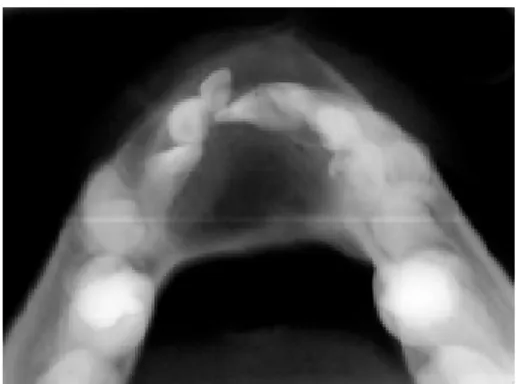
Figure 1. Panoramic radiograph showing a well-defined unilocular radiolucent area in the anterior mandibular region and divergence of the dental roots.
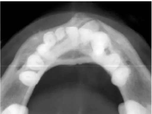
Figure 2. Occlusal radiograph showing expansion of the buccal and lingual cortical plates.
Figure 3. Periapical radiograph showing discrete resorption of the roots of the primary teeth in contact with lesion.
Figure 4. Photomicrograph of the biopsy tissue showing cells with long fusiform palisade nuclei, characteristic of intraosseous Antoni-A schwannoma alternating with homogeneous acellular zones (Verocay bodies). H&E (Original magnification X200). causing root divergence (Fig. 1), expansion of the
buccal and lingual cortical plates (Fig. 2) and discrete resorption of the roots of the primary teeth in contact with mass (Fig. 3). Agenesia of the central permanent incisors was also observed (there was not history of extraction of these teeth).
Under local anesthesia, a buccal mucoperiosteal flap was elevated and a bone window was made to gain access to the tumor. Because of its soft consistency, the tumor could not be removed in toto, and thus lesion remnants were carefully curetted . There were not signs of bone invasion. After adequate hemostasis was ob-tained, the flap was repositioned with 3-0 silk suture.
Histopathologic examination showed neoplastic connective tissue arranged in short fascicles, composed of cells with aligned long nuclei, similar to Schwann
cells. Some of these nuclei formed typical palisades around an acellular eosinophilic area (Verocay bodies) (Fig. 4). Antoni A tissue was the predominant micro-scopic pattern, alternating with Antoni B areas occa-sionally. No signs of hemorrhage, necrosis, calcifica-tion or hyalinizacalcifica-tion were observed. Although some nuclear atypia could be noticed, typical and atypical mitotic activity was absent. The histopathologic diagno-sis was intraosseous schwannoma.
Braz Dent J 17(3) 2006
Intraosseous schwannoma 257
DISCUSSION
Schwannoma rarely occurs in the oral cavity. Intraosseous schwannomas are rare (less than 1%) (1,6), but when they occur, the mandible is the most commonly affected site (7,8). Most cases reported in the mandible had a more posterior location, corre-sponding to the intraosseous course of the inferior alveolar nerve (10,11).
In the case presented in this paper, the schwannoma was probably associated with the periph-eral nervous plexus of the anterior region of the man-dible. In 39 cases of intraosseous schwannoma of the mandible reviewed by Chi et al. (9), 28 were localized on the posterior body/ascending ramus region and 11 involved the anterior body, but only three cases affect-ing the symphysis. The present case is somewhat unusual because of its anterior location (mandibular symphysis).
There are 3 mechanisms by which schwannomas may involve bone: 1) a tumor may arise centrally within the bone, 2) a tumor may arise within the nutrient canal and produce canal enlargement, or 3) a soft tissue or periosteal tumor may cause secondary erosion and penetration into bone (12). In case we hereby report, the third mechanism can be excluded because there was no cortical bone perforation.
The clinical presentation of this case was a painless swelling tumor in the region of mandibular symphysis in a boy on the second decade of life. The radiographic appearance of a well-defined unilocular nonspecific radiolucent lesion, with root divergence (expansive growth) and root resorption only in teeth contacting the lesion, therefore suggestive of a benign
process, had a difficult preoperative diagnosis. The possibility of intraosseous schwannoma was not con-sidered at first because of the extreme rarity of this location. The histopathologic examination provided a definitive diagnosis for the case.
The microscopic aspect was characteristic and easily distinguishable from other lesions. There are two types of tissue arrangement: Antoni-A and Antoni-B. Alternation between Antoni A and B regions is common. Antoni-A type is composed of aligned fusiform cells, forming a typical palisade. Between the fibrils there are small eosinophilic masses called Verocay bodies. Antoni-B type is composed of a smaller number of cells and the spindle cells are randomly arranged within a loose myxomatous stroma (1,13). In this case, Antoni A tissue was the predominant microscopic pattern.
Because it is a well-encapsulated lesion, the treatment of choice for schwannomas is the conserva-tive surgical enucleation with periodic follow-up. Re-currence is uncommon. In the present case, the patient has been followed up for five years with no clinical or radiographic signs of lesion recurrence. The slow (3 years) and expansive growth (root divergence was more accentuated than resorption), the well-delimited borders, the encapsulation and the lack of invasion signs, significant atypia, necrosis and recurrence after 5 years suggest benignity from both clinical and histo-pathological standpoints.
As diagnostic tools, ultrasonography, computed tomography and magnetic resonance imaging may be helpful for estimation of tumor margins as well as infiltration of surrounding structures. Nevertheless, they should not be considered as routine or indispens-able procedures (1).
Figure 5. Panoramic radiograph showing normal bone tissue and
Braz Dent J 17(3) 2006
258 S.A. Lacerda et al.
RESUMO
O schwannoma (neurilemoma) é um neoplasma benigno que se origina da bainha neural e ocorre mais freqüentemente na cabeça e pescoço. Schwannomas intra-ósseos são raros. O local mais comumente afetado é a mandíbula. Este artigo documenta um caso de schwannoma intra-ósseo localizado na sínfise mandibular de um menino com 11 anos de idade. A lesão foi removida cirurgicamente e não houve evidências radiográficas de recorrência após 5 anos de acompanhamento.
REFERENCES
1 . Asaumi J, Konouchi H, Kishi K. Schwannoma of the upper lip: ultrasound, CT and MRI findings. J Oral Maxillofac Surg 2000;58:1173-1175.
2 . Pfeifle R, Baur DA, Paulino A, Helman J. Schwannoma of the tongue: report of 2 cases. J Oral Maxillofac Surg 2001;59: 802-804.
3 . Murphy J, Giunta JL. Atypical central neurilemmoma of the mandible. Oral Surg Oral Med Oral Pathol 1984;59:275-278. 4 . Belli E, Becelli R, Matteini C, Iannetti G. Schwannoma of the
mandible. J Craniofac Surg 1997;8:413-416.
5 . Villanueva J, Gigoux C, Sole F. Central neurilemmoma of maxilla. Oral Surg Oral Med Oral Pathol Oral Radiol Endod
1995;79:41-43.
6 . Unni KK. Neurilemmoma and related tumors. In: Dahlin’s bone tumors: general aspects and data on 11,087 cases. Unni KK (editor). 5th ed. Philadelphia: Lippincott-Raven, 1996. 343-347.
7 . Artzi Z, Taicher S, Nass D. Neurilemmoma of the mental nerve. J Oral Maxillofac Surg 1991;49:196-200.
8 . Minic AJ. Central schwannoma of the maxilla. Int J Oral Maxillofac Surg 1992;21:297-298.
9 . Chi AC, Carey J, Muller S. Intraosseous schwannoma of the mandible: A case report and review of the literature. Oral Surg Oral Med Oral Pathol Oral Radiol Endod 2003;96:54-55. 10. Ellis GL, Abrams AM, Melrose RJ. Intraosseous benign neural
sheath neoplasms of the jaws. Report of seven new cases and review of the literature. Oral Surg 1977;44:731-742. 11. Gordon EJ. Solitary intraosseous neurilemmoma of the tibia:
review of intraosseous neurilemmoma and neurofibroma. Clin Orthop 1976;117:271-282.
12. Park Y, Kim YW, Yang MH, Kim EJ, Ryu DM. Neurilemmoma of the mandible. Skeletal Radiol 1999;28:536-539.
13. Neville BW, Damm DD, Allen CM, Bouquot JE. Oral & Maxillofacial Pathology, 2nd ed. Philadelphia: WB Saunders Company, 2004.