© 2012 Sociedade Brasileira de Hemodinâmica e Cardiologia Intervencionista. Published by Elsevier Editora Ltda. All rights reserved.
Clinical Profile and Late Follow-up of Patients
with Bare-metal Stent Thrombosis
Wersley Araújo Silva
1, J. Ribamar Costa Junior
2, Roberto Ramos Barbosa
3, Jackson Stadler
4,
Ricardo A. Costa
5, Dimytri A. Siqueira
6, Rodolfo Staico
7, Fausto Feres
8, Áurea J. Chaves
9,
Alexandre Abizaid
10, Amanda G. M. R. Sousa
11, J. Eduardo Sousa
12ABSTRACT
Background: Stent thrombosis is the sudden occlusion of a stented coronary artery due to thrombus formation. Our objective was to identify variables associated to definite stent thrombosis (ST) and assess the outcomes of patients treated with bare-metal stents. Methods: Consecutive patients treated between December 2007 and August 2012 were analyzed. Those with ST were compared to those without ST as to clinical and angiographic characteristics, and early and late outcomes. Results: Of a total of 6,495 percutaneous coronary interventions (PCIs), 36 cases of ST (0.55%) were observed, of which 18 were early (50%), 14 (38.9%) late and 4 (11.1%) very late ST. Patients with ST were younger, with a greater prevalence of chronic renal failure and acute coronary syndromes. ST was more frequent in bifurcation lesions (11% vs. 4%; P = 0.03) or lesions with visible thrombus at angiography (55.5% vs. 2.8%; P < 0.01). All patients were submitted to emergency PCI, and in the in-hospital phase, myocardial infarction (MI) and death were observed in 33.3% and 16.6%, respectively. Mean follow-up was 30.2 + 16.3 months and early discontinuation of dual antiplatelet therapy was observed in 6 of the 36 cases (16.7%). In the late follow-up target vessel revascularization was observed in 33.3%, MI in 20% and no additional deaths were observed. Conclusions: ST proved to be an event with high in-hospital mortality and late morbidity. The occurrence of this event was associated to more complex clinical and
1 Improving Interventionist Cardiologist Physician at the Invasive
Cardiology Service of Instituto Dante Pazzanese de Cardiologia. São Paulo, SP, Brazil.
2 Doctor. Interventionist Cardiologist Physician at the Invasive
Car-diology Service of Instituto Dante Pazzanese de Cardiologia. São Paulo, SP, Brazil.
3 Interventionist Cardiologist physician at the Hemodynamics and
In-terventional Cardiology Services at Instituto de Cardiologia do Espírito Santo. Vitória, ES, Brazil.
4 Interventionist Cardiologist Physician at Instituto Dante Pazzanese de
Cardiologia. São Paulo, SP, Brazil.
5 Doctor. Interventionist Cardiologist Physician at the Invasive
Car-diology Service of Instituto Dante Pazzanese de Cardiologia. São Paulo, SP, Brazil.
6 Doctor. Interventionist Cardiologist physician at the Invasive
Car-diology Service of Instituto Dante Pazzanese de Cardiologia. São Paulo, SP, Brazil.
7 Doctor. Interventionist Cardiologist Physician at the Invasive
Car-diology Service of Instituto Dante Pazzanese de Cardiologia. São Paulo, SP, Brazil.
8 Doctor. Interventionist Cardiologist Physician at the Invasive
Car-diology Service of Instituto Dante Pazzanese de Cardiologia. São Paulo, SP, Brazil.
9 Doctor. Cardiologist Physician at the Invasive Cardiology Service of
Instituto Dante Pazzanese de Cardiologia. São Paulo, SP, Brazil.
10 Full Professor. Director of the Invasive Cardiology Service of Instituto
Dante Pazzanese de Cardiologia. São Paulo, SP, Brazil.
11 Full Professor. CEO of Instituto Dante Pazzanese de Cardiologia.
São Paulo, SP, Brazil.
12 Full Professor. Director of the Heart Structural Diseases Interventions
Center of Coração do Instituto Dante Pazzanese de Cardiologia. São Paulo, SP, Brazil.
Correspondence to: J. Ribamar Costa Jr. Serviço de Cardiologia Invasiva do Instituto Dante Pazzanese de Cardiologia. Av. Dr. Dante Pazzanese, 500 – Ibirapuera – São Paulo, SP, Brazil – CEP 04012-909
E-mail: rmvcosta@uol.com.br
Received on: 10/10/2012 • Accepted on: 12/4/2012
Original Article
RESUMO
Perfil Clínico e Evolução Tardia de Pacientes com Trombose de Stent Não-Farmacológico
Introdução: A trombose de stent é a oclusão súbita de uma artéria tratada com stent em decorrência da formação de trombos. Nosso objetivo foi identiicar variáveis associadas à trombose deinitiva de stent (TS) em stents não-farmacológicos e avaliar a evolução dos pacientes que apresentaram esse evento. Métodos: Foram analisados pacientes tratados con-secutivamente entre dezembro de 2007 e agosto de 2012. Aqueles com TS foram comparados ao grupo sem TS quanto às características clínicas e angiográicas, e evoluções inicial e tardia. Resultados: De um total de 6.495 intervenções coronárias percutâneas (ICPs), foram observados 36 (0,55%) casos de TS, sendo em 18 (50%) precoce, em 14 (38,9%) tardia e em 4 (11,1%) muito tardia. Pacientes com TS mostr-aram ser mais jovens, com maior prevalência de insuiciência renal crônica e síndrome coronária aguda. TS ocorrereu mais frequentemente em lesões de bifurcação (11% vs. 4%; P = 0,03) ou lesões com presença de trombo visível à angiogra-ia (55,5% vs. 2,8%; P < 0,01). Todos os pacientes foram
submetidos a ICP de emergência, sendo observados, na fase intra-hospitalar, infarto do miocárdio (IM) e óbito em 33,3% e 16,6%, respectivamente. O tempo médio de seguimento foi de 30,2 ± 16,3 meses e a descontinuação precoce da terapia
angiographic characteristics and lower compliance with dual antiplatelet therapy.
DESCRIPTORS: Coronary thrombosis. Angioplasty. Stents
The present study included all patients that were treated at this institution for ST with bare-metal stents (BMS), and compared their clinical, angiographic, and procedural characteristics with the characteristics of cases without ST. Patients treated with drug-eluting stents (DES) were excluded, as they represented a mi-nority of this population, and because they comprised part of research protocols that did not always include PCI in everyday clinical practice, which was the aim of this analysis.
Definitions
ST is deined, according to the Academic Research Consortium (ARC),1 as the angiographic conirmation of
the presence of a thrombus that originated inside the stent or in the segment surrounding the 5 mm proximal or distal to the stent, and that was associated with at least one of the following criteria within a 48-hour time window: acute ischemic symptoms at rest; new ischemic alterations on ECG; typical increases and decreases in markers of myocardial necrosis; and the presence of an occluding or non-occluding thrombus. ST was also deined through the pathological conirmation of a recent thrombus within the stent during autopsy or through the analysis of tissue removed during thrombectomy.
ST was also classiied according to the time of oc-currence after stent implantation, as acute (24 hours), subacute (> 24 hours to 30 days), late (> 30 days to
one year), or very late (> one year). The acute and
subacute cases of ST were grouped together as early stent thrombosis.1
Periprocedural MI was deined by an increase in the biomarker creatine phosphokinase MB-fraction of >
three times the normal upper limit in a test performed at 48 hours post-procedure.1 Major adverse clinical events
(MACEs) were deined by the combined outcomes of death from any cause, MI, and emergency heart surgery. Angiographic success was deined by the achievement of Thrombolysis in Myocardial Infarction (TIMI) low grade 3, absence of a thrombus, dissection or perforation with active extravasation of contrast material, and residual stenosis < 20% (quantitative coronary angiography) in the
treated segment at the end of the procedure. Procedural success was deined as angiographic success, together with the absence of MACEs during the index hospitalization.
S
tent thrombosis (ST) is the sudden occlusion of anartery treated with a stent due to thrombus formation, which presents, in most patients, as sudden death (in approximately 20% to 40% of cases), myocardial infarction (MI) (in approximately 50% to 70%), or the need for revascularization.1-8
Despite the development of better stenting tech-niques, and more effective and more potent antiplate-let therapy, ST continues to occur in 1% to 2% of elective cases and in up to 5% of patients with acute coronary syndrome.3,6,9-12 Several observational studies
have identiied a number of clinical, angiographic, and procedure-related risk factors associated with the occurrence of ST, contributing to the identiication of at-risk populations and the use of preventive measures against these events.3,6,9-11,13
As a result of myocardial ischemia caused by the sudden occlusion of an epicardial vessel by a thrombus, emergency revascularization by percutaneous coronary intervention (PCI) is the treatment of choice to restore vessel patency. Manual thrombus aspiration is an attrac tive option that is associated with higher rates of myocardial reperfusion. Emergency coronary artery bypass graft (CABG) surgery is required in a minority of cases (< 10%) when recanalization of the occluded
vessel cannot be achieved.1
The present study aimed to identify the clinical and angiographic variables and to assess the late evolution of patients with ST treated at a tertiary hospital in the public healthcare network of the state of São Paulo.
METHODS
Study design and population
A retrospective data analysis was performed of patients treated in the Invasive Cardiology Department of Instituto Dante Pazzanese de Cardiologia (São Paulo, SP, Brazil) from December 2007 to August 2012. Dur-ing this period, the clinical and angiographic data, as well as those related to procedural and in-hospital evolution were prospectively collected and stored in a dedicated electronic database, according to a pre-es-tablished protocol, for all patients undergoing elective or emergency PCI.
(16,7%). Na evolução tardia observou-se revascularização do vaso-alvo em 33,3%, IM em 20% e nenhum óbito adicional.
Conclusões: A TS mostrou ser evento com elevada mortali-dade hospitalar e alta morbimortali-dade tardia. A ocorrência desse evento foi associada a características clínicas e angiográicas de maior complexidade e a menor aderência à terapêutica antiagregante dupla.
The diagnosis of renal failure was established in patients with creatinine clearance < 60 mL/min/1.73 m2.
Procedure
The PCI procedures were performed according to the current guidelines, aiming for optimal angiographic results after coronary device implantation.14 The choices
of arterial access route, the material used in the PCI (guidewire and balloon catheter, among others), the type of stent used, the stent implantation technique, and the adjunctive medical therapy were at the sur-geon’s discretion. Regarding antithrombotic therapy, the pre-treatment included acetylsalicylic acid, at a dose of 100 mg/day in cases of chronic use (> seven
days) or at a loading dose of 200 mg administered
> 24 hours prior to PCI, and clopidogrel, at a loading
dose of 300 mg > 24 hours before the intervention
or 600 mg before the procedure (preferably > two
hours) in patients with acute coronary syndrome. After the procedures, the patients were instructed to maintain dual antiplatelet therapy (acetylsalicylic acid 100 mg and clopidogrel 75 mg) for at least one month in cases of BMS placement, and for one year for DES. Regarding antithrombin therapy during the procedure, heparin was administered intravenously at a dose of 70 U/kg to 100 U/kg of body weight to maintain activated clotting time > 250 seconds, or >
200 seconds in cases of concomitant administration of glycoprotein IIb/IIIa inhibitors, prescribed at the surgeon’s discretion.
Complementary examinations were conducted ac-cording to the institutional protocol, and they included 12-lead electrocardiography before, immediately after and once daily after the procedure until hospital dis-charge. The laboratory tests included analysis of cardiac biomarkers pre-procedure, at the irst 24 hours post-procedure, and once daily until hospital discharge.
Angiographic analysis
Qualitative and quantitative angiographic analyses were performed before and after the procedures. A qualitative assessment was performed in accordance with the criteria used for American College of Cardiology/ American Heart Association (ACC/AHA) classiication.15
Lesions were considered as type C when they had at least one of the following characteristics: extension >
20 mm (diffuse injury); signiicant tortuosity (three or more angles > 75° in the segment proximal to the
le-sion); signiicant lesion angulation (> 90°); bifurcation
lesions with the inability to protect the side branch with a guidewire; degenerated saphenous vein graft lesions; and chronic occlusion (> three months). The analysis of
quantitative coronary angiography was performed of-line by experienced professionals, using validated and commercially available software (QAngio XA, release 7.3 – Medis Medical Imaging Systems BV – Leiden, the Netherlands). Lesion extent was deined by the distance
between points immediately before and after the target stenosis was considered as free of atheromatous disease, that is, the transition between the stenotic segment and the normal reference. The minimal luminal diameter (MLD) and the reference vessel diameter (RVD) were used to calculate the stenosis diameter (SD) using the following formula: SD (%) = (1 – [MLD/RVD]) × 100. Immediate gain was deined as the pre- and post-procedure difference in MLD (post-post-procedure MLD – pre-procedural MLD). Quantitative variables were reported in the intrastent and intra-segment segments, which included the intrastent segment combined with the 5-mm borders in the peristent regions, according to the previously described methodology.
Statistical analysis
For comparative purposes, the total population was divided into two groups, according to the presence or absence of thrombosis, aiming to identify factors as-sociated with this adverse event. Qualitative variables were expressed as absolute frequencies and percentages and were compared by the chi-squared test or Fisher’s exact test, as appropriate. The quantitative variables are expressed as the means and standard deviations and were compared using Student’s t-test. A p-value < 0.05 was considered signiicant.
RESULTS
Of the total of 6,495 PCIs with BMS performed during the study period, there were 36 (0.55%) cases of ST, with 18 (50%) early (acute or subacute), 14 (38.9%) late, and four (11.1%) very late events.
Patients with ST were younger (58.6 ± 11.4 years
old vs. 63.7 ± 11.9 years old; P < 0.01) and had a
higher prevalence of previous PCI (100% vs. 14.1%; P < 0.01), chronic renal failure (41.7% vs. 28%; P < 0.01),
and procedures performed in the presence of acute coronary syndrome when compared to patients without ST. There were no differences between the groups regarding gender, comorbidities, or the prevalence of risk factors for atherosclerosis (Table 1).
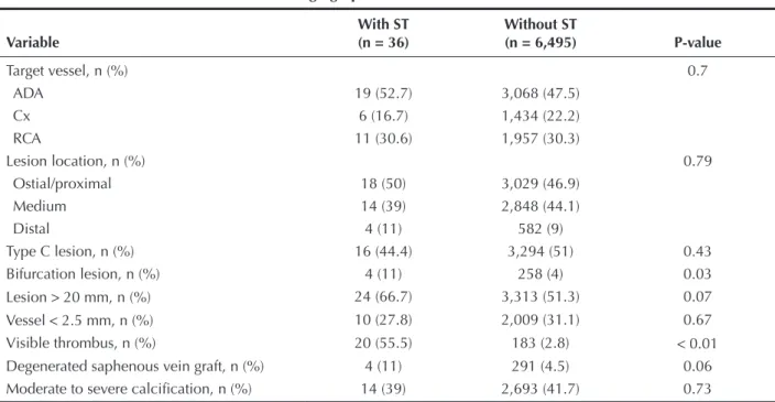
ST occurred more frequently in bifurcation lesions (11% vs. 4%; P = 0.03) and in lesions with visible thrombi on angiography (55.5% vs. 2.8%; P < 0.01)
(Table 2). ST also tended to occur more often in lesions located in degenerated saphenous vein grafts (11% vs. 4.5%; P = 0.06) and in lesions > 20 mm (66.7% vs.
51.3%; P = 0.07).
Table 3 presents the results of the quantitative coronary angiography analysis of patients with ST.
Among the patients with ST, the mean clinical follow-up duration was 30.2 ± 16.3 months. Early
discontinuation of dual antiplatelet therapy (< one
Table 1
Basal clinical characteristics
Variable
With ST (n = 36)
Without ST
(n = 6,495) P-value
Age, years 58.6 ± 11.4 63.7 ± 11.9 < 0.01
Female gender, n (%) 14 (38.9) 2,020 (31.3) 0.33
Systemic arterial hypertension, n (%) 30 (83.3) 5,524 (85.5) 0.73
Dyslipidemia, n (%) 22 (61.1) 4,360 (67.5) 0.45
Current smoking, n (%) 10 (27.8) 1,214 (19.2) 0.15
Diabetes, n (%) 13 (36.1) 2,002 (31) 0.51
Insulin use 2 (5.6) 221 (3.4) 0.48
Family history of CAD, n (%) 3 (8.3) 625 (9.7) 0.79
Previous PCI, n (%) 36 (100) 904 (14.1) < 0.01
Previous CABG, n (%) 1 (2.8) 258 (4) 0.71
Previous stroke, n (%) 1 (28) 137 (2.1) 0.79
Renal failure, n (%) 15 (41.7) 1,806 (28) < 0.01
COPD, n (%) 1 (2.8) 123 (1.9) 0.83
Clinical picture, n (%) < 0.01
ACSNSTE 12 (33.3) 1,155 (17.9)
ACSSTE 18 (50) 688 (10.7)
Stable angina 6 (16.7) 3,025 (46.8)
Asymptomatic/ischemic equivalent 0 1,591 (24.6)
ST, stent thrombosis; CAD, coronary artery disease; PCI, percutaneous coronary intervention; CABG, coronary artery bypass graft surgery; COPD, chronic obstructive pulmonary disease; ACSSTE, acute coronary syndrome with ST-segment elevation; ACSNSTE, acute coronary syndrome without ST-segment elevation.
Table 2
Angiographic characteristics
Variable
With ST (n = 36)
Without ST
(n = 6,495) P-value
Target vessel, n (%) 0.7
ADA 19 (52.7) 3,068 (47.5)
Cx 6 (16.7) 1,434 (22.2)
RCA 11 (30.6) 1,957 (30.3)
Lesion location, n (%) 0.79
Ostial/proximal 18 (50) 3,029 (46.9)
Medium 14 (39) 2,848 (44.1)
Distal 4 (11) 582 (9)
Type C lesion, n (%) 16 (44.4) 3,294 (51) 0.43
Bifurcation lesion, n (%) 4 (11) 258 (4) 0.03
Lesion > 20 mm, n (%) 24 (66.7) 3,313 (51.3) 0.07
Vessel < 2.5 mm, n (%) 10 (27.8) 2,009 (31.1) 0.67
Visible thrombus, n (%) 20 (55.5) 183 (2.8) < 0.01
Degenerated saphenous vein graft, n (%) 4 (11) 291 (4.5) 0.06
Moderate to severe calciication, n (%) 14 (39) 2,693 (41.7) 0.73
the irst month. The irst reports of late ST with BMS appeared only after the recognition of late thrombosis with brachytherapy.16,17 Wenaweser et al.17 demonstrated,
in one of the largest series of patients treated with BMS, that ST occurred in 1.6% of cases; it was acute in 11%, subacute in 64%, and late in 25% of patients. ST is a complex phenomenon, and its etiology is multifactorial. According to some studies,3,13,18,19 early
discontinuation of dual antiplatelet therapy (< one
month for BMS) is one of the most important factors associated with ST. Iakovou et al.3 demonstrated, in
patients treated with drug-eluting stents, that the most important independent predictor of ST was the premature discontinuation of antiplatelet therapy (hazard ratio [HR] = 89.78; 95% conidence interval [95% CI] = 29.9 to 269.6), followed by renal failure (HR 6.49; 95% CI = 2.6 to 16.15), bifurcation lesions (HR 6.42; 95% CI = 2.93 to 14.07), diabetes (HR 3.71, 95% CI = 1.74 to 7.89), and ejection fraction (HR 1.09; 95% CI = 1.05 to 1.36 for every 10% reduction).
Renal failure is associated with increased mortality, despite a successful PCI.20-22 Renal failure is associated
with metabolic and microcirculatory abnormalities, which can predispose the patient to thrombus formation.23,24
Regarding bifurcation lesions, histopathology studies have suggested that the locations of arterial branching All patients were submitted to emergency PCI;
du-ring the in-hospital phase, the incidences observed of periprocedural myocardial infarction, contrast-induced nephropathy and death were 33.3%, 22.2% and 16.6% of cases, respectively (Table 4). During the late fol low-up period, target-vessel revascularization was observed in 33.3% of cases, which was achieved by CABG in most cases. A new MI was observed in six (20%) cases, two resulting from recurrent ST. No additional deaths were observed.
DISCUSSION
The main indings of the present study were: ST after BMS implantation occurred in the late phase (> 30 days) in 50% of cases; early discontinuation of
dual antiplatelet therapy was observed in one-sixth of patients with ST; the presence of renal failure, bifurca-tion lesions, or evident thrombus on angiography, and of long lesions or of lesions located in degenerated saphenous vein grafts was more frequent in patients with ST; and in-hospital mortality in this population was high (16.7%), as was the incidence of new MACEs during the late clinical follow-up period.
Historically, ST in BMS has been an event that occurs more frequently during the initial days after surgery, and, by deinition, has not been reported after
Table 3
Quantitative coronary angiography of cases with coronary stent thrombosis
Variable n = 36
Pre-procedure
Reference vessel diameter, mm 2.9 ± 0.7
Minimal luminal diameter, mm 0.55 ± 0.51
Stenosis diameter, % 84.1 ± 11.8
Lesion extent, mm 21.1 ± 7.2
Post-procedure
Reference vessel diameter, mm 3.2 ± 0.6
Intra-segment
– Minimal luminal diameter, mm 2.37 ± 0.53
– Stenosis diameter, % 24.1 ± 26.8
– Immediate gain, mm 1.89 ± 0.62
Intrastent
– Minimal luminal diameter, mm 2.7 ± 0.46
– Stenosis diameter, % 14.1 ± 6.7
– Immediate gain, mm 1.89 ± 0.53
Table 4
In-hospital and late clinical follow-up of cases with coronary stent thrombosis
Variable
In-hospital n = 36
Mean hospitalization time, days 6.3
MI, n (%) 12 (33.3)
Death, n (%) 6 (16.6)
CIN, n (%) 8 (22.2)
Late n = 30
Time of follow-up (months) 30.2 ± 16.3
MI, n (%) 6 (20)
TVR, n (%) 10 (33.3)
– PCI 2 (6.7)
– CABG 8 (27)
Death, n (%) 0
have low shear regions and low speed low, predispo-sing the vessels to the development of atherosclerotic plaques and thrombi.25-27
Study limitations
The limitations of the present study include the retrospective analysis of the data from two cohorts with non-adjusted clinical variables and the performance of the study in a single centre. Additionally, only indivi-duals with ST treated at this institution were included, which may have resulted in the underestimation of the true incidence.
CONCLUSIONS
In this contemporary real-world experience, ST was shown to be an event with high rates of hospital mortality and late morbidity. As demonstrated by other studies, the occurrence of this event was associated with clinical and angiographic characteristics of greater complexity, and with lower adherence to the prescribed antiplatelet therapy.
CONFLICT OF INTEREST
The authors declare no conlicts of interest.
REFERENCES
1. Cutlip DE, Windecker S, Mehran R, Boam A, Cohen DJ, van Es GA, et al. Clinical end points in coronary stent trials: a case for standardized deinitions. Circulation. 2007;115(17):2344-51. 2. Holmes DR Jr, Kereiakes DJ, Laskey WK, Colombo A, Ellis SG,
Henry TD, et al. Thrombosis and drug-eluting stents: an objec-tive appraisal. J Am Coll Cardiol. 2007;50(2):109-18. 3. Iakovou I, Schmidt T, Bonizzoni E, Ge L, Sangiorgi GM,
Stankovic G, et al. Incidence, predictors, and outcome of thrombosis after successful implantation of drug-eluting stents. JAMA. 2005;293(17):2126-30.
4. Cutlip DE, Baim DS, Ho KK, Popma JJ, Lansky AJ, Cohen DJ, et al. Stent thrombosis in the modern era: a pooled analy-sis of multicenter coronary stent clinical trials. Circulation. 2001;103(15):1967-71.
5. Doyle B, Rihal CS, O’Sullivan CJ, Lennon RJ, Wiste HJ, Bell M, et al. Outcomes of stent thrombosis and restenosis during extended follow-up of patients treated with bare-metal coronary stents. Circulation. 2007;116(21):2391-8.
6. Daemen J, Wenaweser P, Tsuchida K, Abrecht L, Vaina S, Morger C, et al. Early and late coronary stent thrombosis of sirolimus-eluting and paclitaxel-eluting stents in routine clinical practice: data from a large two-institutional cohort study. Lancet. 2007;369(9562):667-78.
7. Roukoz H, Bavry AA, Sarkees ML, Mood GR, Kumbhani DJ, Rabbat MG, et al. Comprehensive metaanalysis on drug-eluting stents versus bare-metal stents during extended follow-up. Am J Med. 2009;122(6):581.e1-10.
8. Kirtane AJ, Gupta A, Iyengar S, Moses JW, Leon MB, Applegate R, et al. Safety and eficacy of drug-eluting and bare metal stents: comprehensive meta-analysis of randomized trials and observational studies. Circulation. 2009;119(25):3198-206. 9. Kuchulakanti PK, Chu WW, Torguson R, Ohlmann P, Rha SW,
Clavijo LC, et al. Correlates and long-term outcomes of
angiographically proven stent thrombosis with sirolimus- and paclitaxel-eluting stents. Circulation. 2006;113(8):1108-13. 10. Smit JJ, van’t Hof AW, de Boer MJ, de Boer MJ, Hoorntje JC,
Dambrink JH, et al. Incidence and predictors of subacute throm-bosis in patients undergoing primary angioplasty for an acute myocardial infarction. Thromb Haemost. 2006;96(2):190-5. 11. Rinaldi MJ, Kirtane AJ, Piana RN, Caputo RP, Gordon PC,
Lopez JJ, et al. Clinical, procedural, and pharmacologic cor-relates of acute and subacute stent thrombosis: results of a multicenter case-control study with 145 thrombosis events. Am Heart J. 2008;155(4):654-60.
12. Pisterer M, Brunner-La Rocca HP, Buser PT, Rickenbacher P, Hunziker P, Mueller C, et al. Late clinical events after clopi-dogrel discontinuation may limit the beneit of drugeluting stents: an observational study of drug-eluting versus bare-metal stents. J Am Coll Cardiol. 2006;48(12):2584-91.
13. Airoldi F, Colombo A, Morici N, Latib A, Cosgrave J, Buellesfeld L, et al. Incidence and predictors of drug-eluting stent thrombosis during and after discontinuation of thienopyridine treatment. Circulation. 2007;116(7):745-54.
14. Mattos LAP, Lemos Neto PA, Rassi Jr A, Marin-Neto JA, Sousa AGMR, Devito FS, et al. Diretrizes de intervenção coronária per-cutânea e métodos adjuntos diagnósticos em cardiologia intervencionista (II edição 2008). Rev Bras Cardiol Invasiva. 2008;16(2 Supl 2):9-88.
15. Ryan TJ, Faxon DP, Gunnar RM, Kennedy JW, King SB 3rd, Loop FD,
et al. Guidelines for percutaneous transluminal coronary angioplasty: a report of the American College of Cardiology/ American Heart Association Task Force on Assessment of Diag-nostic and Therapeutic Cardiovascular Procedures (Subcom-mittee on Percutaneous Transluminal Coronary Angioplasty). Circulation. 1988;78(2):486-502.
16. Wang F, Stouffer GA, Waxman S, Uretsky BF. Late coronary stent thrombosis: early vs. late stent thrombosis in the stent era. Catheter Cardiovasc Interv. 2002;55(2):142-7.
17. Wenaweser P, Rey C, Eberli FR, Togni M, Tüller D, Locher S, et al. Stent thrombosis following bare-metal stent implanta-tion: success of emergency percutaneous coronary intervention and predictors of adverse outcome. Eur Heart J. 2005;26(12): 1180-7.
18. McFadden EP, Stabile E, Regar E, Cheneau E, Ong AT, Kinnaird T, et al. Late thrombosis in drug-eluting coronary stents after dis continuation of antiplatelet therapy. Lancet. 2004; 364(9444): 1519-21.
19. Jeremias A, Sylvia B, Bridges J, Kirtane AJ, Bigelow B, Pinto DS, et al. Stent thrombosis after successful sirolimus-eluting stent implantation. Circulation. 2004;109(16):1930-2.
20. Gruberg L, Mintz GS, Mehran R, Gangas G, Lansky AJ, Kent KM, et al. The prognostic implications of further renal function deterioration within 48 h of interventional coronary procedures in patients with pre-existent chronic renal insuficiency. J Am Coll Cardiol. 2000;36(5):1542-8.
21. Rubenstein MH, Harrell LC, Sheynberg BV, Schunkert H, Bazari H, Palacios IF. Are patients with renal failure good candidates for percutaneous coronary revascularization in the new device era?. Circulation. 2000;102(24):2966-72.
22. Mehran R, Aymong ED, Nikolsky E, Lasic Z, Iakovou I, Fahy M, et al. A simple risk score for prediction of contrast-induced nephropathy after percutaneous coronary intervention: de-velopment and initial validation. J Am Coll Cardiol. 2004; 44(7):1393-9.
24. Amann K, Ritz E. Cardiac disease in chronic uremia: patho-physiology. Adv Ren Replace Ther. 1997;4(3):212-24. 25. Glagov S, Zarins C, Giddens DP, Ku DN. Hemodyna mics
and atherosclerosis: insights and perspectives gained from studies of human arteries. Arch Pathol Lab Med. 1988;112(10): 1018-31.
26. Glagov S, Weisenberg E, Zarins CK, Stankunavicius R, Kolettis GJ. Compensatory enlargement of human atherosclerotic coronary arteries. N Engl J Med. 1987;316(22):1371-5.