Comparison of Classical and Secondary Cytologic
Criteria Relative to Hybrid Capture for Diagnosing
Cervical-vaginal Infection by
Human Papillomavirus
Comparação entre os critérios citológicos clássicos
e secundários para o diagnóstico de infecção
cérvico-vaginal por papiloma vírus humano em relação à
captura híbrida
Renata Margarida Etchebehere
1Élia Cláudia Souza Almeida
2Eliângela Castro Côbo
3Ana Cristina da Rocha Duque
4Eddie Fernando Cândido Murta
2Sheila Jorge Adad
21Surgical Pathology Service, Clinical Hospital, Hospital de Clínicas da Universidade Federal do Triângulo Mineiro (UFTM), Uberaba, Minas Gerais, Brazil
2Post Graduate Program in Health Sciences, UFTM, Uberaba, Minas Gerais, Brazil
3Special Pathology Discipline, Universidade Federal do Triângulo Mineiro (UFTM), Uberaba, Minas Gerais, Brazil
4Maternal and Infant Department, Discipline of Gynecology and Obstetrics, UFTM, Uberaba, Minas Gerais, Brazil
Rev Bras Ginec Obst 2016;38:41–46.
Address for correspondence Renata Margarida Etchebehere, PhD, Universidade Federal do Triângulo Mineiro–UFTM, Rua Getúlio Guaritá, 130–Abadia, 38025-440 - Uberaba, MG, Brazil (e-mail: [email protected]).
Keywords
►
HPV
►
cervical neoplasia
►
vaginal smears
►
papillomavirus
►
DNA
Abstract
Objective
To compare the diagnostic accuracy of the classic Meisels cytologic criteria
and the Schneider secondary criteria relative to the hybrid capture method for
diagnosing HPV infection.
Methods
This was a retrospective study performed at a public university hospital. A
total of 41 patients with a cytologic diagnosis of HPV infection and 40 HPV-negative
patients were selected for review of the cervical-vaginal smears seeking to classical and
secondary criteria. A single pathologist reviewed the slides in search of the criteria. The
classical and secondary cytologic criteria were compared with the hybrid capture for
diagnosing HPV infection. Bartleti test was applied for the age analysis, and Fisher
’
s
exact test was used to compare proportions. The tests were considered signi
fi
cant
when the probability of rejecting the null hypothesis was less than 5% (
p
<
0.05).
Results
The Meisels criteria were less sensitive (34.0%) than the secondary Schneider
criteria (57.5%) when compared with the hybrid capture (
p
<
0.0001), although the
speci
fi
city of the former criteria was non-signi
fi
cantly higher (91.2% and 67.7%,
respectively). In cases of moderate or intense in
fl
ammation, the sensitivity and
speci
fi
city of the Schneider criteria were decreased, 33.3% and 50.0% respectively
(
p
¼
0.0115).
received
September 18, 2015
accepted
October 28, 2015
DOIhttp://dx.doi.org/ 10.1055/s-0035-1570105.
ISSN 0100-7203.
Copyright © 2016 by Thieme Publicações Ltda, Rio de Janeiro, Brazil
Introduction
Cervical cancer is the third most common cancer among women. In developed countries, the diagnosis is made ear-lier; consequently, the 5-year survival rate is higher. In the last few decades, the incidence of cervical cancer has de-creased in countries with effective screening systems, prob-ably as result of the early treatment of precancerous lesions. Thus, it is important to understand the minimal abnormal-ities in cervical-vaginal cytology.1–3
With the development of molecular biological techniques, epidemiological and laboratoryfindings have identified
hu-man papillomavirus(HPV) as the principal agent involved in
the geneses of cervical cancer and cervical intraepithelial neoplasia. This evidence has increased the importance of the morphological identification of HPV infection in cervical-vaginal cytology.1,4–8Classically, the natural history of cer-vical cancer starts with infection by HPV. Subsequent pro-gressive intraepithelial transformations can evolve into invasive neoplasia in the long term. This connection has raised interest in establishing and perfecting the diagnosis of this infection, as well as identifying risk factors for it, to
detect populations susceptible to HPV-induced cervical carcinogenesis.4
Although cellular biological tests are more sensitive in diagnosing HPV infection than cytological tests, access be-come larger and the cost somewhat lower, some factors limit their routine use, including cost and methodological diffi -culty, particularly in poorer countries.9,10Thus, the cytologic Papanicolaou exam (also known as the Pap smear) remains the main screening method for cervical cancer.3,11Where as Papanicolaoufirst described the exfoliated squamous cells of vaginal and cervical condyloma acuminatum,12 it was Meisels and Fortin13 and Meisels, Fortin and Roy14 who identified the cell changes that, currently, are considered pathognomonic for HPV infection (i.e., classic koilocytosis and dyskeratosis). These criteria for HPV infection are very specific, but not very sensitive.3,11To increase the sensitivity of the test without substantially reducing specificity, various authors have sought other “secondary” cytologic indices.3,15–19
Among the secondary criteria proposed, the most ac-cepted are those of Schneider et al. (1987),15which include:
Conclusions
Compared with hybrid capture for diagnosis of HPV infection, the
sensitivity of the secondary Schneider criteria was higher than the classical Meisels
criteria. Moderate or intense in
fl
ammation reduces the sensitivity and speci
fi
city of the
secondary Schneider criteria for diagnosing HPV infection using the hybrid capture as
the gold standard.
Resumo
Objetivo
Comparar a acurácia diagnóstica dos critérios citológicos clássicos de
Meisels com a dos critérios secundários de Schneider em relação a captura híbrida
para o diagnóstico de infecção pelo HPV.
Métodos
Trata-se de estudo retrospectivo realizado em hospital público
universi-tário. Quarenta e uma pacientes com diagnóstico citológico de infecção pelo HPV e 40
pacientes HPV-negativas foram selecionadas para avaliação dos esfregaços
cervicais-vaginais em busca dos critérios clássicos e secundários. Um único patologista reviu as
lâminas. Os critérios citológicos clássicos e secundários foram comparados com a
captura híbrida para o diagnóstico de infecção pelo HPV. O teste de Bartleti foi aplicado
para a análise das idades e o teste exato de Fisher para comparar proporções. Os testes
foram considerados signi
fi
cativos quando a probabilidade de rejeitar a hipótese de
nulidade foi menor que 5% (
p
<
0,05).
Resultados
Os critérios de Meisels foram menos sensíveis (34,0%) que os secundários
de Schneider (57,5%) quando comparados com a captura híbrida (
p
<
0,0001), embora
a especi
fi
cidade dos critérios de Meisels não tenha sido signi
fi
cativamente superior
(91,2% e 67,7%, respectivamente). Em casos de in
fl
amação moderada ou intensa, a
sensibilidade e especi
fi
cidade dos critérios secundários de Schneider foram diminuídas,
33,3% e 50,0%, respectivamente (
p
¼
0,0115).
Conclusões
Comparado a captura híbrida para o diagnóstico da infecção pelo HPV, a
sensibilidade dos critérios secundários de Schneider foi maior que a dos critérios
clássicos de Meisels. In
fl
amação moderada ou intensa reduziu a sensibilidade e
especi
fi
cidade dos critérios secundários de Schneider para o diagnóstico de infecção
pelo HPV utilizando a captura híbrida como padrão-ouro.
Palavras-chave
►
HPV
►
neoplasia do colo do
útero
slight koilocytosis or an outline of koilocytosis, slight dysker-atosis, cleared cytoplasm, keratin hyaline granules, conden-sation of filaments in the cytoplasm, fusiform cells, hyperchromatic nuclei, bi- or multinucleation, and perinuc-lear halo.
Nevertheless, there is still some controversy about the use of secondary criteria.16–19Thus, we wanted to compare the use of classical and secondary cytologic criteria to the hybrid capture (HC) molecular biological test for diagnosing HPV infection. The HC method was chosen because it is easily performed, yields rapid results, has good sensitivity for latent, subclinical, and clinical infections, and can detect HPV infection anywhere in the woman’s lower genital tract.5,9,20
The objective of this study was to compare the classic cytologic Meisels criteria (CMC)13,14 and the secondary criteria proposed by Schneider et al.15(SSC) to the molecular biological HC method for diagnosing HPV infection.
Methods
The Research Ethics Committee at the Universidade Federal Triângulo Mineiro approved this research (protocol no. 0281 on 12.20.2002). We performed a pilot study to determine the sample size. The sample calculation was performed for two proportions, following Arango.21 Applying the formula to compare two samples, we calculatedn¼27. Using the value 1.96 with an α level of 0.05 and β level of 0.084, we determined that the sample size for this study was sufficient tofind significant differences when they actually exist.
This retrospective study included 41 patients with a cytologic diagnosis of HPV infection by Pap smear. Patients received care between June 2000 and October 2002 at the Gynecology and Obstetrics outpatient clinic of a public university hospital. All patients signed an informed consent form to demonstrate that they agreed to participate in the study. These cases were paired with 40 control cases with a normal routine or inflammatory cervical-vaginal cytology, collected during the same sample period. When the 81 patients returned to the clinic, we collected material to perform the HC test to diagnose HPV infection.
The same observer reviewed the slides according to the CMC13,14and SSC.15The observer was unaware of the pre-vious results of the Papanicolaou and HC exams. We con-sidered the cytology as positive for a diagnosis of HPV infection based on the CMC when classic koilocytosis or
dyskeratosis was present. The cytology was considered positive according to the SSC when a minimum offive of the nine criteria were present.
We also analyzed the presence of inflammation and infection by other agents. To diagnose inflammation, we analyzed the presence of neutrophils and cell changes, such as an increase in nuclear volume, binucleation, hyper-chromasia, margination of chromatin, small perinuclear halo, and cytoplasmic vacuolization.3We classified the
in-flammatory process as slight, moderate or intense, based on the intensity of the inflammatory exudate and the frequency of inflammation-related cell changes. Diagnosis of infection was based only on the morphological identification of the agent.22
The obtained results were entered into a database for statistical analysis via Microsoft Access2000®. GRAPHPAD INSTAT® (version 3.0) was used to perform the statistical calculations. Bartleti test was applied for the age analysis, and Fisher’s exact test was used to compare proportions. The tests were considered significant when the probability of rejecting the null hypothesis was less than 5% (p<0.05).
Results
In the HC exam, 47 (58%) of the 81 cases tested positive for HPV infection. The age of patients with positivity for HPV-DNA by HC ranged from 14 to 47 years, with an average of 24.16.5 years. In the HPV-DNA–negative group, the age ranged from 14 to 50 years, with an average age of 25.186.56 (p¼0.4693 between groups).
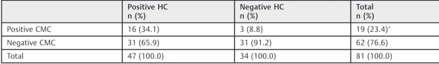
Using the CMC and considering the CH as gold standard for diagnosing HPV infection, of the 81 cases, 19.8% were true positives and 38.3% true negatives. The specificity was 91.2% and the sensitivity by 34% (►Table 1).
Using the SSC, 33.3% of the 81 cases were classified as true positives and 23 (28.39%) as true negatives. The sensitivity was 57.5%, and the specificity 67.7% (►Table 2).
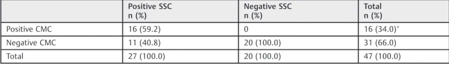
All cases that were positive by HC and CMC were also positive by SSC (►Table 3). Thus, use of the SSC increased the sensitivity for diagnosing HPV infection from 34% to 57.5% (p<0.0001). The difference in specificity between the SSC and CMC was not statistically significant.
The most frequently encountered SCC were: bi-or multi-nucleation (68 cases), nuclear hyperchromasia (61 cases), perinuclear halo (58 cases), slight koilocytosis (54 cases), and dyskeratosis (36 cases). These criteria were also common in
Table 1 Cytologic analysis of 81 cases based on the classic Meisels criteria (CMC), using hybrid capture (HC) as the gold standard for diagnosing HPV infection in patients accompanied on ambulatory of Universidade Federal do Triângulo Mineiro
Positive HC n (%)
Negative HC n (%)
Total n (%)
Positive CMC 16 (34.1) 3 (8.8) 19 (23.4)
Negative CMC 31 (65.9) 31 (91.2) 62 (76.6)
Total 47 (100.0) 34 (100.0) 81 (100.0)
cases with a negative HC result, without any significant difference. Cleared cytoplasm was the only criteria that, in isolation, showed a significant difference for the groups with positive and negative HC results; however, it was present in only 30.86% of positive cases and was a commonfinding (11.12%) in negative cases.
Some of the SSC were also often found in cases of infl am-mation or infection, especially in more accentuated cases, which may have been related to the increased number of false positives. In reviewing the slides, 76 of the 81 cases (93.8%) showed varying degrees of inflammation, including 81.5% of slight, 14.5% of moderate, and 4% of intense inflammation. Among patients with inflammatory changes, most had non-specific inflammation (lactobacilli), 15.8% had a diagnosis of infection by Gardnerella vaginalis, 14.5% had a diagnosis of infection by Candida sp, and 6.6% had predominance of cocobacilli.
We separated the cytologies into two groups: cases with slight or no inflammation, and cases with moderate or intense inflammation. Of the 67 cases with slight or no inflammation, 47.8% hadfive or more SSC (i.e., were considered positive for HPV infection by SSC). Of these cases, 21.9% had a negative HC result, and 78.1% had a positive HC result. The sensitivity of the SSC relative to HC was 60.98%, and the specificity was 73.08%. There was a statistically significant difference (p¼0.0115) between the groups, indicating that the SSC were good indica-tors of infection by HPV in cases with slight or no inflammation. Among the 14 cases with moderate or intense infl amma-tion, 42.9% were considered positive for HPV infecamma-tion, according to the SSC. Of these 6 cases, 33.3% had a positive HC result, and 66.7% had a negative HC result. The sensitivity of the SSC in relation to HC was 33.3%, and the specificity 50%. The sensitivity and specificity of the SSC in more intense inflammatory situations was lower.
Discussion
The Pap test has worked as well as it has despite the poor sensitivity of a single test because it is repeated periodically during the span of a woman’s lifetime.23
Despite the high specificity, the sensitivity of the CMC is low. Compared with HC for diagnosis of HPV infection, the sensitivity of the SSC was higher than CMC, but the specifi -city was lower, agreement with other authors.15–18Thus, the secondary criteria described by Schneider et al.15appear to have a better ability to detect HPV infection in true-positive patients.
Other authors found sensitivity for non-classic criteria of only 15.8% and the specificity of 100% in samples previously diagnosed by polymerase chain reaction (PCR). However, they used different cytologic criteria from our study, exam-ining only nuclear hyperchromasia, pleomorphism, and the nucleus/cytoplasm relationship in HIV infected patients.24In other study, after including secondary criteria, they observed that the diagnostic frequency for HPV using cytology in-creased from 24.4% to 75.6%.18 Again, these authors used different secondary cytologic criteria from those used in our study. In comparing cytology with molecular hybridization, thefirst study obtained an agreement of 48% when they used only koilocytosis as a cytologic criterion. However, when they also included dyskeratosis, dyskariosis, binucleation, and multinucleation agreement increased to 75%.24Different research indicated that the inclusion of non-classical cyto-morphologic signs increased the sensitivity of the cytologic test for detecting HPV when compared with PCR, although they used monobed, rather than conventional, cytology.17 Using PCR for HPV 16, an study concluded that the classic cell changes were not the only ones that permitted a diagnosis of HPV infection.11
Table 2 Cytologic analysis of 81 cases based on Schneider’s secondary criteria (SSC), relative to hybrid capture (HC) as the gold standard for diagnosing HPV infection in patients accompanied on ambulatory of Universidade Federal do Triângulo Mineiro
Positive HC n (%)
Negative HC n (%)
Total n (%)
Positive SSC 27 (57.4) 11 (32.4) 38 (46.9)
Negative SSC 20 (42.6) 23 (67.6) 43 (53.1)
Total 47 (100.0) 34 (100.0) 81 (100.0)
Fisher’s exact test;p¼0.0417.
Table 3 Distribution of the 47 HPV-DNA–positive cases, according to cytologic analysis by Schneider’s secondary criteria (SSC) and the classic Meisels criteria (CMC) in patients accompanied on ambulatory of Universidade Federal do Triângulo Mineiro
Positive SSC n (%)
Negative SSC n (%)
Total n (%)
Positive CMC 16 (59.2) 0 16 (34.0)
Negative CMC 11 (40.8) 20 (100.0) 31 (66.0)
Total 27 (100.0) 20 (100.0) 47 (100.0)
Fisher’s exact test;
We found a reduction in the specificity and sensitivity of the SSC in cases with moderate or intense inflammation. This
finding probably stems from the superimposition of cytolo-gic changes related to inflammation with certain secondary criteria involved in the HPV diagnosis. Moreover, the infl am-matory exudate may have made it difficult to visualize the cytologic criteria.3Thus, we believe that it is better not to use the SSC in cases with moderate or intense inflammation, to avoid an increase in the number of false positives.
Studing the importance of the application of the non-classical criteria compared with HC, they found infl amma-tion in 61.5% of cases, although they did not classify the intensity of inflammation.19 Other research identified
in-flammation in only 4.9% of cases when comparing non-classical criteria with PCR.18 Nevertheless, their findings conflict with those in the literature, as inflammation is a very commonfinding in cervical-vaginal cytology, particu-larly unspecific cervicitis in young patients.3,4,22In addition, the authors did not classify the intensity of the inflammation. Despite the introduction of the HPV vaccines, screening programs using cervical-vaginal cytology must continue. The vaccine does not completely protect against cervical cancer and is still not universally used.25–27
Compared with HC for diagnosis of HPV infection, the sensitivity of the SSC was higher than CMC. Moderate or intense inflammation reduces the sensitivity and specificity of the SSC for diagnosing HPV infection using the HC as the gold standard. We believe that, with greater use of the vaccine against HPV, there will be an increase in the number of cytologies with minimal cell changes, making it even more important to recognize the non-classical changes that are associated with HPV infection.
Conflicts of Interest None.
Acknowledgments
To National Council for Scientific and Technological Devel-opment (Conselho Nacional de Desenvolvimento Científico e Tecnológico–CNPq) forfinancial assistance (Application no477539/2001
–7/Research Productivity Grant: Applica-tion no520 629/99
–0 NV); FIOCRUZ of Rio de Janeiro, RJ, for donation of the Hybrid Capture machine; and Doctor Uilho Antônio Gomes of the Ribeirão Preto São Paulo University (FMRP - USP), for reviewing the statistical analysis.
References
1 Guarisi R, Hardy E, Derchain SFM, Fonsechi-Carvasan GA, Borges JBR. Rastreamento, diagnóstico e tratamento das lesões precur-soras e do câncer invasor do colo uterino no município de Franco da Rocha, SP. Rev Bras Cancerol 2004;50(1):7–15
2 Brasil. Ministério da Saúde. Instituto Nacional de Câncer José Alencar Gomes da Silva [Internet]. Tipos de câncer: colo do útero. Brasília (DF): Ministério da Saúde; 2014 [citado 2015 Jul 10]. Disponível em:<http:/ www.inca.gov.br/wps/wcm/connect/tiposdecancer/site/home/ colo_utero>
3 DeMay RM. The art and science of cytopathology. 2nd ed. Vol. 1, Exfoliative cytology. Chicago: American Society of Clinical Pathol-ogists Press; 2012. Chapter 1, The pap test; p. 2–149
4 Silva C, Almeida EC, Côbo EdeC, Zeferino VF, Murta EF, Etchebe-here RM. A retrospective study on cervical intraepithelial lesions of low-grade and undetermined significance: evolution, asso-ciated factors and cytohistological correlation. Sao Paulo Med J 2014;132(2):92–96
5 Cope JU, Hildesheim A, Schiffman MH, et al. Comparison of the hybrid capture tube test and PCR for detection of human papillo-mavirus DNA in cervical specimens. J Clin Microbiol 1997;35(9): 2262–2265
6 Curry CL, Sage YH, Vragovic O, Stier EA. Minimally abnormal Pap testing and cervical histology in HIV-infected women. J Womens Health (Larchmt) 2012;21(1):87–91
7 Cuzick J, Arbyn M, Sankaranarayanan R, et al. Overview of human papillomavirus-based and other novel options for cervical cancer screening in developed and developing countries. Vaccine 2008; 26(Suppl 10):K29–K41
8 Muñoz N. Human papillomavirus and cancer: the epidemiological evidence. J Clin Virol 2000;19(1–2):1–5
9 Nomelini RS, Guimarães PD, Candido PA, Campos AC, Michelin MA, Murta EFC. Prevention of cervical cancer in women with ASCUS in the Brazilian Unified National Health System: cost-effectiveness of the molecular biology method for HPV detection. Cad Saude Publica 2012;28(11):2043–2052
10 Akbar S, Pervez SN, Shah W. Manual liquid based cytology for Pap smear preparation and HPV detection by PCR in Pakistan. Asian Pac J Cancer Prev 2015;16(2):579–583
11 Kashyap V, Hedau S, Bhambhani S. Defining the validity of classical and non-classical cellular changes indicative of low-grade squamous intraepithelial lesion encompassing human pa-pillomavirus infection in relation to human papa-pillomavirus deox-yribonucleic acid testing. J Cytol 2011;28(4):159–164
12 Papanicolaou GN. Atlas of exfoliative cytology. Cambridge: Harvard University Press; 1954
13 Meisels A, Fortin R. Condylomatous lesions of the cervix and vagina. I. Cytologic patterns. Acta Cytol 1976;20(6):505–509
14 Meisels A, Fortin R, Roy M. Condylomatous lesions of the cervix. II. Cytologic, colposcopic and histopathologic study. Acta Cytol 1977;21(3):379–390
15 Schneider A, Meinhardt G, De-Villiers EM, Gissmann L. Sensitivity of the cytologic diagnosis of cervical condyloma in comparison with HPV-DNA hybridization studies. Diagn Cytopathol 1987; 3(3):250–255
16 Roteli-Martins CM, Alves VA, Santos RT, Martinez EZ, Syrjänen KJ, Derchain SF. Value of morphological criteria in diagnosing cervical HPV lesions confirmed by in situ hybridization and hybrid capture assay. Pathol Res Pract 2001;197(10): 677–682
17 Bollmann M, Bánkfalvi A, Trosic A, Speich N, Schmittt C, Bollmann R. Can we detect cervical human papillomavirus (HPV) infection by cytomorphology alone? Diagnostic value of non-classic cyto-logical signs of HPV effect in minimally abnormal Pap tests. Cytopathology 2005;16(1):13–21
18 Kaneshima EN, Suzuki LE, Irie MMT, Yoshida CS, Silva SFM, Consolaro MEL. Importância da aplicação de critérios morfológi-cos não clássimorfológi-cos para o diagnóstico citopatológico de Papiloma-vírus humano (HPV) previamente detectado por PCR. Acta Bioquím Clin Latinoam. 2005;39(1):61–68
19 Jordão AV, Ruggeri LS, Chiucheta GIR, Piva S, Consolaro MEL. Importância da aplicação de critérios morfológicos não clássicos para o diagnóstico citomorfológico de papilomavírus humano. J Bras Patol Med Lab. 2003;39(1):81–89
21 Arango HG. Bioestatística: teórica e computacional. 3a ed. Rio de Janeiro: Guanabara-Koogan; 2009
22 Adad SJ, de Lima RV, Sawan ZT, et al. Frequency of Trichomonas vaginalis, Candida sp and Gardnerella vaginalis in cervical-vaginal smears in four different decades. Sao Paulo Med J 2001;119(6): 200–205
23 Thaxton L, Waxman AG. Cervical cancer prevention: immuniza-tion and screening 2015. Med Clin North Am 2015;99(3):469–477
24 de Faria IM, Melo VH, de Castro LP, et al. [Accuracy of oncotic cytology for HPV infection diagnosis on the cervix uteri of
HIV-infected women]. Rev Bras Ginecol Obstet 2008;30(9):437–444 Portuguese
25 Wentzensen N, Klug SJ. Cervical cancer control in the era of HPV vaccination and novel biomarkers. Pathobiology 2009;76(2):82–89
26 Sopracordevole F, Cigolot F, Mancioli F, Agarossi A, Boselli F, Ciavattini A. Knowledge of HPV infection and vaccination among vaccinated and unvaccinated teenaged girls. Int J Gynaecol Obstet 2013;122(1):48–51