1 Universidade Federal do Paraná, Faculdade de Odontologia, Departamento de Estomatologia. Av. Lothário Meissner, 632, 80210-170, Jd. Botânico,
Curitiba, PR, Brasil. Correspondência para / Correspondence to: AM SEBASTIANI. E-mail: <[email protected]>.
2 Universidade Federal do Paraná, Programa de Residência em Cirurgia e Traumatologia Buco-maxilo-faciais. Curitiba, PR, Brasil.
3 Universidade Federal do Paraná, Programa de Mestrado em Odontologia, Programa de Residência em Cirurgia e Traumatologia Buco-maxilo-faciais.
Curitiba, PR, Brasil.
Le Fort III osteotomy for severe dentofacial deformity correction associated
with hypoplasia of the midface
Osteotomia Le Fort III para correção de deformidade dentofacial severa associada a hipoplasia de terço médio da face
Aline Monise SEBASTIANI1
Nelson Luis Barbosa REBELATTO2
Leandro Eduardo KLÜPPEL3
Delson João da COSTA3
Fernando ANTONINI4
Rafaela Scariot de MORAES5
ABSTRACT
The combination of orthodontic therapy and orthognathic surgery is a well-established treatment modality for the correction of dentofacial deformities. When these deformities are more severe, involving hypoplastic midface, surgical techniques not used routinely in the treatment of facial changes are required, such as the Le Fort III osteotomy or variations of this technique. Few studies have reported the use of this technique or its modiications in non-syndromic patients. This paper demonstrates the orthodontic-surgical resolution of a patient with dentofacial deformity with severe malocclusion Class III, involving midface hypoplasia, with a modiication technique of a Le Fort III osteotomy associated with Le Fort I and sagittal of the rami osteotomies. After three years of postoperative follow-up, the patient demonstrates signiicant improvement in chewing ability, no functional complaints, and high satisfaction with the aesthetics and improved quality of life.
Indexing terms: Face. Malocclusion. Orthognathic surgery.
RESUMO
A combinação da terapia ortodôntica com a cirurgia ortognática é uma modalidade de tratamento bem estabelecida para a correção de deformidades dentofaciais. Quando estas deformidades apresentam maior severidade, envolvendo a hipoplasia do terço médio da face, exigem técnicas cirúrgicas não utilizadas como rotina no tratamento das alterações faciais, como a osteotomias Le Fort III ou as variações destas técnicas. Poucos estudos relatam o uso desta técnica ou de suas modiicações em pacientes não sindrômicos. Este trabalho tem como objetivo demonstrar uma resolução ortodôntica-cirúrgica de um paciente apresentando deformidade de face com má-oclusão Classe III severa, envolvendo hipoplasia do terço médio facial, com a realização de uma técnica modiicada da osteotomia Le Fort III, associada as osteotomias Le Fort I e osteotomia sagital dos ramos mandibulares. O paciente encontra-se com três anos de acompanhamento pós-operatório, com melhora signiicativa na sua habilidade mastigatória, sem queixas funcionais, relatando alta satisfação com a estética e melhora na qualidade de vida.
Termos de indexação: Face. Má oclusão. Cirurgia ortognática.
INTRODUCTION
The combination of orthodontic and orthognathic surgery therapy is a well-established treatment modality to correct moderate and severe dentofacial deformities1. The most common surgery techniques are the bilateral sagittal split osteotomy (BSSO) in mandible surgeries and the Le Fort I osteotomy in surgeries involving the maxilla.
After the orthodontic-surgical planning, teeth 14 and 24 were extracted to enable the maxillary teeth decompensation. After two years of orthodontic treatment, he showed a 15-mm maxillo-mandible discrepancy (Figure The Le Fort III osteotomy has been widely used in dentofacial deformity treatment, primarily in syndromic patients2-5. It was irst reported in 1950 by Gilles and Harrison6, and after this, Tessier7 described the technique in more reined way, making it more applicable and predictable in craniofacial deformity treatment and revolutionizing the management in patients with total deiciency of the midface.
Several modiications of the technique were realized with osteotomy alterations8 or Le Fort I osteotomy association9-10. Fewer studies related the use of the technique or one of its modiications in non-syndromic patients with midface hypoplasia10.
This paper reports the orthodontic-surgical resolution in one non-syndromic patient with dentofacial
deformity and severe Class III malocclusion on which a Le Fort III modiied osteotomy associated with the Le Fort I and BSSO osteotomies was performed.
CASE REPORT
A 23-year-old male patient with extensive mandible prognathism associated with midface hypoplasia was referred due to a functional complaint with restriction in feeding, esthetic complaints and bad quality of life. In the facial analysis, he exhibited a concave proile with midface depression, a wide chin-cervical distance and his lower face extended. The patient had a mandible asymmetry and deviated septum, both on the right side (Figure 1A, B).
Figure 1. Figure 1. Photos in the preoperative period. A) Front view. B) Proile view. C) Occlusal view.
Figure 2. Planning in semi-adjustable articulator and model surgery. A) Initial occlusion. B) Simulation of the midface advance. C) Simulation with the maxilla advancement. C) Simulation with the mandibular setback.
1C).
4-mm midface advance, 5-mm maxilla advance and 7-mm mandible setback with medium line correction and genioplasty to a 6-mm vertical reduction. The model surgery was performed with the models repositioning in
three segments to make the surgery splints: intermediate splint 1 (after midface reposition), intermediate splint 2 (midface and maxilla operated) and inal splint (Figure 2).
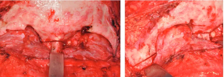
Figure 3. A) Horizontal osteotomy below the fronto-nasal suture. B) Oblique osteotomy of the zygomatic bone (right side).
Figure 4. Stable internal ixation. A) In the fronto-nasal region. B) In oblique osteotomy.
Operative technique
The surgery was begun through bicoronal access; the incision line was drawn with methylene blue through the head vertex to the preauricular area bilaterally. After subcutaneous iniltration (bupivacaine 0.5% with
lines was performed. The dissection continued under the periosteum, and the supraorbital vascular-nervous bundle was released from its foramen through an osteotomy with the piezoelectric motor; the orbital content was moved away to perform detachment of the orbital borders until the inferior orbital issure was identiied. During periorbital dissection, the cantal ligament remained intact, and the dissection extended back to the lacrimal apparatus.
Sideways, the outer layer of the deep temporal fascia was incised superiorly to the zygomatic arch and continuously joined with the incision of the pericranium. The dissection was then performed through the layer of fat to achieve the zygomatic arch and extended back to the anterior to expose the zygoma and the lateral wall of the orbit. Afterwards, subperiosteal detachment was conducted over the nasal bones.
Figure 5. Photos in the postoperative period of 03 years. A) Front view. B) Proile view. C) Occlusal view.
Figure 6. Postoperative CT.
A horizontal osteotomy was performed right below the frontonasal suture, extending laterally through the median orbital surface and posteriorly to the lacrimal
osteotomy was performed on the side of the orbita to the zygomatic bone inferior area (Figure 3B). Through the transconjunctival access bilaterally, a third osteotomy was performed, joining the other two osteotomies through the orbital loor carefully so as not to rupture the infraorbital branch. All osteotomies were performed with the piezoelectric motor and completed with chisels. The chisel was introduced in the nasal bone, perpendicular to the cribiform plate when the separation of the nasal septum from the skull was performed.
In order, the maxilla, mandible and chin were iniltrated with a local anesthetic, and the maxilla was incised bilaterally (Figure 4A, B). The detachment was extended to expose the side walls in the nasal cavity, the posterior area of the maxilla and the inferior area of the zygoma. Using chisels, the osteotomy was completed in the inferior area of the zygoma and posteriorly of the maxilla tuberosity on both sides. A curved chisel was used to promove the pterygoid plates’ disjunction.
Rowe forceps were used to mobilize the segment. After the complete manipulation, the maxillo-mandible block with the intermediate split 1 was performed, promoting a 4-mm midface straightforward (including the maxilla). The ixation was performed with two plates and monocortical screws in the frontonasal suture and one straight plate in the oblique osteotomy in the zygoma body bilaterally. A small undesirable fracture occurred in the infraorbital rim on both sides and requiring ixation with miniplates and screws. The maxillo-mandible block was removed, and intermediate occlusion, the symmetry of the advance and the segment stability were checked.
This was followed by the Le Fort I osteotomy and maxilla down fracture to 5 mm advance planned, using as a reference intermediate splint 2, which was ixed with four L plates and screws. After the maxillo-mandible lock was removed, the positioning of the maxilla, upper-incisor exposure and new intermediate occlusion were checked. In order, through BSSO, the mandible was set back to 7 mm, correcting the midline deviation. The segments were ixed with a straight plate and monocortical screws bilaterally. Finally, the genioplasty was held with a 6-mm vertical reduction, and the ixation was performed with plate and monocortical screws. The blocking was removed, and the inal occlusion was checked.
The intraoral and transconjunctival approaches were sutured with continuous sutures and absorbable thread. The bicoronal approach was repositioned, and after suspensory sutures in the cantal lateral ligament and
temporal muscle with absorbable thread, the scalp was closed in two layers, the deepest with absorbable sutures and shallowest with nylon 3-0 with portovac drain suction installation. The scalp was held using a compressive bandage around the head and chin.
The patient was kept for the irst 48 hours in the intensive care unit. The drain portovac was removed after 48 hours. After ive days, the patient was discharged from the hospital.
Currently, after three years of postoperative follow-up he shows signiicant improvement in chewing ability, denies respiratory and visual complaints and has no pain or functional complaints. In addition, patient reports high satisfaction with the aesthetics (Figure 5A, B, C).
Postoperative computed tomography demonstrates the stability of ixation (Figure 6).
DISCUSSION
Maxilla hypoplasia is a common diagnosis in the spectrum of dentofacial deformities and is usually corrected by a Le Fort I osteotomy to maxilla advance. However, when we encounter a severe midface hypoplasia, we should consider that facial appearance is heavily inluenced by the periorbital area. This area includes the eyeballs, eyelids, eyebrows and cheeks. The symmetry, form and position of these components are extremely important because little alterations in this area contribute signiicantly to an individual’s appearance11.
Depending on the extent of midface hypoplasia, the surgical treatment of the deformity could be performed by a quadrangular Le Fort I or by Le Fort II osteotomy. However, these techniques do not properly correct the malar hypoplasia that could be presenting10. Other approaches have been used, such as the Le Fort I osteotomy with an increased infraorbital region. Procedures like higher Le Fort I osteotomies, without addressing the infraorbital region, can lead to the patient having a sunken appearance in the upper portion of the midface. The simultaneous increase in the infraorbital area with alloplastic associated with Le Fort I, leads to graft communication with the maxillary sinus and may subsequently become infected. Bone grafts used to increase the infraorbital rim are unpredictable in their resorption rate and are uncomfortable to the patient. Thus, for these cases, a Le Fort III osteotomy or one of its modiications is more appropriate12.
the naso-orbit-malar level, could result in enophthalmos, and in cases of normal nasal projection, it can result in an undesirable increase in nasal prominence. In addition, even with the greatest advances, a deformity in the lateral orbital arches can be found. To avoid this, modiications in the Le Fort III osteotomy were proposed. In 1971, Kufner13 proposed a modiication to correct midface projection deiciencies without involving the nasal subunit. The modiication involves an osteotomy in the orbital lateral border to the zygoma body, through the orbital loor, through the inferior orbital issure, and through the maxilla to the side wall of the nasal cavity. Other advantages of this modiication include midface stabilization, avoiding the need for bone grafting, facilitating the ixing of the plates, allowing greater bone interfacing and the postoperative protection of the orbital sclera.
Cheung et al.10 described the application of an oblique modiied Le Fort III osteotomy that included the bones in the nose in addition to a Le Fort I osteotomy with segmentation for treatment of non-syndromic patients with maxillar hypoplasia in three patients, obtaining satisfactory results.
The patient with the midface deiciency associated with maxillary deiciency has been well described in the literature. Technical problems arise when the maxilla and midface require different movements, when different midline deviation presents or when the maxilla should be segmented12. When the maxilla and midface movements are confronted, it is necessary to perform the Le Fort I associated with the Le Fort III osteotomy. Le Fort I osteotomy association is also especially useful in cases in which there is a difference in the extent of the midface hypoplasia in the orbital region in relation to a maxillo-mandibular discrepancy14.
The biggest advantage of simultaneous Le Fort III and Le Fort I osteotomies is that both, midface deformity and maxilla, can be addressed in a predictable manner and simultaneously in separate segments, resulting in optimal aesthetic results when the horizontal maxilla deiciency differs signiicantly from the infraorbital areas12. In addition, it minimizes the advancement in the Le Fort III osteotomy, reducing the need for bone grafts14-15.
The patient in this report had midface hypoplasia involving the nasal bones, zygoma, inferior orbital rims and maxilla bone. His maxillo-mandibular discrepancy was greater than his midface deiciency, mainly due to its severe mandibular prognathism. However, a very large mandibular
setback could damage the patient’s airway. Thus, for the midface approach, the surgical technique was chosen based on the modiication performed by Cheung et al.10 with a Le Fort III osteotomy involving the nasal bones and the Le Fort I osteotomy, allowing further advancement of the maxillary alveolar segment.
The Le Fort III osteotomy is considered a complex technique, representing a challenge to surgeons due to the risk associated with several complications that could be a mild recurrence and extending to blindness and death14. Therefore, it should be considered only in speciic cases in which it is impossible to resolve the patient’s deformity and complaints with other less complex techniques. In addition, the patient must have be in good physical condition to support the procedure due to prolonged operative time and extensive blood loss.
The patient who will be subjected to these extensive reconstructions should be aware of the possible transoperative and postoperative complications. In the preoperative period, the preparation for possible trans operative complications should be performed, a bed should be booked in the intensive care unit and erythrocyte concentrates reservation. The patient should also be accompanied by a multidisciplinary team including an ophthalmologist, otolaryngologist, nutritionist, psychologist, physiotherapist and speech therapist.
Moreover, it is important to clarify that dentofacial deformities arise from skeletal defects involving the three planes of space, and careful planning is required for the correct three-dimensional positioning of all segments. Any planning error can result in subsequent errors, preventing proper correction of the deformity. Thus, both the surgeon and orthodontist have a fundamental function in surgical planning for the correct management before and after the intervention, enabling occlusion, oral function and appropriate aesthetic appearance.
Collaborators
REFERENCES
1. Santos R, Sebastiani AM, Todero SRB, Moraes RF, Costa DJ, Rebelatto NLB, et al. Complicações associadas à osteotomia sagital dos ramos mandibulares. Rev Cir Traumatol Bucomaxillofac. 2012;12(1):77-84.
2. Vu DD, Tiwana OS. Le Fort III and Le Fort II osteotomies. Atlas Oral Maxillofacial Surg Clin N Am. 2016;24:15-25.
3. Hariri F, Lan TH, Cheung LK. Simultaneous Le Fort III and Le Fort I osteotomies for correction of midface hypoplasia in Crouzon syndrome. Asian J Oral Maxillofac Surg. 2011;23(3):128-33. doi: 10.1016/j.ajoms.2011.04.002
4. Saltaji H, Altalibi M, Major MP, Al-Nuaimi MH, Tabbaa S, Major PW, et al. Le Fort III distraction osteogenesis versus conventional Le Fort III osteotomy in correction of syndromic midfacial hypoplasia: a systematic review. J Oral Maxillofac Surg. 2014;72(5):959-972. doi: 10.1016/j.joms.2013.09.039
5. Dai J, Wang X, Yu H, Cheng J, Yuan H, Gui H, et al. Simultaneous Le Fort I, II, and III osteotomies for correction of midface deiciency in apert disease. J Craniofac Surg. 2012;23(5):1391-5. doi: 10.1097/SCS.0b013e3182565a5d
6. Gilles H, Harrison SH. Operative correction by osteotomy of recurred malar-maxillary compound in a case of oxycephaly. Br J Plast Surg. 1950;3(2):123.
7. Tessier P. Total osteotomy of the middle third of the face for faciostenosis or for sequelae of Le Fort 3 fractures. Plast Reconstr Surg. 1971;48(6):533-41.
8. Magraw CB, Garaas R, Shaw A, Phillips C, Turvey TA. Changes in scleral exposure following modiied Le Fort III osteotomy. Oral Surg Oral Med Oral Pathol Oral Radiol. 2015 Aug;120(2):119-24. e1. doi: 10.1016/j.oooo.2015.04.005
9. Schmitz JP, Tiner BD, van Sickels JE. Stability of simultaneous modiied LeFort III/LeFort I osteotomies. J Craniomaxillofac Surg. 1995 Oct;23(5):287-95.
10. Cheung LK, Ow A, Chua HD. Simultaneous modiied oblique Le Fort III and segmentalized Le Fort I osteotomies. J Oral Maxillofac Surg. 2010 Apr;68(4):915-23. doi: 10.1016/j.joms.2009.06.009 11. Soydan SS, Bayram B, Sar C, Uckan S. Change in inferior sclera
exposure following Le Fort I osteotomy in patients with midfacial retrognathia. J Oral Maxillofac Surg. 2014;72(166):e161-e165 12. van Sickels JE, Tiner BD. A combined Le Fort I and bilateral
zygomatic osteotomy for management of midface and maxillary deiciency. J Oral Maxillofac Surg. 1994 Mar;52(3):327-31. doi: 10.1016/0278-2391(94)90312-3
13. Kufner J. Four-year experience with major maxillary osteotomy for retrusion. J Oral Surg. 1971;29(8):549-53.
14. Nout E, Cesteleyn LL, van der Wal KG, van Adrichem LN, Mathijssen IM, Wolvius EB. Advancement of the midface, from conventional Le Fort III osteotomy to Le Fort III distraction: review of the literature. Int J Oral Maxillofac Surg. 2008 Sep;37(9):781-9. doi: 10.1016/j.ijom.2008.04.006
15. Saltaji H, Altalibi M, Major MP, Al-Nuaimi MH, Tabbaa S, Major PW, et al. Le Fort III distraction osteogenesis versus conventional Le Fort III osteotomy in correction of syndromic midfacial hypoplasia: a systematic review. J OralMaxillofac Surg. 2014 May;72(5):959-72. doi: 10.1016/j.joms.2013.09.039
Received on: 16/3/2016 Final version resubmitted on: 23/6/2016