ABSTRACT
Objective
To quantify the color variation of two glass ionomer cements and a composite resin used in pediatric dentistry, after being immersed in different pigments agents.
Methods
Using two glass ionomer cements (Ketac™ Molar and Photac™ Fil) and a microhybrid composite resin (Filtek™ z250), were produced 40 disks of each material (10 mm in diameter and 2 mm thick). The samples were soaked in artiicial saliva (control group), coke, peach Ice Tea®
and chocolate milk, for 72 hours in an oven at 37ºC. After this period, the samples were washed in 50 ml of distilled water. Finally, using the spectrophotometer, it was made the reading of results. The color change was measured according to the CIE L * a * b * system. Color changes were statistically analyzed using parametric one-way ANOVA and ANOVA with Welch correction, the nonparametric Kruskal-Wallis tests and post-hoc Tukey and Dunnet T3 with p≤ 0.05.
Results
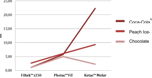
The immersion of restorative materials in different pigmentation agents caused a signiicant color variation on the samples. The agent who presented higher results was the Peach Ice Tea®. The chocolate milk was the luid with lowest pigmentation capacity of all restorative materials under study. The
greater color variation was found on the Ketac™ Molar submerged in Coca-Cola® and the smallest on the Filtek™ z250 in chocolate milk.
Conclusion
All restorative materials were shown to be susceptible to pigmentation by all agents. The Filtek™ z250 proved to have better color stability, followed by Photac™ Fil and inally by Ketac™ Molar.
Indexing terms: Composite resins. Glass ionomer cements. Pigmentation.
RESUMO
Objetivo
Quantiicar a variação da cor de materiais restauradores utilizados emodontopediatria, após serem imersos em agentes pigmentantes.
Métodos
Recorrendo aos cimentos de ionómero de vidro Photac™ Fil e Ketac™ Molar, e à resina composta microhíbridaFiltek™ z250, foram efectuados 40 discos de cada material (10 milímetros de diâmetro e 2 milímetros de espessura). As amostras foram imersas em saliva artiicial (grupo controlo), Coca-Cola®, Ice Tea® de pêssego e Leite com chocolate, durante 72 horas, numa estufa a 37ºC. Decorrido este período, as amostras
foram lavadas em 50 mililitros de água destilada. Por último, através da utilização do Espectrofotómetro, procedeu-se à leitura dos resultados. A variação da cor foi medida de acordo com o sistema CIE L* a* b* e analisada estatisticamente utilizando os testes paramétricos ANOVA one-way e ANOVA com correcção de Welch, o teste não paramétrico Kruskal-Wallis e os testes post-hoc Dunnet T3 e Tukey com p≤ 0,05.
Resultados
A imersão das amostras nos luidos, provocou uma variação de cor signiicativa das mesmas. O agente que apresentou resultados de pigmentação mais elevados foi o Ice Tea®de pêssego. O leite com chocolate foi o luido que apresentou menor capacidade pigmentante em todos os materiais em estudo. A maior variação de cor veriicou-se no Ketac™ Molar em Coca-Cola® e a menor no Filtek™ z250 em leite com chocolate.
Conclusão
Todos os materiais restauradores demonstraram ser suscetíveis à pigmentação, por parte dos agentes. O Filtek™ z250 demonstrou ter uma melhor estabilidade de cor, seguido pelo Photac™ Fil e pelo Ketac™ Molar.
Termos de indexação: Pigmentação. Cimentos de ionómero de vidro. Resinas compostas.
Quantiication of color variation of restorative materials used on
pediatric dentistry after pigmentation
Quantiicação da variação da cor de materiais restauradores utilizada em Odontopediatria após
pig-mentação
Luísa Bandeira Pires Monteiro LOPES1
Andreia Soia Lopes de ARAÚJO1
Virginia Barreiros MILAGRE1
with regard to the physical properties and maintaining
advantages such as adhesion and luoride release7. As in the
conventional GIC, the modiied resins exhibit polyacrylic acid and a basic powdered glass in their constitution. However resinous hydrophilic monomers such as group
2-hydroxyethyl-methacrylate (HEMA) and photoinitiator5 are added in its
constitution. This material possess a wide variety of uses and can be applied as a restorative material, cementing material,
indirect pulp protector and also as a pit and issure sealant8,
making it easy to understand why the RMGIC have gained great popularity and currently being widely used in the
practice of odontopediatrics dentistry9.
The Composite Resins were irst introduced in the
mid-1960’s10. This material possesses several applications
such as: direct restorations, cavity lining, sealing of pits and issures, core buildings, inlays, onlays, crowns, provisional restorations, cementing unit or multiple prostheses and
orthodontic appliances, among others11. In its constitution
we can ind a polymeric matrix (organic matter), charged particles (inorganic matter), a silane, canforoquinonas that promote or modulate the polymerization reaction, and
pigments12. Despite the fact that the composite resins
have been suffering several changes over the time, the
pigmentation of material still constitutes a problema11.
METHODS
The Restorative materials used in this study were a Conventional Glass Ionomer Cement (Ketac Molar, 3M ESPE, Minnesota, USA), a Glass Ionomer Cement Modiied by Resin (Photac Fil, 3M ESPE, Minnesota, USA) and a Microhybrid Composite Resin (Filtek z250, 3M ESPE, Minnesota, USA). One hundred and twenty samples in disc form were prepared, using a metallic matrix with 10mm diameter and 2mm of thickness. A sheet of acetate was placed between the glass plate and the metallic matrix. Forty discs were then produced with each restorative material according to the manufacturer’s instructions, photopolymerizating the Glass Ionomer Cement for 20 seconds and the Composite Resin for 40 seconds. The forty discs of each restorative material were then divided into four groups, ten discs from each placed into a different immersion liquid: artiicial
saliva (control group), Coca-Cola®, chocolate milk
and peach Ice-Tea®. After immersed in different liquid
pigments, in speciic containers, the one hundred and twenty samples were stored in an oven at 37 ºC, to reproduce the oral environment for 72 hours. At the end of 72 hours, the samples were washed in 50ml of
INTRODUCTION
The teeth are usually identiied as an important feature in the beauty of the face, performing a major role in the relationship and iteractions between the individuals
and the society1. In modern times we have to consider the
color change that teeth are exposed to, since the aesthetic demands by the patients have grown at an exponential level. Due to this growing interest in regard to dental esthetics, it is considered important to carry out investigations with the purpose of studying the characteristics of various restorative materials to subsequently proceed to the improvement of their physical properties, including color stability.
The theet color is determined by a combination between the intrinsic color of the tooth and the extrinsic
pigments that can be formed on its surface2. Teeth
discoloration can be classiied as intrinsic, extrinsic or a
combination of both3. The intrinsic pigmentation results from
structural changes related to the thickness or composition of the dental hard tissues; the causes can be related to metabolic diseases and systemic factors that alter the tooth
development and consequently its pigmentation2-3. In the
extrinsic pigmentation, the responsible materials are located in the surface of the tooth or in the acquired pellicle. This pigmentation can be removed through dental prophylaxis. Among the materials responsible for the pigmentation we can ind: food ingridients containing tannis, coffee, tea, red
wine, tobacco, iron salts, chlorhexidine and others2-3.
Over the past 50 years there have been several changes in the development and availability of restorative materials used in odontopediatrics dentistry, with the appearance of the Composite Resins and the Glass Ionomer Cement. Thus, currently the dentist has at his disposal several materials, from which he can choose the most appropriate for
each type of restoration4.
The irst Glass Ionomer Cement (GIC) was developed by Wilson and Kent in the mid- 70’s. These so-called conventional Ionomers, present in its composition an aqueous solution of polyacrylic acida at a 45% concentration and a powder consisting of Fluoramino Silicate Glass, Silicon,
Aluminum, Phosphorus, Fluorine, Sodium and Calcium5. The
conventional GIC are widely used in children and can be used as sealants for pits and issures, cavity liner, cementing
agents and as illing material5. They are also profusely used in
Atraumatic Restorative Treatment, since they have the ability
to remineralize the dental hard tissues6.
In response to the disadvantages related to
convencional GIC, the Ionomer glass Modiied Resins (RMGIC)5
In the matter related with the comparison of solutions in the same restorative material, by statistical analysis, it can be concluded that there is a signiicant difference between the pigmentante capacity of all agents containing pigment, when applied to the Ketak™ Molar.
The same does not apply between Coca-Cola® and the
chocolate milk when the Filtek™ z250 is subjected to its action, since the pigmentation values caused by them in this material are quite close. In the case of Photac™ Fil there is only signiicant differences of pigmentation between the
chocolate milk and the peach Ice-Tea®, being Ice-Tea® the
luid who presents the higher value of pigmentation. In general, it is possible to claim that the peach
Ice-Tea® was the luid who showed the highest pigmentante
capacity in the restorative materials, the exception being the Ketac™ Molar. In this case, the pigmentante agent that caused greater chromatic change in the restorative
material was the Coca-Cola®. In the opposite perspective,
the chocolate milk was the pigmentante agent who showed the lowest pigmentation values.
Focusing on the comparison of restorative materials, when subjected to the same pigmentante agent, after statistical analysis, it can be concluded that the difference between the pigmentation caused by the pigmentante agents is statistically signiicant. This fact applies for all materials when they are submitted to the action of any luid.
distilled water, to remove possible residues existing on the surface. The reading of the samples was performed using the spectrophotometer Spectro - Shade Micro (MHT - Niederhasli, Switzerland) and the variation of the color was mesured using the CIE L* a* b*. The chromatic variation was determined by the difference between the coordinates L*, a* and b* obtained after the immersion of the disks in different pigmentation agents and the coordinates L*, a* and b* calculated to an average of 10 discs immersed in artiicial saliva (the control group). Thus, the formula used to determine the color stability
was: ΔE = [(inal L* - L* mean of the control group)2 +
(inal a* - a* mean of the control group)2 + (inal b* - b*
mean of the control group)2]1/2. To test the differences
of chromatic variation between groups of restorative materials and within the same group, but with different pigmentation agents, it was used the one-way ANOVA test. When the assumption of normality was not satisied, the nonparametric test of Kruskal - Wallis was used as an alternative. When the assumption of homogeneity in the variances was not satisied, the ANOVA test with Welch correction was applied. Mutiple comparison tests were also conducted, the Tuckey test, applied when the null hypothesis in the ANOVA test was rejected and Dunnett T3 test, when the same happened in the ANOVA test with Welch correction. The degree of signiicance to accept or reject the null hypothesis was p ≤ 0,05. The statistical analysis was performed using SPSS (Statistical Package for the Social Sciences) version 20.0 for Windows.
RESULTS
The mean values of color variation (ΔE) and the standard deviations are shown in Table 1.
Tabel 1. Mean values of ΔE and standard deviations.
ΔE Filtek™ z250 Ketac™ Molar Photac™ Fil
Coca-Cola® 1,11 (0,40) 22,29 (1,55) 5,76 (0,83)
Chocolate
milk 1,07 (0,40) 2,28 (0,55) 5,00 (0,41) Peach
Ice Tea® 2,67 (0,46) 9,33 (0,53) 6,10 (0,47)
Coca-Cola ®
Peach
Ice-Tea ®
Chocolate milk
pigmentation values were higher for the Photac™ Fil. The Filtek™ z250, proved to be the material that suffered less color variation after being subjected to the different agents of pigmentation. In general, the material that suffered the greatest
chromatic change was the Ketac™ Molar, however when the restorative materials were submitted to the chocolate milk,
Figure 2. Compararison of pigments agents.
Coca‐Cola® Chocolate milk Peach Ice‐Tea®
DISCUSSION
In modern times the general population shows an increasingly concern not only with the oral health but also with their appearance. The teeth are seen as an importante feature in the beauty of the face, performing a crucial role in the interactions and the individual´s relationship
with society1. For the restorative material to be clinically
acceptable it is important that this presents a most similar color as possible to the tooth color after the application, in order to give an aesthetic aspect, and it is also essencial
that it remains over time13.
This study evaluates the color stability of three restorative materials used in the practice of pediatric dentistry, two glass ionomer cements (Ketac™ Molar and Photac™ Fil) and a microhybrid composite (Filtek™ z250), after being subjected to the action of different drinks commonly ingested by children and adolescentes. The null hypothesis suggested for this study was rejected, since there have been signiicant changes in the color of restorative materials before and after immersion in different agents of pigmentation.
The color determination of a tooth by visual comparison with color guides is the most often used method in clinical destistry. However during recent years there has been development of new technologies directed
to analysis, communication and veriication of color14.
In this investigation it was used the spectrophotometer, as it eliminates the subjectivity of the analysis done by visual comparison and considering the fact that it is one of the most useful and accurate tools in
matching colors15. Since the American Dental Association
recommends using the CIE L* a* b* to evaluate possible
changes in color, applying the formula of color variation (ΔE) which correlates the coordinates L* a* and b*, this was
chosen to be used in this investigation16. The coordinate L*
represents the lightness of the color, while the coordinates a* and b* refer to the chromatic characteristics of color. The coordinate a* represents the range of color from green
until red, while b* refers to the blue-yellow axis17.
As it was mentioned before, an essential feature for a restorative material to be clinically acceptable is the stability of its color. Several authors state that ΔE values between 1 and 3 are visible to the naked eye and above
3.3 are clinically unacceptable13.
Based on the literature, it is clear that all restorative materials in study, experienced a color change noticeable to the naked eye and apart from this fact, it is still possible to verify that considering the three materials, only the composite resin Filtek™ z250 achieved clinically acceptable results.
The color changes in dental structures and restorative materials may occur due to intrinsic and extrinsic factors. Several studies suggest that diet is one of the factors that leads to the development of extrinsic pigmentation. Restorative materials being constantly subjected to oral environment and daily exposed to salive, food and beverage, can therefore suffer changes
in their color18-19.
The presence of soft drinks in the diet of the population has increased considerably in recent years, being the children and adolescentes the largest consumers of such drinks. In addition to soft drinks also chocolate milk is often present in eating habits of this age group, hence the choice
of Coca-Cola®, peach Ice Tea® and chocolate milk to join
the effect of Coca-Cola® in chromatic stability of various
restorative materials13,18,20, the number of investigations
using chocolate milk as pigmentante agent is quite limited18.
In this study the peach Ice-Tea® demonstrated
a superior pigmentante capacity when compared with
the other agents containing pigment, Coca-Cola® and
chocolate milk. Several investigations who studied this same characteristic also show that tea has more pigmentante
effect on restorative materials than Coca-Cola®18,20.
A study by Tunc et al.19 that as this research
evaluated the susceptibility of Glass Ionomer Cements and one Composite Resin to the pigmentation, veriied
that Coca-Cola® was the more pigmented solution; on the
other hand, the chocolate milk was the agent that showed lower value of pigmentation, lying in accordance with what was found in this study.
The susceptibility of resinous restorative materials for pigmentation can be related to the type of polymerization, iller particles, type of pigmentant agents,
among others20. This susceptibility also comes from the
presence of monomers such as α - glycidyl methacrylate
bisphenol (bis - GMA), urethane dimethacrylate (UDMA) and triethyleneglycol dimethacrylate (TEGDMA) on their matrix, making them possess a hydrophilic médium, leading to a greater absorption of water and other solutions,
thereby providing a color change in the material18.
The Conventional Glass Ionomer Cements (CGIC) and the ones modiied by resin (RMGIC), possess different compositions, so their susceptibility to suffer pigmentation
will naturally be different. Cattani-Lorente et al.21 and
Small et al.22 conducted studies that show that water
absorption by RMGIC is higher than that absorbed by the Conventional due to the absorption of this by the HEMA, a resinous component present in the constitution of RMGIC. Another reason that explains this difference may reside in the fact that the CGIC has greater amount of water in its composition as compared to the RMGIC, therefore it will absorb less water and consequently less pigments.
In the present investigation, the microhybrid composite resin Filtek™ z250 proved to be the restorative material less susceptible to pigmentation, and the CGIC Ketac™ Molar the material that suffers the biggest change in
the color. However in the study by Tunc et al.19, the results do
not match with this investigation because despite the fact that they show lower pigmentation values from Composite Resin Filtek™ z250 in comparison with RMGIC, the CGIC was the restorative material which showed lower values. This may be due to an increase in surface roughness of the material used in this study, thereby retaining a larger amount of pigment.
A study by Austregésilo & Filho23, shows that the
RMGIC have a smaller roughness in the surface as compared with CGIC. For a better evaluation of the materials studied, speciically with regard to the color variation after pigmentation, and in order to combat some of its limitations, it would have been interesting to introduce the analysis of surface roughness of different materials.
Another property which differentiates CGIC from RMGIC is the microhardness, since the CGIC presents
higher values in that matter24. In the future, it would be
interesting to conduct an investigation similar to this one, introducing the analysis of physical property, in order to check if there is any relationship between this and the susceptibility of pigmentation of restorative materials.
Several studies by different researchers, still give us account of pigmentation differences between the same
materials but of different brands13,18,20. In a future study it
would be advantageous to add several brands of the same material in order to analyze this difference.
CONCLUSION
It has occurred change in the color of all materials tested in this investigation, after their immersion in the different agents of pigmentation. The material that has suffered major change in the color after pigmentation was the Ketac™ Molar and the one who suffered minor change in the color after pigmentation was the Filtek™ z250. The
Ice-Tea® was the agent containing pigment that, in a general
way, caused greater color variation and the chocolate milk proved to be the one who caused less change in all the tested materials. The Microhybrid Composite Filtek™ z250 was the material that showed higher color stability, and although the color variation caused by the three agents of pigmentation is visible to naked eye, It is considered clinically acceptable. In a general way the color change observed in the Ketac™ Molar and Photac™ Fil is not only visible to the naked eye as it is not clinically acceptable.
Collaborators
REFERENCES
1. Tin-Oo MM, Saddki N, Hassan N. Factors inluencing patient satisfaction with dental appearance and treatments they desire to improve aesthetics. BioMed. 2011;11(1):6.
2. Watts a, Addy M. Tooth discolouration and staining: a review of the literature. Br Dent J. 2001;190(6):309-16.
3. Azer SS, Hague AL, Johnston WM. Effect of pH on tooth discoloration from food colorant in vitro. J Dent. 2010; 38(2):106-9.
4. Berg JH. The continuum of restorative materials in pediatric dentistry - a review for the clinician. Pediatr Dent. 1998;20(2):93-100.
5. Sidhu SK. Glass-ionomer cement restorative materials: a sticky subject? Aust Dent J. 2011;56(1):23-30.
6. Mickenautsch S, Mount G, Yengopal V. Therapeutic effect of glass-ionomers: an overview of evidence. Aust Dent J. 2011;56(1):10-15.
7. Sidhu SK. Clinical evaluations of resin-modiied glass-ionomer restorations. Dent Mater. 2010;26(1):7-12.
8. Maneenut C, Sakoolnamarka R, Tyas MJ. The repair potential of resin-modiied glass-ionomer cements. Dent Mater. 2010;26(7):659-65.
9. Aguiar FHB, Georgetto MH, Soares GP, Catelan A, Dos Santos PH, Ambrosano GMB, et al. Effect of different light-curing modes on degree of conversion, staining susceptibility and stain’s retention using different beverages in a nanoilled composite resin. J Esthet Restor Dent. 2011;23(2):106-14.
10. Barutcigil Ç, Yıldız M. Intrinsic and extrinsic discoloration of dimethacrylate and silorane based composites. J Dent. 2012;40(1):57-63.
11. Ferracane JL. Resin composite--state of the art. Dent Mater. 2011;27(1):29-38.
12. Hervás-García A, Martínez-Lozano MA, Cabanes-Vila J, Barjau-Escribano A, Fos-Galve P. Composite resins. A review of the materials and clinical indications. Med Oral Patol Oral Cir Bucal. 2006;11(2):215-20.
13. Mundim FM, Garcia LDFR, Pires-de-Souza FDCP. Effect of staining solutions and repolishing on color stability of direct composites. J Appl Oral Sci. 2010;18(3):249-54.
14. Chu SJ, Trushkowsky RD, Paravina RD. Dental color matching instruments and systems. Review of clinical and research aspects. J Dent. 2010;38(2):2-16.
15. Paul S, Peter A, Rodoni L, Pietrobon N. Conventional visual vs spectrophotometric shade taking for porcelain-fused-to-metal crowns: a clinical comparison. Int J Periodontics Restorative Dent. 2004;24(3):222-31.
16. Ayad NM. Susceptibility of restorative materials to staining by common beverages: an in vitro study. Eur J Esthet Dent. 2007; 2(2):236-47.
17. Moreira AD, Mattos CT, De Araújo MVA, Ruellas ACDO, Sant’Anna EF. Chromatic analysis of teeth exposed to different mouthrinses. J Dent. 2013;41(5):24-7.
18. Bagheri R, Burrow MF, Tyas M. Inluence of food-simulating solutions and surface inish on susceptibility to staining of aesthetic restorative materials. J Dent. 2005;33(5):389-98.
19. Tunc E Sen, Bayrak S, Guler AU, Tuloglu N. The effects of children’s drinks on the color stability of various restorative materials. J Clin Pediatr Dent. 2009;34(2):147-50.
20. Guler AU, Yilmaz F, Kulunk T, Guler E, Kurt S. Effects of different drinks on stainability of resin composite provisional restorative materials. J Prosthet Dent. 2005;94(2):118-24.
21. Cattani-Lorente MA, Dupuis V, Payan J, Moya F, Meyer JM. Effect of water on the physical properties of resin-modiied glass ionomer cements. Dent Mater. 1999;15(1):71-8.
22. Small ICB, Watson TF, Chadwick AV, Sidhu SK. Water sorption in resin-modiied glass-ionomer cements: an in vitro comparison with other materials. Biomaterials. 1998;19(6):545-50.
23. Austregésilo SC, Filho PFM. Avaliação da rugosidade supericial de cimentos ionoméricos convencionais e modiicados. XVII Congresso de Iniciação Cientíica. 2009. p. 27-30.
24. Xie D, Brantley WA, Culbertson BM, Wang G. Mechanical properties and microstructures of glass-ionomer cements. Dent Mater. 2000;16(2):129-38.