ABSTRACT
The ameloblastic ibro-odontoma is a benign, mixed, asymptomatic and rare odontogenic tumor that can easily be confused radiographically and histologically with other diseases. Usually it affects a population between the irst and second decades of life, is more frequent in the mandible and shows predilection for males. This lesion, characterized by dental agenesis at the tumor site, has no predilection for anatomical region; however, an increase of intraoral volume may cause mild facial asymmetry. It shows slow but expansive growth, and is a well-deined radiolucent lesion, usually unilocular, with radiopacity inside. Normally surgical removal is conservative and the prognosis is favorable. This article reports a case of ameloblastic ibro-odontoma treated by surgical removal and follow-up of two years. The aim of this work was to study the ameloblastic ibro-odontoma in its entirety, seeking to inform clinicians about the best diagnostic and treatment methods for this type lesion.
Termos de indexação: Diagnosis. Odontoma. Therapeutics.
RESUMO
O fibro-odontoma ameloblástico é um tumor odontogênico misto benigno, assintomático e raro, que pode ser facilmente confundido radiograficamente e histopatologicamente com outras enfermidades. Normalmente atinge uma população entre a primeira e a segunda décadas de vida, sendo mais frequente em mandíbula e com predileção pelo sexo masculino. Este tumor caracteriza-se pela agenesia dentária no local do tumor, sem predileção por região anatômica, e por um aumento de volume intrabucal, podendo causar leve assimetria facial. Apresenta crescimento lento, porém expansivo; revela-se como uma lesão radiolúcida bem definida, normalmente unilocular, com radiopacidades no seu interior. Normalmente a remoção cirúrgica é conservadora e o prognóstico é favorável. O presente trabalho relata um caso clínico de fibro-odontoma ameloblástico tratado por meio de remoção cirúrgica e com acompanhamento de dois anos. O objetivo deste trabalho foi estudar o fibro-odontoma ameloblástico em sua integralidade, buscando expor ao cirurgião-dentista os melhores métodos de diagnóstico e tratamento para tal lesão.
Termos de indexação: Diagnóstico. Odontoma. Terapêutica
Ameloblastic ibro-odontoma: case report
Fibro-odontoma ameloblástico: relato de caso
Cintia Gollo PIVA1
Daniela Cristina MIYAGAKI1
Maria Salete LINDEN1
Ferdinando DE CONTO1
Isadora RINALDI1
João Paulo DE CARLI1
1 Universidade de Passo Fundo, Faculdade de Odontologia. Campus Universitário, São José, 99052-900, Passo Fundo, RS, Brasil. Correspondência para
with the formation of dentin and enamel matrix.
AOF affects a young population with a mean age of 19 years. They are more frequent in the posterior region of the mandible, followed by the posterior region of the maxilla4,6-7.
Buchner et al.7, in their research showed that
AFO has a discrete predilection for the male gender4,8,
however, some studies, and the WHO affirm that there is no predilection for gender and localization5,9-10.
Lesion growth is slow, but expansive, and may cause an increase in volume within the mouth, and also
INTRODUCTION
The ameloblastic fibro-odontoma (AFO) is an uncommon, mixed odontogenic tumor that represents from 1 to 3% of all the odontogenic tumors1-5 and
years. A logical sequence of diagnosis, treatment and clinical follow-up of the AFO was established, with the aim of studying the ameloblastic fibro-odontoma in its entirety, to inform dentists about the best methods of intervention in this pathology.
CASE REPORT
The patient, a 19-year-old Caucasian woman, sought dental care in April 2012 for third molar extractions. In the intra and extraoral exams no increase in volume of change in gingival color were verified. The patient denied any changes in health, and presented to local discomfort.
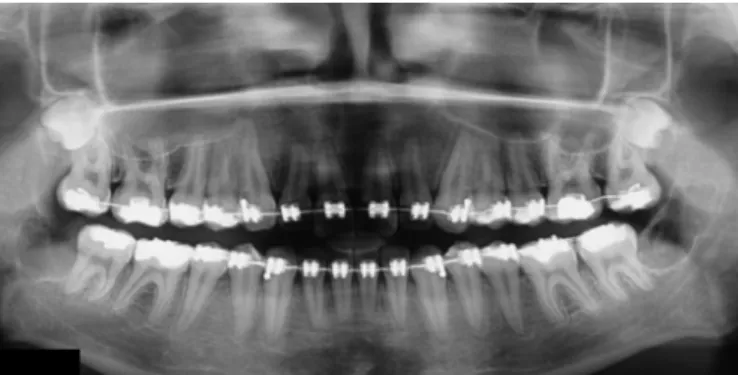
In the panoramic radiograph (Figure 1) agenesis of tooth 38 was identified. A closer observation revealed a well-defined radiolucent image of about 25 mm in diameter, localized in the distal portion of tooth 37, without present a direct relationship with this tooth. Within this image, discrete radiopaque foci were noted. During anamnesis, the patient informed that she had had tooth 48 extracted three months previously, and that she had been using an orthodontic appliance for the last three years.
Figure 1. Panoramic radiograph showing a radiolucent image with discrete radi-opaque point inside it and well defined margins in the region of the ascending ramus of the mandible on the left.
In May 2012, with a presumed diagnosis of AFO, calcifying odontogenic and calcifying epithelial odontogenic tumor, an incisional biopsy of the lesion was performed, and seven fragments of soft tissue were removed, of which two were white and the remainder brown, all grossly spherical and friable, together measuring 4x3x1mm.
In the histopathological exam results, the cut stained by the HE technique showed an odontogenic epithelium sometimes in cords, and sometimes in islands (Figure 1), with juxtaposed ameloblasts that resembled the cause slight asymmetry in the patient’s face7,11. Even with
the intraoral increase in volume, the color of the mucosa is not changed, but its consistency becomes hardened. The lesion is normally asymptomatic, ad is not associated with histories of traumas or local infections2,5,11. According to
De Riu et al.11 and de Souza Tolentino et al.12, The tumor
may inhibit eruption of the tooth involved, or displace it without affecting its vitality, and the tooth may be found in the midst of the tumor mass.
Because AFO is asymptomatic, it is normally only diagnosed when the patient has a routine panoramic radiograph taken, or when the parents perceive that eruption of some tooth is delayed3-4. Cavalcante et al.13
related that the increase in volume is also a reason for the children’s families to seek dentists, although the studies of Boxberger et al.4 showed that only a small portion of the
cases presented expansion.
Radiographically, the AFO presents as a well-circumscribed unilocular or multilocular radiolucent lesion that may contain a mass in it, with radiopaque foci or variable radiodensity compatible with the material of the dental structure14-15. Buchner et al.7 conducted a study
in which they revealed that approximately 90% of the lesions were unilocular and that multilocular lesions were uncommon, however, present in 10% of the cases.
In histopathological exams, odontogenic tumors tend to reproduce their tissue of origin; that is, they may resemble the enamel or dental pulp organ, as well as contain immature mineralized tissue fragments, such as enamel, dentin and cement15-16.
As differential diagnosis of this lesion, according to the WHO and Buchner et al.7 we may consider the
odontoma and ameloblastic fibroma.
As the AFO lesion is encapsulated, non-invasive and easily becomes detached, in the majority of cases, the treatment is conservative surgery that may be an excision or nucleation by curettage3-4,7,10-11.
Buchner et al.7 showed that recurrence of AFO is
uncommon and normally occurs due to a residual lesion resulting from inadequate surgical removal at the time of initial treatment. The prognosis is favorable, bearing in mind that transformation into the ameloblastic fibro-sarcoma is rare15.
tooth germ, superimposed on loose connective tissue with starred fibroblasts. Also observed, was the presence of mineralized tissue similar to cement and areas resembling enamel. With these characteristics, the initial definitive diagnosis was ameloblastic fibro-odontoma (AFO).
Figure 2. Photomicrograph revealing epithelial islands and cords in mesenchymal stroma similar to that of dental papilla. HE Staining, 100 x magnifications.
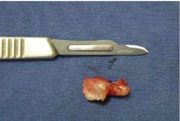
Conservative surgical treatment (Figure 3) was performed under general anesthesia, follow by meticulous curettage at the tumor site. The surgical part removed was composed of a fragment of elongated, light soft tissue with a papillary, elastic surface, containing a capsule around it, measuring 19x7x5mm (Figure 4). This was sent for histopathological exam, to obtain confirmation of the initial diagnosis.
Figure 3. Conservative surgery and curettage for removal of the odontogenic tumor.
Figure 4. Surgical part removed in the second biopsy.
The cuts examined (Figure 5) confirmed the diagnosis of AFO, sowing an odontogenic epithelium in ample cellular proliferation, forming numerous islands with shoots or cellular cords proliferating on the loose connective tissue that resembled dental pulp. Also observed, were mineralized tissues similar to dental cement and spongy, hemosiderin, trabecular bone fragments.
Figure 5. Photomicrograph exhibiting epithelial island containing cylindrical cells at the periphery and cubic cells in the central area, surrounded by a white halo, suggesting area occupied by enamel. HE Staining, 400x magnifica-tion.
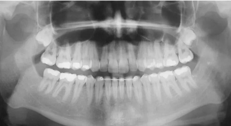
Figure 6. Follow-up panoramic radiograph (25 months) showing complete bone remodeling.
DISCUSSION
The ameloblastic fibro-odontoma is a rare disease that affects a young population, with a mean age of 2l years, according to Buchner et al.7, and the area most
affected is the posterior mandibular region. In the present study, the case related is compatible with the mean age, since the patient was 19 years old when the AFO was discovered in the posterior mandibular region on the left.
The tumor diagnosis was confirmed by association of the panoramic radiograph, which showed a well circumscribed radiolucent image with varying radiodensity within it, with the intraoral exam and biopsy performed. There are other exams that may help with differential diagnosis, such as computed tomography. The treatment for AFO is conservative surgery with curettage, since the lesion is presented encapsulated and is non-invasive. Thus, its prognosis is favorable and the rate of recurrence is low. In the case of the tumor described, the differential diagnosis included lesions with mixed radiographic patterns, such as: Calcifying Odontogenic Epithelial Tumor (COET), Calcifying Odontogenic Cyst (COC), Complex Orthodontia and Adenomatoid Odontogenic Tumor (AOT)15.
Boxberger et al.4, Buchner et al.7 and Zouhary et
al.8 affirmed that this tumor presented predilection for the
male sex, however, the World Health Organization and authors such as Nelson & Thompson5, Hunter et al.9 and
Lúcio et al.10 affirmed that there was no predilection for
gender or localization.
The patient in this case was Caucasian, however, on analyzing the literature of the area, no information was found about the prevalence of AOF in different ethnic groups.
The study of Buchner et al.7 affirmed that even
when faced with a slow growing AOF, intra and extraoral exam must be performed, because this lesion may cause an intrabucal increase in volume, with a hardened consistency, however, the color of the mucosa remains pink, and in general, there is slight facial asymmetry11. Nelson &
Thompson5, Franco et al.2and De Riu et al.11 alleged that
the patient feels no pain and reports no previous history of infections or local traumas. The patient in the case reported presented no intraoral increase in volume or change in color of the gingiva, had no local discomfort and systemic changes in health.
According to De Riu et al.11 and Tolentino et al.12,
the tumor may inhibit eruption of the tooth involved. In the present case, agenesis of tooth 38 was diagnosed, and the tumor had not affected tooth 37, which was found in the routine panoramic radiograph taken for the purpose of having third molars extracted3-4.
In the panoramic radiograph, according to Mummolo et al.3 and Boxberger et al.4, and from the aspects
visualized in the case presented, the ameloblastic fibro-odontoma appeared as a well circumscribed radiolucent lesion, with an radiopaque mass inside it.
The histopathological exam of the biopsy performed on the patient showed an epithelium that was sometimes in cords and sometimes in islands, with the presence of juxtaposed ameloblasts that resembled the tooth germ. After surgical removal of the tumor, a new histopathological exam confirmed the diagnosis of AFO, showing the odontogenic epithelium in ample cellular proliferation, with numerous islands and cell cords on the loose conjunctive tissue that resembled dental pulp.
Santos et al.16 affirmed that it was possible to
diagnose an AFO only by correlating the clinical data with imaging exams and performing a biopsy, and the case presented is in agreement with this author, because it was diagnosed by means of these three exams. In turn, Mummolo et al.3 based their diagnosis on the presence
or absence of elements indicative of differentiation of the dental germ in the histopathological exam, which was confirmed by the biopsy performed in the case here reported, in which mineralized tissue similar to dental cement was observed.
10. Lucio PS, Cavalcante RB, Maia RN, Santos ES, Godoy GP. Aggressive ameloblastic fibro-odontoma assessment with CBCT and treatment. Eur Arch Paediatr Dent. 2013;14(3):179-84. doi: 10.1007/s40368-013-0032-9
11. De Riu G, Meloni SM, Contini M, Tullio A. Ameloblastic fibro-odontoma. Case report and review of the literature. J Craniomaxillofac Surg. 2010 Mar;38(2):141-4. doi: 10.1016/j. jcms.2009.04.009
12. de Souza Tolentino E, Centurion BS, Lima MC, Freitas-Faria P, Consolaro A, Sant’ana E. Ameloblastic fibro-odontoma: a diagnostic challenge. Int J Dent. 2010;2010: 104630. doi: 10.1155/2010/104630
13. Cavalcante AS, Anbinder AL, Costa NC, Lima JR, Carvalho YR. Ameloblastic fibro-odontoma: a case report. Med Oral Patol Oral Cir Bucal. 2009;14(12):e650-3.
14. Nascimento JE, Araujo LD, Almeida LY, De Paula AM, Bonan PR. Ameloblastic fibro-odontoma: a conservative surgical approach. Med Oral Patol Oral Cir Bucal. 2009;14(12):e654-7. doi: 10.4317/medoral.14.e654
15. Wewel J, Narayana N. Ameloblastic fibro-odontoma of the anterior mandible in a 22-month-old boy. Indian J Dent Res. 2010;21(4):618-20. doi: 10.4103/0970-9290.74237
16. Santos TS, de Carvalho RW, Avelar RL, Dias de Oliveira e Silva E, Frota R, Anjos ED. Ameloblastic fibro-odontoma in children: report of 2 cases. J Dent Child (Chic). 2011 Sep-Dec;78(3):173-7.
Received on: 17/1/2017 Final version resubmitted on: 29/6/2017 Approved on: 5/7/2017
REFERENCES
1. Hegde V, Hemavathy S. A massive ameloblastic fibro-odontoma of the maxilla. Indian J Dent Res. 2008;19(2):162-4.
2. Franco A, Riscala S, Kahoudji M, Croue A. Endoscopic removal of a mandibular ameloblastic fibro-odontoma. Rev Stomatol Chir Maxillofac. 2009;110(6):359-61. doi: 10.1016/j. stomax.2009.03.011
3. Mummolo S, Marchetti E, Di Martino S, Scorzetti L, Marzo G. Ameloblastic fibro-odontoma: a case report. Ann Stomatol (Roma). 2010;1(2):11-3.
4. Boxberger NR, Brannon RB, Fowler CB. Ameloblastic fibro-odontoma: a clinicopathologic study of 12 cases. J Clin Pediatr Dent. 2011;35(4):397-403.
5. Nelson BL, Thompson LD. Ameloblastic fibro-odontoma. Head Neck Pathol. 2014;8(2):168-70. doi: 10.1007/s12105-013-0501-9
6. Howell RM, Burkes EJ Jr. Malignant transformation of ameloblastic fibro-odontoma to ameloblastic fibrosarcoma. Oral Surg Oral Med Oral Pathol. 1977;43(3):391-401.
7. Buchner A, Kaffe I, Vered M. Clinical and radiological profile of ameloblastic fibro-odontoma: an update on an uncommon odontogenic tumor based on a critical analysis of 114 cases. Head Neck Pathol. 2013;7(1):54-63. doi: 10.1007/s12105-012-0397-9
8. Zouhary KJ, Said-Al-Naief N, Waite PD. Ameloblastic fibro-odontoma: expansile mixed radiolucent lesion in the posterior maxilla: a case report. Oral Surg Oral Med Oral Pathol Oral Radiol Endod. 2008;106(4):e15-21. doi: 10.1016/j.tripleo.2008.05.038
9. Hunter AK, Muller S, Kalathingal SM, Burnham MA, Moore WS. Evaluation of an ameloblastic fibro-odontoma with cone beam computed tomography. Tex Dent J. 2012;129(6):619-24.
indicated, considering that normally recurrences arise from a residual lesion resulting from initially inadequate surgery7.
Santos et al.16 recommended long-term follow-up,
although the AFO presents a low rate of recurrence. The patient in the present article has been followed up on a six-monthly basis by means of clinical radiographic exam for 2 years, without signs of recurrence
CONCLUSION
In the case presented, with exception of the patient’s gender, the remaining clinical-radiographic and histopathological characteristics were similar to those
described in the literature. The treatment proposed was shown to be effective and the post-surgical follow-up of two years presented no evidence of recurrence of the lesion.