ALMA MATER STUDIORUM UNIVERSITÀ DI BOLOGNA DIPARTIMENTO DI MEDICINA SPECIALISTICA,
DIAGNOSTICA E SPERIMENTALE UNIVERSIDADE NOVA DE LISBOA FACULDADE DE CIÊNCIAS MÉDICAS
THOMSEN-FRIEDENREICH ANTIGENS IN
BLADDER CANCER: EVALUATION OF THEIR
PROGNOSTIC VALUE
PAULO FILIPE SEVERINO
Thesis submitted for the Degree of Doctor in Life Sciences in the Specialty of Immunology
in the Faculdade de Ciências Médicas
and for the Degree of Doctor in Oncology and Experimental Pathology in the Specialty of Experimental Pathology
in the Dipartimento di Medicina Specialistica, Diagnostica e Sperimentale
ALMA MATER STUDIORUM UNIVERSITÀ DI BOLOGNA DIPARTIMENTO DI MEDICINA SPECIALISTICA,
DIAGNOSTICA E SPERIMENTALE UNIVERSIDADE NOVA DE LISBOA FACULDADE DE CIÊNCIAS MÉDICAS
THOMSEN-FRIEDENREICH ANTIGENS IN
BLADDER CANCER: EVALUATION OF THEIR
PROGNOSTIC VALUE
Paulo Filipe Severino
Portuguese Supervisor: Professor Doctor Paula Videira
Italian Supervisor: Professor Doctor Fabio Dall’Olio
Thesis submitted for the Degree of Doctor in Life Sciences in the Specialty of Immunology
and for the Degree of Doctor in Oncology and Experimental Pathology in the Specialty of Experimental Pathology
as co-first author:
Lima L., Severino P. F., Silva M., Miranda A., Tavares A., Pereira S., Fernandes E., Cruz R.,
Amaro T., Reis C. A., Dall'Olio F., Amado F., Videira P. A., Santos L. and Ferreira J. A.,
2013. Response of high-risk of recurrence/progression bladder tumours expressing
sialyl-Tn and sialyl-6-T to BCG immunotherapy. Br J Cancer 109, 2106-14.
as second author:
Carrascal M. A., Severino P. F., Cabral M. G., Silva M., Ferreira J. A., Calais F., Quinto H.,
Pen C., Ligeiro D., Santos L. L, Dall’Olio F. and Videira P. A., 2014.
Sialyl Tn-expressing bladder cancer cells induce a tolerogenic phenotype in innate and
adaptive immune cells. Mol Oncol published online.
Ferreira J. A., Videira P. A., Lima L., Pereira S., Silva M., Carrascal M., Severino P. F.,
Fernandes E., Almeida A., Costa C., Vitorino R., Amaro T., Oliveira M. J., Reis C. A.,
Dall'Olio F., Amado F. and Santos L. L., 2013. Overexpression of tumour-associated
carbohydrate antigen sialyl-Tn in advanced bladder tumours. Mol Oncol 7, 719-31.
and for the book chapter:
Severino P., Silva M., Carrascal M., Calais F., Dall'Olio F. and Videira P., 2012.
Bladder cancer-glycosylation insights, Vol. 38, The Royal Society of Chemistry,
pp. 156-175.
The following scientific paper is under preparation:
Severino P. F., Silva M., Carrascal M., Astolfi A., Catera M., Forleo R., Chiricolo M.,
Malagolini N., Videira P. A. and Dall’Olio F., 2014. Thomsen-Friedenreich antigens in bladder cancer: evaluation of their prognostic value.
This study was fulfilled at the
Faculdade de Ciências Médicas (Lisboa) and
à minha Família
This PhD thesis results from a long hard work that would not be possible without such a large number of contributions, and for this reason I wish to thank:
both to the Faculdade de Ciências Médicas from Universidade Nova de Lisboa and to the
Department of Experimental, Clinical and Specialty Medicine from Università di Bologna,
for hosting me, giving me all the conditions to develop my PhD project;
to Professor Paula Videira and Professor Helder Trindade for the inviting and accepting me as a PhD student under the supervision of Professor Paula Videira in the Department
of Immunology at Faculdade de Ciências Médicas; with a special thank to Professor
Paula Videira for all the critic spirit during my PhD project;
to Professor Fabio Dall’Olio for accepting me as his PhD student in the Department of
Experimental, Clinical and Specialty Medicine from Università di Bologna, for all the
friendship and all the wise teaching passed to me;
to Doctor José Alexandre Ferreira and Doctor Lucio Santos from the Experimental
Pathology and Therapeutics Group of Instituto Português de Oncologia at Porto and
Doctor Celso Reis from the Institute of Molecular Pathology and Immunology of the
Universidade of Porto (IPATIMUP) for all the work in collaboration;
to Doctor Fernando Calais for RIVM-BCG donation, to Professor Yi Luo, Harvard Medical School for Pasteur-BCG donation;
to my laboratory colleagues (the PhD students Mariangela Catera and Roberto Forleo, Doctor Mariella Chiricolo, Doctor Guadalupe Cabral and others) and to all the Bologna department colleagues for the good work environment with a very special thank to Doctor Nadia Malagolini and the PhD student Mariana Silva for all the good work environment, friendship and technical supporting;
to MSc Milena Urbini and Doctor Annalisa Astolfi from Interdepartmental Centre for
Cancer Research – Giorgio Prodi of Università di Bologna, at Policlinico
Sant’Orsola-Malpighi Hospital, for all the technical support in the achieving of the array results and all the friendship;
to Doctor Francesca Borsetti and Doctor Enzo Spisni from Department of Biological,
Geological and Environmental Sciences of Università di Bologna, for all the technical
support in the achieving of the secretome results and all the friendship;
to my home mates, Manuela Ianni, Rossella Davenia and Federico Giamporcaro, for the great home environment and all the friendship, as well to all my friends from Morgani and Ghigi residentials Rosalba Gabriele, Roberto ed Ignazio Valario, Manuela De Luca, Daniele Pesare, Antonio Aprile, Valeria Perrino, Gabriela Falsea, Daniel Simon, Eleonora Conti, Claudia Mannino, Riccardo Damiano, Ada Civetta, Stefano Pacella, Jessica Scicchitani, Filomena De Fino and others for all the Italian funny moments;
to my ever friends Ana Rita Piteira, Gustavo Patrício, Ana Catarina Bolotinha, Pedro Miranda, Inês Ramos, Mafalda Santos, Nelson Baptista, Nuno Marques, Ana Duarte and
others, for all the “sporadic friendship meetings”;
“A nossa vida não nos pertence. Desde o ventre materno ao túmulo,
estamos
entreligados,
ligados
ao
passado
e
presente,
e através de cada ação perversa, de cada ação amável,
criamos o nosso futuro.”
“La nostra vita non ci appartiene. Dal grembo materno alla tomba,
siamo legati agli altri, passati e presenti, e da ogni crimine,
da
ogni gentilezza, generiamo il nostro futuro.”
“Our lives are not our own. From womb to tomb, we are bound to
others, past and present, and by each crime and every kindness,
we birth our future.”
Resumo --- 15
Riassunto --- 17
Abstract --- 19
List of figures --- 21
List of Tables --- 24
List of Abbreviations --- 25
Chapter I – Introduction I.1 Bladder Cancer --- 29
Epidemiology --- 29
Aetiology and risk factors --- 30
Classification and pathological staging --- 33
Diagnosis, prognosis and therapy --- 35
Bladder cancer burden --- 39
I.2 Insights into Glyco-Oncology --- 40
Introduction to Glycobiology --- 40
Glyco-Oncology of bladder cancer --- 48
I.3 BCG Immunotherapy --- 50
Adhesion and Internalization of BCG --- 50
Immune response triggered by BCG --- 51
Purpose of the work --- 52
Chapter II – Materials and Methods Cell lines --- 57
Flow cytometry --- 57
Preparation of cell lines constitutively expressing sialyltransferases --- 58
Sialyltransferase activity assays --- 59
Neuraminidase treatment --- 60
Growth of Mycobacterium bovis Bacillus Calmette-Guérin (BCG) --- 60
Real time RT-PCR --- 61
Whole transcriptome analysis by expression microarray --- 62
Preparation of monocyte-derived macrophages --- 63
Induction of cytokine secretion --- 64
Detection of secreted cytokines --- 64
Phagocytosis assay --- 65
Chapter III – Results III.1 Cellular Models of Bladder Cancer (BC) --- 69
FACS characterization of cells highly expressing sT or sTn antigens --- 69
Sialyltransferase expression by selected cell populations --- 73
III.2 Response of BC cell lines to BCG --- 77
BCG Internalization --- 77
Transcriptome analysis of BC cell lines --- 79
Effect of ST3GAL1 expression – Gene Signatures of HT1376 cells --- 79
Effect of ST6GALNAC1 expression – Gene Signatures of MCR cells --- 93
Cytokine secretion by BCG-challenged BC cells --- 102
III.3 Response of Macrophages to BCG-challenged BC cell lines --- 104
Cytokine secretion by macrophages in response to BC secretome --- 104
Phagocytosis of apoptotic BC cells by macrophages --- 107
Chapter IV – Discussion and Conclusion IV.1 Cellular Models of Bladder Cancer --- 111
IV.2 Response of Bladder Cancer to BCG --- 116
IV.3 Response of Macrophages to BCG-challenged BC cells --- 119
IV.4 Conclusions --- 121
Bibliography --- 123
Supplement I --- 151
Paulo Severino | Thomsen-Friedenreich antigens in bladder cancer: evaluation of their prognostic value 15
bexiga: avaliação do seu valor prognóstico
Resumo
Introdução. O cancro de bexiga é uma patologia comum que representa o 6° e o 5° cancro mais incidente em Portugal e na Itália, respetivamente. Em mais de metade dos casos ocorre reincidência durante o primeiro ano, requerendo acompanhamento clínico ao longo da vida. A instilação intravesical de Bacillus Calmette-Guérin (BCG)
(uma estirpe atenuada do Mycobacterium bovis) representa uma imunoterapia eficaz no
combate ao cancro de bexiga, no entanto, muitos aspetos da interação de BCG com as células tumorais bem como com as células do sistema imunitário permanecem por desvendar. As células tumorais de bexiga expressam frequentemente as formas sialiladas
dos antigénios de Thomsen-Friedenreich (TF), i.e., sialil-T (sT) e sialil-Tn (sTn).
Contudo ainda se desconhece o significado da sua expressão na malignidade tumoral e se
afeta a eficácia da terapêutica BCG. Objetivo do estudo. Investigar o papel dos
antigénios sT e sTn no fenótipo maligno de células de cancro de bexiga bem como na
resposta mediada pelo sistema imunitário à terapia com BCG. Metodologia. Para tal,
foram utilizadas as linhas celulares de cancro da bexiga HT1376 e MCR, geneticamente
modificadas por transdução com vetores codificantes para as sialiltransferases ST3GAL1
ou ST6GALNAC1, de forma a expressar homogeneamente os antigénios sT ou sTn respetivamente. Estes modelos celulares foram estudados após confronto com BCG. O nível de BCG internalizado foi avaliado por citometria de fluxo. O perfil global de expressão genética dos modelos celulares antes e após incubação com BCG foi analisado
pela tecnologia de microarray. O perfil de citocinas secretadas pelos modelos celulares
após incubação com BCG, bem como de macrófagos estimulados pelo secretoma de células de cancro de bexiga que por sua vez foram estimuladas previamente por BCG,
foi estudado pelo sistema multiplex de “imuno-esferas”. Resultados. A análise do
transcritoma dos modelos celulares revelou que grupos de genes envolvidos em funções específicas foram modulados em paralelo nos dois modelos celulares, após transdução, independentemente da sialiltransferase expressa. Ou seja, em células que expressavam a
16 Thomsen-Friedenreich antigens in bladder cancer: evaluation of their prognostic value | Paulo Severino
negativamente. Genes descritos na literatura como marcadores para o cancro de bexiga foram também modulados. A incubação com BCG resultou numa tendência ao aumento da expressão de genes relevantes na preservação e estabilidade genómica e menor malignidade, no entanto, apenas em células que expressavam sT ou sTn. Entre as dez citocinas testadas, apenas a IL-6 e IL-8 foram expressas pelas linhas celulares de cancro da bexiga, com indução destas após estimulação com BCG, e principalmente em células
que expressavam ST3GAL1 ou ST6GALNAC1. Em macrófagos, citocinas inflamatórias,
tais como IL-1β, IL-6 e TNFα, e a citocina anti-inflamatória IL-10, foram induzidas
apenas pelo secretoma de células de cancro da bexiga confrontadas com BCG, com maior
relevância quando estas expressavam ST3GAL1 ou ST6GALNAC1, prevendo a
estimulação de macrófagos semelhantes aos de tipo M1 e uma melhor resposta à terapia
com BCG. Conclusões. O efeito geral da expressão destas sialiltransferases e dos
Paulo Severino | Thomsen-Friedenreich antigens in bladder cancer: evaluation of their prognostic value 17
vescica: valutazione del valore prognostico
Riassunto
Introduzione. Il cancro della vescica è una patologia comune che rappresenta la 6ª e la 5ª neoplasia a maggiore incidenza in Portogallo ed in Italia, rispettivamente. Più della metà dei casi presentano una recidiva entro un anno, richiedendo quindi il monitoraggio a vita.
L’instillazione intravescicale di Bacillus Calmette Guerin (BCG)
(un ceppo attenuato di Mycobacterium bovis) rappresenta una immunoterapia efficace
contro il cancro alla vescica, ma molti aspetti dell’interazione del BCG con le cellule
tumorali e con le cellule del sistema immunitario sono ancora da comprendere. Le cellule tumorali di vescica spesso esprimono le forme sialilate degli antigeni
Thomsen-Friedenreich (TF), i.e., sialil-T (sT) e sialil-Tn (sTn). Però il significato
biologico della loro espressione e nel determinare l’efficacia terapeutica del BCG è
ancora sconosciuto. Obiettivo dello studio. Studiare il ruolo degli antigeni sT e sTn nel
determinare il fenotipo maligno delle cellule tumorali della vescica e la risposta
immunitaria alla terapia con BCG. Metodologia. Sono state utilizzate le linee cellulari di
cancro alla vescica HT1376 e MCR, geneticamente modificate mediante trasduzione
genica con vettori codificanti le sialiltrasferasi ST3GAL1 o ST6GALNAC1, allo scopo di
ottenere l’espressione omogenea degli antigeni sT o sTn. Questi modelli cellulari sono
stati studiati dopo l’incubazione col BCG. Il livello di internalizzazione di BCG è stato
valutato mediante citometria a flusso. Il profilo di espressione genica delle linee cellulari
prima e dopo incubazione col BCG è stato studiato mediante tecnologia microarray.
Mediante un sistema immunobiglie multiplex, è stato possibile studiare simultaneamente
l’espressione di numerose citochine secrete o dalle cellule di cancro della vescica dopo
incubazione con BCG e o dai macrofagi stimulati dal secretoma delle cellule tumorali di
vescica dopo incubazione col BCG, . Risultati. Le analisi del trascrittoma delle linee
cellulari hanno rivelato che vi erano gruppi di geni accomunati dal fatto di svolgere una analoga funzione, che venivano regolati in parallelo nelle due linee cellulari dopo
trasduzione con l’una o l’altra sialiltrasferasi. In particolare, in cellule che esprimevano
18 Thomsen-Friedenreich antigens in bladder cancer: evaluation of their prognostic value | Paulo Severino
letteratura come marcatori del cancro della vescica venivano modulati nelle cellule
trasdotte con l’una o l’altra sialiltrasferasi. L’incubazione col BCG induceva una tendenza alla sovraespressione di geni coinvolti nel mantenimento della stabilità genomica e nella riduzione della malignità, ma soltanto in cellule che esprimevano sT o sTn. Tra le dieci citochine analizzate, solo IL-6 e IL-8 erano secrete dalle linee cellulari. Il contatto con
BCG ne stimolava la secrezione, in particolare nelle cellule che esprimevano ST3GAL1 o
ST6GALNAC1. Nei macrofagi, la secrezione delle citochine infiammatorie IL-1β, IL-6 e
Paulo Severino | Thomsen-Friedenreich antigens in bladder cancer: evaluation of their prognostic value 19
evaluation of their prognostic value
Abstract
Background. Bladder cancer is a common malignancy representing the 6th and the 5th most incident cancer in Portugal and in Italy, respectively. More than half of the cases relapse within one year, requiring though a lifelong follow-up. Intravesical instillation of
Bacillus Calmette-Guérin (BCG) (an attenuated strain of Mycobacterium bovis)
represents an effective immunotherapy of bladder cancer, although many aspects of the interaction of BCG with cancer cells and host immune cells remain obscure. Bladder cancer cells often express the sialylated forms of the Thomsen-Friedenreich (TF),
i.e., sialil-T (sT) e sialil-Tn (sTn). However, it’s still unknown the sense of such
expression in tumour malignancy and in the BCG therapy efficacy.
Aim of the study. To investigate the role of the sT and sTn antigens on the malignant phenotype of bladder cancer cells and the immune mediated response to BCG therapy. Experimental. We have utilized populations of the bladder cancer cell lines HT1376 and
MCR, genetically modified by transduction with the sialyltransferases ST3GAL1 or
ST6GALNAC1 to express homogeneously sT or sTn antigens. The level of BCG internalized was assessed by flow cytometry. The whole gene expression profile of BCG-challenged or unchallenged bladder cancer cell lines was studied by microarray technology. The profile of cytokines secreted by BCG-challenged bladder cancer cells and that of macrophages challenged by the secretome of BCG-challenged bladder cancer
cells was studied by multiplex immune-beads assay. Results. Transcriptome analysis of
20 Thomsen-Friedenreich antigens in bladder cancer: evaluation of their prognostic value | Paulo Severino
In macrophages, inflammatory cytokines, such as IL-1β, IL-6 and TNFα, and the
Paulo Severino | Thomsen-Friedenreich antigens in bladder cancer: evaluation of their prognostic value 21
Chapter I pp
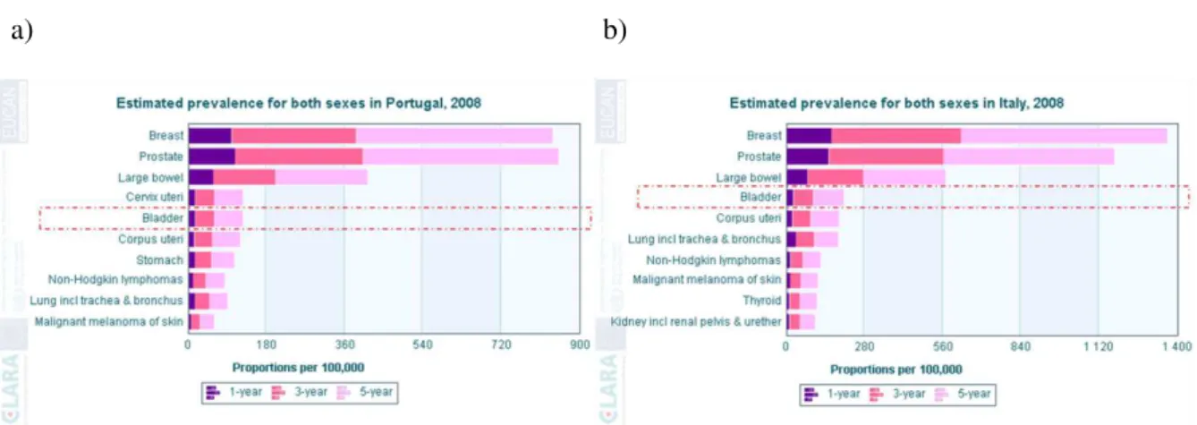
Figure 1.1. Incidence and mortality rates per 100 000 for Bladder Cancer. a) Bladder cancer is the 8th most incident cancer in the world, b) showing high incidence rates in Southern and Western Europe. Bladder cancer is c) the 6th most frequent in Portugal and d) the 5th in Italy. From GLOBOCAN database available at http://globocan.iarc.fr (Ferlay, Shin et al. 2010).
29
Figure 1.2. Prevalence rates per 100 000 of the ten most prevalent cancers in a) Portugal, and b) in Italy. Bladder cancer is the 5th most prevalent cancer in Portugal and the 4th In Italy. From EUCAN database available at http://eco.iarc.fr/ (Bray, Ren et al. 2012).
30
Figure 1.3. Bladder cancer incidence rates by age in Europe according to GLOBOCAN database (Ferlay, Shin et al. 2010).
33
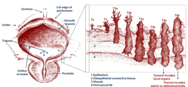
Figure 1.4. Representation of the anatomy of a male bladder and the different stages of bladder cancer according to the TNM classification. In the left is represented the anatomy of the human male bladder and prostate, and in the right is represented a small portion of the epithelium containing al states of bladder cancer. This figure was elaborated by both the author and the MSc Filipe Miguel Severino.
34
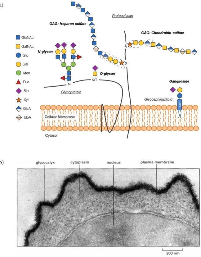
Figure 1.5. Glycoconjugates in mammalian cells. a) Glycans can be attached to proteins as in glycoproteins and proteoglycans. Glycoproteins are decorated with N- and/or O-glycans and proteoglycan with glycosaminoglycans (GAG). Glycosphingolipids are mainly associated with membranes and are formed by a glycan attached to the lipid moiety ceramide. A ganglioside contains one or more residues of sialic acid (Sia). The glycan structures presented for each N-linked, O-linked, glycosaminoglycan (GAG) and glycosphingolipids exemplify one of the possible structures (Severino, Silva et al. 2012). b) The electron micrograph of the surface of a lymphocyte stained with ruthenium red emphasizes the thick carbohydrate layer surrounding the cell, a.k.a. glycocalyx (Alberts, Johnson et al. 2002).
42
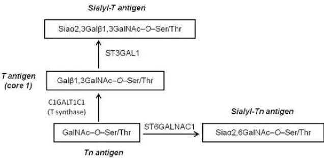
Figure 1.6. Structures of Thomsen-Friedenreich related antigens. Glycan structures and the main enzymes responsible for their synthesis are shown.
45
Chapter III
Figure 3.1. Flow Cytometry analysis of transduced cells. a) T antigen expression byHT1376 cells. Mock transduced HT1376NC cells (green line) were strongly reactive for PNA because of the large number of Gal1,3GalNAc termini. The ST3GAL1-transduced HT1376ST3G1 cell line presented a bimodal reactivity for PNA (blue line). b) Sialyl Tn expression by MCR cells. sTn expression on mock-transduced MCRNC cells (green line) was negligible, while sTn expression on ST6GALNAC1-transduced MCRST6GN1 cells was bimodal (blue line) with a minority of sTn negative cells. Black line: unlabelled cell lines or isotype control antibody when labelling with lectins or antibodies was used, respectively.
70
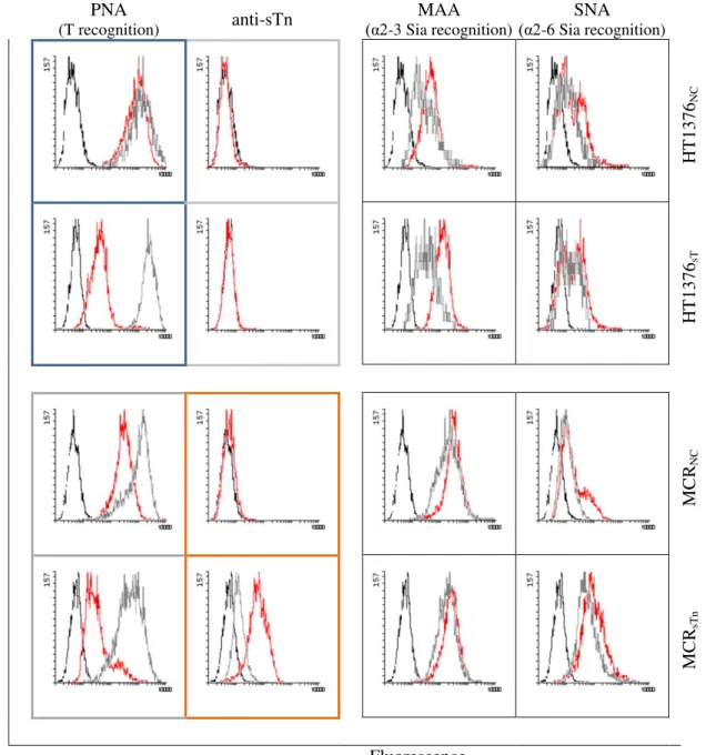
Figure 3.2. Characterization of transduced bladder cancer cell lines by Flow Cytometry.
Bladder cancer cell lines were labelled with PNA-FITC, MAA-FITC, SNA-FITC and anti-sTn (labelled with anti-Ig-FITC) before (red line) or after (grey line) sialidase treatment. Black line: unlabelled cell lines or isotype control antibody when labelling with lectins or antibodies was used, respectively.
71
Figure 3.3. Different PNA reactivity in mock- or ST6GALNAC1-transduced MCR cells. PNA reactivity data indicate that the number of accessible T structures is much lower in MCRsTn cells than in MCRNC cells. This is due to the fact that ST6GALNAC1 acts not only on Tn antigen but also on T and sT structures, resulting in an almost complete masking of the T antigen by
α2,6-linked sialic acid. GalNAc: yellow square; Gal: yellow circle; Sia: purple diamond.
22 Thomsen-Friedenreich antigens in bladder cancer: evaluation of their prognostic value | Paulo Severino
homogenates. The sialyltransferase activity was negligible in untransduced HT1376 or MCR, and in mock-transduced HT1376NC or MCRNC cells. Both selection procedures led to increase the respective sialyltransferase activities. Consonant expression of b) ST3GAL1 in HT1376sT cells and of d) ST6GALNAC1 in MCRsTn cells was found. Data are the mean ±SD of 3 experiments. ** p < 0.001; * p < 0.05.
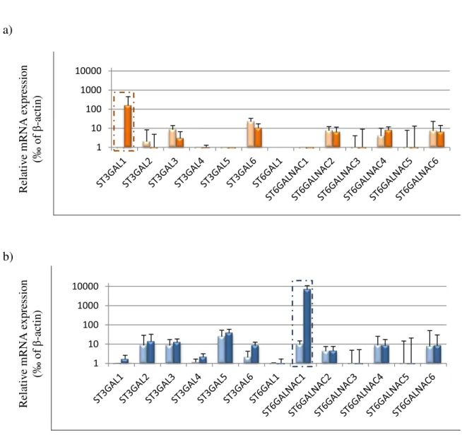
Figure 3.5. Sialyltransferase mRNA expression in transduced cells. Relative mRNA levels of several sialyltransferases were analysed in a) HT1376NC (pink bars), HT1376sT (orange bars) and b) MCRNC (light blue bars) and MCRsTn (dark blue bars) cells by RT-qPCR. Results were normalized for 1000 molecules of the endogenous control β-actin. High levels of ST3GAL1 and ST6GALNAC1 mRNA were detected in MCRsTn and in HT1376sT respectively, while the expression of other sialyltransferases were low or undetectable e was not interfered significantly after transduction. Data are the mean ±SD of 3 experiments. Student’s t test revealed that sialyltransferases ST3GAL1 and ST6GALNAC1 were the only statistically modulated in HT1376sT and MCRsTn, respectively. Boxed expression data are the same reported in Figure 3.4 and are repeated here for comparison.
75
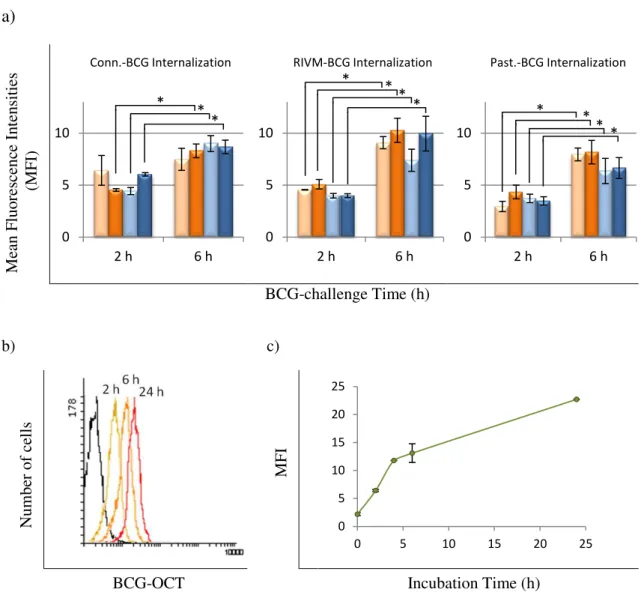
Figure 3.6. Time course of BCG internalization by bladder cancer cell lines. a) Analysis by FACS of the internalization of Connaught-BCG, RIVM-BCG or Pasteur-BCG by HT1376NC cells (light orange bars), HT1376sT cells (orange bars), MCRNC cells (light blue bars) and MCRsTn cells (dark blue bars), after 2 or 6 h of incubation with BCG. Internalization values were normalized with the respective controls incubated at 4 ºC. Data are the mean ± SD of 3 experiments. * p < 0.05. b) and c) An example of time-dependent Connaught-BCG internalization by MCRsTn cells MFI2h = 7, MFI6h = 13 and MFI24h = 23. Black line: internalization at 4 ºC. Over longer periods of time, internalization of BCG displayed a two slope curve with tendency to saturation. OCT stands for orange cell tracker.
78
Figure 3.7. Gene signatures in HT1376 cells. Expression variation of relevant genes in a) HT1376sT cells vs. HT1376NC cells, in b) HT1376NC cells and in c) HT1376sT cells after BCG-challenging. Genes were grouped in “bladder cancer markers” and “caretaker” categories, according to putatively increased or decreased malignant phenotypes. Only variations greater then ± 0.5 were considered. Data are the mean of 3 experiments. * p < 0.05.
81
Figure 3.8. Modulation of MHC genes by BCG internalization in HT1376 cells. Percentage of expression variation of MHC related genes in a) HT1376NC, b) HT1376sT, after BCG-challenging. Only variations greater then ± 0.5 were considered. Data are the mean of 3 experiments. * p ≤ 0.05.
92
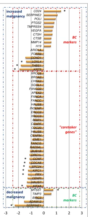
Figure 3.9. Gene signatures in MCR cells. Expression variation of relevant genes in a) MCRsTn cells vs. MCRNC cells in b) MCRNC cells and in c) MCRsTn cells after BCG-challenging. Genes were grouped in "BC markers" and "caretaker" according to their putative effect on malignancy. Only differential variations greater then ± 0.5 were considered. Data are the mean of 3 experiments. * p < 0.05.
95
Figure 3.10. Modulation of MHC genes by BCG internalization. Percentage of expression variation of MHC genes in a) MCRNC, b) MCRsTn cells after BCG-challenging. Only variations greater then ± 0.5 were considered. Data are the mean of 3 experiments. * p ≤ 0.05.
101
Figure 3.11. Cytokine secretion by HT1376 cells. An induction of both IL-6 and IL-8 was found in both HT1376NC (pink bars) and HT1376sT (orange bars) 12 h after BCG-challenging. IL-8 induction in HT1376sT cells was stronger than in HT1376NC cells. Data are the mean ± SD of 3 experiments. *** p < 0.0001; * p < 0.05.
102
Figure 3.12. Cytokine secretion by MCR cells. Twelve hours after BCG-challenging, a significant induction of both IL-6 and IL-8 secretion was observed in MCRsTn (dark blue bars), but not in MCRNC (light blue bars). Data are the mean ± SD of 3 experiments. *** p < 0.0001; ** p < 0.001.
103
Figure 3.13. Cytokine secretion by macrophages stimulated by HT1376 secretome. Secretome from BCG-challenged HT1376sT (dark orange bars) induced stronger secretion than that of HT1376NC (pink bars) of all cytokines except IL-8. Gray bars represent cytokine secretion by unstimulated macrophages. Data are the mean ± SD of 3 experiments. *** p < 0.0001; ** p < 0.001; * p < 0.05.
Paulo Severino | Thomsen-Friedenreich antigens in bladder cancer: evaluation of their prognostic value 23 from unchallenged MCRsTn (dark blue bars) or MCRNC (light blue bars) cells induced little secretion of IL-6 and TNFα and higher levels of IL-8. Secretome from BCG-challenged MCR cells induced all five cytokines with stronger induction of IL-10, IL-1β, IL-6 and TNFα when secretome from MCRsTn was tested. Gray bars represent cytokine secretion by unstimulated macrophages. Data are the mean ± SD of 3 experiments. *** p < 0.0001; ** p < 0.001; * p < 0.05.
Figure 3.15. Phagocytosis of apoptotic bladder cancer cell lines by macrophages. a) Phagocytosis of apoptotic BCG-challenged or unchallenged HT1376NC (pink bars) or HT1376sT (orange bars) cells. Phagocytosis was not affected by BCG-challenging but was higher with HT1376sT cells. b) Phagocytosis of apoptotic BCG-challenged or -unchallenged MCRNC (light blue) or MCRsTn cells (dark blue). Phagocytosis was not affected either by BCG-challenging or sTn expression. Data are the mean ± SD of 3 experiments. * p < 0.05.
107
Supplement I
Figure I.1. Genes showing up- or down-regulation in HT1376sT cells compared with
HT1376NC cells. Heat-maps are presented for the 2log expression ratio (≤ -0.9 in green or ≥ 0.9 in
red) between HT1376sT cells and HT1376NC cells. Constitutive expression of ST3GAL1 induced
significant (p ≤ 0.05) over-expression of 197 genes and down-regulation of 107 genes. Data are the mean of 3 experiments.
153
Figure I.2. Genes showing up- or down-regulation in both HT1376NC and HT1376sT after
BCG-challenging. Heat-maps are presented for the 2log expression ratio (≤ -0.5 in green or ≥ 0.5 in red) between BCG-challenged and unchallenged HT1376 (NC and sT) cell lines. BCG-challenging induced down-regulation of 18 genes and up-regulation of 68 genes. p ≤ 0.05.
154
Figure I.3. Genes showing differential modulation after BCG-challenging in HT1376NC or
HT1376sT cells. Heat-maps are presented for the 2log expression ratio (≤ -1.0 in green or ≥ 1.0 in
red) between BCG-challenged HT1376NC or HT1376sT cells and unchallenged cells. a) Group of genes modulated by BCG-challenging in HT1376NC, but not in HT1376sT cells. b) Group of genes modulated by BCG-challenging in HT1376sT, but not in HT1376NC cells. Twenty five genes showed modulation (12 up-regulation, 13 down-regulation) in HT1376NC, cells, but not in HT1376sT. On the contrary, 107 genes displayed modulation in HT1376sT cells but not in HT1376NC. Of these, 27 displayed up-regulation and 80 down-regulation. Data are the mean of 3 experiments. p ≤ 0.05.
155
Figure I.4. Genes showing up- or down-regulation in MCRsTn cells compared with MCRNC.
Heat-maps are presented for the 2log expression difference (≤ -0.9 in green or ≥ 0.9 in red) between MCRsTn cells and MCRNC cells. In MCRsTn cells, constitutive ST6GALNAC1 overexpression induced the down-regulation of 114 genes and the up-regulation of 83 genes. Data are the mean of 3 experiments. p ≤ 0.05.
156
Figure I.5. Genes showing up- or down-regulation in both MCRNC and MCRsTn after
BCG-challenging. Heat-maps are presented for the 2log expression ratio (≤ -0.5 in green or ≥ 0.5 in red) between BCG-challenged and unchallenged MCR (NC and sTn) cell lines. BCG-challenging, 24 genes are down-regulated and 25 genes are significantly up-regulated. Data are the mean of 3 experiments. p ≤ 0.05.
157
Figure I.6. Genes showing differential modulation after BCG-challenging in MCRNC or
MCRsTn cells. Heat-maps are presented for the 2log expression difference (≤ -1.0 in green or ≥ 1.0
in red) between BCG-challenged MCRNC or MCRsTn cells and their respective unchallenged cells. a) Group of genes modulated by BCG-challenging in MCRNC, but not in MCRsTn cells. b) Group of genes modulated by BCG-challenging in MCRsTn, but not in MCRNC cells. In MCRsTn bladder cancer cells, after BCG-challenging, a) 17 genes failed to respond to BCG whether in MCRNC these responded negatively (11 genes) or positively (6 genes). On the other hand b) 38 new genes differentially respond to BCG-challenging (29 genes were up-regulated and 9 were down-regulated). Data are the mean of 3 experiments. p ≤ 0.00).
24 Thomsen-Friedenreich antigens in bladder cancer: evaluation of their prognostic value | Paulo Severino
Chapter I pp
Table 1.1. 2009 TNM classification of urinary bladder cancer (Babjuk, Oosterlinck et al. 2011). 34
Chapter II
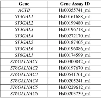
Table 2.1. Set of housekeeping gene and sialyltransferases gene assays ID from Applied Biosystems.
62
Chapter III
Table 3.1. Description of up- or down-regulated genes of HT1376sT cells vs. HT1376NC cells. 82
Table 3.2. Description of up- or down-regulated genes by BCG-challenging in HT1376NC
cells.
89
Table 3.3. Description of up- or down-regulated genes by BCG-challenging in HT1376sT cells. 89
Table 3.4. Genes with analogous or identical functions showing parallel down-regulation in sialyltransferase-transduced bladder cancer cell lines.
93
Table 3.5. Description of up- or down-regulated genes in MCRsTn cells vs. MCRNC cells. 96
Table 3.6. Description of up- or down-regulated genes by BCG-challenging in MCRNC cells. 99
Table 3.7. Description of up- or down-regulated genes by BCG-challenging in MCRsTn cells. 100
Chapter IV
Table 4.1. Molecular classification of HT1376 and MCR bladder cancer cells according to Sjodahl et al.
Paulo Severino | Thomsen-Friedenreich antigens in bladder cancer: evaluation of their prognostic value 25 BC Bladder cancer
BCG Bacillus Calmette-Guérin (an attenuated strain of Mycobacterium bovis) C1GALT1 Glycoprotein-N-acetylgalactosamine 3-beta-galactosyltransferase 1 (or core 1
synthase)
C1GALT1C1 C1GALT1-specific chaperone 1 (or Cosmc) CD4 Cluster of differentiation 4 molecule CD8 Cluster of differentiation 8 molecule
Fuc Fucose
Gal Galactose
GalNAc N-acetylgalactosamine
Glc Glucose
GlcNAc N-acetylglucosamine
GU Genomic unstable bladder cancer molecular subtype
HT1376 Bladder cancer cell line isolated from a 58 year old Caucasian woman with invasive TCC
HT1376NC HT1376 cell line mock-transduced with the virapower lentiviral expression system HT1376ST3G1 HT1376 cell line ST3GAL1-transduced with the virapower lentiviral expression system
(heterogeneously expressing sT)
HT1376sT HT1376 cell line ST3GAL1-transduced with the virapower lentiviral expression system (homogeneously expressing sT)
IL-1β/6/8/10 Interleukin 1 beta/ 6 / 8 / 10 INFγ Interferon gamma
MAA Maackia amurensis lectin
MCR Bladder cancer cell line isolated from a subcutaneous metastatic lesion of a 51 year old male diagnosed with invasive TCC
MCRNC MCR cell line mock-transduced with the virapower lentiviral expression system MCRST6GN1 MCR cell line ST6GALNAC1-transduced with the virapower lentiviral expression
system (heterogeneously expressing sTn)
MCRsTn MCR cell line ST6GALNAC1-transduced with the virapower lentiviral expression system (homogeneously expressing sTn)
MHC Major histocompatibility complex
MUC Mucin
NMIBC Non-muscle invasive bladder cancer PNA Peanut lectin
s6T Galβ1,3(Siaα2,6)GalNAc TF related antigen
SCCL Squamous cell cancer-like bladder cancer molecular subtype Sia Sialic acid
SNA Sambucus nigra lectin sT Sialyl-T antigen
ST3GAL1 ST3 beta-galactoside alpha-2,3-sialyltransferase 1
ST6GALNAC1 ST6 (alpha-N-acetyl-neuraminyl-2,3-beta-galactosyl-1,3)-N-acetylgalactosaminide alpha-2,6-sialyltransferase 1
sTn Sialyl-Tn antigen
T Galα1,3GalNAc-O-Ser/Thr TF related antigen (or core 1 structure) TCC Transitional cell carcinoma
TF Thomsen Friedenreich antigens Tis Bladder carcinoma in situ
Tn GalNAc-O-Ser/Thr TF related antigen TNFα Tumour necrosis factor
TUR Transurethral resection
CHAPTER I
Paulo Severino | Thomsen-Friedenreich antigens in bladder cancer: evaluation of their prognostic value 29
I.1 Bladder Cancer
Epidemiology
Bladder cancer (BC) is a common malignancy in the world (Figure 1.1a), presenting the
8th highest incidence in 2008, with more than 10/100000 new cases. Particularly high
incidence rates are observed in Southern and Western Europe (Figure 1.1b) with more
than 20/100 000 new cases. In Portugal (Figure 1.1c) and in Italy (Figure 1.1d),
bladder cancer represents the 6th and the 5th most incident cancer
(Ferlay, Shin et al. 2010).
a) c)
b) d)
Figure 1.1. Incidence and mortality rates per
30 Thomsen-Friedenreich antigens in bladder cancer: evaluation of their prognostic value | Paulo Severino
Women have approximately a 4-fold lower incidence and mortality rate for bladder
cancer (Fajkovic, Halpern et al. 2011). A decreasing trend in mortality has been evident
in most European countries over the last decades (Ferlay, Randi et al. 2008). Still, bladder
cancer displays the highest recurrence rates among solid tumours with a significant
percentage of progression to muscle invasion (Jacobs, Lee et al. 2010). More than half of
the cases relapse within one year, requiring though a lifelong follow-up (Babjuk,
Oosterlinck et al. 2011). Bladder cancer shows also the highest prevalence rates,
representing the 5th and 4th most prevalent cancer in Portugal (Figure 1.2a) and in Italy
(Figure 1.2b) respectively (Bray, Ren et al. 2012).
a) b)
Figure 1.2. Prevalence rates per 100000 of the ten most prevalent cancers in a) Portugal, and b) in Italy. Bladder cancer is the 5th most prevalent cancer in Portugal and the 4th In Italy. From EUCAN database available at http://eco.iarc.fr/ (Bray, Ren et al. 2012).
Aetiology and risk factors
There are multiple risk factors associated with the development and progression of bladder cancer. Tobacco smoking is the principal risk factor (relative risk of about 4, according to a follow-up occurring between 1995 and 2006), being responsible for
approximately 50% of death caused by bladder cancer (Freedman, Silverman et al. 2011).
The incidence of bladder cancer is directly related to the lifetime tobacco use and the
number of cigarettes smoked per day (Brennan, Bogillot et al. 2000). A recent study
showed that bladder cancer in smokers displayed a younger age of diagnosis, larger tumours, a higher tumour stage and a higher grade than in never smokers (van Roekel,
Cheng et al. 2013). The precise mechanism by which cigarette smoking causes bladder
Paulo Severino | Thomsen-Friedenreich antigens in bladder cancer: evaluation of their prognostic value 31 the risk of this cancer is related to the large number of chemicals present in smoke, such
as 4-aminobiphenyl, polycyclic aromatic hydrocarbons (PAH), N-nitroso compounds,
heterocyclic amines, arylamines and various epoxides agents (Zeegers, Tan et al. 2000).
Variability in susceptibility for chemically induced urinary bladder cancer is due to the individual genetic background (Roos 2008). The initial step in chemically induced bladder carcinogenesis formation of reactive and DNA-affecting metabolites from bladder procarcinogens occurs via the catalytic activity of cytochromes P450 (CYP). For instances, cigarette smoking leads to an increase in exfoliated urothelial cells
expressing CYP1A1 cytochrome which catalyses the formation of reactive metabolites
from PAH (Dorrenhaus, Muller et al. 2007). The induction of CYP1A1 can be regarded as
an indicator of cancer initiation. Moreover, Roos suggested that some individuals with different CYP alleles background may present different susceptibility to bladder carcinogenesis (Roos 2008). A clear polymorphism-dependent effect on bladder cancer
susceptibility is known for two phase II enzymes though, namely N-acetyltransferase 2
(NAT2), which is involved in arylamine metabolism (Golka, Prior et al. 2002) and
glutathion S-transferase M1 (GSTM1) (Dong, Potter et al. 2008). In addition, bladder
cancer was the first cancer to be related to certain occupations.
An increased risk for bladder cancer was observed among workers in the industries of dye, printing, rubber, transportation and in the electrical/gas/sanitary services. Some
compounds related to professional exposure, such as o-toluidine, aniline, nitrobenzene,
4,4’-methylenebis(2-chloroaniline) and the aromatic amines 2-naphthylamine,
4-aminobiphenyl and benzidine (Samanic, Kogevinas et al. 2008, Sorahan 2008, de Vocht,
Sobala et al. 2009, Talaska, Gaultney et al. 2012, Burger, Catto et al. 2013) are
responsible for the majority of occupational bladder cancer cases. An increased risk of bladder cancer has been reported in people living in areas close to industrial
waste-treatment spots (Garcia-Perez, Fernandez-Navarro et al. 2013). The ingestion of
water with high levels of arsenic or nitrate, or the intake of some drugs, such as cyclophosphamide, analgesics containing phenacetin are also associated with the risk of
developing bladder tumours (Guo, Chiang et al. 1997, Letasiova, Medve'ova et al. 2012,
Wang, Fan et al. 2012). In addition, several studies reported a correlation between the
ingestion of food such as barbecued meat, salted meat, fried eggs and fat meals, and an increased risk for bladder cancer. On the other hand, the ingestion of fruits and vegetables
was reported to exert a protective effect (Steinmaus, Nunez et al. 2000, Balbi, Larrinaga
32 Thomsen-Friedenreich antigens in bladder cancer: evaluation of their prognostic value | Paulo Severino
multicentre cohort study with 521468 subjects who showed no statistically significance between fruit and vegetable consumption and a lower risk for bladder cancer
(Buchner, Bueno-de-Mesquita et al. 2011).
Gender is also a risk factor for bladder cancer. Interestingly Jiang et al reported in a study
on 1586 women a lower risk of invasive high-grade bladder cancers in those who experienced multiple infections. This could be due to the immune response triggered by
the bladder infection and/or to the infection therapy (Jiang, Castelao et al. 2009).
For this reason, the lower risk in women for bladder cancer might be directly correlated with the higher risk in women for bladder infection. In some parts of Africa and the Middle East, higher rates of squamous cell carcinoma of the bladder, rather than TCC, were correlated though to high prevalence of the parasitic chronic infection with
Schistosoma haematobium (IARC 1994, Michaud 2007).
As occurs for other solid tumours, the incidence of bladder cancer increases with age
(Ferlay, Shin et al. 2010) (Figure 1.3). In fact, tumours of the bladder rarely occur before
the age of 40-50, arising most commonly in the seventh decade of life (Shariat, Milowsky
et al. 2009). Individuals older than 65 years have a 3-fold increase in the incidence of bladder cancer, and the mean age of newly diagnosed cases is approximately 66, 69, 69
and 72 years in the World, Europe, Portugal and Italy respectively (Ferlay, Shin et al.
2010). The relative late onset of bladder cancer has been hypothesized to be the result of the accumulation of exposures to a variety of carcinogens over time, mainly cigarette smoking and occupational exposure. However, other factors may also be involved. For instance, urothelial enzymes responsible for inactivation of carcinogens may deteriorate over time, effectively increasing exposure of the urothelium to active carcinogens, or in general physiologic and immunological changes that occur with aging
Paulo Severino | Thomsen-Friedenreich antigens in bladder cancer: evaluation of their prognostic value 33
Figure 1.3. Bladder cancer incidence rates by age in Europe according to GLOBOCAN database
(Ferlay, Shin et al. 2010).
Classification and pathological staging
The most common histological types of bladder tumours (90% of cases) are originated in the urothelial epithelial cells, and are referred to as transitional cell carcinoma (TCC)
(Jacobs, Lee et al. 2010). Approximately 5% are squamous cell carcinoma, and less than
2% are adenocarcinoma (Jacobs, Lee et al. 2010). Bladder cancer can be classified into
different stages, depending on the level of invasion. The TNM (tumour, node, metastasis) classification system takes into consideration the depth of the bladder wall reached by the tumour (T), the invasion of lymph nodes (N) and the metastatic spread to other parts of
the body (M) (Table 1.1 and Figure 1.4) (Babjuk, Oosterlinck et al. 2011).
0 50 100 150 200 250
0-14 15-39 40-44 45-49 50-54 55-59 60-64 65-69 70-74 75+
rate
s
p
e
r
100 000
age group
Europe estimated bladder cancer incidence by age
34 Thomsen-Friedenreich antigens in bladder cancer: evaluation of their prognostic value | Paulo Severino
Table 1.1: 2009 TNM classification of urinary bladder cancer (Babjuk, Oosterlinck et al. 2011).
T – Primary tumour
TX Primary tumour cannot be assessed T0 No evidence of primary tumour Ta Non-invasive papillary carcinoma Tis Carcinoma in situ: ‘flat tumour’
T1 Tumour invades subepithelial connective tissue T2 Tumour invades muscle
T2a Tumour invades superficial muscle (inner half) T2b Tumour invades deep muscle (outer half) T3 Tumour invades perivesical tissue
T3a Microscopically
T3b Macroscopically (extravesical mass)
T4 Tumour invades any of the following: prostate, uterus, vagina, pelvic wall, abdominal wall T4a Tumour invades prostate, uterus or vagina
T4b Tumour invades pelvic wall or abdominal wall
N – Lymph nodes
NX Regional lymph nodes cannot be assessed N0 No regional lode metastasis
N1 Metastasis in a single lymph node in the true pelvis (hypogastric, obturator, external iliac, or presacral)
N2 Metastasis in a multiple lymph nodes in the true pelvis (hypogastric, obturator, external iliac, or presacral)
N3 Metastasis in common iliac lymph node(s)
M – Distant metastasis
MX Distant metastasis cannot be assessed M0 No distant metastasis
M1 Distant metastasis
Paulo Severino | Thomsen-Friedenreich antigens in bladder cancer: evaluation of their prognostic value 35 At diagnosis, more than 75% of TCC cases are non-muscle invasive (NMIBC), including those that are superficial papillary tumours (Ta), invasive tumour of the subepithelial
connective tissue (T1), and carcinoma in situ (Tis), which are rarely lethal, but show a
high recurrence rate of 50-70%. In about 10-20% of patients with Ta/T1 TCC, the disease
progresses to muscle-invasion (≥T2 lesions), which can lead to metastasis and death
(Jacobs, Lee et al. 2010). The level of differentiation (inversely related with grade) of a
tumour is also an important parameter for the prognosis assessment and treatment strategies. According to the World Health Organization (WHO) 2004 bladder cancer
grading, it can be distinguished in flat lesions (hyperplasia – flat lesion without atypia or
papillary, reactive atypia – flat lesion with atypia, atypia of unknown significance,
Urothelial dysplasia and Urothelial – Tis) and papillary lesions (urothelial papilloma –
which is a completely benign lesion, papillary urothelial neoplasm of low malignant
potential – PUNLMP, low-grade papillary urothelial carcinoma and high-grade papillary
urothelial carcinoma) (Babjuk, Oosterlinck et al. 2011). Over time, great efforts have
been done toward a better molecular characterization of bladder cancer (Lindgren,
Sjodahl et al. 2012, Sjodahl, Lovgren et al. 2013). Recently, Sjödahl et al elaborated a
more precise classification of bladder cancer based on several RNA and protein molecular markers. Tree major molecular subtypes of bladder cancer have been defined; urobasal A and B (Uro A and B), genomically unstable (GU), and squamous cell cancer-like (SCCL), in which Uro A tumours are associated with a good prognosis, and UroB and SCCL
tumours with the worst outcome (Sjodahl, Lovgren et al. 2013).
Diagnosis, prognosis and therapy
Haematuria is the most common symptom in NMIBC. Lower urinary tract symptoms may appear in patients with Tis. Diagnosis of bladder cancer depends mainly on urine cytology, cystoscopic examination of the bladder and histological evaluation of multiple
tissue biopsies (Babjuk, Oosterlinck et al. 2011). Like in any other cancer, the
identification of specific biomarkers allowing the screening of the patients has been a
principal goal of several research groups (Tilki, Burger et al. 2011). Reliable biomarkers
36 Thomsen-Friedenreich antigens in bladder cancer: evaluation of their prognostic value | Paulo Severino
heterogeneity provide overwhelming odds against the existence of a single marker with the sensitivity and specificity desired for diagnosis and prognosis of bladder cancer
(Stenzl, Cowan et al. 2009). Still, the use of multiple biomarkers can be used to improve
the detection and classification of the disease (Catto, Abbod et al. 2010, Guo, Che et al.
2011). Until now, six urine markers have been approved for clinical use in the detection of bladder cancer. Based on these, commercial urine tests were developed, namely the bladder tumour antigen (BTA) stat test (which measures complement factor H and related glycoprotein), nuclear matrix protein 22 test, ImmunoCyt (detecting a high molecular weight form of carcinoembryonic antigen and two bladder tumour cell-associated mucins
in exfoliated urothelial cells) and the UroVysion (a fluorescence in situ hybridization
(FISH) assay that detects chromosomal aneuploidy and the loss of the p16 tumour
suppressor gene) (Van Tilborg, Bangma et al. 2009, Mowatt, Zhu et al. 2010, Tilki,
Burger et al. 2011). Urine markers are highly sensitive, however, less specific than urine
cytology (Lokeshwar, Habuchi et al. 2005, van Rhijn, van der Poel et al. 2009).
Photodynamic diagnosis (PDD) based on blue-light cystoscopy (BLC) after intravesical instillation of 5-aminolevulinic acid or hexaminolaevulinic acid, is the most recent endoscopic technique used for detection of bladder lesions. BLC at the time of transurethral resection (TUR) facilitates a more complete resection and prolongs
recurrence-free survival (Cheung, Sahai et al. 2013). At diagnosis, classically patients
with TaT1 tumours are categorized into low-, intermediate-, and high-risk groups of both recurrence and progression according to the European Organisation for Research and Treatment of Cancer (EORTC) scoring system. Further treatment strategy of bladder cancer depends largely on its risk classification and invasive status (Figure 1.5). The primary approach to the management in cases of low risk of recurrence and progression is the TUR of the bladder tumour followed by one immediate instillation of chemotherapy. This procedure reduces the odds of recurrence when compared with TUR
alone (Gudjonsson, Adell et al. 2009). Intravesical chemotherapy is usually approached
with the instillation of mitomycin C, epirubicin or doxorubicin drugs that show equivalent
efficacies (Sylvester, Oosterlinck et al. 2004). More recently gemcitabine has been
introduced as a chemotherapeutic agent for metastatic bladder cancer (Shelley, Cleves et
al. 2011). Owing to the likelihood of recurrence and/or progression in patients diagnosed
Paulo Severino | Thomsen-Friedenreich antigens in bladder cancer: evaluation of their prognostic value 37 In tumours evaluated with higher risk for progression (solid lesions, positive urine cytology), other than an immediate chemotherapy intravesical instillation, a subsequent intravesical immunotherapy with the bacillus Calmette-Guérin (BCG, an attenuated strain of Mycobacterium bovis) is an essential treatment option (Babjuk, Oosterlinck et al.
2011). Invasive forms are more likely treated by cystectomy (Kaufman, Shipley et al.
2009).
Aggressive therapy can also be considered for those patients who are at high risk of
progression and failure of BCG treatment (Raj, Herr et al. 2007). Therapeutic approaches
in elderly have been discussed by several research groups, since the elderly and, in particular, the octogenarian have a worse tolerance to aggressive therapies (Destefanis,
Bisconti et al. 2010). Is important to note that innate and adaptive immunity deteriorate
with age (Gomez, Nomellini et al. 2008), resulting in a hypothetical less durable response
to BCG therapy. In a retrospective analysis of 805 patients with multiple or recurrent high-grade Ta, T1, and/or Tis, it was reported that age did not affect the initial response to BCG therapy (Herr 2007) but patients older than 70 were less likely to maintain that
response and remain free of tumour recurrence (Joudi, Smith et al. 2006). Moreover, the
potential side effects or complications of intravesical BCG may not be well tolerated in elderly individuals (Herr 2007). The decision to undergo treatment for cancer is a trade off between loss of function and/or independence and extension of life. Comorbid medical conditions, functional declines and “frailty”, family dynamics, and social and psychological issues are important when deciding for the bladder cancer therapy (Shariat,
Sfakianos et al. 2010). In general, quality of life tended to be better in patients with
preservation of the bladder (Miyanaga, Akaza et al. 1999). Cystectomy patients reported
more fatigue, appetite loss and decreased role functioning (Singer, Ziegler et al. 2012).
In a large single centre study of 1054 patients treated uniformly with radical cystectomy and pelvic lymph node dissection for invasive bladder cancer, the recurrence-free and overall survival was 68% and 66% at 5 years and 60% and 43%, at 10 years, respectively
(Stein, Lieskovsky et al. 2001). The 10-year disease-specific and overall survival rates in
node-positive patients after cystectomy have been reported to be 28% and 21%,
respectively (Gschwend, Dahm et al. 2002). In another study, 5-year recurrence-free
survival was 76% in patients with T1 tumours, 74% for T2, 52% in T3, and 36% in T4
38 Thomsen-Friedenreich antigens in bladder cancer: evaluation of their prognostic value | Paulo Severino
According to a multi-institutional database of 888 consecutive patients undergoing cystectomy and lymphadenectomy for bladder cancer the outcome at 5 years was 58% for a mean recurrence-free survival and 66% for bladder cancer-specific survival
(Shariat, Karakiewicz et al. 2006).
Owing to its anatomy, the bladder offers advantages to adjuvant therapy because it allows
high local concentrations of either BCG or any chemotherapeutic drug (Bevers, Kurth et
al. 2004). BCG and/or chemotherapeutic agent instillations are important to reduce the
risk for recurrence and progression of NMIBC (Sylvester, Oosterlinck et al. 2008).
The efficacy of BCG in preventing recurrences of TaT1 tumours has been confirmed, having a long lasting effect. The advantages of BCG were also observed in patients with intermediate-risk tumours, being more effective than other agents (Malmstrom, Sylvester
et al. 2009, Sylvester, Brausi et al. 2010). Moreover, BCG therapy has been correlated with prevention of tumour progression, fewer metastases, a better overall and
disease-specific survival (Sylvester, Brausi et al. 2010). In cases of recurrence,
maintenance schedule of BCG therapy showed an optimal efficacy (Houghton, Chalasani
et al. 2012). BCG instillations are classically given according to the empirical 6-weekly induction schedule, and different maintenance schedules have been used with up to 30
instillations given over 3 years (Lamm, Blumenstein et al. 2000). Regarding cases of Tis,
there are no reliable prognostic factors that can be used to predict the course of the
tumour (Babjuk, Oosterlinck et al. 2011). Tis cannot be resolved by endoscopic procedure
alone, being necessary histological evaluation followed by further treatment, either intravesical instillations or radical cystectomy. Yet no consensus exists about whether conservative therapy (intravesical BCG instillations) or aggressive therapy (cystectomy)
should be performed (Sylvester, van der Meijden et al. 2005). Recent in vitro studies on
bladder cancer cell lines suggests that different commercial BCG trains could exert
different anti-tumour activities (Secanella-Fandos, Luquin et al. 2013). However, data
from a meta-analysis of the published data of 24 randomized clinical trials with progression information on 4863 patients suggests that at least the five most commonly
used BCG strains, i.e. Tice, Pasteur, Connaught, RIVM and A. Frappier strains; do not
differ in terms of preventing tumour progression (Sylvester, van der Meijden et al. 2002).
Paulo Severino | Thomsen-Friedenreich antigens in bladder cancer: evaluation of their prognostic value 39
survival (RFS) or adverse events (Sengiku, Ito et al. 2013). Nevertheless, up to 90% of all
patients will experience some sort of side effect of BCG therapy among which common cystitic symptoms are by far the most frequent. Sepsis and even death following
intravesical BCG have been reported (Witjes, Palou et al. 2008). For this, full acceptance
of this therapy is still debated. In addition, despite BCG therapy success, patients either fail to respond to the intravesical pharmacotherapy or recur in 50-70% of the cases, being 10-15% of these patients prone to develop progression of the disease to muscle invasion within a 5-years period (Witjes and Hendricksen 2008). A continuous follow-up of bladder cancer patients is though required due to high recurrence rates
(Babjuk, Oosterlinck et al. 2011).
Bladder cancer burden
The social and economic burden of bladder cancer is expected to increase dramatically
with the increase of the life expectancy (Shariat, Milowsky et al. 2009). Bladder cancer
has the highest lifetime treatment costs per patient of all cancers. The high recurrence rate and ongoing invasive monitoring requirement are the key contributors to the economic
and human toll of this disease (Sievert, Amend et al. 2009). The bladder cancer burden is
highest in developed communities but with the increasing age and exposure to risk factors of the population in the developing Countries, it is expected an increase in such Countries
in the forthcoming years (Ploeg, Aben et al. 2009). Long-term cost benefits can be
achieved through reduced tumour recurrence and potentially reduced progression rates
40 Thomsen-Friedenreich antigens in bladder cancer: evaluation of their prognostic value | Paulo Severino
I.2 Insights into Glyco-Oncology
Introduction to Glycobiology
Glycosylation is one of the most frequent modifications of proteins and lipids. This process consists in the covalent attachment of one or more glycans to a protein or a
lipid, forming a glycoconjugate (Figure 1.5) (Varki, Kannagi et al. 2009). Unlike the
biosynthesis of nucleic acids and proteins, which are deterministic, template-driven processes, glycosylation is a stochastic process, regulated mainly by the relative abundance, cellular localization and specificities of biosynthetic enzymes (glycosyltransferases) and catabolic enzymes (glycosidases). The stochastic nature of the glycosylation process is at the basis of the phenomenon known as microheterogeneity, which means that the structure of the sugar chains attached to a specific glycosylation site
in a given glycoconjugate displays a certain degree of variability (Nairn, York et al. 2008,
Lauc, Rudan et al. 2010). The synthesis of glycoconjugates occurs mainly in the lumen of
the endoplasmic reticulum (ER) and in the Golgi apparatus (Li and Richards 2010) and is mediated by glycosyltransferases, a family of enzymes which transfer a sugar residue
from a donor, which is frequently a nucleotide-sugar (e.g. GDP-fucose, UDP-galactose or
CMP-sialic acid) to an acceptor which can be a sugar, an amino acid or a lipid. Glycosyltransferases are classified on the basis of the sugar they transfer
(e.g. fucosyltransferases, galactosyltransferases, sialyltransferases). Moreover, members
of each glycosyltransferase family are distinguished on the basis of the structure they recognize as acceptor and of the isomeric linkage they form. Glycoconjugates can be grouped in glycolipids, proteoglycans and glycoproteins. In glycolipids, the sugar portion is usually attached through a glucose (Glc) residue to the hydrophilic portion of a membrane lipid, which is often ceramide. In this case the glycolipid is referred to as
glycosphingolipid. Usually, a galactose (Gal) residue is 1,4-linked to Glc. Based on their
basic glycan structures, glycosphingolipids are classified into four groups, namely, globo-series, lacto-series, neolacto-series and ganglio-series, classified according to the
sugar types linked to the (Gal) residue (Schnaar, Suzuki et al. 2009). The basic structure
of proteoglycans is comprised of a core protein and one or more covalently attached glycosaminoglycan (GAG) side chains. GAGs are polysaccharides composed of repeating
Paulo Severino | Thomsen-Friedenreich antigens in bladder cancer: evaluation of their prognostic value 41
heparan sulphate or N-acetylgalactosamine (GalNAc) in chondroitin sulphate) linked to
D-glucuronic acid (GlcA) or L-iduronic acid (IdoA). The linkage of GAGs to the protein
core involves a specific trisaccharide composed of two Gal and one xylose (Xyl) sugars
(GAG-GalGalXyl-O-CH2-protein). GAGs exist also as free molecules, as is the case of
hyaluronan (Esko and Lindahl 2001). In glycoproteins, the two types of glycans attached
to the peptide are the N- and O-glycans, which may co-exist in the same protein.
The N-linked chains are attached through a GlcNAc residue to the asparagine residue of
the sequence N-X-S/T (X represents any amino acid, except proline), while the O-linked
chains are usually linked through a GalNAc residue to serine or threonine.
The biosynthesis of N-linked chains requires the assembly of an oligosaccharide precursor
comprised of two GlcNAc, nine mannose (Man) and three Glc residues associated to the
membrane lipid dolichol-phosphate. This structure is then transferred en bloc to an
asparagine residue of a the nascent polypeptide chain (Kornfeld and Kornfeld 1985). Successively, the protein-linked oligosaccharide first undergoes trimming of the glucose and of some of the mannose residues. Then GlcNAc, Gal, sialic acid (Sia) and fucose (Fuc) residues are added forming "complex type" glycans. The trisaccharide units
comprised of Sia-Gal-GlcNAc are referred to as branches or antennae. When a GlcNAc
residue is β1,4-linked to the innermost Man residue is referred to as “bisecting GlcNAc”
and is not elongated further. The presence of a Fuc residue α1,6-linked to the innermost GlcNAc of the core is referred to as “core fucosylation” (Takahashi, Kuroki et al. 2009).
O-linked glycans are attached to the hydroxyl group of serine or threonine in the Golgi
apparatus, through the stepwise addition of single monosaccharides. After the addition of the first GalNAc residue, the addition of other sugars is a stepwise process which
accompanies the maturation of the glycoprotein (Brockhausen 1999). In the ‘mucin-type’
O-glycans, the first GalNAc is further extended with Gal, GlcNAc, and Sia. There are
also several types of non-mucin O-glycans, in which the peptide-bound sugar can be
O-Fuc, or O-Xyl, O-Man or O-GlcNAc (Varki, Kannagi et al. 2009). In this thesis,
42 Thomsen-Friedenreich antigens in bladder cancer: evaluation of their prognostic value | Paulo Severino a)
b)
![Figure 3.4. ST3GAL1 and ST6GALNAC1 expression. Incorporation of radioactive sialic acid ([ 3 H]-Sia) into a) benzyl-T by HT1376 cell homogenates or into c) asialo-BSM by MCR cell homogenates](https://thumb-eu.123doks.com/thumbv2/123dok_br/15745985.637160/73.892.100.739.295.863/figure-galnac-expression-incorporation-radioactive-sialic-homogenates-homogenates.webp)