Phage display technology - Applications and innovations
Marco Antonio Arap
Universidade de São Paulo, Hospital das Clinicas da Faculdade de Medicina,
Departamento de Cirurgia, Disciplina de Urologia, São Paulo, SP, Brazil.
Abstract
The expression of exogenous peptides on the surface of filamentous bacteriophage was initially described by Smith in 1985. Since his first study, different molecules such as small peptides and antibodies have been displayed on coat proteins of phage, greatly expanding the applications of the technology. The past decade has seen considerable progress in the techniques and applications of phage libraries. In addition, different screening methods have allowed isolation and characterization of peptides binding to several molecules in vitro, in the context of living cells, in animals and in humans. Here we review the applications, recent innovations, and future directions of phage display technology.
Key words:phage display, applications.
Received: March 15, 2004; Accepted: October 19, 2004.
Introduction
Phage display technology was first introduced in 1985 by George Smith . It was used as an expression vector, capable of presenting a foreign amino acid sequence acces-sible to binding an antibody. Since then, a large number of phage displayed peptide and protein libraries have been constructed (Bass et al. 1990, McCafferty et al. 1990, Barbas et al. 1991, Smith 1991, Smith and Scott 1993, Hoogenboom 2002, Szardenings 2003), leading to various techniques for screening such libraries. This technology has had a major influence on the work and discoveries done in the fields of immunology, cell biology, pharmacology and drug discovery.
Phage display allows the presentation of large peptide and protein libraries on the surface of filamentous phage, which leads to the selection of peptides and proteins, in-cluding antibodies, with high affinity and specificity to al-most any target. The technology involves the introduction of exogenous peptide sequences into a location in the ge-nome of the phage capsid proteins. The encoded peptides are expressed or “displayed” on the phage surface as a fu-sion product with one of the phage coat proteins. This way, instead of having to genetically engineer different proteins or peptides one at a time and then express, purify, and ana-lyze each variant, phage display libraries containing up to
1010variants can be constructed simultaneously. Phage par-ticles withstand very harsh conditions, such as low pH and low temperatures, without losing bacterial infectivity. Thus, protocols using low pH and high concentration urea have been used to dissociate bound phage from a target. In addition, bound phage does not need to be eluted from a microtiter well or animal tissue before bacterial infection. Instead, infection can proceed after addition of bacteria di-rectly into the well or to the homogenized organ or tissue.
The strength of phage technology is its ability to iden-tify interactive regions of proteins and other molecules without preexisting notions about the nature of the interac-tion. The past decade has seen considerable progress in the applications of phage display technology. Different screen-ing methods have allowed isolation and characterization of peptides binding to several molecules in vitro, in the con-text of living cells, in animals and in humans (Arap 2002b). Here we review the applications, as well as recent innova-tions and future direcinnova-tions of phage display technology.
Bacteriophage - Structure and biology
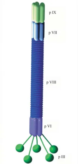
The bacteriophage (or simply phage) mostly used in phage display technology, are single-stranded DNA viruses that infect a number of gram-negative bacteria. The fila-mentous phage particles mostly used for display purposes are known as Ff and include strains M13, f1, Fd and ft. Fd phage particles consist of a long cylindrical protein capsid, 930 nm in length and 6.5 nm in diameter, enclosing a sin-gle-stranded DNA genome of about 6400 nucleotides, con-sisting of 11 genes. The viral mass is approximately
Send correspondence to Marco Antonio Arap. Universidade de São Paulo, Hospital das Clinicas da Faculdade de Medicina, Depar-tamento de Cirurgia, Disciplina de Urologia, R. Adma Jafet 50, 3o
andar, Bela Vista 01308-050 São Paulo, SP, Brazil. E-mail: marcoarap@hotmail.com.
16.3 MDa, and consists mainly of about 2700 copies of the pVIII, a 50 aa residue protein encoded bygene VIII. On one side of the phage particle there are 3 to 5 copies of the pro-teins pVII and pXIX (genes VII and XIX) and on the other side there are 3 to 5 copies of the proteins pIII and pVI (Fig-ure 1) (Webster 2001). In most display applications, pIII, a 406 aa adsorption protein, is the protein used for peptide ex-pression. The pIII protein appears to have two functional domains: an exposed N-terminal domain that binds the F pilus, but is not required for phage particle assembly, and a C-terminal domain that is buried in the particle and is an in-tegral part of the capsid structure. The C-terminal portion of pVIII is inside the phage particle, close to the DNA, while the N-terminal part is exposed to the surroundings.
Fd particles are able to infect a variety of Gram-negative bacteria, includingE. coli,using pili (F pilus inE. coli) as receptors. Filamentous phage infection does not produce lytic infection inE coli., but rather induces a state in which the infected bacteria produce and secrete phage particles into the growing medium. Infection begins by the attachment of phage pIII to the F pilus of a maleE. coli. The circular single-stranded DNA enters the bacteria where it is converted by the host DNA replication machinery into dou-ble-stranded plasmid replicative form. By rolling circle replication, the replicative form makes single-stranded DNA and the templates for expression of proteins pIII and pVIII are formed. Phage descendants are assembled by packaging of the single-stranded DNA into protein coats and extruded through the bacterial membrane (Russel 1991).
Most of the currently used phage display vectors use the N-terminus of pIII protein or pVIII protein to display the foreign peptide or protein (Smith and Scott 1993). The pIII libraries display 3-5 copies of each individual peptide (Scott and Smith 1990), whereas pVIII libraries can display up to 2700 copies of small (up to six amino acids) peptides (Greenwoodet al.1991). The pIII and pVIII proteins can display peptides of various lengths and cysteine residues can be introduced to the fusion peptide to create conformational constraints by the formation of “loops” be-tween disulfide bridged cysteine residues. Furthermore, the exogenous peptides are well exposed, facilitating the in-sert-target interactions. Large peptide inserts of up to 38 amino acids can be introduced into the amino terminus of pIII protein without the loss of phage infectivity or particle assembly.
Working with phage display technology
Detailed description of the materials, methods and space needed for those who want to start phage work may be found in specialized textbooks (Barbas et al. 2000, Pasqualini and Arap 2002). The basic protocols used with phage studies can also be obtained in these books, whereas its variations are found in published studies. Initial invest-ment is relatively small, as most of the fundainvest-mental
materi-als are common laboratory devices, such as Petri dishes, Falcon tubes and centrifuges. However, one cannot forget the most important tool for phage work: k91kan E. coli and peptide or antibody phage libraries. The construction of a peptide phage library involves a detailed protocol and, for those who are not experienced with phage work, one way to obtain reliable phage libraries and start a “bio-panning” is to establish a collaboration with a more experienced labora-tory. This is a very important step, as the panning results de-pend directly on the quality of the library. After the “bio-panning” is started, the most expensive step is to se-quence the clones obtained after a few rounds of selection, as hundreds of clones are usually sequenced in each pan-ning.
Peptide libraries offer the possibility of characteriz-ing peptide bindcharacteriz-ing specificity of important targets, such as proteins, cells and vascular endothelium. In general, the af-finity selection of ligands from phage display random pep-tide libraries involves 5 fundamental steps:
i) preparation of a primary library or amplification of an existing library,
ii) exposure of the phage particles to a target (immo-bilized protein/cell surface protein/vascular endothelium) for which specific ligands are planned to be identified,
iii) removal of non-specific binders (washing/perfu-sion),
iv) recovery of the target bound phage by elution or direct bacterial infection and amplification of the recovered phage,
v) back to step i two to four times.
This “bio-panning” can be repeated several times un-til a population of best binders is enriched. By sequencing the phage genome encoding the displayed peptide, one can determine and reproduce its sequence as recombinant or synthetic peptides, and finally determine specific and selec-tive ligands to target receptors (Koivunenet al.1999).
It is a common observation that the binding motif of a targeting peptide is a tripeptide motif appearing several times in different sequence contexts. Three amino-acid res-idues seem to provide the minimal framework for structural formation and protein-protein interaction (Vendruscoloet al. 2001). This is therefore the minimal searched frame-work, although it is obvious that repeated peptides consist-ing of more residues may mean a stronger interaction.
Applications
Identification of receptor-ligandsin vitro
Peptide libraries have been used to determine the epitope to which an antibody binds. Antibodies recognize peptide motifs based on only three or four conserved resi-dues. Therefore, it is possible to define the region of a pro-tein recognized by an antibody based on the motif revealed by phage display (Scott and Smith 1990). Folgori and col-leagues selected antigenic mimics (mimotopes) of two dif-ferent epitopes from the human hepatitis B virus envelope protein (HBsAg) and showed that a humoral response to these mimotopes was widespread in the immunized popu-lation, suggesting that the strategy identifies displayed pep-tides with a potential role as diagnostic reagents (Folgoriet al.1994).
Phage libraries were used to define peptide structures recognized by major histocompatibility (MHC) molecules. Peptides with binding specificity for lymphoblastoid-derived DR1 molecules, determined by phage display li-braries (Hammeret al.1992), may have important applica-tions as MHC-specific antagonists. Other examples of binding of ligand motifs to their receptors discovered by phage display technology include a family of cell surface integrins that recognize the tripeptide RGD (Koivunenet al.2001, Ruoslahti 1996). These integrins mediate cell at-tachment to many types of extracellular matrix proteins that contain the RGD motif, such as fibronectin, vitronectin and fibrinogen (Ruoslahti 1991, Ruoslahti 1996). Phage display
peptide libraries have also been used to find ligands to the SH2, a common domain in protein kinases and signaling proteins. The SH2 domain recognizes peptides containing a phosphorylated tyrosine residue (Denteet al.1997, Gram et al.1997).
Using phage display in combination with other meth-ods, potent mimetics of proteins and minimal active pep-tides have been found. A dimmer of the 14-amino acid cyclic thrombopoietin mimetic, for example, was found to be as active as the parental polypeptide (Cwirlaet al.1997). More recently, Cardo-Vilaet al. introduced an approach based on phage display technology to identify molecules that specifically interact with the cytoplasmic domain of the beta 5 integrin subunit. Initially, they isolated beta 5 specific peptides by screening a phage library on a recom-binant fusion protein containing the beta 5 cytoplasmic do-main. Next, they designed and synthesized an internalizing version of the peptide by using the penetratin system for intracellular delivery, and showed that it triggered cell apoptosis in a caspase-dependent manner, suggesting a functional link between the alpha v beta 5 integrin, annexin V, and programmed cell death (Cardo-Vilaet al.2003).
Besides proteins, peptides affecting biologically sig-nificant protein-DNA interactions (Chenget al.1996), pep-tides binding to carbohydrates (Matsubara et al. 1999, Noda et al. 2001, Peletskaya et al. 1996), to carbon nanotubes (Wanget al.2003) and to small chemical com-pounds such as taxol (Rodiet al.1999) have been isolated from phage display random peptide libraries.
Phage display has also been applied as a tool for di-rected evolution for more than a decade. In enzymology, it has been applied for mechanistic-based studies and to gen-erate enzyme variants with new or improved properties. Enzymes with a broader range of substrates, for instance, would have improved function and allow certain advan-tages to its host. A strategy described by Petersen and col-leagues makes possible thein vitroisolation of enzymes for almost any reaction. Moreover, this strategy theoretically allows one to functionally clone natural enzymes based on their ability to catalyze specific reactions rather than their structure or binding ability (Pedersenet al.1998). Using a combination of phage display selection and high-throughput screening methods, Wirsching and colleagues generated variants of hirudin, a thrombin-specific inhibitor, with increased protease resistance that may prove useful for hematologic disorders (Wirschinget al.2003). Phage dis-play technology has also allowed the development of DNA-binding proteins with novel specificities, energetics of protein folding and directed evolution of antibodies (O’Neil and Hoess 1995).
Selection of ligand-receptors in complex
biological systems
during recent years. When using such complex targets, en-hancement of specific binding above the background phage adherence is usually necessary, as unspecific binding to common molecules such as albumin is expected and may interfere with the panning results.
Selection on living cells can be done on either monolayers of adherent cells or on cells in suspension. Un-bound phage must be washed away and phage recovery is done by bacterial infection. Goodson and colleagues iso-lated ligands to the urokinase receptor after transfecting cells with the gene for the same receptor (Goodsonet al. 1994). Human platelets have been used as targets for the se-lection of a peptide antagonist of the thrombin receptor (Doorbar and Winter 1994). Another group isolated two an-tibody clones that recognize melanoma cells by subjecting an antibody phage library to human melanoma cells (Kupschet al.1999). More recently, Ardelt and colleagues screened 2 phage display random peptide libraries on hu-man urothelium and peptides. Those selected in this bio-panning were tested for their binding abilities to normal and malignant urothelial cells. Two classes of peptide mo-tifs shared the same amino acid sequence bound to normal urothelium and to 2 transitional carcinoma cells, and were therefore suitable for translation into targeted intravesical therapy (Ardeltet al.2003).
In a very elegant study, Giordano and colleagues de-scribed a new approach for the screening, selection and sorting of cell-surface binding peptides from phage librar-ies. The method, termed bio-panning and rapid analysis of selective interactive ligands (BRASIL), allows separation of complexes formed by the cells and bound phage from the remaining unbound phage still in the suspension. This tech-nique is based on a differential centrifugation of the aque-ous phage/cell suspension through a non-miscible organic lower phase. Centrifugation will drive the cells from a hy-drophilic phase into a non-miscible (hydrophobic) organic phase. The passage of cells from a hydrophilic to a hydro-phobic setting will separate water-soluble materials, such as the unbound phage. The cell/phage pellet is then recov-ered from the bottom of the tube after immediate freezing in liquid nitrogen. Next, the cell pellet is thawed and bound phage are recovered by bacterial infection. As the method involves one centrifugation and does not require repeated washes, it allows a simpler and more convenient phage re-covery from cell membranes than other cell-panning tech-niques. As a proof of the principle, they screened human endothelial cells stimulated with vascular endothelial growth factor (VEGF), constructed a peptide-based ligand-receptor map of the VEGF family and validated a chimeric ligand-mimic that binds specifically to VEGF re-ceptor-1 and neuropilin-1 (Giordanoet al.2001).
One of the most important tumor targets identified us-ing bio-pannus-ing on cultured cells is the Glucose-regulated protein-78 (GRP78) (Mintzet al.2003), a chaperone pro-tein initially found to be expressed in the endoplasmic
retic-ulum of various cell types (Munro and Pelham 1986, Lee 1992 and Morimoto 1993). GRP78 induction is markedly increased in a variety of cellular stress conditions, such as glucose starvation, oxygen deprivation and accumulation of unglycosylated proteins (Lee 1987, Liet al.1993). This induction is a cellular protective response against stress (Li et al.1992, Sugawaraet al.1993, Jamoraet al.1996) and prevents apoptosis (Myiakeet al.2000).
The relatively hypoxic environment found in some solid tumors is one of the possible mechanisms involved in the over-expression of GRP78 in human cancers. GRP78 over-expression triggers an immune response against the protein that has proven to be related to androgen-independent prostate cancer and reduced overall patient survival (Mintzet al.2003). Our group is now conducting in vivo studies aiming to develop targeted therapies for hu-man cancers via GRP78. Initial results showed that the pep-tides used for targeted delivery are capable of killing cells in vitro, based on the GRP78 surface expression. In addi-tion, in vivo assays with the same peptides showed that the protein was successfully used as a molecular target for di-rected therapy against breast and prostate cancers.
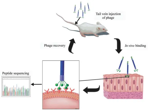
In vivoselection
In this technique phage libraries are injected intrave-nously into animals and then organs or tissues are collected and examined for phage bound to tissue-specific endothe-lial cell markers (Figure 2). Pasqualini was the first to de-scribe this method and to isolate peptides that home to renal and cerebral vascular endothelium in vivo (Pasqualini and Ruoslahti 1996). Since Pasqualini’s study, identification of receptor-ligand pairs has been described for several organs such as lung, kidney, pancreas, adrenal gland, muscles, in-testines and uterus. Selection in vivo has several advan-tages: first, peptides displayed on the phage particles are identified and tested functionally and must overcome natu-ral mechanisms of degradation. Second, peptides recogniz-ing unspecific molecules are depleted from circulation. Finally, in vivo selection proved to be able to identify re-ceptors expressed selectively on tumor endothelium (Arap et al.2002). These receptors may serve as molecular targets for the development of diagnostic techniques and targeted therapies.
al. 2002). This may mean a reduction in prostate cancer risk, as well as alternatives to surgical prostate ablation.
Recently, a novel breast-homing peptide was selected from a phage library, and Aminopeptidase P was identified as the peptide receptor (Essleret al. 2002). As it is ex-pressed in normal and malignant breast tissue, this aminopeptidase-P-binding peptide may be used for the de-velopment of drugs directed to prevent and treat breast can-cer. Perhaps the most striking and promising contribution of in vivo use of phage display comes with the work of Kolonin and colleagues, who recently published a study in which a fat-targeting peptide was used to cure high-calorie related obesity in living animals (Kolonin et al. 2004). These findings not only mean hope for the treatment of sev-eral high-incidence diseases, but also demonstrate the ver-satility of phage display technology.
Molecular diversity of receptors in human blood ves-sels remains largely unexplored due to the lack of safe tech-niques applicable for human research. The first report of in vivo screening of a phage peptide library in humans was published in 2002 (Arapet al.2002b). They developed a se-lection method in which peptides that home to specific hu-man vascular beds are identified after intravenous administration of a peptide library. Moreover, they selected and isolated a mimic motif of interleukin 11 (IL-11) from prostate biopsies after the administration of phage. Analy-sis of the selected motifs revealed similarities to ligands for
differentially expressed cell-surface proteins. Recently, the same group showed that IL-11 receptor alpha is a candidate target for translational clinical trials against advanced and metastatic prostate cancer (Zuritaet al.2004). Table 1 rep-resent some of the peptides with therapeutic potential iden-tified by phage display.
Recent innovations in phage display technology
Selective infective phage (SIP)
Enrichment for phage displaying high-affinity mole-cules over non-specific binders is one of the most difficult tasks in phage display technology. In SIP technology (Krebberet al.1997), the N-terminal domains of pIII are re-placed by the gene for a peptide or a protein leading to the generation of noninfective phage particles. The missing N-terminal domains, necessary for phage infectivity, are supplied within adapter molecules consisting of the ligand coupled covalently to these N-terminal domains. Infectivity is restored when noninfective phage and adapter molecules are mixed only to phage particles displaying peptides that are capable of binding the ligand with the lat-ter, providing the missing N-terminal domains of pIII to the phage. This is a method that eliminates the need for physi-cal separation of specific and unspecific binders, therefore providing an efficient and rapid procedure for selection of high-affinity interactions (Krebberet al.1997).
Landscape phage
Phage particles with amino acids 2 to 4 on every wild-type pVIII coat protein replaced with random octamers are called landscape phage (Petrenkoet al.1996). This substitution leads to a fixed peptide framework that al-lows phage to have properties dependent on the introduced variable peptides. Moreover, additional properties may arise owing to the global architecture of the phage particle. Clones that can bind to dioxin, streptavidin, avidin, and beta-galactosidase with nanomolar affinity were selected from landscape phage libraries against immobilized targets and were named “substitute-antibody filaments” (Petrenko et al. 1996 and Petrenko and Smith 2000). Due to their novel surface properties, these filaments may have binding advantages over their immunoglobulin counterparts.
Isolation of allergens by phage display
Rhyner and colleagues described the isolation of al-lergens that induce IgE production using the association of robotic-based high-throughput screening technology and the selective enrichment of cDNA libraries expressed on phage surface with serum IgE from allergic individuals. Fu-sion proteins created by the principle of linking the phage phenotype (expressed as a gene product displayed on its coat) to its genetic information are covalently associated with the phage particle. Therefore, cDNA libraries dis-played on phage surface can be screened for the presence of specific clones by affinity purification. Phage clones that bind to IgE may be selected by screening and enrichment of phage libraries against serum IgE immobilized in a solid phase. The amino acid sequence of surface-expressed
aller-gens can be clarified by sequencing the DNA of the inte-grated section of the phage, as there is a physical linkage between its genotype and phenotype (Rhyneret al.2004).
Use of phage display for gene delivery
Gene delivery to mammalian cells has also been ac-complished by the use of single- (Yokoyama-Kobayashi and Kato 1993) and double-stranded phage (Ishiuraet al. 1982, Okayama and Berg 1985). The modification of exist-ing gene therapy vectors by selectexist-ing alternative ligands may increase their selectivity and therefore improve effi-cacy and reduce toxicity of gene delivery. In order to in-crease vector potency and selectivity, growth factors such as fibroblast growth factor (FGF) and other ligands that bind to cell surface receptors have been used for targeting viral and non-viral vectors (Goldmanet al.1997, Rogerset al.1998). Larocca and colleagues successfully displayed FGF2, a growth factor ligand, on the phage surface as a pIII fusion (Laroccaet al.1999). It was the first report to dem-onstrate gene transfer to mammalian cells by genetically targeted filamentous phage. It also supported previous sug-gestions that peptide-displaying phage can be used them-selves as gene delivery vectors (Barry et al. 1996 and Laroccaet al.1998). Therefore, the combination of gene delivery techniques and the power of combinatorial phage libraries allowed the creation of genetically altered phage, displaying a known gene targeting ligand. This may result in safer and more efficient gene transfer into mammalian cells.
Tumor targeting
Tumor targeting peptide ligands found by phage dis-play technology have also been used for delivery of cytotoxic chemotherapy (Arap et al.1998), proapoptotic peptides (Arap et al. 2002, Ellerby et al. 1999), and cytokines (Curniset al.2000) to receptors in the angiogenic vasculature showing marked therapeutic efficacy in tu-mor-bearing mouse models. Tumor targeting peptide lig-ands can also deliver imaging agents to tumor vasculature (Hong and Clayman 2000).
Final Remarks
Here we reviewed basic concepts of the structure and use of phage display technology. The applications of the technology are being actively explored, yielding different peptides that may prove useful for basic and clinical re-search. Recombinant antibodies, new diagnostic proce-dures, targeted therapies and several other phage display-based applications have arisen, facilitating diagno-sis and treatment of benign and malignant conditions. Some of the peptides described here will probably be part of dif-ferent therapies directed towards human diseases. In this context, human phage screenings must be continued so that other treatment options for conditions such as cancer and atherosclerosis may take place.
Table 1- Peptides with potential therapeutic implications identified by phage display.
Organ/tissue/ molecule
Phage sequence recovered
Reference
αvβ5 Integrin VVISYSMPD Cardo-Vilaet al.(2003)
Aminopeptidase N CNGRCVSGCAGRC Pasqualiniet al.(2000)
Mouse lung CGFECVRQCPERC Rajotteet al.(1998) Mouse brain CSSRLDAC Pasqualini and
Ruoslahti (1996) Mouse kidney CLPVASC Pasqualini and
Ruoslahti (1996)
Mouse retina CSCFRDVCC Rajotteet al.(1998) Mouse pancreas SWCEPGWCR Rajotteet al.(1998)
Mouse skin CVALCREACGEGC Rajotteet al.(1998) Mouse white fat CKGGRAKDC Koloninet al.(2004) Mouse placenta TPKTSVT Koloninet al.(2002)
Human prostate CGRRAGGSC Arapet al.(2002b)
References
Arap W, Pasqualini R and Ruoslahti E (1998) Cancer treatment by targeted drug delivery to tumor vasculature in a mouse model. Science 279:377-380.
Arap W, Haedicke W, Bernasconi M, Kain R, Rajotte D, Krajewski S, Ellerby HM, Bredesen DE, Pasqualini R and Ruoslahti E (2002) Targeting the prostate for destruction through a vascular address. Proc Natl Acad Sci USA 99:1527-1531.
Arap W, Kolonin MG, Trepel M, Lahdenranta J, Cardo-Vila M, Giordano RJ, Mintz PJ, Ardelt PU, Yao VJ, Vidal CI, Chen L, Flamm A, Valtanen H, Weavind LM, Hicks ME, Pollock RE, Botz GH, Bucana CD, Koivunen E, Cahill D, Troncoso P, Baggerly KA, Pentz RD, Do KA, Logothetis CJ and Pasqualini R (2002b) Steps toward mapping the human vasculature by phage display. Nat Med 8:121-127. Ardelt PU, Wood CG, Chen L, Mintz PJ, Moya C, Arap MA,
Wright KC, Pasqualini R and Arap W (2003) Targeting urothelium:Ex vivoassay standardization and selection of internalizing ligands. J Urol 169:1535-1540.
Azzazy HM and Highsmith WE Jr (2002) Phage display technol-ogy: Clinical applications and recent innovations. Clin Biochem 35:425-445.
Barbas CF 3rd, Kang AS, Lerner RA and Benkovic SJ (1991) As-sembly of combinatorial antibody libraries on phage sur-faces: the gene III site. Proc Natl Acad Sci USA 88:7978-7982.
Barbas CF, Burton DR, Scott JK and Silverman GJ (2000) Phage display: A laboratory manual. Cold Spring Harbor Labora-tory Press, Cold Spring Harbor, New York, pp 1-24. Barry MA, Dower WJ and Johnston SA (1996) Toward
cell-targeting gene therapy vectors: selection of cell-binding peptides from random peptide-presenting phage libraries. Nat Med 2:299-305.
Bass S, Greene R and Wells JA (1990) Hormone phage: An en-richment method for variant proteins with altered binding properties. Proteins 8:309-314.
Cheng X, Kay BK and Juliano RL (1996) Identification of a bio-logically significant DNA-binding peptide motif by use of a random phage display library. Gene 171:1-8.
Cardo-Vila M, Arap W and Pasqualini R (2003) Alpha v beta 5 integrin-dependent programmed cell death triggered by a peptide mimic of annexin V. Mol Cell 11:1151-1162. Curnis F, Sacchi A, Borgna L, Magni F, Gasparri A and Corti A
(2000) Enhancement of tumor necrosis factor alpha antitumor immunotherapeutic properties by targeted deliv-ery to aminopeptidase N (CD13). Nat Biotechnol 18:1185-1190.
Cwirla SE, Balasubramanian P, Duffin DJ, Wagstrom CR, Gates CM, Singer SC, Davis AM, Tansik RL, Mattheakis LC, Boytos CM, Schatz PJ, Baccanari DP, Wrighton NC, Barrett RW and Dower WJ (1997) Peptide agonist of the thrombopoietin receptor as potent as the natural cytokine. Science 276:1696-1699.
Dente L, Vetriani C, Zucconi A, Pelicci G, Lanfrancone L, Pelicci PG and Cesareni G (1997) Modified phage peptide libraries as a tool to study specificity of phosphorylation and recogni-tion of tyrosine containing peptides. J Mol Biol. 269:694-703.
Doorbar J and Winter G (1994) Isolation of a peptide antagonist to the thrombin receptor using phage display. J Mol Biol 244:361-369.
Ellerby HM, Arap W, Ellerby LM, Kain R, Andrusiak R, Rio GD, Krajewski S, Lombardo CR, Rao R, Ruoslahti E, Bredesen DE and Pasqualini R (1999) Anti-cancer activity of targeted pro-apoptotic peptides. Nat Med 5:1032-1038.
Essler M and Ruoslahti E (2002) Molecular specialization of breast vasculature: A breast-homing phage-displayed pep-tide binds to aminopeptidase P in breast vasculature. Proc Natl Acad Sci USA 99:2252-2257
Folgori A, Tafi R, Meola A, Felici F, Galfre G, Cortese R, Monaci P and Nicosia A (1994) A general strategy to identify mimotopes of pathological antigens using only random pep-tide libraries and human sera. EMBO J 13:2236-2243. Giordano RJ, Cardo-Vila M, Lahdenranta J, Pasqualini R and
Arap W (2001) Biopanning and rapid analysis of selective interactive ligands. Nat Med 7:1249-1253.
Goldman CK, Rogers BE, Douglas JT, Sosnowsky BA, Ying W, Siegal GP, Baird A, Campain JA and Curiel DT (1997) Tar-geted gene delivery to Kaposi’s sarcoma cells via the fibroblast growth factor receptor. Cancer Res 57:1447-1451. Goodson RJ, Doyle MV, Kaufman SE and Rosenberg S (1994) High-affinity urokinase receptor antagonists identified with bacteriophage peptide display. Proc Natl Acad Sci USA 91:7129-7133.
Gram H, Schmitz R, Zuber JF and Baumann G (1997) Identifica-tion of phosphopeptide ligands for the Src-homology 2 (SH2) domain of Grb2 by phage display. Eur J Biochem 246:633-637.
Greenwood J, Hunter GJ and Perham RN (1991) Regulation of fil-amentous bacteriophage length by modification of electro-static interactions between coat protein and DNA. J Mol Biol 217:223-227.
Hammer J, Takacs B and Sinigaglia F (1992) Identification of a motif for HLA-DR1 binding peptides using M13 display li-braries. J Exp Med 176:1007-1013.
Hong FD and Clayman GL (2000) Isolation of a peptide for tar-geted drug delivery into human head and neck solid tumors. Cancer Res 60:6551-6556.
Hoogenboom HR (2002) Overview of antibody phage-display technology and its applications. Methods Mol Biol 178:1-37.
Ishiura M, Hirose S, Uchida T, Hamada Y, Susuki Y and Okada Y (1982) Phage particle-mediated gene transfer to cultured mammalian cells. Mol Cell Biol 2:607-616.
Jamora C, Dennert G and Lee AS (1996) Inhibition of tumor pro-gression by suppression of stress protein GRP78/BiP induc-tion in fibrosarcoma B/C10ME. Proc Natl Acad Sci USA 93:7690-7694.
Kay BK, Adey NB, He YS, Manfredi JP, Mataragnon AH and Fowlkes DM (1993) An M13 phage library displaying ran-dom 38-amino-acid peptides as a source of novel sequences with affinity to selected targets. Gene 128:59-65.
Koivunen E, Arap W, Rajotte D, Lahdenranta J and Pasqualini R (1999) Identification of receptor ligands with phage display peptide libraries. J Nucl Med 40:883-888.
leucine-leucine-glycine motif-containing peptides. J Cell Biol 153:905-916.
Kolonin MG, Pasqualini R and Arap W (2002) Teratogenicity in-duced by targeting a placental immunoglobulin transporter. Proc Natl Acad Sci USA 90:13055-13060.
Kolonin MG, Saha PK, Chan L, Pasqualini R and Arap W (2004) Reversal of obesity by targeted ablation of adipose tissue. Nat Med 10:625-632.
Krebber C, Spada S, Desplancq D, Krebber A, Ge L and Pluckthun A (1997) Selectively-infective phage (SIP): A mechanistic dissection of a novel in vivo selection for pro-tein-ligand interactions. J Mol Biol 268:607-18.
Kupsch JM, Tidman NH, Kang NV, Truman H, Hamilton S, Patel N, Newton Bishop JA, Leigh IM and Crowe JS (1999) Isola-tion of human tumor-specific antibodies by selecIsola-tion of an antibody phage library on melanoma cells. Clin Cancer Res 5:925-31.
Larocca D, Witte A, Johnson W, Pierce GF and Baird A (1998) Targeting bacteriophage to mammalian cell surface receptor for gene delivery. Hum Gene Ther 9:2393-2399.
Larocca D, Kassner PD, Witte A, Ladner RC, Pierce GF and Baird A (1999) Gene transfer to mammalian cells using geneti-cally targeted filamentous bacteriophage. FASEB J 13:727-734.
Lee AS (1987) Coordinated regulation of a set of genes by glucose and calcium ionophores in mammalian cells. Trends Biochem Sci 12:20-23.
Lee AS (1992) Mammalian stress response: induction of the glu-cose-regulated protein family. Curr Opin Cell Biol 4:267-273.
Li LJ, Li X, Ferrario A, Rucker N, Liu ES, Wong S, Gomer CJ and Lee AS (1992) Establishment of a Chinese hamster ovary cell line which express grp78 antisense transcripts and sup-presses A23187 induction of both GRP78 and GRP94. J Cell Physiol 153:575-582.
Li WW, Alexandre S, Cao C and Lee AS (1993) Transactivation of the grp78 promoter by Ca2+ depletion: A comparative analysis with A23187 and the endoplasmic reticulum Ca2+-ATPase inhibitor thapsigargin. J Biol Chem 268:12003-12009.
Matsubara T, Ishikawa D, Taki T, Okahata Y and Sato T (1999) Selection of ganglioside GM1-binding peptides by using a phage library. FEBS Lett 456:253-256.
McCafferty J, Griffiths AD, Winter G and Chiswell DJ (1990) Phage antibodies: Filamentous phage displaying antibody variable domains. Nature 348:552-554.
Mintz PJ, Kim J, Do KA, Wang X, Zinner RG, Cristofanilli M, Arap MA, Hong WK, Troncoso P, Logothetis CJ, Pasqualini R and Arap W (2003) Fingerprinting the circulating reper-toire of antibodies from cancer patients. Nat Biotechnol 21:57-63.
Miyake H, Hara I, Arakawa S and Kamidono S (2000) Stress pro-tein GRP78 prevents apoptosis induced by calcium ionophore, ionomycin, but not by glycosylation inhibitor, tunicamycin, in human prostate cancer cells. J Cell Biochem 77:396-408.
Morimoto RI (1993) Cells in stress: Transcriptional activation of heat shock genes. Science 259:1409-1410.
Munro S and Pelham HR (1986) An Hsp70-like protein in the ER: Identity with the 78 kd glucose-regulated protein and immu-noglobulin heavy chain binding protein. Cell 46:291-300.
Noda K, Yamasaki R, Hironaka Y and Kitagawa A (2001) Selec-tion of peptides that bind to the core oligosaccharide of R-form LPS from a phage-displayed heptapeptide library. FEMS Microbiol Lett 205:349-354.
O’Neil KT and Hoess RH (1995) Phage display: Protein engineer-ing by directed evolution. Curr Opin Struct Biol 4:443-9. Okayama H and Berg P (1985) Bacteriophage lambda vector for
transducing a cDNA clone library into mammalian cells Mol Cell Biol 5:1136-1142.
Pasqualini R and Ruoslahti E (1996) Organ targeting in vivo using phage display peptide libraries. Nature 380:364-366. Pasqualini R, Kovunen E, Kain R, Lahdenranta J, Sakamoto M,
Stryhn A, Ashmun RA, Shapiro LH, Arap W and Ruoslahti E (2000) Aminopeptidase N is a receptor for tumor-homing peptides and a target for inhibiting angiogenesis. Cancer Res 60:722-727
Pasqualini R and Arap W (2002) Vascular targeting. In: Bertino JR (ed) Encyclopedia of Cancer. Academic Press, New Jer-sey, pp 501-508.
Pedersen H, Holder S, Sutherlin DP, Schwitter U, King DS and Schultz PG (1998) A method for directed evolution and functional cloning of enzymes. Proc Natl Acad Sci USA 95:10523-10528.
Peletskaya EN, Glinsky G, Deutscher SL and Quinn TP (1996) Identification of peptide sequences that bind the Thom-sen-Friedenreich cancer-associated glycoantigen from bacteriophage peptide display libraries. Mol Divers 2:13-18. Petrenko VA, Smith GP, Gong X and Quinn T (1996) A library of organic landscapes on filamentous phage. Protein Eng 9:797-801.
Petrenko VA and Smith GP (2000) Phages from landscape librar-ies as substitute antibodlibrar-ies. Protein Eng 13:589-592. Rajotte D, Arap W, Hagedorn M, Koivunen E, Pasqualini R and
Ruoslahti E (1998) Molecular heterogeneity of the vascular endothelium revealed byin vivophage display. J Clin Invest 102:430-437.
Rhyner C, Weichel M, Fluckiger S, Hemmann S, Kleber-Janke T and Crameri R (2004) Cloning allergens via phage display. Methods 32:212-218.
Rodi DJ, Janes RW, Sanganee HJ, Holton RA, Wallace BA and Makowski L (1999) Screening of a library of phage-displayed peptides identifies human bcl-2 as a taxol-binding protein. J Mol Biol 285:197-203.
Rogers BE, Douglas JT, Sosnowsky BA, Ying W, Pierce GF, Buchsbaum DJ, Manna DD, Baird A and Curiel DT (1998) Enhanced in vivogene delivery to human ovarian cancer xenografts utilizing a tropism-modified adenovirus vector. Tumor Targeting 3:25-31.
Ruoslahti E (1991) Integrins. J Clin Invest 87:1-5.
Ruoslahti E (1996) RGD and other recognition sequences for integrins. Annu Rev Cell Dev Biol 12:697-715.
Russel M (1991) Filamentous phage assembly. Mol Microbiol 5:1607-1613.
Scott JK and Smith GP (1990) Searching for peptide ligands with an epitope library. Science 249:386-390.
Smith GP (1985) Filamentous fusion phage: Novel expression vectors that display cloned antigens on the virion surface. Science 228:1315-1317.
Smith GP and Scott JK (1993) Libraries of peptides and proteins displayed on filamentous phage. Methods Enzymol 217:228-257.
Sugawara S, Takeda K, Lee AS and Dennert G (1993) Suppres-sion of stress protein GRP78 induction in tumor B/C10ME eliminates resistance to cell mediated cytotoxicity. Cancer Res 53:6001-6005.
Szardenings M (2003) Phage display of random peptide libraries: Applications, limits, and potential. J Recept Signal Transduct Res 23:307-349.
Vendruscolo M, Paci E, Dobson CM and Karplus M (2001) Three key residues form a critical contact network in a protein folding transition state. Nature 409:641-645.
Wang S, Humphreys ES, Chung SY, Delduco DF, Lustig SR, Wang H, Parker KN, Rizzo NW, Subramoney S, Chiang YM and Jagota A (2003) Peptides with selective affinity for carbon nanotubes. Nat Mater 2:196-2000.
Webster R (2001) Filamentous phage biology. In Phage Barbas CF, Burton DR, Scott JK and Silverman (eds) Phage Dis-play: A Laboratory Manual. Cold Spring Harbor Laboratory Press, Cold Spring Harbor pp 1.1-1.37.
Wirsching F, Keller M, Hildmann C, Riester D and Schwienhorst A (2003) Directed evolution towards protease-resistant hirudin variants. Mol Genet Metab 80:451-462.
Yokohama-Kobayashi M and Kato S (1993) Recombinant f1 phage particles can transfect monkey COS-7 cells by DEAE dextran method. Biochem Biophys Res Commun 192:935-939.
Zurita A, Troncoso P, Cardo-Vila M, Logothetis C, Pasqualini R and Arap W (2004) Combinatorial screenings in patients: The interleukin-11 receptor alpha as a candidate target in the progression of human prostate cancer. Cancer Res 64:435-439.