Molecular characterization and T and B cell epitopes prediction of
Mycoplasma synoviae
53 strain VlhA hemagglutinin
Ilana Lopes Camargo
1, Cristina Toscano Fonseca
1, Santuza Ribeiro Teixeira
1, Vasco Azevedo
2,
Anderson Myioshi
2and Sergio Costa Oliveira
11
Departamento de Bioquímica e Imunologia, Instituto de Ciências Biológicas,
Universidade Federal de Minas Gerais, Belo Horizonte, MG, Brazil.
2
Departamento de Biologia Geral, Instituto de Ciências Biológicas, Universidade Federal de Minas Gerais,
Belo Horizonte, MG, Brazil.
Abstract
Mycoplasma sinoviae is a major pathogen of poultry causing synovitis and respiratory infection. M. synoviae hemagglutinin (VlhA) is a lipoprotein encoded by related multigene families that appear to have arisen by horizontal gene transfer. It is an abundant immunodominant surface protein involved in host-parasite interaction mediating binding to host erythrocytes. Herein, we have performed in silico analysis of the vlhA gene product from the Mycoplasma synoviae 53 strain and compared it to the VlhA protein of M. synoviae WUV1853 strain. The VlhA of the M. synoviae 53 strain possesses 569 amino acids and showed 85% identity with the VlhA protein of the M. synoviae WUV1853 strain. Further, a signal peptide was identified from amino acid M1to D28and a cleavage site between D28
and Q29, both located in the N-terminal domain of the molecule. Additionally, an insertion of PAPT amino acids was
observed between T30-P35and a deletion of the amino acids GTPGNP within the PRR region of the VlhA from theM.
synoviae 53 strain, which may be related to its reduced virulence. Finally, we have identified 17 B cell epitopes and 22 T cells epitopes within the VlhA from theM. synoviae 53 strain. The B cell epitope S263-D277and the T cell epitopes
N45-N54 and G58-N67 showed 100% and 87-100% identity, respectively, with regions of VlhA protein of tested
Mycoplasma synoviae and Mycoplasma galisepticum strains. Thus, these peptides represent new candidate mole-cules for the development of efficient diagnostic assays and new subunit vaccines.
Key words: Mycoplasma synoviae, hemagglutinin, epitopes, host-parasite interaction, vaccine.
Received: April 12, 2006; Accepted: October 16, 2006.
Introduction
Mycoplasma synoviaeis one of the smallest and sim-plest bacteria lacking a cell wall known to exist, and it is a major pathogen of chickens and turkeys, causing respira-tory tract infection and arthritis worldwide (Kleven, 1997). Although the basis of mycoplasma pathogenicity remains unknown, it is widely accepted that most of the damage re-sulting from mycoplasma infections in humans and animals is due to host immune and inflammatory responses rather than to direct toxic effects of mycoplasma virulence factors (Razinet al., 1998).
M. synoviaeisolates differ in their infectivity, tissue tropism and pathogenicity (Rottem, 2003). Many animal mycoplasmas depend on adhesion to host tissues for
colo-nization and infection. In these mycoplasmas, adherence is the major virulence factor and adherence-deficient mutants are avirulent (Baseman and Trully, 1997). Current theory holds that mycoplasma remains attached to the surface of epithelial cells, although some mycoplasmas have evolved mechanisms for entering host cells that are not naturally phagocytic (Razinet al., 1998). The intracellular localiza-tion is obviously a privileged niche, well protected from humoral mechanisms of the host immune system and from the action of many antibiotics. The finding that some myco-plasmas can reside intracellularly opens up new horizons to the study of the role of mycoplasma and host surface mole-cules in invasion. Although, the ability of internalized my-coplasmas to multiply within the host cell remains to be convincingly demonstrated, reports describing myco-plasma invasive phenotypes have offered new insights into the potential virulence strategies employed by these bacte-ria (Rottem, 2003).
Send correspondence to Sergio Costa Oliveira. Universidade Fe-deral de Minas Gerais, Instituto de Ciências Biológicas, Departa-mento de Bioquímica e Imunologia, Av. Antonio Carlos 6627, Pampulha, Belo Horizonte, MG, Brazil, 31270-901. E-mail: [email protected].
Escaping the host immune system is of critical impor-tance to mycoplasma survival within its host. The major survival mechanisms that have been extensively studied are molecular mimicry and phenotype plasticity which ensure that mycoplasmas are not fully nor efficiently recognized by the host immune system (Markhamet al., 1994; Wren, 2000). Molecular mimicry refers to antigenic epitopes that are shared by different mycoplasmas and host cells and they are considered as putative factors involved in the eva-sion of host defense mechanisms (Rottem, 2003). Myco-plasmas are also endowed with phenotypic plasticity defined as the ability of a single genotype to change its anti-genic make-up to produce more than one morphology, physiological state, and/or behavior in response to environ-mental conditions (Rottem, 2003). The common way to achieve phenotype plasticity in mycoplasma is by antigenic variation. Additionally, membrane lipoproteins are the ma-jor components of intact mycoplasmas and are able to acti-vate macrophages, thus playing an important role in cytokine production and consequently in the inflammatory response during infection (Chambaudet al., 1999).
VlhA is a variable protein encoded by thevlhAgene inM. synoviaethat is post-translationally cleaved into the N-terminal lipoprotein fragment MSPB (Major Surface Protein B) and the C-terminal fragment MSPA (Major Sur-face Protein A) which is directly involved in hemadherence (Noormohammadiet al., 1997). There is only one copy of the completevlhAgene in the genome of theM. synoviae
WUV1853 strain. Other copies are not functional genes and lack the 5’ end of the expressed gene (Noormohammadiet al., 1997, 2000). Comparing differentM. synoviaestrains, it was possible to observe differences in length and anti-genic determinants of MSPB proteins (Noormohammadiet al., 1997). The complete genome sequence ofMycoplasma synoviae53 strain revealed the organization of hemagglu-tinin genes with a single locus comprising 70 coding DNA sequences (CDS) (Vasconceloset al., 2005). In this study, we have characterized the vlhA gene product from the
Mycoplasma synoviae 53 strain and compared it to the VlhA protein of the Mycoplasma synoviae WUV1853 strain (Noormohammadi et al., 1997). The VlhA of the
Mycoplasma synoviae53 strain possesses 569 amino acids, a signal peptide and a cleavage site located in the N-terminal domain of the molecule. Additionally, using bioinformatic search tools, we have identified 17 B cell epitopes and 22 T cells epitopes that may be involved in host immune response against this microorganism.
Materials and Methods
DNA and amino acid sequences
The DNA and translated amino acid sequences of
vlhAgenes fromMycoplasma synoviae53 (Vasconceloset al., 2005) and Mycoplasma synoviae WUV1853 strains (Noormohammadi et al., 1997) were retrieved from
GenBank under accession no. NC007294 and AF035624, respectively. ThevlhAgene is located between the nucleo-tides of number 292135 and 293844 of the genome se-quence of theMycoplasma synoviae53 strain sequenced by our group (Vasconceloset al., 2005).
Characterization ofM. synoviaeVlhA by bioinformatics
These amino acid sequences for VlhA from
Mycoplasma synoviae 53 and Mycoplasma synoviae
WUV1853 strains were aligned by CLUSTALW Multiple Sequence Alignment available online. SOSUISignal and SOSUI were used to identify motifs in these proteins, such as peptide signal and hydrophobic domains. Additionally, SignalP 3.0 software was used for prediction of cleavage sites.
T and B cell epitopes prediction
The B cell epitope prediction was performed using the program Predicting Antigenic Peptides available on-line. The software for the detection of antigenic peptides is based on Kolaskar’s and Tongaonkar’s method previously described (Kolaskar and Tongaonkar, 1990). The T cell epitope prediction was performed using RANKPEP soft-ware. This software uses Position Specific Scoring Ma-trices (PSSMs) or profiles from a set of aligned peptides known to bind to a given MHC molecule as the predictor of MCH-peptide binding. We used the mouse MHC system H-2, as a model for this study and tested the I-Aband I-Ak alleles for MHC class II. Herein, we have selected several peptides that had high scores of binding to these MHC class II alleles. Predicted T and B cell epitopes shared between
M. synoviae53 andM. synoviae WUV1853 strains were also analyzed for their identity with other Mycoplasma synoviae strains (Mycoplasma synoviae B133-96, B154-02, B2700, B31-88, B38-96-170, B94-91, J26-85, J151-85, K1, K4, K1968, K2581, K27, MS-H, TN/427, ULB925 and ULB925KF) and Mycoplasma gallisepticum strains (M. gallisepticumR and S6) using the BLAST computer pro-gram blastp.
Results and Discussion
re-peats (PRR) region which has been identified in immu-nodominant surface antigens involved in the interaction between pathogens and host cells (Noormohammadiet al., 1997; Bencinaet al., 2001). A longer PRR region has been associated with higher invasiveness for theM. synoviae
strain K1968 (Bencinaet al., 2001). Further,M. synoviae
clonal populations can synthesize size and antigenic vari-ants of MSPB proteins and their expression can be associ-ated with transition from HA+to HA-phenotype (Noormo-hammadiet al.,1997).
The 569 amino acid sequence of VlhA from theM. synoviae53 strain and the 785 amino acid sequence of the VlhA protein derived from the M. synoviae WUV1853 strain were retrieved from the GenBank and aligned in or-der to compare their identity. VlhA protein ofM. synoviae
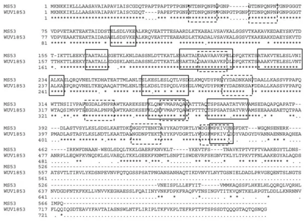
53 strain showed 85% identity with VlhA of the M. synoviaeWUV1853 strain, despite the difference in length (Figure 1). Amino acid sequence analysis performed by SOSUISignal and SignalP 3.0 computer programs resulted in the identification of a region encoding a signal peptide from amino acid M1to D28and a cleavage site between D28
and Q29in the VlhA of theM. synoviae53 strain that are
also present and they correspond to the same positions in the VlhA of theM. synoviaeWUV1853 strain. No variation in the signal peptide domains of both proteins was detected. The cleavage site which results in the MSPB and MSPA membrane antigens is located at S306in the VlhA of theM.
synoviae53 strain and at S308in the VlhA of theM. synoviae
WUV1853 strain (Figure 2). Among the differences be-tween the two sequences, there is an insertion of four amino acids (PAPT) after T30of VhlA from the M. synoviae53
strain This insertion is similar to an insertion observed in the M. synoviae FMT strain which is considered mildly pathogenic (Bencinaet al., 2001). The MSPB domain of theM. synoviae53 strain has also a deletion of six amino acids within the proline-rich repeats (PRR) (GTPGNP) in the N-terminal region which correlates to amino acids G54
to P59of theM. synoviaeWUV1853 strain (Figures 1 and
2). This deletion may affect bacterial virulence. The live at-tenuatedM. synoviaeMS-H strain vaccine has a similar de-letion of the amino acids PGNPGT within the PRR region
Figure 1- Comparison of the deduced amino acid sequences of VlhA ofMycoplasma synoviaestrain 53 (GenBank accession number NC007294) and
Mycoplasma synoviaestrain WUV1853 (GenBank accession number AF035624). Asterisks indicate identical residues and gaps are indicated by dashes. Squares with dashed-lines represent T cell epitopes and squares with solid-lines represent B cell epitopes.
Figure 2- Schematic representation of VlhA protein ofM. synoviae53 strain. Signal peptide region (SP) comprises amino acids from M1to D28.
The MSPB region contains two proline-rich repeats region (PRR) and has a PAPT insertion at T30and a GTPGNP deletion at P57. D28and S306
(Noormohammadiet al., 2002). Since this deletion was not observed in the VlhAM. synoviae WUV1853 strain, we speculate that the M. synoviaeWUV1853 strain may be more virulent than theM. synoviae53 strain.
Identification of immunodominant epitopes within a vaccine candidate antigen is extremely useful, since it is possible to formulate a vaccine composed of relevant epitopes from different antigens.In silicoepitope predic-tions resulted in the identification of 17 B cell epitopes, ranging from 7 to 23 mers (Table 1), and 22 T cell epitopes of 9 mers (Table 2). The predicted epitopes were distributed along the entire protein sequence. Comparing the epitopes predicted from the VlhA protein sequence of the M. synoviae53 strain with theM. synoviaeWUV1853 strain, we observed that 13 of them are shared by both strains as in-dicated in Figure 1. Additionally, amino acids from S195to
L211, K345to A356, and N440to E446represent epitopes that
could be recognized by either T and B cells, as shown in Figure 1. These B and T cells epitopes can improve efficacy of these peptides in vaccine design.
B and T cell epitopes comprised in the VlhA protein and shared byM. synoviae53 andM. synoviaeWUV1853 strains were also compared to sequences of other
Mycoplasma synoviae and Mycoplasma gallisepticum
strains. These analyses revealed that the B cell epitope S263-D277 and the T cell epitopes N45-N54 and G58-N67
showed 100% and 87-100% identity, respectively, with corresponding regions of the VlhA protein ofMycoplasma synoviae andMycoplasma gallisepticumstrains (Table 3 and 4).
Althoughin vivoor in vitro assays have to be per-formed to confirm these selected peptides,in silicoepitope prediction has been used in many studies in the develop-ment of new immunodiagnostic and vaccine formulations (Panigadaet al., 2002; Iwai et al., 2003; Fonseca et al., 2004). Identification of T and B cell epitopes on different
Mycoplasmastrains become even more relevant since eva-sion mechanisms used by these bacteria to escape host im-mune response are based on antigen mimicry and antigenic variability (Markhamet al., 1994; Wren, 2000). Among B cell predicted epitopes, peptides E94-E101, T165-L171, S195
-L211, A220-A237, S263-D277, K345-A356, T364-V376and M440
-E446are shared between VhlA ofM. synoviae53 andM.
synoviaeWUV1853 strains (Figure 1). The S263-D277
pep-tides represent the most conserved B cell epitope which possesses 100% identity with peptides of the VhlA from the
M. synoviae K1968, MS-H, ULB925 and ULB925KF strains and also withM. gallisepticumS6 and R strains (Ta-ble 3).
Regarding T cells epitopes, N45-N54and G58-N67are
the most conserved epitopes (with identity ranging from 87-100%) found in the VhlA of the majority ofM. synoviae
strains tested and also within theM. gallisepticumS6 strain (Table 4). Finally, the analysis performed here demon-Table 1- B cell epitopes predicted in the VlhA protein ofM. synoviae53
strain.
Sequence Position
ELSDLVKE 94-101
AEALVSAVKALSGSVTKAKAVKE 123-145
EYSKVTD 148-154
TAATALL 165-171
SAKTALDAAVAAVKPEL 195-211* ATAKVTELESLVNIALKA 220-237 SLKESLESLQTLVSD 263-277 LKMQVDYPR 279-287
NSIIVPA 317-323
KLQNFVMAPAQA 345-356* TSPSAAATATVRV 364-376 QAPQAPATPDLASTVSYLKSLSD 383-405
MPKIVLE 440-446*
KNVKLTYKEV 489-498 AVKTPTVTFTVAA 504-516 NEISPLLLEFYITV 527-540 QSSFLHKELHLQQRQLVQRHL 545-565 *Protein regions containing B and T cell epitopes (Figure 1).
Table 2- T cell epitopes predicted in the VlhA protein ofM. synoviae53 strain.
Sequence Position Scorea Haplotype
PAPTPAPTP 31-39 12.8 I-Ab APTPTPGNP 36-44 13.5 I-Ab
NTDNPQNPN 45-53 20.7 I-Ak DNPQNPNPG 47-55 9.36 I-Ak GTDNPQNPN 58-66 18.2 I-Ak
NTGNPGGGT 66-74 9.63 I-Ak KTALDAAVA 197-205 12.69 I-Ab
DAAVAAVKP 201-209 12.6 I-Ab YYDADNKAN 289-297 13.9 I-Ab DADNKANFD 291-299 14.2 I-Ak
EGDALPNPR 325-333 11.8 I-Ak QNFVMAPAQ 347-355 9.5 I-Ak
FVMAPAQAA 349-357 20.2 I-Ab DAQAPQAPA 381-389 20.65 I-Ak AATTTAQTS 357-365 12.8 I-Ab
AATATVRVA 369-377 13.04 I-Ab NGDTTENKT 415-423 12.5 I-Ak
DGFMPKIVL 437-445 14.7 I-Ak
FDKTWGQNS 448-456 16.3 I-Ab DNSVNEISP 523-531 11.6 I-Ak
strated that there are conserved B and T cell epitopes mainly in the N-terminal region of the VhlA protein from theM. synoviae53 strain, which may represent potential targets for the development of new diagnostic assays and subunit vaccines.
Acknowledgements
This work was supported by CNPq.
Abbreviations
Vlha: Variably expressed lipoprotein and hemagglutinin. PRR: Protein rich region.
MSPB: Major surface protein B.
MSPA: Major surface protein A.
PSSMS: Position specific scanning matrices.
HA: Hemagglutination.
References
Baseman JB and Tully JG (1997) Mycoplasmas: Sophisticated, reemerging and burdened by their notoriety. Emerg Infect Dis 3:21-31.
Bencina D, DrobnicValic M, Horvat S, Narat M, Kleven SH and Dovc P (2001) Molecular basis of the length variation in the N-terminal part of Mycoplasma synoviae hemagglutinin. FEMS Microbiol Lett 203:115-123.
Table 3-Mycoplasma synoviae53 B cell epitopes identity with other mycoplasma strains and species.
Strain GenBank accession number E94-E101 T165-L171 S195-L211 A220-A237 S263-D277 K345-A356 T364-V376 M440-E446
M. synoviae K1 CAE45740 100% — — — — — — —
M. synoviae K1968 AF314230.1 — — 100% 82% 100% — — —
M. synoviae K2581 AAG48110 — — 100% 77% — — — —
M. synoviae K27 KAE46393 100% — — — — — — —
M. synoviae MS-H AF464936.1 — 100% 94% 88% 100% 91% 91% —
M. synoviae TN/427 AAX84496 — 100% — — — — — —
M synoviae ULB925 AF488712.1 — — 94% 76% 100% — — —
M. synoviae ULB925KF AF314228.1 — — 94% 77% 100% — — —
M.gallisepticum R NB853210 — 100% 88% 83% 100% 100% 65% 85%
M gallisepticum S6 AAB 50153 — — — — 100% 100% 69% 85%
Table 4-Mycoplasma synoviae53 predicted T cell epitopes identity with other mycoplasma strains and species.
Strains GenBank accession number N45-N53 G58-N66 D201-P209 Y289-N297 E325-R333 F349-A357 N415-T423 D437-L445
M. synoviae B133-96 CAE45733 100% 100% — — — — — —
M. synoviae B154-02 CAE45738 100% 100% — — — — — —
M. synoviae B2700 CAE45735 100% 100% — — — — — —
M. synoviae B31-88 CAE45737 90% 87% — — — — — —
M. synoviae B38-96-170 CAE45732 100% 100% — — — — — —
M. synoviae B94-91 CAE45739 87% — — — — — — —
M. synoviae J26-85 CAE45729 90% 87% — — — — — —
M. synoviae J151-85 CAE45728 90% 87% — — — — — —
M. synoviae K1 CAE45740 100% 100% — — — — — —
M. synoviae K4 CAE45731 100% 100% — — — — — —
M. synoviae K1968 AF314230 100% 100% 100% 90% 100% — — —
M. synoviae K2581 AAG48110 100% 100% 100% 80% — — — —
M. synoviae K27 CAE46393 100% 100% — — — — — —
M. synoviae MS-H AF464936.1 100% 100% 90% 90% 100% 90% — 90%
M. synoviae TN/427 AAX84496 100% 100% — — — — — —
M. synoviae ULB925 AF488712.1 90% 87% 90% 80% 100% 88% 80% 100%
M. synoviae ULB925KF AF314228.1 90% 87% 90% 80% 90% — — —
M.gallisepticum R NB853210 — — 100% 80% 100% 100% 90% 100%
Chambaud I, Wroblewski H and Blanchard A (1999) Interections between mycoplasma lipoproteins and the host immune sys-tem. Trends Microbiol 7:493-499.
Fonseca CT, Cunha-Neto E, Kalil J, Jesus AR, Correa-Oliveira R, Carvalho EM and Oliveira SC (2004) Identification of immunodominant epitopes ofSchistosoma mansonivaccine candidate antigens using human T cells. Mem Inst Oswaldo Cruz 99:63-66.
Iwai LK, Yoshida M, Sidney J, Shikanai-Yasuda MA, Goldberg AC, Juliano MA, Hammer J, Juliano L, Sette A and Kalil J (2003)In silicoprediction of peptides binding to multiple HLA-DR molecules accurately identifies immunodominant epitopes from gp43 of Paraccocidioides brazilienses fre-quently recognized in primary peripheral blood mononu-clear cell responses from sensitized individuals. Mol Med 9:1-12.
Kleven SH (1997)Mycoplasma synoviaeinfection. In: Calneck BW, Barnes HJ, Beard CW, MacDougald LR and Saif YM (eds) Diseases of Poultry. 9thedition. Iowa State University
Press, Ames, pp 220-228.
Kolaskar AS and Tongaonkar PC (1990) A semi-empirical me-thod for prediction of antigenic determinants on protein anti-gens. FEBS Lett 276:172-174.
Markham PF, Glew MD, Sykes JE, Bowden TR, Pollocks TD, Browning GF, Withear KG and Walker ID (1994) The orga-nization of the multigene family which encodes the major cell surface protein pMGA, ofMycoplasma gallisepticum. FEBS Lett 352:347-352.
Noormohammadi AH, Markham PF, Whithear KG, Walker ID, Gurevich VA, Ley DH and Browning GF (1997).
Mycoplasma synoviaehas two distinct phase-variable major membrane antigens, one of which is a putative hemagglu-tinin. Infect and Immun 65:2542-2547.
Noormohammadi AH, Markham PF, Kanci A, Whithear KG and Browning GF (2000) A novel mechanism for control of anti-genic variation in the haemagglutinin gene family of
Mycoplasma synoviae. Mol Microbiol 35:911-923.
Noormohammadi AH, Browning GF, Jones J and Whithear KG (2002) Improved detection of antibodies to Mycoplasma synoviae vaccine MS-H using an autologous recombinant MSPB enzyme-linked immunosorbent assay. Avian Pathol 31:611- 617.
Panigada M, Sturniolo T, Besozzi G, Boccieri MG, Sinigaglia F, Grassi GG and Grassi F (2002) Identification of a promiscu-ous T cell epitope inMycobacterium tuberculosisMce pro-teins. Infect Immun 70:79-85.
Razin S, Yogev D and Naot Y (1998) Molecular biology and pathogenicity of mycoplasmas. Microbiol Mol Biol Rev 62:1094-1156.
Rottem S (2003) Interaction of Mycoplasmas with host cells. Physiol Rev 83:417-423.
Vasconcelos AT, Ferreira HB, Bizarro CV, Bonatto SL, Carvalho MO, Pinto PM, Almeida DF, Almeida LG, Almeida R, Alves-Filho L,et al.(2005) Swine and poultry pathogens: The complete genome sequences of two strains of
Mycoplasma hyopneumoniaeand a strain ofMycoplasma synoviae. J Bacteriol 187:5568-5577.
Wren BW (2000) Microbial genome analysis: Insights into viru-lence host adaptation and evolution. Nat Rev Genet 1:30-39.
Internet Resources
CLUSTALW Multiple Sequence Alignment, http://www.ebi.ac. uk/clustalw. (September 19th2005).
SOSUISignal and SOSUI, http://bp.nuap.nagoya-u.ac.jp/sosui/ (September 19, 2005).
SignalP 3.0 software, http://www.cbs.dtu.dk/services/SignalP/ (September 19, 2005).
Predicting Antigenic Peptides, http://bio.dfci.harvard.edu/Tools/ antigenic.html (September 19, 2005).
RANKPEP software, http://mif.dfci.harvard.edu/Tools/rankpep. html (September 19, 2005).
Basic Local Alignment Search Tool, http://www.ncbi.nlm.nih. gov/BLAST/ (September 19, 2005).