UNIVERSIDADE NOVA DE LISBOA
THE INFLUENCE OF EXORIBONUCLEASES IN THE
REGULATION OF STRESS RELATED SMALL RNAs
AFONSO MARTINS BRAVO
UNIVERSIDADE NOVA DE LISBOA
THE INFLUENCE OF EXORIBONUCLEASES IN THE
REGULATION OF STRESS RELATED SMALL RNAs
AFONSO MARTINS BRAVO
DISSERTATION PRESENTED TO OBTAIN THE MASTER DEGREE IN MEDICAL MICROBIOLOGY
SUPERVISOR: PROFª. DRª. CECÍLIA M. ARRAIANO
CO-SUPERVISOR: DRª. VÂNIA POBRE
INSTITUTO DE TECNOLOGIA QUÍMICA E BIOLÓGICA ANTÓNIO XAVIER
CONTROL OF GENE EXPRESSION LABORATORY
Published Elements
C. Bárria, V. Pobre, A.M. Bravo, and C.M. Arraiano, Stress and Environmental Control
of Gene Expression in Bacteria. Chapter 2.13: Ribonucleases as modulators of bacterial
Acknowledgements
Gostava de agradecer a todos os que me apoiaram durante este ano de tese, especialmente à professora Cecília, que me deu um lugar no seu laboratório, e à Vânia, que me ajudou e aturou todos os dias. Um obrigado também a todos os professores, que possibilitaram a existência deste mestrado. Um grande agradecimento ao Jorge, por ser um grande amigo. Um obrigado ao Ricardo, que nunca se queixou (muito) por ter de partilhar a secretária comigo, principalmente tendo em conta que eu usurpei metade sem pedir autorização. Ao Zé, agradeço não ter cobrado a utilização do espaço de bancada extra que às vezes era necessário. Um especial obrigado também a todos os restantes membros do laboratório pelos momentos de descontração e diversão.
Abstract
Escherichia coli must be able to withstand anaerobic conditions and pH as low as
2 for several hours when it colonises a human. As a consequence of this selective pressure, and in order to be able to survive in such rapid changing environments, complex networks of genetic regulation have emerged.
Post-transcriptional regulatory mechanisms are crucial in bacterial adaptation. Based on the concerted actions of both the ribonucleases and the sRNAs, these regulatory networks allow the cell to quickly and efficiently change their genetic programs.
In this work, we determined how the E. coli exoribonucleases (RNase II, RNase
R and PNPase) and the Hfq RNA chaperone influence the cellular anaerobic and acidic response pathways. We discovered that RNase II appears to be essential in the general acid shock response mechanism, with RNase II deficient cells indefinitely stopping their growth after acid shock. Moreover, we report that both RNase II and Hfq are required for the expression of the acid related sRNA ArrS. Surprisingly, we also discovered that in the RNase II and, to a lesser extent, in the RNase R mutant strains, the anaerobically induced sRNA FnrS is highly expressed in aerobic conditions. Until now, the expression of this sRNA had only been observed in an anaerobic environment.
Resumo
De modo a poder colonizar um ser humano, a bactéria Escherichia coli tem de ser
capaz de resistir tanto a condições de anaerobiose como a um pH de 2. Como resultado directo desta pressão selectiva, e de modo a permitir uma adaptação eficiente em ambientes instáveis, várias redes de regulação genética emergiram. Entre estas, a regulação pós-transcricional é crucial. Baseada nas acções concertadas de tanto as ribonucleases como dos sRNAs esta rede de regulação permite que a célula altere o seu programa genético de forma rápida e eficiente.
Neste trabalho determinámos como é que as exoribonucleases de E. coli (RNase
II, RNase R e PNPase) e a proteína Hfq influenciam as vias de resposta a ambientes acídicos e anaeróbios. De facto, descobrirmos que a RNase II parece ser essencial no mecanismo geral de resposta ao choque acídico, com as células deficientes em RNase II a parar o seu crescimento após este. Determinámos ainda que tanto a RNase II como a Hfq são essênciais à expressão do sRNA ArrS, envolvido na adaptação a ambientes acídicos. Surpreendentemente, descobrimos também que tanto no mutante da RNase II como no da RNase R, o sRNA FnrS é expresso em condições aeróbias. Até hoje, este sRNA apenas tinha sido detectado em ambiente anaeróbio.
Index
Published Elements ... i
Acknowledgements ... ii
Abstract ... iii
Resumo ... iv
Index ... v
Figure Index ... viii
Table Index ... ix
1. Introduction ... 1
1.1. From RNA to DNA: The Beginning ... 1
1.2. From DNA to RNA and Protein: Transcription and Translation ... 2
1.3. Quality Control... 3
1.4. RNA Degradation ... 4
1.5. Ribonucleases ... 7
1.5.1. Exoribonucleases ... 8
1.5.1.1. Ribonuclease II ... 8
1.5.1.2. Ribonuclease R ... 9
1.5.1.3. Polynucleotide Phosphorylase ... 10
1.5.2. Endoribonucleases ... 11
1.5.2.1. Ribonuclease III ... 11
1.5.2.2. Ribonuclease E ... 12
1.6. Regulatory RNAs ... 13
1.6.1. Protein binding sRNAs ... 14
1.6.2. Antisense sRNAs ... 15
1.6.3. Cis encoded sRNAs ... 16
1.6.3.1. Cis-RNAs and the gad system: an intricate story ... 16
1.6.4. Trans encoded sRNAs ... 18
1.7. Hfq... 19
1.8. Introductory Remarks ... 22
1.8.1. Objectives ... 22
2.2. Growth curves ... 24
2.2.1. Acid shock growth curves ... 24
2.2.2. Anaerobic growth curve ... 25
2.3. Acid shock survival assay ... 25
2.4. Total RNA extraction ... 26
2.4.1. Total RNA extraction for RNA half-life determination ... 26
2.4.2. Total RNA extraction of steady-state RNAs ... 27
2.4.3. Determination of the degradation rate of FnrS sRNA under induction of anaerobic conditions. ... 27
2.4.4. Total RNA extraction: Phenol/chloroform method ... 27
2.5. Genomic DNA extraction ... 29
2.6. Electrophoresis ... 30
2.7. Northern Blot ... 30
2.7.1. Northern Blot sample preparation ... 30
2.7.2. Northern Blot ... 31
2.8. Synthesis and labelling of probes for Northern Blot analysis ... 33
2.8.1. Primer labelling ... 33
2.8.2. Synthesis of the FnrS probe ... 34
2.8.2.1. FnrS gene PCR and purification ... 34
2.8.2.2. In vitro transcription ... 35
2.9. Northern blot membrane hybridisation and exposure ... 36
2.10. Quantitative PCR ... 37
2.10.1. cDNA synthesis ... 37
2.10.2. qPCR Reaction ... 38
3. Results ... 40
3.1. Acid Adaptation ... 40
3.1.1. The growth of the RNase II mutant is inhibited after acid shock ... 40
3.1.2. The survival of RNase II mutant is not affected by acid shock ... 43
3.1.3. ArrS expression is growth phase dependent, GadY is not ... 44
3.1.4. ArrS levels are dependent on RNase II ... 45
3.1.4.1. RNase II complement strain rescues ArrSlevels ... 46
3.1.6. Hfq influences the levels of the sRNA ArrS ... 48
3.1.7. Exoribonucleases influence the levels of mRNAs related with acid response mechanisms ... 49
3.2. Anaerobic Adaptation ... 52
3.2.1. RNase II stabilises the sRNA FnrS in aerobic conditions ... 52
3.2.2. The sRNA FnrS levels are differentially degraded before and after anaerobic induction ... 53
3.2.3. RNase II influences the levels of gpmA, an FnrS target ... 55
3.2.4. Anaerobic growth rate is not affected by exoribonucleases ... 56
4. Discussion and Conclusions ... 57
4.1. Acid Adaptation ... 57
4.2. Anaerobic Adaptation ... 63
4.3. Final Remarks ... 65
References ... 67 Appendix ... I
Figure Index
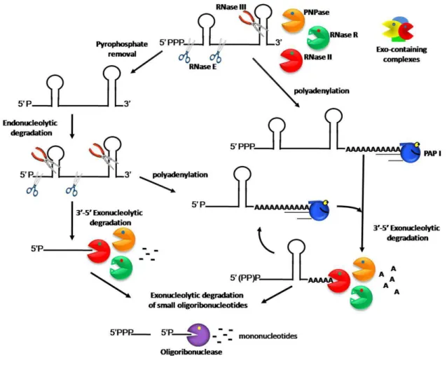
Fig. 1 - Model of RNA degradation pathways in E. coli. ... 6
Fig. 2 - Simplified model of the gad system regulatory network. ... 18 Fig. 3 - Widely accepted modes of Hfq activity. ... 21
Fig. 4 - Growth curves of the WT, Δrnb, Δrnr and Δpnp strains when submitted to
acid shock. ... 42 Fig. 5 - Relative Survival of the WT, Δrnb, Δrnrand Δpnp strains after acid shock... 43
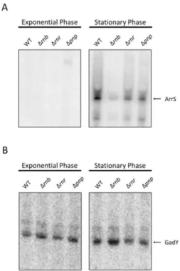
Fig. 6 - Growth phase expression of acid response related sRNAs in the WT, Δrnb, Δrnr
and Δpnp strains. ... 45
Fig. 7 - Northern blot analysis of the stability of the sRNA ArrS in the WT, Δrnb, Δrnr
and Δpnp strains. ... 46
Fig. 8 - Northern blot analysis of the stability of the sRNA ArrS in the WT, Δrnb and
Δrnb + pCMA01 strains and strain specific fold-change values. ... 47
Fig. 9 - Northern blot analysis of the stability of the sRNA GadY in the WT, Δrnb, Δrnr
and Δpnp strains. ... 48
Fig. 10 – Northern blot analysis of the ArrS and GadY sRNA in the WT and Δhfq strain. ... 49
Fig. 11 - Northern blot analysis of the stability of the sRNA FnrS in the WT, Δrnb, Δrnr
and Δpnp strains incubated in aerobic conditions. ... 52
Fig. 12 – Strain specific sRNA FnrS fold-change values in aerobic and anaerobic conditions. ... 53
Fig. 13 - Northern blot analysis of the stability of the sRNA FnrS in the WT, Δrnb, Δrnr
and Δpnp strains after anaerobic induction. ... 55
Table Index
Table 1. Major Ribonucleases involved in RNA degradation in E. coli. …...13
Table 2. Plasmids and E. coli strains used in this work ...24
Table 3. Luria-Agar and Luria-Broth Mediums recipe for 1L of medium. H2O Milli-Q® was added until 1L and the mixture was sealed and autoclaved. ... 24
Table 4. Rifampicin solution recipe for 50ml of cell culture. ...26
Table 5. Nalidixic acid solution recipe for 2ml. ...27
Table 6. TM STOP buffer recipe for 100ml. ...27
Table 7. Lysis buffer recipe for 20ml. ...29
Table 8. Electrophoretic Agarose-gel (1.5%) recipe for 100ml. Agarose was dissolved in boiling 1x TBE buffer...,...30
Table 9. PAA loading buffer recipe. ...31
Table 10. 10% polyacrylamide gel solution recipe for 500ml. Urea was dissolved at 42ºC and the solution filtered with a 0.45μm filter. ...33
Table 11. 10x TBE buffer recipe for 1L. ... 33
Table 12. 20x TAE buffer recipe for 1L. ... 33
Table 13. Primer labelling mix used for labeling with [γ-32P] the ArrS and GadY DNA oligonucleotides. ... 34
Table 14. PCR mix used for FnrSamplification... 35
Table 15. Thermocycler program used for FnrSamplification. ... 35
Table 16. In vitro transcription mix used for the synthesis of RNA from the FnrS PCR product using the Riboprobe®in vitro transcription systems (Promega)... 36
Table 17. Wash solution (wash sol.) I, II and III recipe for 1L. ... 37
Table 18. 20X SSC buffer recipe for 1L. pH was adjusted for 7.0 with HCl. ... 37
Table 19. TURBOTM DNase digestion for cDNA synthesis protocol ...38
Table 20. gDNA wipeout step (QuantitectTM reverse transcription Kit, Qiagen). The samples were incubated at 42ºC for 2 minutes. ... 38
Table 21. cDNA synthesis mix (QuantitectTM reverse transcription Kit, Qiagen)... 38
Table 23. Thermocycler (Rotor Gene RG-3000, Corbett) program
used for the qPCRs... 40 Table 24. Fold-change values of the gadE, gadX, gadW and hdeA mRNAs in the Δrnb,
Δrnr and Δpnp strains. ... 52
Table 25. Fold-change values of the gpmA mRNA in the
Δrnb, Δrnr and Δpnp strains. ... 56
1. Introduction
In an existence that spans more than 3.5 billion years on planet Earth [1], bacteria have endured drastic environmental changes and survived several extinction events. As a consequence of this selective pressure, bacteria have evolved into versatile organisms, capable of thriving in almost every known niche [2]. For example, by the means of infecting and proliferating in a host, pathogenic bacteria are able to survive due to the abundant source of nutrients [3]. Nonetheless, in order to evade the hostile background created by the host’s immune response, an invading bacteria must possess machinery capable of swiftly adapting to rapid environmental changes. Overall, the pressure to survive and quickly adapt to stress conditions drives forward the selection for complex regulatory mechanisms, such as RNA based regulators [3].
1.1. From RNA to DNA: The Beginning
The RNA World hypothesis is a conceptual idea about the origin of life. In this hypothesis, RNA was the first “life form” to appear, capable of both processing information and catalysing chemical reactions [4]. Evidence for the latter was established with the discovery of ribozymes, such as self-splicing introns [5], the ribonuclease P RNA [6] or even the ubiquitous ribosomal RNA [7]. These RNA catalysts could be RNA “fossils” from a simpler RNA based metabolism, the first version of the far more complex DNA/RNA/protein system. Furthermore, the realisation that RNA can evolve in an acellular environment in response to selective pressure [8] and the discovery of both RNA riboswitches [9] and a self-catalytic system based on RNA ligases [10] have all contributed to the plausibility of the RNA World hypothesis.
opened the door for the stable formation of larger genomes and, consequently, to the emergence of cells [11].
1.2. From DNA to RNA and Protein: Transcription and Translation
Transcription and translation are the fundamental processes associated with the phenomenon of genetic expression, the process by which the information contained in a DNA sequence - a gene - is used to modulate the synthesis of a functional product [12, 13].
Gene expression initiates with gene transcription, characterised as the mechanism by which an RNA polymerase transcribes the DNA sequence of a gene into an RNA molecule. Ultimately, this phenomenon yields an RNA sequence complementary to that of the strand of DNA used as a template. The swift release of the nascent RNA from the coding DNA strand results in a highly efficient process in which several RNA copies can be transcribed simultaneously from the same gene in a short amount of time, effectively increasing the cell’s proficiency when a change in genetic program is required. The newly synthesised RNAs often fold into specific tri-dimensional structures. This is due to both the single stranded nature of RNA and the existence of complementary base pairing within the same RNA molecule [12, 13].
The process of translation represents the conversion of the information found in the nucleotide sequence of an mRNA into the amino acid sequence that, ultimately, constitutes a protein. Translation begins with a ribosome binding to the Shine-Dalgarno sequence of an mRNA. The nucleotide information encoded in the mRNA is then read in the 5´ to 3´ direction in consecutive groups of three nucleotides, the codons. Depending on the genetic code of the organism in question, each codon is either associated with a specific amino acid or a stop in translation. The codons in the mRNA are recognised by the transfer RNAs (tRNAs), another type of non-coding RNA. tRNAs act as adaptor molecules, recognizing the codon and associating it with the correspondent amino acid. When a correct match is obtained, the amino acid is added to the nascent protein. Ribosomes are extremely efficient, with bacterial ribosomes adding about 20 amino acids per second to a growing polypeptide chain [12, 13].
The end of the mRNA protein coding region is signalled by the presence of a stop codon, which isn’t recognised by a tRNA. The binding of a release factor to the empty site created by the lack of a tRNA signals the ribosome to stop translation, thus freeing the polypeptide chain and promoting the dissociation of the ribosome/mRNA pair. Lastly, the polypeptide undergoes folding and becomes an integral part of the protein pool [12, 13].
1.3. Quality Control
Despite the existence of several proofreading mechanisms, the process of transcription and translation, like all biological processes, is not errorproof. Incorrectly processed or transcribed RNAs may, inadvertently, commence translation or, in the case of non-coding RNAs, function improperly. The danger of producing both aberrant proteins and RNAs has led to the emergence of quality control mechanisms. One such mechanism is the trans-translation process, which operates when a ribosome stalls due to
ribosome/mRNA dissociation. This particular situation is resolved by the transfer-messenger RNA (tmRNA), which acts both as a tRNA and an mRNA. In order to be functional, tmRNA requires the presence of the SmpB protein, which mimics the anticodon structure naturally found in tRNAs and that is lacking in the tmRNA. This process of trans-translational is initiated by the formation of the tmRNA-SmpB complex,
which then binds to the ribosome in the tRNA binding site and ejects the defective mRNA. The tmRNA is then translated as a normal mRNA would be, tagging the nascent defective protein with an amino acid tag that directs the polypeptide chain to degradation. The existence of a STOP codon in the tmRNA promotes ribosome dissociation, allowing the complex to be recycled [12, 13]. Finally, the defective mRNA is degraded [15].
1.4. RNA Degradation
The RNA degradation pathway is a major component of the cellular metabolism, being required for ribonucleotide turnover and RNA quality control mechanisms. Moreover, by modulating the kinetic rate of RNA decay, RNA degradation is able to influence the intracellular level of a RNA species, thereby facilitating the continuous adjustment of the RNA population to the needs of the cell [16, 17].
Several factors dictate RNA stability in prokaryotes. Among these, the best studied are the secondary structures found in the 5´ and 3´ UTRs [18]. Considerable differences in stability control have been observed in both these regions, with specific structures conferring stabilizing properties and others promoting instability.
The ribonucleases are the main effectors of the RNA degradation pathway. Due to the existence of functional overlaps between these enzymes, it is now clear that several RNases can simultaneously participate, or even substitute each other, in the degradation of a given RNA molecule [16, 19]. Indeed, some RNases are known to form RNA-degrading multiprotein complexes in order to degrade extensively structured RNAs. These complexes are believed to expedite and facilitate RNA turnover by promoting the cooperation of different enzymes in RNA degradation [17, 20, 21].
In E. coli, mRNA degradation usually begins with an endoribonucleolytic
Fig. 1 - Model of RNA degradation pathways in E. coli.
The decay of the majority of transcripts starts with an endoribonucleolytic cleavage by RNase E. This endoribonuclease prefers a monophosphorylated 5´ end, but not in a strict way. RNase III is another enzyme responsible for the initial endoribonucleolytic cleavage of structured RNAs. However, unlike RNase E, RNase III cleaves dsRNAs. After endoribonucleolytic cleavages, the linear transcripts are rapidly degraded by the 3´ to 5´ degradative exoribonucleases, RNase II, RNase R and PNPase. RNase R, unlike RNase II and PNPase, is efficient against highly structured RNAs. PNPase, in association with other proteins, namely RNA helicases, can also unwind RNA duplexes. A minor pathway in the cell is the exoribonucleolytic degradation of full-length transcripts. Poly(A) polymerase (PAP I) adds a poly(A) tail to the short 3´ overhang. These tails provide a ‘toe-hold’ to which exoribonucleases can bind. Cycles
1.5. Ribonucleases
Ribonucleases (RNases) are RNA specific hydrolases, or phosphorylases, that are capable of catalysing the cleavage of the RNA phosphodiester bonds in a reaction that, ultimately, yields monoribonucleotides. Depending on the manner in which they cleave RNA, RNases can be subdivided into two classes: exoribonucleases, which cleave the RNA from its 5´ or 3´ extremity, and endoribonucleases, witch cleave the RNA’s internal phosphodiester bonds [21, 25] (Table 1).
Besides intervening in RNA degradation, RNases are also the main effectors of the post-transcriptional regulation network. RNases thus directly intervene in the degradation, processing, maturation and quality control of all RNA molecules. For example, the cellular concentration of an RNA molecule arises from the dynamic balance established between the transcription frequency and the RNA decay rate. By controlling the latter, RNases display a fundamental role in gene expression, quickly adjusting the cellular RNA levels.
RNases also participate in RNA maturation. For instance, a few RNAs, such as tmRNA, tRNA and rRNA, require ribonuclease mediated processing from a longer transcript prior to becoming active [19, 26]. Unexpectedly, RNase action can also inhibit RNA degradation. For example, the enzyme ribonuclease II (RNase II) stabilizes the rpsO
mRNA by removing its poly(A) tail, which blocks degradation by other exoribonucleases [27].
1.5.1. Exoribonucleases
In E. coli there are eight characterised exoribonucleases, and they all degrade
RNA in the 3´ to 5´ direction [25]. Among these, the three main exoribonucleases involved in RNA decay are RNase II, polynucleotide phosphorylase (PNPase) and ribonuclease R (RNase R) (Table 1).
1.5.1.1. Ribonuclease II
E. coli RNase II, encoded by the rnb gene, is a processive 3´- 5´ ssRNA hydrolase
[29]. RNase II is sensitive to structured RNAs, easily stalling 7 nucleotides (nt) before a double stranded region.
Regardless of being sequence independent, RNase II displays increased reactivity in the presence of poly(A) tails [30], a RNA degradation marker in bacteria. Paradoxically, this augmented degradation of poly(A) tails by RNase II can hinder the activity of other exoribonucleases, thereby partly protecting some RNAs from the decay process [27, 31]. In fact, it is estimated that around 31% of all cellular mRNAs are protected, to some degree, by RNase II, which is considered to be the major cellular hydrolytic exoribonuclease [21, 32].
Among the antisense RNAs, RNase II has only been found to protect the RNA-OUT from degradation [31]. This RNA regulates Tn10/IS10 transposition, a transposon involved in tetracycline resistance [33]. Furthermore, it has been recently described in a
eukaryote, Plasmodium falciparum, an enzyme with an RNase II domain, PfRNase II,
capable of regulating several non-coding RNAs. This report not only attributes to the enzyme a role in the post transcriptional regulation of several non-coding RNAs, but also suggests a liaison between PfRNase II and virulence related genes [34]. Moreover, both in Salmonellathyphimurium and E. coli, RNase II was demonstrated to be involved in
1.5.1.2. Ribonuclease R
E. coli RNase R, encoded by the rnr gene, belongs to the RNase II family and, as
such, is a processive 3´- 5´ hydrolase [37]. Nonetheless, unlike, RNase II, RNase R is capable of degrading highly structured RNAs, required that there is a 3´ single-stranded RNA (ssRNA) overhang, such as a poly(A) tail [38, 39]. RNase R thus displays a fundamental role in the degradation of several structured types of RNA, such as tRNAs, rRNAs, sRNAs and mRNAs, especially when stable stem loops are present [15, 40, 41]. This ribonuclease also plays an important role in RNA quality control mechanisms [17, 21]. For example, both RNase R and PNPase intervene in the degradation of aberrant 16S and 23S rRNAs, thereby affecting ribosome maturation and assembly [17, 42]. RNase R further participates in defective tRNA degradation [15, 21]. Regarding the process of
trans-translation, RNase R was associated with both the maturation of the regulatory
RNA, tmRNA, and with the degradation of the defective mRNA ejected by the tmRNA in the trans-translation process [15, 43].
Regarding stress adaptation responses, RNase R is a general stress induced enzyme, with its levels being increased in stationary phase and in both cold and heat shock [17, 41, 44]. Indeed, in cold conditions, the RNase R levels increase several fold due to the stabilisation of both the rnr operon and of the protein itself. Interestingly, this latter is
dependent on the acetylation of a lysine residue in the RNase R protein. In both cold shock and stationary phase cells, the lysine residue is not acetylated and the free form of the RNase R is stabilised. In fact, when acetylated, both tmRNA and SmpB bind to the enzyme, associating it to the ribosome where it participates in trans-translation [45, 46].
Ultimately, the modulation of RNase R activity allows the cell to efficiently respond to both the increase in RNA secondary structures that normally arise in lower temperatures and to the higher requirement for RNA quality control mechanisms in the growing phase [44, 47].
RNase R has been implicated in the establishment of virulence in both Shigella
flexneri and enteroinvasive E. coli [48]. RNase R was also shown to be important in the
adhesion and invasion of Campylobacter jejuni into eukaryotic cells [49]. Moreover, in
1.5.1.3. Polynucleotide Phosphorylase
E. coli PNPase is an exoribonuclease encoded by the pnp gene. PNPase is
characterised for displaying processive, sequence independent, phosphorolytic activity, being capable of both synthetizing and degrading ssRNA, depending on the conditions. For instance, at a low nucleotide diphosphate (ndp)/high inorganic phosphate (iP) concentration, PNPase catalyses the 3´- 5´ phosphorolytic degradation of RNA in a reaction that increases the cellular ndp pool. Interestingly, at a high ndp/low iP concentration, the enzyme yields iP by catalysing the polymerisation of heteropolymeric RNAs in a template-free manner [21, 25, 51, 52]. Due to this intriguing action mechanism, PNPase was originally described as a “RNA Polymerase” [53]. Indeed, this erroneous characterisation awarded Severo Ochoa the 1959 Nobel Prize.
The phosphorolytic activity exerted by PNPase requires a 3´ overhang of 7 to 9nt of RNA, frequently staling in the presence of structured RNAs [52]. Nonetheless, folded RNAs, such as sRNAs, can also be targeted by PNPase if a 3´ polyadenylated tail is present. In this case, PNPase typically associates with other enzymes in a multiprotein complex called the degradosome, capable of degrading highly structured RNAs [17]. PNPase can also associate and form complexes with Hfq and poly(A) polymerase I (PAP I). In fact, in an Hfq mutant in stationary phase and in the presence of low concentrations of iP, PNPase becomes the primary polyribonucleotide polymerase, adding heteropolymeric tails to 3´ truncated mRNAs [54].
PNPase activity is required in several stress related responses. For example, PNPase is important in cold conditions, with its levels increasing about 2 fold during cold shock [55]. Moreover, after the acclimatisation phase, PNPase acts as a post-transcriptional regulator by degrading the mRNAs of several cold shock proteins (CSP), thus helping in the transition from the cold shock acclimation phase to the cell growth resumption stage [56]. Additionally, PNPase has likewise been established as a virulence factor in several pathogens, such as Salmonella enterica and Yersiniaspp [50, 57].
PNPase has also been linked to stationary phase associated stress, with the steady-state levels of several sRNAs being increased in pnp mutants [58, 59]. Moreover, PNPase
preponderant role in their respective degradation [59, 60]. Indeed, PNPase is the main ribonuclease involved in the decay of hfq-free-sRNAs [61]. These findings shine new light on the importance of the exoribonucleolytic turnover in the RNA networks, opening the door for future work on this yet poorly understood sRNA regulation mechanism.
1.5.2. Endoribonucleases
In E. coli, the process of RNA degradation usually begins with an
endoribonucleolytic cleavage at one or more internal sites of the RNA molecule. Two endoribonucleases have been associated with these initial cleavage events: ribonuclease III (RNase III) and ribonuclease E (RNase E) [21] (Table 1).
1.5.2.1. Ribonuclease III
Ribonuclease III was the first dsRNA specific endoribonuclease to be discovered [62]. Since then, it has been shown that RNase III is widely distributed among other organisms, being the prototype of the RNase III family [21].
RNase III, like all members of its family, is a hydrolytic enzyme capable of degrading dsRNAs and yielding both a 5´ monophosphate end and a 2nt overhang at the 3´ hydroxyl terminus [63]. In E. coli, RNase III is encoded by the rnc gene and plays an
essential role not only in rRNA and tRNA maturation, but also in the decay of some mRNAs, such as the pnp mRNA [21].
RNase III action is required for a plethora of distinct bacterial stress responses [50], being necessary for both heat and cold shock adaptation [64], cobalt and nickel resistance [65] and osmotic stress adaptation [66]. Furthermore, it is also important for biofilm formation [36], S. enterica motility and proliferation inside the host cells [67] and
acid adaptation [68].
In similar fashion to the Eukaryotic RNase III orthologues, Dicer and Drosha, the
E. coli RNase III is also involved in the translational silencing and degradation of
(miRNA) maturation, RNase III is likewise involved in the processing of some described bacterial sRNAs [63, 66, 69, 70].
1.5.2.2. Ribonuclease E
Encoded by the rne gene, RNase E is an ssRNA endoribonuclease that primarily
cleaves A/U rich sequences [21]. RNase E is essential for cell growth, being primarily involved in RNA decay, 5S and 16S rRNA processing and in the maturation of tmRNAs and several tRNAs.
In E. coli, RNase E also functions as the scaffold for the assembly of the
degradosome [21]. This multi protein complex is assembled around the C-terminal region of RNase E, which localises the complex to the inner cytoplasmic membrane and acts as a scaffold for protein association [19, 21, 71]. Depending on the growth conditions, and among other minor components [20], the degradosome encompasses the exoribonuclease PNPase [72], the endoribonuclease RNase E [72], the DEAD-box helicase RhlB and the glycolytic enzyme enolase [73]. Furthermore, a recent report by Feng Lu and Aziz Taghbalout associated the exoribonuclease RNase II with the complex [74].
During cold conditions, RNase E further interacts with another DEAD-box helicase, DeaD, which is incorporated in the degradosome and probably assists in the degradation of structured RNAs [47, 75].
Curiously, in Pseudomonas syringae, RNase E only interacts with the
Table 1
Major Ribonucleases involved in RNA degradation in E. coli [21].
Ribonuclease
Gene
Notes
RNase II rnb
Exoribonuclease. Stalls in the presence of RNA secondary structures and can protect RNAs from degradation.
RNase R rnr
Exoribonuclease. Efficiently degrades RNA secondary structures. Important in RNA quality control and stress adaptation.
PNPase pnp
Exoribonuclease. Forms multiprotein complexes with other enzymes. Major ribonuclease involved in the degradation of Hfq-free sRNAs.
RNase E rne Endoribonuclease. Scafold for the assembly of the degradosome. Cleaves ssRNA.
RNase III rnc Endoribonuclease. Cleaves dsRNA. Associated
with stress conditions.
1.6. Regulatory RNAs
RNA based regulators were initially discovered in 1981 as a replication control mechanism for the colE1 plasmid, a process based on the hybridisation of the regulator RNA, RNA I, with the colE1 replication initiation primer [77, 78]. Three years later, in 1984, Mizuno et al described the first chromosomally encoded small RNA regulator,
micF, a 174 bp transcript responsible for the premature translation termination of the
OmpF gene mRNA [79]. Taken together, these findings have opened the door for the
discovery, firstly in bacteria and later in eukaryotes, of a previously unknown RNA based post-transcription regulation network, capable of coordinating a multitude of physiological responses in a variety of changing environments. For example, the OxyS sRNA, which is induced under oxidative stress, controls the expression of as many as 40 genes [80]. Another example is the sRNA FnrS, which helps in the transition from an
110 sRNAs have been experimentally proven to exist in Escherichia coli [83], with
several others waiting validation.
When compared with other regulatory effectors, such as proteins, sRNAs display the clear advantage of being more cost-effective. For example, sRNAs do not require translation, thereby both avoiding the need for more regulatory proteins and saving time and energy on the translational process itself. Moreover, sRNAs generally act at the post-transcriptional level, which not only allows a much faster control of gene expression, but a better management of the RNA pool as well. sRNAs are also less stable than regulatory proteins and can be rapidly degraded if needed [18, 84].
In a similar fashion to the functional mechanics of the bacterial sRNAs, the eukaryotic counterparts, the miRNAs and short interfering RNAs (siRNA), can also efficiently regulate their targets, thereby acting as an extra layer of cellular regulation [85]. Nonetheless, striking differences emerge. For example, while miRNAs and siRNAs typically have between 21 and 25 nt in length and require processing from a longer single or double stranded precursor by a RNase III like enzyme, sRNAs tend to be more heterogenic both in size and in structure. For instance, although some sRNAs may require the action of different ribonucleases to be activated, the majority is commonly produced as a highly structured, single, unprocessed primary transcript, with an average length varying from 50 to 250 nt. [19, 86].
sRNA mediated regulation can act by two distinct mechanisms. One based on the direct interaction of a sRNA with a protein, thereby modulating its activity, the other, more frequent, on the base pairing of a sRNA (antisense) with a target RNA (sense), resulting in an alteration of the target stability and/or translation [19, 58].
1.6.1. Protein binding sRNAs
There are only a few known examples of sRNA mediated protein regulation in E.
coli. Nonetheless, all share a common trait: the sRNA operates as a protein antagonizer;
The 6S RNA, which was the first sRNA proven to impact transcription, can be considered the perfect example of the previously described mechanism. This sRNA operates by mimicking a promoter, which binds to the holoenzyme RNA polymerase-σ70,
thereby impairing its activity. This process ultimately translates in the transcriptional inhibition of several “housekeeping” σ70 promoters, especially in the stationary phase,
where the 6S RNA is more abundant [87].
Another interesting example is the RNA binding CsrA (carbon storage regulator) protein. CsrA represses several carbon related metabolic pathways by binding to the mRNA of its targets and inhibiting their translation [86, 88]. This activity is counteracted by the CsrB sRNA, which sequesters multiple copies of the CsrA protein by mimicking the shine-dalgarno sequence of the CsrA mRNA targets. [89]. Considering that CsrA activates csrB, this mechanism creates a negative feed-back loop in which CsrA
modulates its own activity [90].
1.6.2. Antisense sRNAs
Unlike what happens with protein binding sRNAs, most of the known sRNAs act on other RNAs by an antisense mechanism: A process that can be mediated by either
trans-encoded (trans-sRNAs) or cis-encoded (cis-sRNAs) sRNAs. When considering
their genetic expression, almost all known trans/cis-sRNAs are preferentially expressed
under specific growth conditions, such as limiting carbon or oxidative stress [86].
Regarding the base-pairing mechanisms, sRNAs frequently bind
stoichiometricallynear the mRNA 5´ end. This process can either result in the degradation or stabilisation of the sRNA/mRNA pair [22]. For instance, in the case of the ryhB sRNA,
which is involved in the regulation of the iron metabolism, the sRNA represses the target expression by binding to the target mRNA and inducing the degradosome degradation pathway [91]. On the other hand, in the less frequent case of transcript stabilisation, the binding of an activator sRNA prompts a conformational change that commonly results in the exposure of the mRNA Shine-Dalgarno sequence [18, 19, 86, 92].
concentration is higher than that of the mRNA, gene expression if shut off. However, when the sRNA concentration is lower than that of the mRNA, little impact is observed. This critical sRNA threshold phenomenon suggest than sRNAs are more effective at repressing mRNA translation when the sRNA activator signal is persistent and abundant, whereas in the case of weak and transient signals, proteins are more efficient [84].
1.6.3.
Cis
encoded sRNAs
Cis-sRNAs are encoded on the opposite DNA strand from which their RNA target
is transcribed. This results in the existence of extended sections of complementarity between the cis-sRNA and the respective target, frequently 75 nt or more [84, 93].
Nonetheless, despite being transcribed from the same DNA region, both antisense and sense RNAs act as independent molecules, each subject to individual reaction kinetics in the cellular environment.
Most of the known cis-sRNAs regulate the replication of mobile elements such as
the colE1 plasmid [78, 93]. Others repress the translation of deadly toxic proteins, thereby acting as antitoxins by inhibiting cell death in the presence of the sRNA antitoxin genetic carrier [84]. A few cis-sRNA can also influence the expression of chromosomally
encoded genes, such as those in the glutamate acid response (gad) operon [19, 84].
1.6.3.1.
Cis
-RNAs and the gad system: an intricate story
The gad system is the most prominent acid response mechanism in E. coli. The
gad system main effectors, GadA and GadB, lower the intracellular pH by consuming protons during the decarboxylation of glutamate. This reaction ultimately originates γ -aminobutyric acid, which is then exported out of the cell by GadC [94]. This system’s regulatory network is complex and still under debate, however, it has been proven that it is targeted by several layers of post-transcriptional control that include, at least, two cis
-RNA activators, GadY and ArrS.
GadY is a cis-RNA encoded in the 3´ UTR of the gadX mRNA. Its expression is
pH conditions [95, 96]. Despite being cis encoded, GadY is able to bind the RNA
chaperone Hfq, which is normally associated with trans-RNAs, as discussed in the next
section [28]. This intriguing sRNA binds to the intergenic region of the GadX-GadW
mRNA and promotes RNase III mediated processing of the transcript, which results in an increased level of both the GadX and GadW mRNAs and in a decreased concentration of
the longer transcript GadX-GadW mRNA [28, 58, 68]. GadX, among others, partly
activates the expression of the main gad activator, GadE, and of the GadA, GadB and GadC proteins. Inversely, GadW appears to be primarily involved in negatively regulating the transcription of both gadX and GadY [97, 98].
ArrS is a cis-RNA encoded in the unusually long 5´ UTR of the T3 gadE form.
ArrS is normally expressed during stationary phase and is dependent upon the factors gadE and σS. ArrS expression is further increased in acidic conditions [99, 100]. ArrS
controls the levels of the gadE mRNA T3 form, which abruptly decrease and give rise to
its smaller T2 active form when the sRNA availability increases [100]. The GadE transcription factor, which is originally transcribed in its apparently inactive T3 form, is a major gad system activator, being required for gadA, gadB and gadC expression (Fig.
2). Curiously, both gadX and gadW are only needed in some circumstances [101]. The existence of a monophosphorilated 5´ extremity (instead of a triphosphorilated one) in the
gadE T2 form and the lack of T2 expression in RNase III deficient cells suggest a
ribonuclease involvement in both the gadE mRNA processing and in the cellular
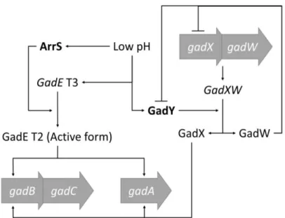
Fig. 2 - Simplified model of the gad system regulatory network.
Lines indicate regulation. The processing from the gadE T3 mRNA to the gadE T2 mRNA form is
catalysed by the sRNA ArrS in an RNase III dependent reaction. The GadXW transcript cleavage is
catalysed by the sRNA GadY in a reaction that yields both gadX and gadW.
1.6.4.
Trans
encoded sRNAs
Trans-sRNAs are, in their majority, characterised for being encoded in a distinct
chromosomal location than the one the target mRNA is transcribed from. When compared with the cis-sRNA, they have a more limited, non-contiguous complementarity between
the mRNA-sRNA pair. By having a more discrete base-pair complementation with its targets, a trans-sRNA molecule can, therefore, carry more than one mRNA binding site,
thus explaining the trans-sRNA’s capacity to regulate multiple mRNAs [19, 84, 86].
Unfortunately, the mechanisms that determine the specificity between the sRNAs and their target mRNAs are still largely unknown. For this reason, the several sRNA/mRNA interaction predicting algorithms that today exist frequently display erroneous results. In order to increase their accuracy at predicting productive, in vivo, sRNA/mRNA
interactions, further work on this subject thus needs to be done. This will effectively increase experimental efficiency by avoiding the study of false targets [84].
Trans-sRNAs are the most abundant and well-studied class of sRNAs,
to produce energy from fermentation and/or anaerobic respiration. These changes in gene expression are partly regulated by the trans-sRNA FnrS, whose expression is dependent
on the transcriptional fumarate and nitrate reductase regulator (FNR), an oxygen availability sensor. When in anoxic conditions, the sRNA FnrS is highly expressed in order to repress the translation of several aerobic metabolic enzymes, hence increasing the efficiency of the anaerobic metabolism [81, 102]. Trans-sRNAs thus have the
capability of swiftly regulating entire physiological pathways, quickly forwarding large quantities of obsolete mRNAs into degradation and effectively stopping a metabolic program at the RNA level. Furthermore, due to differences in binding affinities, sRNAs help in prioritizing the regulation of different target mRNAs and, consequently, in controlling the various stages of a changing gene expression program [84, 103].
Unlike the Eukaryotic miRNAs and siRNAs, which require complex proteic machinery to operate, the Enterobacteriaceae trans-sRNAs’s have only been found to
require the presence of the Hfq RNA chaperone (Hfq).
1.7. Hfq
Hfq, originally described as HF-1, was firstly discovered in E.coli cells infected
by the Qβ RNA bacteriophage as a key host factor involved in viral RNA synthesis [104, 105]. Since then, Hfq has been found to be the E.coli counterpart of a ubiquitous family
of RNA-binding proteins, the Sm and Sm-like protein family. Hfq thereby shares, alongside the rest of the members of the family, what is considered to be the family’s main feature, the presence of a multimeric ring-like quaternary conformation in the active protein form. This peculiar organisation facilitates the discontinuous and imperfect interactions that are established between a trans-sRNA and its respective target mRNA.
Hfq thus acts as a chaperone, mediating the formation of the RNA-RNA pair, expediting its assembly and, consequently, increasing the efficiency of the trans-sRNA regulatory
network [106-108]. In fact, Hfq seems to be the limiting factor for sRNA mediated regulation, being essential for productive trans-sRNA-mRNA base pairing [109, 110].
example, an induced sRNA can affect the action of another, unrelated, sRNA. Indeed, this mechanism was firstly described as an explanation for the oxyS dependent negative
regulation of the σS factor, whose translation is dependent on an activator sRNA [111].
In Hfq mutant cells, pleiotropic effects such as impairment of stress response pathways, metabolic regulation deficiencies and loss of virulence can occur. However, several of this consequences may be amplified by the multitude of interactions that Hfq establishes with other proteins, such as RNase E, PNPase and PAP I [54, 60, 109].
Hfq seems to bind to both A/U and poly(A) rich sequences [112], with its activity being dependent on the tri-dimensional arrangements found in the same RNAs with which it interacts. Theoretically, this RNA structural information renders a conformational change in the Hfq protein, resulting in the formation of different complexes and, thus, in the display of several distinct modus operandi [107] (Fig. 3). Hfq action can, thereby,
cause different outcomes depending on the RNA-RNA interactions. For example, Hfq is involved not only in mediating the interactions that are established between an mRNA-sRNA pair, but also on its recycling, mostly through the recruitment of ribonucleases such as RNase E [54, 106, 107, 113]. Paradoxically, by binding to poly(A) tails and RNase E cleavage sites, Hfq can likewise help protect a few described RNAs, such as dsrA, ryhB
and rpoS, from ribonuclease activity [61, 110, 112-115] (Fig. 3). Ultimately, the
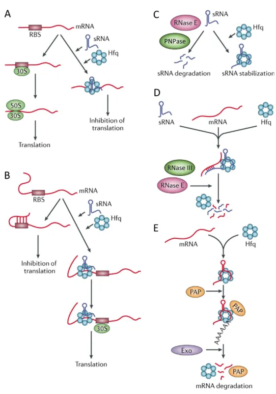
Fig. 3 - Widely accepted modes of Hfq activity.
(A) Hfq in association with a sRNA may sequester the ribosome-binding site (RBS) of a target mRNA, thus blocking binding of the 30S and 50S ribosomal subunits and repressing translation. (B) In some
1.8. Introductory Remarks
It is now becoming increasingly clear that RNases, and not just regulatory RNAs, play a predominant role in the post-transcriptional regulation of several distinct stress activated pathways. While sRNAs are capable of efficiently regulating entire metabolic pathways, RNases can act on both the sRNAs and their RNA targets, effectively regulating the regulators. Indeed, both are required to effectively control the vast interconnected network that constitutes the cellular RNA pool [19, 36]. Ultimately, post-transcriptional regulators not only allow the cell to swiftly respond to changing environments, but also anticipate the regulatory requirements needed for a change in genetic expression.
1.8.1. Objectives
2. Materials and Methods
2.1. Strains and Growth Conditions
E. coli K-12 MG1693 and all derivative strains are listed in Table 2. All strains
were grown at 37ºC with agitation at 180 rpm in Luria-Broth medium (LB) (Table 3) supplemented with thymine (50µg ml-1). When required, antibiotics where supplemented
in the following concentrations: Tetracycline (tet) (20µg ml-1), kanamycin (kan) (50µg
ml-1), streptomycin (str) (25µg ml-1), spectinomycin (spc) (25µg ml-1), ampicillin (amp)
(100µg ml-1) and chloramphenicol (cam) (20µg ml-1).
All cell cultures inoculated in Luria-Agar medium (LA) (Table 3) were supplemented with thymine (50µg ml-1) and incubated at 37ºC until individualised
colonies were visible. When necessary, antibiotics were also added to the medium in the previously referred concentrations.
Overnight cultures were performed by inoculating isolated colonies in LB medium (supplemented as required), followed by incubation with agitation for 16h (overnight) at 37ºC and 180 rpm.
Optical density values were obtained by pippeting 1ml of cell culture/medium to a disposable plastic cuvette of 1.5mL (Sarstedt) and measuring the respective absorbance
at 600nm (OD600) in a spectrophotometer (BioPhotometer plus, Eppendorf).
Exponential phase cells were obtained by inoculating fresh LB medium (supplemented as required) with overnight cultures to an initial OD600 of ~ 0.03. The cell
cultures were then incubated with agitation at 37ºC and 180 rpm until an OD600 of 0.5
(exponential phase).
Stationary phase cells were obtained by inoculating fresh LB medium (supplemented as required) with overnight cultures to an initial OD600 of ~ 0.03. The new
cultures were then incubated overnight (16h) with agitation at 37ºC and 180 rpm. Stationary phase was confirmed by measuring the OD600 of the cell cultures after
Table 2
Plasmids and E. coli strains used in this work
Strains
Relevant Genotype
Reference
MG1693 (WT) thyA715 rph-1 [116]
CMA201 (Δrnb) thyA715 rph-1 Δrnb-201::tetR [117]
HM104 (Δrnr) thyA715 rph-1Δrnr::kanR [44]
SK10019 (Δpnp) thyA715 rph-1 pnpΔ683::strR/spcR [32]
CMA599(ΔHfq) thyA715 rph-1 Δhfq::camR Unpublished
pCMA01 6.5kb HindIII-XhoI fragment of
pDK07 cloned into pBluescript SK+ [118]
Table 3
Luria-Agar and Luria-Broth Mediums recipe for 1L of medium. H2O Milli-Q® was
added until 1L and the mixture was sealed and autoclaved.
Luria-Agar (LA)
Reagents
Quantities
Tryptone 10g
Yeast Extract 5g
NaCl 10g
Agar 15g
Luria-Broth (LB)
Reagents
Quantities
Tryptone 10g
Yeast Extract 5g
NaCl 10g
2.2. Growth curves
2.2.1. Acid shock growth curves
Overnight cultures for the WT, Δrnb, Δrnr and Δpnp strains were performed as
described. Respectively, each cell culture was then used to inoculate to an initial OD600
of ~ 0.03, 50ml of fresh LB medium in triplicate (supplemented as required). Following inoculation, all cultures were incubated at 37ºC with agitation at 180 rpm. The culture’s OD600 was measured hourly and plotted against time of measurement. In each strain, one
of the triplicates was used as the control growth curve. In the other two, acid shock was performed by adding 150µl of HCl 4N (pH drop from ~ 7 to ~ 4) at an OD600 of either
2.2.2. Anaerobic growth curve
Overnight cultures for the WT, Δrnb, Δrnr and Δpnp strains were performed as
described. Each cell culture was then used to respectively inoculate 50ml of fresh LB medium (supplemented as required) to an initial OD600 of ~ 0.03. All cultures were then
transferred to an anaerobic chamber (with a CO2 and argon atmosphere) and incubated at
37ºC in a dry water bath, without agitation. Every hour, cell OD600 was measured as
described above. A final OD600 value was registered after overnight incubation in the
same conditions.
2.3. Acid shock survival assay
Overnight cultures for the WT, Δrnb, Δrnr and Δpnp strains were obtained and
used to respectively inoculate to an initial OD600 of ~ 0.03 50ml of fresh LB medium,
supplemented as required. The newly inoculated cultures were then incubated with agitation at 37ºC and 180 rpm. For each strain, at an OD600 of 0.5, and prior to the acid
2.4. Total RNA extraction
2.4.1. Total RNA extraction for RNA half-life determination
Rifampicin solution (Table 4), a bacterial transcription inhibitor, was prepared immediately before use. NaOH was added to the solution until the rifampicin appeared dissolved. 1.5ml of rifampicin solution per 50ml of LB medium was then added to either stationary or exponential phase cultures. 30s after adding the rifampicin solution, the first time point was taken by decanting either 10 or 20ml (see below) of cell culture to either 10 or 20ml of ice-cold TM STOP buffer (
Table 6). The culture conditions were maintained at 37ºC with agitation at 180 rpm for
the duration of the experiment and at the required time points another 10 or 20ml of culture were decanted to 50bml falcons with ice-cold TM STOP buffer. RNA was then extracted as described below.
Table 4
Rifampicin solution recipe for 50ml of cell culture.
Reagents
Quantities
Rifampicin 0,05g
Nalidixic acid solution (Table 5) 500µl
Methanol 1ml
Table 5
Nalidixic acid solution recipe for 2ml.
Reagents
Quantities
Nalidixic acid 0,0010g per 50ml of cell culture
NaOH 10N 20µl
Table 6
TM STOP buffer recipe for 100ml.
Reagents
Quantities
Tris 1M at pH 7.2 1 ml
MgCl2 1M 0.5 ml
NaN3 1M 2.5 ml
Chloramphenicol (4mg ml-1) 12.5 ml
H2O Milli-Q® 83.5 ml
2.4.2. Total RNA extraction of steady-state RNAs
Total RNA was either extracted from cells in exponential phase (OD600 ~ 0.5) or
in stationary phase (after ~ 16h of growth). RNA from stationary and exponential phase cultures was extracted by respectively decanting 10 or 20ml of culture to a 50ml falcon tube with 10 or 20ml of ice-cold TM STOP buffer.
2.4.3.
Determination of the degradation rate of FnrS sRNA under
induction of anaerobic conditions.
Exponential phase WT, Δrnb, Δrnr and Δpnp cell cultures were obtained as
described. At an OD600 of 0.5, all cultures were transferred to an anaerobic chamber (Plas labs) with a CO2, N2 and H2 atmosphere at 37ºC in order to induce the expression of the
FnrS sRNA (anaerobic shock). After 2h, the cultures were removed from the anaerobic chamber and 10ml of cell culture decanted to ice-cold TM STOP buffer (time 0). All cell cultures were then incubated at 37ºC with agitation at 180rpm in an aerobic environment. The remaining time points (3, 6, 10 and 20 minutes) were extracted as in time 0.
The 50ml falcon tubes with cell culture and cold TM STOP buffer were centrifuged for 20 minutes at 4ºC and 3600 rpm. The supernatants were decanted and the cell pellets ressuspended in 800µl of freshly prepared lysis buffer (Table 7). The cell extracts were then transferred, respectively, into glass COREX tubes and incubated for 5 minutes at 42ºC. Next, three cycles of freezing in liquid nitrogen and thawing at 42ºC were performed. 140µl of acetic acid (20 nM) and 90µl of sodium dodecyl sulphate (SDS) 10% were added in the third cycle and the freezing/thawing cycles continued until the cell solutions appeared lysated (transparent). After the lysis procedure, an enzymatic digestion with DNase (TURBOTM DNase, Ambion®) (2U/µl) was performed for 1h at
37ºC. Either 5µl or 10µl of DNase were added to the cell extracts, depending on whether the cells were obtained from exponential phase or stationary phase cultures, respectively. After DNase digestion, the cell extracts were transferred into 2ml tubes and phenol:chloroform RNA extraction was performed. To this effect, 1ml of phenol at pH 5.2 was added, respectively, to the cell extracts, which were then vortexed for 2 minutes and centrifuged for 10 minutes at 4ºC and 14000 rpm. The supernatants were carefully collected to new 2ml tubes and the previous process repeated. In the third step, 500µl of phenol at pH 5.2 and 500µl of chloroform/alcohol isoamylic (24:1) solution were added to the collected supernatants, which were then vortexed for 1 minute and centrifuged for 5 minutes at 4ºC and 14000 rpm. The supernatants were carefully collected to new 2ml tubes and the previous process repeated one more time. Finally, 1ml of chloroform/alcohol isoamylic (24:1) solution was added to the collected supernatants, which were then vortexed for 1 minute and centrifuged for 5 minutes at 4ºC and 14000 rpm. The supernatants were carefully collected to new 2ml tubes and sodium acetate 3M pH 5.2 and ice-cold ethanol 100% added in a volume corresponding to 0.1x and 2.5x that of the collected supernatants, respectively. The supernatants were left to precipitate overnight at -20ºC.
RNA integrity and DNA contamination were assessed by examining 1µl of the RNA samples in an agarose-gel electrophoresis. In case of DNA contamination, a new DNase digestion was performed by adding to the RNA samples 2.5µl of DNase (TURBOTM DNase, Ambion®), 20µl of DNase buffer and H
2O Milli-Q® to a final volume
of 200µl. After incubating the samples at 37ºC for, at least, 1h, a second phenol:chloroform RNA extraction was performed. To this effect, H2O Milli-Q® was
added to a final volume of 400µl. 200µl of phenol at pH 5.2 and 200µl of chloroform/alcohol isoamylic (24:1) solution were then added to the RNA samples and vortexed for 1 minute, after which a centrifugation for 5 minutes at 4ºC and 14000 rpm was performed. The supernatant was collected and transferred into a new 2ml tube. 400µl of chloroform/alcohol isoamylic (24:1) solution were added to the supernatants and vortexed and centrifuged as before. The supernatants were extracted to new 2ml tubes and sodium acetate 3M pH 5.2 and ice-cold ethanol 100% added as previously described. Precipitation and verification of RNA integrity and DNA contents were also performed as stated. Finally, the RNA samples concentration was quantified using a Nanodrop
Spectrophotometer (Nanodrop ND1000, Alfagene). All RNA samples were stored at
-20ºC.
Table 7
Lysis buffer recipe for 20ml.
Reagents
Quantities
Tris 1M at pH 7.2 200µl
MgCl2 1M 100µl
Turbo DNase (2U/µl) (Ambion) 20µl
Lysozyme from chicken egg white (Sigma-Aldrich) 0.020g
H2O Milli-Q® Until 20ml
2.5. Genomic DNA extraction
Overnight cultures (in stationary phase) for the WT, Δrnb, Δrnr and Δpnp strains
were performed as described and used to extract DNA using the Citogene® DNA Cell &
Tissue Kit (Citogene®). DNA samples were ressuspended in H2O Milli-Q®. DNA
2.6. Electrophoresis
12µl of loading solution (constituted, unless stated otherwise, by 2µl of Orange G loading buffer, 9µl of H2O Milli-Q® and either 1µl of RNA or DNA) was loaded in an
electrophoretic agarose-gel (Table 8). An electrophoresis was then performed at 100V for 30 minutes in 1x TBE buffer. The agarose-gel was then visualised under UV light in a Gel Doc XR+ system (Bio-rad®).
Table 8
Electrophoretic Agarose-gel (1.5%) recipe for 100ml. Agarose was dissolved in boiling 1x TBE buffer.
Reagents
Quantities
Agarose (SeaKem® LE AGAROSE) 1.5g
Ethidium bromide (1mg ml-1) 50µl
1x TBE (Table 11) 100ml
2.7. Northern Blot
2.7.1. Northern Blot sample preparation
Northern blot samples were prepared by pipetting the volume corresponding to 40µg of RNA into a new 1.5ml tube and evaporating the sample to a final volume of 5µl (SpeedVac SVC 100, Savant). 15µl of PAA loading buffer (Table 9) were then added to
all samples.
Table 9
PAA loading buffer recipe.
Reagents
Quantities
Deionized Formamine 5ml
EDTA 0.5M (pH 8) 100µl
Xylene Cyanol 0.005g
2.7.2. Northern Blot
RNase free 1.5mm spacers were assembled in top of a Northern blot glass plate. A notched Northern blot glass plate was then placed in top of the spacers and the sides were sealed with agarose-gel.
In all performed Northern blots, 50ml of 10% polyacrylamide gel solution (Table 10) were prepared and mixed with 500µl of APS (10%) and 50µl of TEMED. The obtained solution was poured into the space between the two Northern blot glass plates and a comb was inserted at the top. After overnight polymerisation, the comb and the bottom spacer were removed and the glass plates with the polyacrylamide gel placed in the Northern blot apparatus. 1x TBE buffer (Table 11) was then added to the Northern blot apparatus´ reservoirs and any air bubbles removed by rinsing both the gel wells and the bottom of the gel with a syringe loaded with 1x TBE. The gel wells were further washed until all residual urea was removed.
A pre-run of the gel (without sample) was then performed for ~ 1h at 420V with wattage limited to either 24 or 48W (PowerPacTM HV, Bio-Rad), depending on whether
one or two Northern blots were being simultaneously performed, respectively.
The Northern blot RNA samples were denatured at 80ºC for 10 minutes and then incubated in ice for another 2 minutes.
After terminating the pre-run, all polyacrylamide gel wells were rinsed with 1x TBE using a syringe and the full volume of the Northern blot RNA samples was applied. The polyacrylamide gel loaded with the RNA samples ran for 2h at 420V, with wattage limited to either 24 or 48W, depending on whether one or two Northern blots were being performed, respectively.
Four litters of 1x TAE buffer (Table 12) was prepared and a positive charged
nylon membrane with 0.45µm pores (Amersham Hybond-N+, GE Healthcare Life
Whatman®). The nylon membrane was activated by submerging it for at least 5 minutes
in Milli-Q® water.
In order to perform the RNA transfer from the polyacrylamide gel to the membrane, two sponges and blot filter papers were saturated with 1x TAE in a RNase free recipient. The transfer apparatus was then assembled in the following order: negative side of transfer cassette, sponge, blot filter papers, polyacrylamide gel, nylon membrane, blot filter papers, sponge and positive side of gel cassette. Air bubbles were removed by manually pressing the saturated blot filter papers with a glass tube. The assembled apparatus was inserted in the transfer chamber (Owl™ VEP-3 Large Tank Electroblotting System, Thermo Scientific™), which was then filled to the top with 1x TAE. The transfer
was then performed at 24V (PS200-HC, Hoefer) for 1h and 45 minutes at 4ºC. When the
transfer had finished, the transfer cassette was disassembled and the transferred RNAs fixed to the nylon membrane by UV crosslinking at 1200μj cm-2 for 3 minutes (UVC 500
Crosslinker, Amersham Biosciences). Transfer efficiency was ascertained by incubating
overnight the polyacrylamide gel in a recipient with bidistilled water and ethidium bromide and examining it under UV light in a Gel DocTM XR+ system (Bio-rad®).
Table 10
10% polyacrylamide gel solution recipe for 500ml. Urea was dissolved at 42ºC and the solution filtered with a 0.45µm filter.
Reagents
Quantities
Urea 210g
1x TBE (Table 12) 50ml
Polyacrylamide 40% 19:1 (RNA) 125ml
Autoclaved H2O Milli-Q® Until 500ml
Table 11
10x TBE buffer recipe for 1L.
Reagents
Quantities
Tris Base 108g
Boric Acid 55g
EDTA 9.3g
Table 12
20x TAE buffer recipe for 1L.
Reagents
Quantities
Tris Base 48.4g
Acetic Acid (100%) 11.4ml
EDTA 0.5M (pH 8) 20ml
Autoclaved H2O Milli-Q® Until 1L
2.8. Synthesis and labelling of probes for Northern Blot analysis
2.8.1. Primer labelling
The ArrS and GadY DNA Northern blot probes were obtained by primer labelling. To this effect, specific DNA oligonucleotides (design in the Clone Manager software, version 9) complementary to the ArrS and GadY genes internal sequences were synthesised (STAB VIDA) (ArrS probe and GadY probe, Appendix, Table I). The obtained DNA oligonucleotides were, respectively, mixed with the reagents depicted in Table 13and incubated at 37ºC for, at least, 1h. The resulting 5´end [γ-32P] labelled DNA
Northern blot probes were purified with a G-25 MicroSpin column (GE Healthcare Life
Sciences). Labelling was confirmed by measuring radioactivity with a Geiger counter
(Mini900 Ratemeter, Thermo Electron Corporation) and the probes stored at -20ºC in a
lead container.
Table 13
Primer labelling mix used for labeling with [γ-32P] ATP
theArrS and GadY DNA oligonucleotides.
Reagents
Quantities
DNA oligo (10nM) 0.5μl
10x T4 PNK Reaction Buffer (Thermo Scientific) 3μl
H2O Milli-Q® 23.5μl
[γ-32P] ATP (
PerkinElmer) 2μl
2.8.2. Synthesis of the FnrS probe
2.8.2.1. FnrS gene PCR and purification
In order to obtain a probe for the sRNA FnrS, the FnrS gene was firstly amplified using the primers FnrS_FW and FnrS_T7 (designed in the Clone Manager software and
synthesised by STAB VIDA) (Appendix, Table I) by polymerase chain reaction (PCR). The FnrS_T7 primer contains a T7 RNA polymerase promoter, which allows the
amplified sequence to be transcribed by this enzyme. The PCR reaction was performed (Table 14) using the DreamTaqTM kit (Thermo ScientificTM) and the MyCycler™ thermal
cycler (Bio-Rad). The PCR program used is depicted in Table 15.
In order to determine whether the PCR was specific, an electrophoresis was performed with 10µl of PCR product. Only one band corresponding to the FnrS amplicon was observed, indicating that the PCR was specific. The PCR product was then purified using the kit “NucleoSpin® Gel and PCR Clean-up” (MACHEREY-NAGEL). To confirm
that the PCR product was not lost in the purification step, an agarose-gel electrophoresis with 1µl of the purified PCR product was, then again, performed.
Table 14
PCR mix used for FnrSamplification.
Reagents
Quantities
Genomic DNA 1µl
FnrS_FW (1pM) 2µl
FnrS_T7 (1pM) 2µl
dNTPs (10nM) 1µl
10x DreamTaq Buffer 5µl
DreamTaq DNA polymerase (5U/µl) 0.25µl