Therapeutic Immunization with HIV-1 Tat Reduces
Immune Activation and Loss of Regulatory T-Cells and
Improves Immune Function in Subjects on HAART
Barbara Ensoli1*, Stefania Bellino1, Antonella Tripiciano1,2, Olimpia Longo1, Vittorio Francavilla1,2, Simone Marcotullio1, Aurelio Cafaro1, Orietta Picconi1, Giovanni Paniccia1,2, Arianna Scoglio1,2, Angela Arancio2, Cristina Ariola2, Maria J. Ruiz Alvarez1,2, Massimo Campagna2, Donato Scaramuzzi2, Cristina Iori2, Roberto Esposito3, Cristina Mussini3, Florio Ghinelli4, Laura Sighinolfi4, Guido Palamara5,
Alessandra Latini5, Gioacchino Angarano6, Nicoletta Ladisa6, Fabrizio Soscia7, Vito S. Mercurio7, Adriano Lazzarin8, Giuseppe Tambussi8, Raffaele Visintini8, Francesco Mazzotta9, Massimo Di Pietro9, Massimo Galli10, Stefano Rusconi10, Giampiero Carosi11, Carlo Torti11, Giovanni Di Perri12, Stefano Bonora12, Fabrizio Ensoli2, Enrico Garaci13
1National AIDS Center, Istituto Superiore di Sanita`, Rome, Italy,2Core Laboratory of Virology and Immunology, San Gallicano Hospital, ‘‘Istituti Fisioterapici Ospetalieri’’, Rome, Italy,3Division of Infectious Diseases, University Policlinic of Modena, Modena, Italy,4Unit of Infectious Diseases, University Hospital of Ferrara, Ferrara, Italy, 5Department of Infectious Dermatology, San Gallicano Hospital, Rome, Italy, 6Division of Infectious Diseases, University of Bari, Policlinic Hospital, Bari, Italy, 7Department of Infectious Diseases, S. Maria Goretti Hospital, Latina, Italy,8Division of Infectious Diseases, S. Raffaele Hospital, Milan, Italy,9Unit of Infectious Diseases, S.M. Annunziata Hospital, Florence, Italy,10Institute of Tropical and Infectious Diseases, University of Milan L. Sacco Hospital, Milan, Italy,11Division of Tropical and Infectious Diseases, Spedali Civili, Brescia, Italy,12Clinic of Infectious Diseases, Amedeo di Savoia Hospital, Turin, Italy,13Istituto Superiore di Sanita`, Rome, Italy
Abstract
Although HAART suppresses HIV replication, it is often unable to restore immune homeostasis. Consequently, non-AIDS-defining diseases are increasingly seen in treated individuals. This is attributed to persistent virus expression in reservoirs and to cell activation. Of note, in CD4+ T cells and monocyte-macrophages of virologically-suppressed individuals, there is
continued expression of multi-spliced transcripts encoding HIV regulatory proteins. Among them, Tat is essential for virus gene expression and replication, either in primary infection or for virus reactivation during HAART, when Tat is expressed, released extracellularly and exerts, on both the virus and the immune system, effects that contribute to disease maintenance. Here we report results of an ad hocexploratory interim analysis (up to 48 weeks) on 87 virologically-suppressed HAART-treated individuals enrolled in a phase II randomized open-label multicentric clinical trial of therapeutic immunization with Tat (ISS T-002). Eighty-eight virologically-suppressed HAART-treated individuals, enrolled in a parallel prospective observational study at the same sites (ISS OBS T-002), served for intergroup comparison. Immunization with Tat was safe, induced durable immune responses, and modified the pattern of CD4+and CD8+cellular activation (CD38 and HLA-DR) together with reduction of
biochemical activation markers and persistent increases of regulatory T cells. This was accompanied by a progressive increment of CD4+T cells and B cells with reduction of CD8+T cells and NK cells, which were independent from the type of
antiretroviral regimen. Increase in central and effector memory and reduction in terminally-differentiated effector memory CD4+and CD8+T cells were accompanied by increases of CD4+and CD8+T cell responses against Env and recall antigens. Of
note, more immune-compromised individuals experienced greater therapeutic effects. In contrast, these changes were opposite, absent or partial in the OBS population. These findings support the use of Tat immunization to intensify HAART efficacy and to restore immune homeostasis.
Trial registration:ClinicalTrials.gov NCT00751595
Citation:Ensoli B, Bellino S, Tripiciano A, Longo O, Francavilla V, et al. (2010) Therapeutic Immunization with HIV-1 Tat Reduces Immune Activation and Loss of Regulatory T-Cells and Improves Immune Function in Subjects on HAART. PLoS ONE 5(11): e13540. doi:10.1371/journal.pone.0013540
Editor:Kim J. Hasenkrug, National Institute of Allergy and Infectious Diseases, United States of America
ReceivedMay 25, 2010;AcceptedSeptember 28, 2010;PublishedNovember 11, 2010
Copyright:ß2010 Ensoli et al. This is an open-access article distributed under the terms of the Creative Commons Attribution License, which permits unrestricted use, distribution, and reproduction in any medium, provided the original author and source are credited.
Funding:This work was funded by the special project for ‘The development of clinical trials of vaccines against HIV/AIDS’ funded by the Italian Ministry of Health. The funders had no role in study design, data collection and analysis, decision to publish, or preparation of the manuscript.
Competing Interests:The authors have declared that no competing interests exist.
* E-mail: [email protected]
Introduction
The use of antiretroviral drugs has changed the quality and expectancy of life of HIV-infected individuals [1]. However, in spite of viral-suppressing drug intervention, immune activation and loss of regulatory T-cells (T-reg), of CD4+ T cells, B cells,
central memory CD4+and CD8+T cells and of immune functions
To block these effects, novel non virus-targeting interventions, such as CCR5 antagonists, are being explored in association with conventional drugs [9,10]. However, this approach appears to be only partially effective, suggesting that pathogenetic factors that maintain HIV disease should be targeted for restoring immune functions.
In this respect, residual virus replication is detected in most patients receiving HAART, likely originating from viral reservoirs, including latently infected CD4+T cells, monocyte-macrophages,
dendritic cells, NK cells, hematopoietic stem cells, mast cells and several cell types in the central nervous system [11–21]. This finding implies that viral gene products are still produced even under a ‘‘successful’’ therapy. Indeed, multi-spliced transcripts encoding HIV regulatory proteins are persistently expressed in viral reservoirs by unintegrated proviral DNA [22,23], and are detected in resting CD4+T cells, monocytes, and hematopoietic
stem cells of HAART-treated individuals in the absence of detectable viremia [12,13,18,22,24–28]. Thus, HIV regulatory proteins are produced in latently infected cells [29], and can contribute to the persistent immune activation, immune system dysfunction, and disease observed in many HAART recipients [2,4,5,17,23,30–32].
In particular, production of the Tat protein in virologically-suppressed individuals is confirmed by evidence of anti-Tat antibody (Ab) seroconversion and increases of Tat-specific T cell responses in HAART-treated patients (B. Ensoli et al., unpub-lished data).
Tat is the transactivator of HIV gene expression, which is essential for viral replication [33–35] and, therefore, for establish-ment of infection or virus reactivation [36–39]. Upon virus entry into cells, Tat is expressed by proviral DNA prior to virus integration [23], and it is released extracellularly early during
Figure 1. Flow diagram of the study participants.One hundred and forty-four HAART-treated patients were screened for enrollment. Of them, 87 met the inclusion criteria and were randomized on schedule and dose of immunization. This represents all the study population prior to the protocol amendment. All recruited individuals were included in the safety analysis (n = 87). Six subjects discontinued the immunization schedule. Of them, 5 were evaluated only for safety, since received at least one immunization, and 1 was evaluated also for immunogenicity since received 3 immunizations out of 5 (as indicated in the Protocol S1). A total of 82 individuals completed the 20-weeks period of the study and 68 have completed the 48-weeks period after the first immunizations.
acute infection or virus reactivation [37,38,40–42] by a leaderless secretory pathway similar to that used by bFGF and IL-Ibto exit cells [40,42,43]. Upon release Tat binds heparan sulphate proteoglycans of the extracellular-matrix and is detected in tissues of infected individuals [40,44]. Extracellular Tat exerts activities on both viral infection and immune activation that are key in acquisition of infection, as well as for virus reactivation and for HIV disease maintenance in HAART treated individuals [23,31,32,38,40,42–51].
By targeting cells expressing RGD-binding integrin receptors such as dendritic cells, macrophages and activated endothelial cells via its RGD-binding site, extracellular Tat enters them very efficiently [44,47,52]. In these cells, Tat activates the proteasome leading to increased antigen processing and presentation thus contributing to Th-1 cell activation [48,53,54]. At the same time, via induction of TNFa, Tat induces the maturation of dendritic cells toward a Th-1 phenotype, again increasing T cell responses [31,47,52]. Tat also activates expression of cytokines with key immunomodulatory effects and/or capable of activating HIV gene expression [31,45,55–60]. Extracellular Tat also induces HIV co-receptor expression [61,62] and can activate virus replication, rescue defective provirus, and facilitate virus transmission to neighbour cells [40,43,50]. Of note, the Tat protein is detected in highly purified virions [63], further supporting its key role in virus transmission and establishment of infection.
Thus, Tat plays key roles any time the virus needs to establish or to reactivate infection, i.e. at the acquisition of infection or under HAART-mediated viral suppression, both of which are accompa-nied by the presence of unintegrated proviral DNA expressing regulatory gene products and RGD-containing Tat protein isoforms [64–66].
Consistently with the roles of Tat in HIV pathogenesis, the presence of anti-Tat immune responses correlates with low or no progression to AIDS. In fact, when present, cellular and Ab anti-Tat responses exert protective roles to control virus replication and to delay disease progression, both in humans and monkeys [67–72]. Recently, a retrospective analysis on 112 monkeys with 67 vaccinees and 45 controls indicated that vaccination with Tat has statistically significant protective effects against acquisition of infection, and, in viremic monkeys, reduces significantly set-point viral load and CD4+T cell decline [73]. Not surprisingly, anti-Tat
Ab are produced by a small fraction (20%) of HIV-infected individuals in the asymptomatic phase and are lost during progression [69,71]. In contrast, high Ab titers are produced against all viral products at all infection stages [74].
With these observations on Tat in mind, and after successful completion of preclinical [75–77] as well as preventative and therapeutic phase I studies [78–81] (http://www.hiv1tat-vaccines. info/), a phase II multicentric open-label clinical trial of immunization with the active Tat protein (ISS T-002, Clinical-Trials.gov NCT00751595) was initiated in anti-Tat Ab negative, HAART-treated and virologically-suppressed individuals. The primary endpoint of the trial was immunogenicity and the secondary endpoint was safety evaluation. In addition, the effect of Tat immunization on the immune activation and dysfunction seen in treated HIV disease was explored as second-line testing.
A parallel and prospective observational study (ISS OBS T-002, ClinicalTrials.gov NCT01024556) conducted on HAART-treated and virologically-suppressed individuals by the same clinical and laboratory platforms of the trial, and stratified to match the trial inclusions criteria served for intergroup comparison (data are shown in Supplementary material).
The open-label design of the phase II trial allowed us to follow up in a ‘‘real time’’ fashion all incoming data and to fine tune-up
the second-line testing focused at assessing biomarkers of HAART efficacy. This exploratory testing was then included also in the observational study (OBS) to verify prospectively the presence or not of the same modifications found in the immunized population.
Due to the encouraging results, anad hoc exploratory interim analysis was conducted on 87 subjects which completed the treatment phase. The results indicated that therapeutic immuni-zation with Tat is safe, immunogenic and reverts biomarkers of HIV disease that persist under virologically-suppressing antiretro-viral treatment [1–5]. In addition, greater therapeutic effects were seen in more immune compromised individuals. After reviewing of these data by the Investigators, the Data Safety Monitoring Board (DSMB), the International Advisory Board (IAB), and the Community Advisory Board (CAB), and in view of the urgency to improve HIV treatment, an amendment was proposed to and approved by the Ethical Committees. This amendment extends the trial to include also more immune compromised individuals and to expand the total sample size from 128 to 160 volunteers. Thus, the trial is still continuing and it is now recruiting according to the broader inclusion criteria (ClinicalTrials.gov NCT0075-1595; Supporting information).
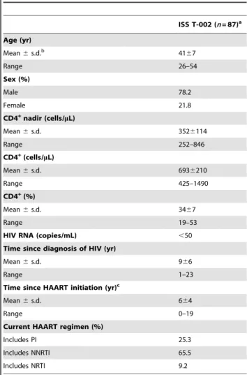
Table 1.Baseline characteristics of the study participants.
ISS T-002 (n= 87)a
Age (yr)
Mean6s.d.b 41
67
Range 26–54
Sex (%)
Male 78.2
Female 21.8
CD4+nadir (cells/mL)
Mean6s.d. 3526114
Range 252–846
CD4+(cells/mL)
Mean6s.d. 6936210
Range 425–1490
CD4+(%)
Mean6s.d. 3467
Range 19–53
HIV RNA (copies/mL) ,50
Time since diagnosis of HIV (yr)
Mean6s.d. 966
Range 1–23
Time since HAART initiation (yr)c
Mean6s.d. 664
Range 0–19
Current HAART regimen (%)
Includes PI 25.3
Includes NNRTI 65.5
Includes NRTI 9.2
aNumber of evaluable individuals;bStandard deviation;cBased on 76 individuals.
The results of the pre-amendment trial population (87 subjects) are reported here and they proof the role of Tat in HIV pathogenesis and disease maintenance under HAART, providing encouragement for combining Tat immunization with conven-tional virus-targeting drugs for an improved treatment of HIV disease.
Methods
The protocol for the ISS T-002 clinical trial and supporting CONSORT checklist are available as supporting information; see Checklist S1 and Protocol S1. The protocol for the ISS OBS T-002 observational study is available as Supporting information; see Protocol S2.
ISS T-002 trial design, conduction and duration
The study ‘‘A phase II randomized, open label, immunogenicity and safety trial of the vaccine based on the recombinant biologically active HIV-1 Tat protein in anti-Tat antibody negative HIV-1 infected HAART treated adult subjects’’, ISS T-002 (EudraCT No. 20072007200216; Clinical-Trials.gov NCT00751595) is a randomized, open-label, phase II
multicentric clinical trial directed at evaluating the immunogenic-ity (primary end-point) and the safety (secondary end-point) of the HIV-1 Tat protein in HIV-1 infected adult subjects, anti-Tat Ab negative, of either gender, 18-55 years-old, HAART-treated with chronic suppressed infection and levels of plasma viremia ,50 copies/mL in the last 6 months prior to screening and without a history of virologic rebound, with CD4+T cell counts
$400 cells/mL and with pre-HAART CD4 nadir.250 cells/mL. The study was approved by the national regulatory body and by the Ethics Committees of each clinical center. All subjects signed the written informed consent prior to enrollment. The trial was conducted in 10 clinical sites in Italy (Policlinico of Modena, Modena;Arcispedale S. Anna, Ferrara; Istituti Fiosterapici Ospitalieri San Gallicano, Rome; Policlinico of Bari, Bari;Ospedale S.M. Goretti
Latina; Fondazione S. Raffaele, Milan;Ospedale S. Maria Annunziata
Florence; Ospedale Luigi Sacco, Milan; Spedali Civili, Brescia;
Ospedale A. di Savoia, Turin). Subjects were randomized, with a competitive enrolment, to one of the four immunization regimens represented by 3 or 5 intradermal administrations of Tat at two doses (7.5mg or 30mg). The study includes 3-weeks screening period, 8-weeks or 16-weeks treatment period, 40-weeks or
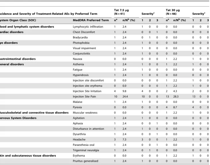
32-Table 2.Adverse Events (AEs) defined ascertainly, probablyorpossiblyrelated to the study medication.
Incidence and Severity of Treatment-Related AEs by Preferred Term
Tat 7.5mg
(N = 41) Severityc Tat 30(N = 46)mg Severityc
System Organ Class (SOC) MedDRA Preferred Term na n/Nb(%) 1 2 3 na n/Nb(%) 1 2 3
Blood and lymphatic system disorders Lymphocytic infiltration 1 2.4 1 0 0 0 0.0 0 0 0
Cardiac disorders Chest Discomfort 1 2.4 0 0 1 0 0.0 0 0 0
Bradycardia 1 2.4 0 1 0 0 0.0 0 0 0
Eye disorders Photophobia 1 2.4 1 0 0 0 0.0 0 0 0
Visual impairment 1 2.4 1 0 0 0 0.0 0 0 0
Conjunctivitis 1 2.4 1 0 0 0 0.0 0 0 0
Gastrointestinal disorders Nausea 0 0.0 0 0 0 1 2.2 1 0 0
General disorders Asthenia 1 2.4 1 0 0 1 2.2 1 0 0
Fatigue 1 2.4 1 0 0 0 0.0 0 0 0
Hyperidrosis 1 2.4 1 0 0 0 0.0 0 0 0
Injection site discomfort 0 0.0 0 0 0 1 2.2 1 0 0
Injection site erythema 0 0.0 0 0 0 1 2.2 1 0 0
Injection Site Irritation 4 9.8 4 0 0 2 4.3 2 0 0
Injection Site Pain 10 24.4 10 0 0 13 28.3 13 0 0
Malaise 1 2.4 1 0 0 0 0.0 0 0 0
Pyrexia 0 0.0 0 0 0 4 8.7 4 0 0
Musculoskeletal and connective tissue disorders Muscular weakness 0 0.0 0 0 0 1 2.2 1 0 0
Nervous System Disorders Agitation 1 2.4 1 0 0 0 0.0 0 0 0
Aphasia 1 2.4 0 0 1 0 0.0 0 0 0
Disturbance in attention 1 2.4 1 0 0 0 0.0 0 0 0
Dysarthria 1 2.4 0 0 1 0 0.0 0 0 0
Headache 3 7.3 3 0 0 1 2.2 1 0 0
Paraesthesia oral 1 2.4 0 0 1 0 0.0 0 0 0
Trigeminal neuralgia 1 2.4 0 1 0 0 0.0 0 0 0
Skin and subcutaneous tissue disorders Erythema 0 0.0 0 0 0 1 2.2 1 0 0
Pruritus generalised 1 2.4 1 0 0 0 0.0 0 0 0
an = number of subjects reporting the events;bN = number of evaluable subjects (%);cSeverity grade: 1 = mild, 2 = moderate, 3 = severe.
All the study participants prior to the protocol amendment (n = 87) have been evaluated for safety. The Medical Dictionary for Regulatory Activities (MedDRA) was used to classify the adverse events occurred during the study.
weeks of follow-up for the 3 or 5 immunizations, respectively, and foresees an extended follow-up of 3 years from the first immunization (Supporting information).
Blood samples from clinical sites were shipped by a certified courier to the Core Laboratory of Immunology and Virology (Ospedale S. Gallicano IFO, Rome, Italy) where all the immunological and virological testing was performed according to the Standard Operating Procedures (SOP) developed within the AIDS Vaccine Integrated Program (AVIP) [82], funded within the FP6 program of the European Community, and implemented in the corresponding phase I preventative (Clinicaltrials.gov Identi-fier NCT00529698) and therapeutic (Clinicaltrials.gov IdentiIdenti-fier NCT00505401) clinical trials [78–81].
All safety assessments were performed at the clinical sites according to the study schedule (see Protocol S1 in Supporting information). The study was monitored and quality assured by an accredited Contract Research Organization (CRO). The study enrolment started on July 2008 and is still open since an amendment has been approved by the DSMB and Ethical Committees to include individuals with a more advanced immune compromission and to expand the sample size from 128 to 160 volunteers (www.clinicaltrials.gov) (Supporting information).
The data shown here refer to the protocol prior to the amendment (Protocol S1) and include all the pre-amendment enrolled volunteers (87).
Study Medication
The study medication is the biologically active recombinant Tat protein administered intradermally in two doses (7.5mg and 30mg) according to two regimens (3 or 5 immunizations) at week 0, 4, 8 or 0, 4, 8, 12, 16, respectively. Before administration, the study medication (Tat 7.5mg/0.5 mL or Tat 30mg/0.5 mL) was thawed, diluted with 1.5 mL of sterile water, swirled gently and then administered by two intradermal injections into the right and left deltoid regions of the upper arms.
Study Outcomes
The primary pre-specified outcome of the study (immunoge-nicity) was measured by the induction, magnitude and persistence of the humoral immune responses to Tat, and by comparing the immunogenicity of the 3 or 5 immunization schedule of the two different vaccine doses (7.5mg and 30mg) at each scheduled time point (see Protocol S1 in Supporting information). ‘‘Responders’’ were defined as those subjects with at least a positive anti-Tat Ab response at any given time point after the first immunization. Cellular immune responses to Tat were a co-primary endpoint.
The anti-Tat humoral immune response was evaluated by determination and titration of IgM, IgG and IgA anti-Tat Ab in sera, while the anti-Tat cellular immune response was evaluated by the assessment of CD4+ and CD8+ lymphoproliferative
responses andin vitroIFN-c, IL-4 and IL-2 production in response to Tat, as detailed below.
The secondary pre-specified outcome of the study (safety) was assessed by the collection of all adverse events occurred during the study, including any significant change in haematological (including coagulation assessment), biochemical (with liver and kidney functional parameters) and immunological parameters (including CD4+, CD8+, CD3+T cells, NK, B cells and
mono-cytes). All the AEs were reported according to the Medical Dictionary for Regulatory Activities (MedDRA) and classified on the basis of the drug relationship as well as by the grade of severity. All the safety data were periodically (every 3 months) evaluated by the DSMB and Annual Safety reports were submitted to the Regulatory Bodies at scheduled times, as by regulatory guidelines (DLgs 211/2003).
Finally, a second-line exploratory testing was performed to characterize in-depth biochemical and immunological biomarkers of disease progression used to assess HAART efficacy, including determination of cellular and biochemical markers of immune activation, regulatory T cells, cell viability, CD4+T cells, CD8+T
cells, B cells and NK cells, central and effector memory CD4+and
Table 3.Relatedorunrelatedserious adverse events occurred during the ISS T-002 trial.
Volunteer Randomization Description Relationship Severity Action taken Outcome
group to treatment
#1 30mg, 5 imm. Hepatitis A Unrelated Severe Hospitalization Resolved
Treatment discontinued
#2 7.5mg, 5 imm. Disarthria and Possible Severe Hospitalization Resolved
Paresthesia Treatment discontinued
of the tongue
#3 7.5mg, 3 imm. Neurosyphilis Unrelated Mild Hospitalization Ongoing
#4 30mg, 3 imm. Uterin fibroma Unrelated Severe Medication required Resolved
Treatment discontinued
#5 30mg, 5 imm. Hodgkin’s Unrelated Severe Medication required Ongoing
lymphoma
#6 7.5mg, 3 imm. Transaminase Unrelated Moderate Hospitalization Resolved
increase
#7 30mg, 3 imm. Right occipital Unrelated Severe Medication required Ongoing
lesion
(ischemic event)
CD8+T cells as well as cellular responses to HIV Env and to recall
antigens, as detailed below.
All data have been reviewed by the Investigators, the DSMB, the IAB and the CAB.
Sample size
The primary objective of the trial was to assess the immuno-genicity of Tat immunization. In particular, ‘‘responders’’ were defined as subjects with at least a positive anti-Tat Ab response at any given time point after the first immunization, as indicated in the Protocol S1 (Supporting information).
To observe a proportion of at least 80% of subjects with Ab responses to vaccination, taking into account a maximum margin of error of 7% and a confidence level of 95%, 112 valuable subjects were found to be required, 28 for each treatment group. In this hypothesis the width of the confidence interval within each of the 4 randomization arms is 15%.
With this sample size a difference of at least 35% of the responders between the two Tat doses with each immunization schedule (3 or 5 inocula, respectively) is detected as statistically significant.
Considering a drop-out rate no greater than 10%, the calculated sample size is 128 subjects, randomized in 4 treatment groups by 32 subjects each.
Randomization
Since the study design foresees different immunization sched-ules, subjects were randomized into 4 treatment groups to ensure unbiased patient allocation.
The Randomization list was generated by the CRO using a block size of 4, according to a randomization scheme of 1:1:1:1 (RND PLUS 2.10 software).
The randomization assignment was carried out by the CRO via web. Each clinical site received a block of 4 numbered treatments (according to the randomization list). A code was assigned at screening at each subject. The code is constituted by the clinical trial code (T2) followed by the clinical site number (01 to 10) and by the progressive patient number within each site. The code was assigned to each volunteer at screening, irrespectively of enrol-ment. Once eligibility was established, the clinical staff contacted the CRO via web to randomize the subject. The drug kit number to be assigned to the subject was provided via web within the
block’s numbers that the clinical site had received. The assigned drug kit number was recorded on the e-CRF.
ISS OBS T-002 observational study enrollment and conduction
The ISS OBS T-002 (ClinicalTrials.gov NCT01024556) is a 5-years prospective observational study, which started before the therapeutic trial and is conducted in parallel at the same clinical centers, and follows the same procedures of the therapeutic trial ISS T-002 (sample collection and certified transportation,
centralized immune and virologic testing, certified CRO manage-ment). The primary objective of this study is to prospectively evaluating the clinical, immunological and virological parameters to determine the impact of naturally occurring (i.e., not induced by vaccination) anti-Tat immunity in HIV-infected individuals, of either gender, $18 years-old, with HAART-suppressed HIV replication (plasma viremia,50 copies/mL for 6 months prior to screening) without a history of virologic rebound and with known CD4+T cell number and nadir. In addition, the study had the
secondary objective of harmonizing the multicentric clinical
platform on common SOPs and procedures in order to both prepare cohorts for trials and to optimize all the operations required for the therapeutic trial conduction. The study is still recruiting. To date 127 patients have been enrolled and are being followed-up, 25 of them are Tat Ab positive and 91 are anti-Tat Ab negative (Fig. S1). Eighty-eight anti-anti-Tat Ab negative (out of the 91) are evaluable subjects (Total OBS Subjects), and 32 out of them meet all the immunological (CD4+ T cell counts
$400 cells/mL, pre-HAART CD4 nadir .250 cells/mL) and virological criteria for eligibility in the ISS T-002 clinical trial, representing therefore the appropriate Reference Group for a comparative assessment of the results with the trial subjects (Fig. S1, Table S1). For clarity, data from the observational study are reported in the Supplementary material section and the protocol is available in Supporting information (Protocol S2).
Anti-Tat antibodies
Ab were assessed as described [76,79,81,83]. Titers are expressed as the reciprocal of sample dilution. Ab titers equal or higher than 25 for IgM and IgA, or 100 for IgG were considered as positive.
IFN-c, IL-2 and IL-4 Elispot
Elispot was performed as previously described [79–81] using commercial kits (EL285, EL202, EL204, R&D Systems), with 4 pools of overlapping 15mer-Tat peptides (5mg/mL each) (UFP Service, Ferrara, Italy), 2 pools of Env peptides (5mg/mL each) (Neosystem), Candida (5mg/mL) (Nanogen Advanced Diagnos-tics), or a combination of Cytomegalovirus, Epstein-Barr and influenza virus (CEF) peptide pool (2mg/mL each) (Anaspec, 01036–05). PHA, (2mg/mL) or medium were the positive and the negative controls, respectively. Tests were considered valid when Spot Forming Cells (SFC)/well were $100 in positive controls. IFN-cElispot was considered positive when SFC/106cells were $30, and fold-increase over control was$3. The IL-2 and IL-4 Elispot were considered positive when fold-increase was$3.
T cell proliferation
Responses to Tat (1–5mg/mL) or Tat peptides (2mg/mL), Env (5mg/mL) (Fitzgerald), Candida (5mg/mL), or CEF peptide pool (2mg/mL each) were assessed by CellTrace CFSE Cell Prolifer-ation kit (Molecular probesTM, Invitrogen), elaborated by ModFit software (Verity Software House, INC.), and expressed as Proliferation Index (PI). Fold-increase (FI), calculated as ratio of
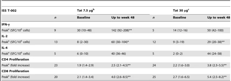
Table 4.Tat-specific cellular immune responses in subjects with a positive response after immunization with Tat.
ISS T-002 Tat 7.5mgb Tat 30mgc
n Baseline Up to week 48 n Baseline Up to week 48
IFN-c
Peaka(SFC/106cells) 9 30 (10–48) 142 (92–208)** 5 14 (12–16) 50 (42–100)
IL-2
Peaka(SFC/106cells) 13 8 (2–30) 60 (30–104)* 12 9 (3–19) 29 (20–38)**
IL-4
Peaka(SFC/106cells) 5 6 (0–10) 40 (36–46) 5 2 (0–2) 44 (24–58)
CD4 Proliferation
Peaka(fold increase) 23 1.9 (1.4–2.9) 2.5 (2.1–4.5)** 24 2.2 (1.6–3.0) 3.8 (2.3–5.5)**
CD8 Proliferation
Peaka(fold increase) 20 2.1 (1.4–3.4) 4.0 (2.6–8.5)** 25 2.7 (1.6–6.5) 5.4 (2.5–8.2)**
The median intensity, with interquartile range of peak of responses, is shown for subjects with at least a positive cellular response at any given time point after the first immunization and up to week 48. Pre-post vaccination median change was evaluated by the Wilcoxon signed-rank test. IFN-c, IL-2, IL-4 production by PBMC and CD4+ or CD8+lymphoproliferative responses were measured at baseline and up to week 48 after the first immunization. Results are stratified by Tat doses, (7.5 and 30mg).n indicates the number of responders.
aMedian (interquartile range) of peak of responses, weeks 8, 12, 20, 48. bTotal subject tested for cytokines: 35; for proliferation: 31.
cTotal subjects tested for proliferation: 33; for proliferation: 31. *P,0.05, **P,0.01.
doi:10.1371/journal.pone.0013540.t004
Table 5.Immune activation markers and T-reg at baseline in study participants.
ISS T-002 n Mean±s.e.
CD38+HLA-DR2on CD8+T cells (%) 38 31.5 62.0
HLA-DR+CD382on CD8+T cells (%) 38 5.5 60.8
CD38+HLA-DR+on CD8+T cells (%) 38 6.0 60.7
CD38+HLA-DR2on CD4+T cells (%) 37 49.7 62.2
HLA-DR+CD382on CD4+T cells (%) 37 4.5 60.5
CD38+HLA-DR+on CD4+T cells (%) 37 3.8 60.5
b2-microglobulin (mg/L) 77 1.760.1
Neopterin (nmol/L) 77 7.561.0
Total IgM (mg/dL) 77 7765
Total IgG (mg/dL) 77 1025629
Total IgA (mg/dL) 77 196611
CD25+on CD4+T cells (%) 66 8.6
60.3
FOXP3+on CD4+CD25+T cells (%) 60 31.2 61.4
CD25+FOXP3+on CD4+T cells (%) 60 2.7 60.2
CD25+FOXP3+on CD4+T cells (cells/mL) 59 19.4 611.7
Mean values (6standard error) of phenotypic and biochemical immune activation markers and T-regs of the study participants at baseline.nindicates the number of individuals tested for each parameter.
PI with antigens versus PI of controls, was considered positive when was$2.
Peripheral Blood Mononuclear Cell (PBMC) viability and lymphocyte subsets
Cell viability was determined by Trypan Blue Dye Exclusion using the Vi-CELLTM XR Counter (Beckman Coulter) [84,85]. Phenotyping was performed with BD Multitest 6-color TBNK reagent with BD Truecount tubes, (BD Biosciences). Samples were processed by FACSCanto flow cytometer (BD Biosciences) and data analyzed by FACSCanto clinical software. CD4+T cell counts were
also performed in parallel at each clinical center, and results were highly consistent with those generated by the Core lab.
Immune activation markers and T-reg
Whole blood was stained with anti-CD8 FITC/CD38 PE/CD3 PerCP/HLA-DR APC (MultiTESTTMBD Biosciences) plus anti-CD4 APC-Cy7 Ab (BD Pharmingen). Collective quadrant gates, based on HLA-DR and CD38 expression on CD4+or CD8+T
cells, were established.
Neopterin, b2-microglobulin and total immunoglobulins (Ig) were determined as described [86].
For CD25+and T-reg, PBMCs were stained with anti-human
APC-Cy7-labeled CD4, APC-labeled CD25 (BD Biosciences) and PE-labeled FOXP3 Ab (Bioscience, USA). Gating was performed on CD4+and CD4
-for CD25 expression on total T cells, and on CD4+T cells for CD25/FOXP3 doubly-positive cells, and then on
CD4/CD25 for FOXP3+lymphocytes.
Naı¨ve, central and effector memory CD4+or CD8+T cells PBMC were stained with anti-human CD3 (PerCP), CD4 (APC-Cy7), CD8 (APC), CD45RA (FITC), CD62L (PE) Ab (MultiTESTTM BD Biosciences), analyzed by FACSCanto flow cytometer (BD Biosciences) with the FlowJo (Tree Star, Ashland, OR) software.
T cell subsets were identified by hierarchical gating (morpho-logical, on CD3+, and then on CD4+or CD8+T cells). Collective
quadrant gates based on CD45RA and CD62L expression on CD3+/CD4+or CD3+/CD8+T cells identified naı¨ve (CD45RA
+/ CD62L+), central memory (CD45RA-/CD62L+, Tcm), effector memory (CD45RA-/CD62L-, Temro), and terminally-differenti-ated effector memory (CD45RA+/CD62L-, Temra) subsets.
HIV-1 viral load
HIV-l RNA was determined with COBAS AmpliPrep/COBAS TaqMan HIV-1 Test, version 2.0, (Roche Diagnostics) [84,87].
Statistical methods
The percentage of ‘‘responders’’ was estimated both for total vaccinees and for each randomization group with a 95% confidence interval.
Cochran-Armitage Trend test was used to compare frequencies of humoral responses. McNemar’s test was used to compare pre-post immunization frequencies of cellular responses within treatment groups. Wilcoxon signed-rank test was applied to evaluate increase of cellular responses intensity. Student’s t-test for paired data was used to assess the mean changes from baseline of
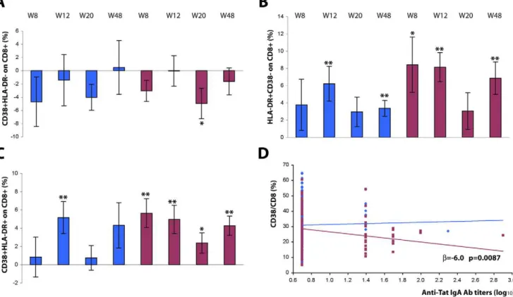
Figure 4. Expression of activation markers on CD8+T cells after Tat immunization.Changes from baseline of CD8+T cells (gating on CD8+ T cells) expressing (A) CD38, (B) HLA-DR, or (C) both CD38 and HLA-DR. Results are shown according to Tat dose and time after the first immunization. Data are presented as the mean % changes (6standard error) at week 8, 12, 20 and 48. Blue bars: Tat 7.5mg, n = 17 up to week 20 and n = 12 at week 48; red bars: Tat 30mg, n = 21 up to week 20, n = 16 at week 48, respectively. The t-Test for paired data was used for the analyses: *p,0.05, **p,0.01. (D) Correlation between CD38+/CD8+T cells (%) and anti-Tat IgA antibody titers (Multivariate regression model for repeated measures). Blue diamond: Tat 7.5mg, n = 39; red square: Tat 30mg, n = 43, respectively.
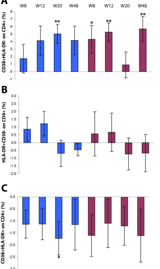
Figure 5. Expression of activation markers on CD4+T cells after Tat immunization.Changes from baseline of CD4+T cells (gating on CD4+ T cells) expressing (A) CD38, (B) HLA-DR, or (C) both CD38 and HLA-DR. Results are shown according to Tat dose and time after the first immunization. Data are presented as the mean % changes (6standard error) at week 8, 12, 20 and 48. Blue bars: Tat 7.5mg, n = 17 up to week 20 and n = 12 at week 48; red bars: Tat 30mg, n = 20 up to week 20, n = 15 at week 48, respectively; The t-Test for paired data was used for the analyses: *p,0.05, **p,0.01.
activation markers, lymphocytic phenotypes and cell viability, after controlling normality assumption of variables distribution (Sa-phiro-wilk test). Multivariate regression model for repeated measures, stratified by Tat dose, was applied to CD38+/CD8+T
cells (%), including anti-Tat Ab titers, cell viability and CD25+
FOXP3+expression on CD4+lymphocytes as explicative factors.
The same model was applied on CD38+/CD8+T cells (%) to all
immunized subjects, to assess the potential relationships with the anti-Tat Ab titers (IgM, IgG, IgA), CD8+central memory (%) and
anti-Tat induced cytokines (IFN-c, IL-2 and IL-4).
Since phase II studies are usually descriptive and are not designed to quantify treatment effects, the statistical analyses were not focused to determine differences in term of efficacy among three or more treatment groups and no adjustments were used for multiple comparisons. Therefore, exploratory analyses were applied to many variables to investigate the potential effect of the Tat immunization on the immunological status of vaccinees.
All statistical tests were carried out at a two-sided 5% significance level. Analyses and data processing were performed using SASHsoftware (SAS Institute, Cary, NC, USA).
Figure 7. Production of total Ig after Tat immunization.Total IgM (A), IgG (B) and IgA (C) serum levels (mg/dL) are shown. Blue bars: Tat 7.5mg, n = 37; red bars: Tat 30mg, n = 40. Data are presented as the mean changes (6standard error). The t-Test for paired data was used for the analyses: *p,0.05, **p,0.01.
Results
Study participants
Study participants were randomized by immunization schedule (3 or 5 times monthly) and Tat doses (7.5 or 30mg) (Fig. 1 and Table 1 and Methods). The results refer to the full pre-amendment trial population (n = 87). Sixty-eight trial participants had also a 48-weeks of follow-up.
Eighty-eight HAART-treated anti-Tat Ab negative and virolog-ically-suppressed individuals (for at last 6 months and without a history of virological rebound) with a known number of CD4+T cells
and CD4 T cell nadir, were enrolled in the prospective observational study (ISS OBS T-002, ClinicalTrials.gov NCT00751595). The results of this study served for intergroup comparison. In particular, 32 of these subjects fully matched the baseline characteristics of the immunized patients and were, therefore, considered as the Reference Group (Fig. S1, Table S1 and Methods). However, since no major differences were seen between the Reference Group and the full OBS population (Total OBS Subjects), results from both these groups are reported (Supplementary material).
Safety Data of therapeutic immunization with the biologically active Tat protein
Safety was assessed in all trial (n = 87) volunteers by monitoring local and systemic adverse events (AEs) as well as hematological,
biochemical and immunological laboratory parameters as per-formed previously in phase I trials [79–81]. Since in immunized subjects no differences in the incidence of adverse events were detected between the number of inocula (3 or 5) with the same Tat dose, the results were stratified by Tat dosages.
No relevant AEs occurred during the study, and most of them were expected both in frequency and type for HIV-infected subjects (Table 2). In particular 51/87 subjects (59%) experienced AEs during the study. Out of them, 26/87 (30%) presented a
certain, probableorpossiblerelationship to the study treatment and were not related to the Tat dosage. These events were mostly local, related to the injection site and mild in severity (Table 2).
Seven serious adverse events (SAE) occurred after Tat immunization, but only one was indicated as possiblyrelated to the study treatment (Table 3).
Based on the first and second year Annual Safety Report and on the periodic meetings and reports, the DSMB of the ISS T-002 deliberated that the immunization with Tat is safe and well tolerated.
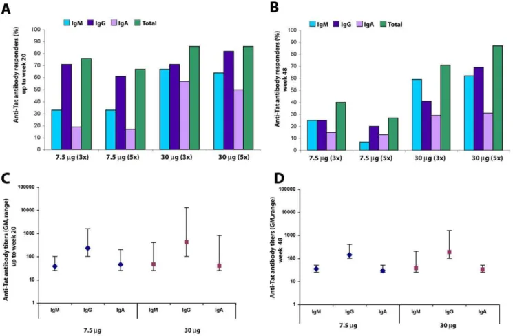
Therapeutic immunization with Tat induces specific humoral and cellular immune responses in HAART-treated individuals
Responders were defined by the induction of anti-Tat Ab. Total responders were 79% (95%C.I. 70–88%), with a higher frequency
Figure 8. CD25 and FOXP3 expression on CD4+T cells after Tat immunization.(A) Changes from baseline of CD4+lymphocytes expressing CD25 are shown according to Tat dose and time after the first immunization (Tat 7.5mg, n = 32 up to week 20 and n = 24 at week 48; Tat 30mg, n = 34 up to week 20 and n = 28 at week 48, respectively). (B) Changes from baseline of the percentage of CD4+CD25+lymphocytes expressing FOXP3+(Tat 7.5mg, n = 31 up to week 20 and n = 23 at week 48; Tat 30mg, n = 29 up to week 20 and n = 28 at week 48, respectively). (C) Changes from baseline of the percentage of CD4+T cells expressing CD25+FOXP3+(Tat 7.5mg, n = 31 up to week 20 and n = 23 at week 48; Tat 30mg, n = 29 up to week 20 and n = 24 at week 48). (D) Changes from baseline of the absolute number of CD4+lymphocytes expressing CD25+FOXP3+(Tat 7.5mg, n = 30 up to week 20 and n = 20 at week 48; Tat 30mg, n = 29 up to week 20 and n = 22 at week 48, respectively). Blue bars: Tat 7.5mg; red bars: Tat 30mg. Data are presented as the mean changes (6standard error) evaluated at 8, 12, 20 and 48 weeks after the first immunization. The t-Test for paired data was used for the analyses: *p,0.05, **p,0.01.
at the Tat 30mg dose (86%; 95%C.I. 76–96%) as compared to the Tat 7.5mg dose (72%; 95%C.I. 58–86%). After stratification by the randomization groups, responders were 76% (95%C.I. 58– 94%) at 7.5mg, 3 inocula, and 67% (95%C.I. 45–88%) after 5 inocula, respectively. At the 30mg Tat dose responders were 86% (95%C.I. 71–100%) after 3 inocula and 86% (95%C.I. 72–100%) after 5 inocula, respectively.
The 30mg dose was, therefore, more potent at inducing anti-Tat Ab and at maintaining long-term humoral responses with little or no differences between the 3 or 5 inoculation regimens (Fig. 2 A–B). Specifically, during the immunization phase (up to week 20) the 30mg Tat dose was the most effective at inducing anti-Tat IgM and IgA subclasses (p = 0.0117 and p = 0.0051, respectively) (Fig. 2A). This Tat dose was also the most effective at inducing a durable Ab response (p = 0.0007), which was still present at approximately 1 year post-immunization (48 weeks), for both IgM and IgG subclasses (p = 0.0024 and p = 0.0048, respectively) (Fig. 2B). In contrast, peak Ab titers did not significantly differ between doses and only slightly decreased at 48 weeks (Fig. 2 C, D).
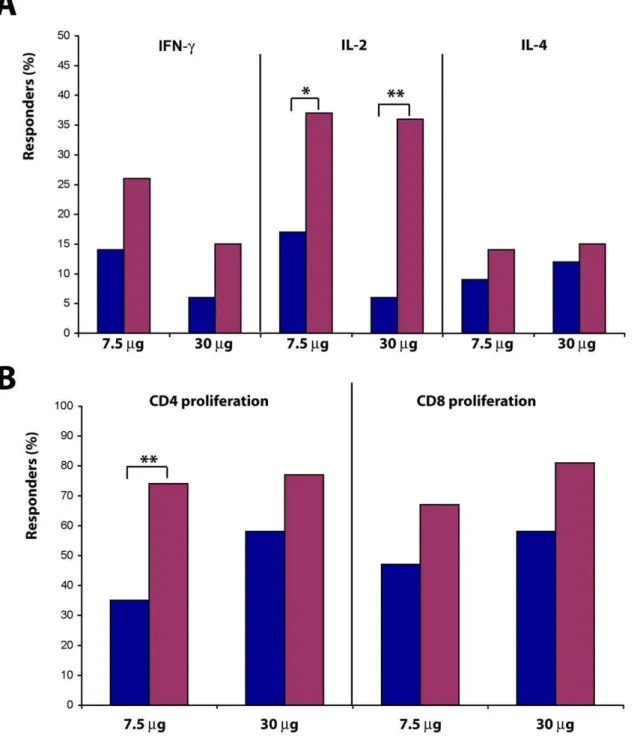
Both Tat doses induced specific cellular responses (Fig. 3 A–B and Table 4). A cumulative analysis of the T cell responses (up to 48 weeks after immunization) indicated an increase in the percentage of responders for production of IL-2 (p = 0.0348 and p = 0.0039 at Tat 7.5 or 30mg, respectively) and, to a lesser extent, IFN-cand IL-4, as well as of Tat-specific CD4+(p = 0.0013 at Tat
7.5mg) and CD8+ T cell proliferation. No relevant differences
were observed between 3 or 5 immunizations.
Further, increased peak values were detected for both cytokines and CD4 +and CD8+T cell proliferation in response to Tat,
which reached significance with Tat at 7.5mg for IFN-c (p = 0.0078), and IL-2 (p = 0.0327), and at Tat 30mg for IL-2 (p = 0.0005), and with both Tat doses for CD4+and CD8+T cell
proliferation (p,0.001) (Table 4).
Immunization with Tat downregulates phenotypic and biochemical markers of immune activation and increases regulatory T cells
To further investigate the effect of immunization with Tat, key biomarkers of AIDS pathogenesis and progression used to evaluate HAART efficacy were determined as second-line exploratory testing. These included phenotypic (CD38, HLA-DR) and biochemical (serumb2-microglobulin, neopterin and total immu-noglobulins) immune activation markers. In addition, T-reg lymphocytes, that are markedly reduced in HIV-infected individ-uals even under HAART, were monitored since these cells are key for controlling immune activation and both the generation and termination of adaptive immune responses. Baseline values of these parameters were determined at study entry (Table 5). Since no differences were detected in immunized subjects between the number of inocula (3 or 5) with the same Tat dose, results were stratified by Tat dosage.
CD38 and HLA-DR expression
Downregulation of CD38 expression was observed on CD8+T
cells from subjects immunized with both Tat doses, and was more pronounced and persistent at Tat 30mg, reaching statistical significance at week 20 (p = 0.0436) to decline thereafter (week 48) (Fig. 4 A and Table 5). At the same time, a significant increase of HLA-DR expression on CD8+T cells, either alone or with CD38,
was also observed in subjects immunized with both Tat doses with a peak at week 12 (Tat 7.5mg, p = 0.0071; Tat 30mg, p = 0.0001), remaining significantly higher at week 48 (Tat 7.5mg, p = 0.0039; Tat 30mg, p = 0.0023) (Fig. 4 B, C). The most evident effects were
again observed with Tat 30mg, for which the average frequency of CD38+/HLA-DR+ doubly-positive CD8+ T lymphocytes
re-mained significantly higher up to week 48 (p = 0.0010).
Of note, a longitudinal analysis showed a significant correlation of CD38 downregulation on CD8+T cells with increasing anti-Tat
IgA titers following the Tat 30mg immunization
[log10 IgA:b= -6.0% (95% CI210.5%;21.5%) p = 0.0087]. The modulation of the expression of these markers was somewhat opposite on CD4+T cells (Fig. 5 A–C). In particular, the percentage
of CD38 singly-positive CD4+T cells was increased in immunized
subjects with both Tat doses and up to week 48, remaining significant for Tat 30mg (p = 0.0038), whereas HLA-DR expression had little changes after immunization. In contrast, the frequencies of CD38+/
HLA-DR+doubly-positive CD4+T lymphocytes were reduced in
immunized subjects at all time points and at both Tat doses.
Biochemical markers of immune activation
The down-regulation of cellular markers of immune activation following Tat immunization was associated with an early decrease (week 4) in the serum levels ofb2-microglobulin (p,0.0001) (Fig. 6 A), and neopterin (Fig. 6 B).
Different changes were observed for total Ig according to the isotype (Fig. 7 A–C). In particular, only total IgG were significantly decreased early (week 4) post-immunization and at both Tat doses (Tat 7.5mg, p = 0.0258; Tat 30mg, p = 0.0111), remaining thereafter below baseline levels only for Tat 30mg.
CD25 expression and T-regulatory Cells
After Tat immunization, the percentage of total CD25+/CD4+T
cells markedly diminished returning to baseline values at week 48. This reduction was greater with Tat 30mg, reaching a nadir at 12
Table 6.Inverse correlation between baseline values and changes after Tat immunization of immune activation markers and T-regs.
ISS T-002 n
Pearson
correlation p-value
coefficient
CD38+HLA-DR2on CD8+T cells (%) 38 r =
20.5 0.0005
CD382HLA-DR+on CD8+T cells (%) 38 r =
20.5 0.0023
CD38+HLA-DR+on CD8+T cells (%) 38 r =
20.4 0.0190
CD38+HLA-DR2on CD4+T cells (%) 37 r =
20.3 0.0623
HLA-DR+CD382on CD4+T cells (%) 37 r =
20.7 ,0.0001
CD38+HLA-DR+on CD4+T cells (%) 37 r =
20.8 ,0.0001
b2-microglobulin (mg/L) 77 r =20.3 0.0017
Neopterin (nmol/L) 77 r =20.9 ,0.0001
Total IgM (mg/dL) 77 r =20.1 0.5371
Total IgG (mg/dL) 77 r =20.4 0.0001
Total IgA (mg/dL) 77 r = 0.0 0.7519
CD25+on CD4+T cells (%) 66 r =
20.5 ,0.0001
FOXP3+on CD4+CD25+T cells (%) 60 r =
20.5 0.0001
CD25+FOXP3+on CD4+T cells (%) 60 r =
20.3 0.0171
The relationship between baseline values and changes at week 20 or week 24 for all immunized subjects was evaluated by the Pearson correlation coefficient (r) after cumulating both Tat doses.nindicates the number of individuals evaluated for each parameter.
weeks post-immunization and remaining significant at week 20 (p = 0.0351) (Fig. 8 A), before returning to baseline levels at week 48. This finding was paralleled by a significant and persistent increase of the percentage of FOXP3 expression in the CD4+/
CD25+ T cell subset as well as of the percentage and absolute
number of CD4+/CD25+/FOXP3+T-reg, which had a significant
and progressive increase at both Tat doses and persisted up to week 48 (T-regs % at Tat 7.5mg, p = 0.0146, at Tat 30mg, p = 0.0002; T-regs number at Tat 7.5mg, p = 0.0090, at Tat 30mg, p = 0.0018) (Fig. 8 C, D).
The changes of immune activation markers and T-reg observed upon immunization were significantly and inversely related to baseline values (Table 6), in that the reduction in immune activation markers was more pronounced in subjects with the highest values at baseline. Conversely, the largest increase in T-reg cell number was seen in individuals with the lowest baseline values.
Immunization with Tat increases PBMC viability and the number of CD4+T cells and B cells
PBMC viability in vitro is reduced in HIV infection [88]. In contrast, progressive and significant increments of cell viability were seen in PBMC early after immunization (since week 8) with both Tat doses and particularly with Tat 30mg. Cell viability continued to increase up to week 48 with both Tat doses (p,0.0001, Fig. 9 A).
The CD4+T cell number increased after Tat immunization, at
all time-points and for both Tat doses (Fig. 9 B), reaching statistical significance with the 7.5mg Tat dose at week 8 (57 cells/mL, p = 0.0206), week 16 (69 cells/mL, p = 0.0188), week 20 (68 cells/ mL, p = 0.0418), week 24 (48 cells/mL, p = 0.045) and week 48 (54 cells/mL, p = 0.0806), and for the 30mg Tat dose at week 8 (59 cells/mL, p = 0.0181) and week 16 (52 cells/mL, p = 0.0388).
Similarly, the B cell number also increased upon immunization with both Tat doses and at all time-points (Fig. 9 C), reaching statistical significance with Tat 7.5mg at week 8 (46 cells/mL, p = 0.0250), week 20 (28 cells/mL, p = 0.0368) and week 48 (66 cells/mL, p = 0.0261), as well as with Tat 30mg at week 8 (26 cells/mL, p = 0.0241) and week 12 (25 cells/mL, p = 0.0200).
Immunization with Tat increases the percentage of CD4+ T cells and B cells and reduces the percentage of CD8+ and NK cells independently from the type of HAART regimen
The determination of the percentage of lymphocyte subsets confirmed the increase of CD4+T cells and B cells observed for
the absolute values, showing significant increases of CD4+T cells,
particularly at week 20 (1%, p = 0.0218) and 48 (2%, p = 0.0010), as well as significant increases of B cells at all time-points starting from week 8 (1%, p,0.01) (Fig. 10 A). In contrast, the percentage of NK and CD8+T cells were significantly reduced at week 20
(21%, p = 0.0288) and 48 (21.4%, p = 0.0217), respectively. These effects were observed with both Tat doses (Fig. 10 B, C).
As a consequence, the CD4/CD8 ratio progressively increased in immunized subjects with the most evident effects at week 20 and
48 from the first immunization, and, particularly, for the 30mg Tat dose (week 20, p = 0.0117; week 48, p = 0.0011).
Stratification of these results according to the type of HAART regimen (NNRTI-based or PI-based) indicated that Tat immuni-zation can overcome the differences seen with different drugs regimens on lymphocyte subsets, suggesting that the effects of Tat immunization are independent from the type of drugs combina-tion (Fig. 11 A, B).
Immunization with Tat increases central memory and reduces terminally- differentiated effector memory (Temra) CD4+and CD8+T cells
Since reduction of central memory T cells (Tcm) with a concomitant increase of effector memory cells (Tem) is commonly observed in HIV infection and this unbalance is only partially restored under effective HAART [5], these T cell subsets were evaluated upon immunization with Tat. The percentage of central (CD45RA2/CD62L+) and, to a lesser extent, effector (CD45RA2/ CD62L+, Temro) memory CD4+and CD8+T cells increased upon
Tat immunization (Fig. 12).
For CD4+ T cells, these findings were concomitant with a
reduction of both terminally-differentiated Tem (CD45RA+/ CD62L2, Temra) and naı¨ve (CD45RA+/CD62L+) T cells
(Fig. 12 A). These changes were evident 8 weeks after the first immunization, peaked at week 12 (Tcm, p = 0.0176; Temra, p = 0.0080; naı¨ve T cells, p = 0.0018) and declined thereafter but did not return to baseline values (Fig. 12 A).
For CD8+T cells the earliest and most prominent changes were
observed for Temra, which were significantly reduced at all time-points post-immunization, reaching a nadir at week 48 (Fig. 12 B). In contrast, the increase in Tcm was less pronounced and slower for CD8+ cells as compared to CD4+ cells, reaching statistical
significance only at week 20 (p,0.0001). As for CD4+T cells, also
the percentage of CD8+ Temro increased after immunization,
reaching statistical significance at week 12 (p = 0.0378). Unlike the CD4+ counterpart, the CD8+ naı¨ve subset remained largely
unaffected by the treatment (Fig. 12 B).
Immunization with Tat increases cellular responses to HIV-Env and to recall antigens
To verify whether the effects of Tat-immunization were accompanied by changes in adaptive immunity against heterolo-gous antigens, T cell responses against HIV-Env, Candida, or CEF antigens were determined by monitoring Th1 and Th2 cytokine production and CD4+and CD8+T cell proliferation. An increase in
both the percentage of responders and the intensity of responses was found in subjects immunized with both Tat doses (Fig. 13 A–F and Table 7–9). In particular, statistically significant increases in the percentage of responders were detected against Env for IFN-c(Tat 7.5mg, p = 0.0348; Tat 30mg, p = 0.0005), and against Candida for IL-2 (Tat 7.5mg, p = 0.0076; Tat 30mg, p = 0.0027) and IL-4 (Tat 7.5mg, p = 0.0114; Tat 30mg, p = 0.0184). Statistically significant increases of CD4+and CD8+T cell proliferation were detected
against Env (for CD4+ at Tat 7.5mg, p = 0.0114, at Tat 30mg,
p = 0.0455; for CD8+at Tat 7.5mg, p = 0.0016), and Candida (for
CD4+at Tat 7.5mg, p = 0.0253, at Tat 30mg, p = 0.0010; for CD8+
Figure 9. Evaluation of PBMC viability, CD4+T cell and B cell counts after Tat immunization.(A) Changes from baseline ofin vitroPBMC viability, stratified by Tat dose. Blue bars: Tat 7.5mg, n = 35 up to week 20 and n = 32 at week 48; red bars: Tat 30mg, n = 40 up to week 20 and n = 33 at week 48, respectively. (B) Changes from baseline of CD4+T cells/mL (data from clinical sites), stratified by Tat dose. Blue bars: Tat 7.5mg, n = 39 up to week 24 and n = 30 at week 48; red bars Tat 30mg, n = 43 up to week 24 and n = 34 at week 48. (C) Changes from baseline of B cells/mL, stratified by Tat dose. Blue bars: Tat 7.5mg, n = 38 up to week 20 and n = 30 at week 48; red bars Tat 30mg, n = 40 up to week 20 and n = 30 at week 48, respectively. The t-Test for paired data was used for the analyses: *p,0.05, **p,0.01.
at Tat 7.5mg, p = 0.0039) (Fig. 13 A–D). Cytokine production in response to CEF was already present in most subjects with no differences after immunization (Fig. 13 E). In contrast, CD4+and
CD8+T cell proliferation to CEF were very low at baseline and
increased after immunization particularly upon vaccination with 30mg of Tat for both CD4+(p = 0.0114) and CD8+(p = 0.0067) T
cell subsets (Fig. 13 F).
The intensity (peak values) of both cytokine production and proliferative responses to Env as well as to recall antigens including CEF were significantly increased after immunization (p,0.05, Table 7–9).
Correlations of Tat immunization with changes in the T cell compartments
A multivariate regression analysis was used to assess the presence of potential correlations among the different parameters investigated after therapeutic immunization. A statistically signif-icant inverse correlation was found between the percentage of CD38+/CD8+T cells with anti-Tat IgA titers (p = 0.0309), CD8+
Tcm lymphocytes (p = 0.0316), and with IL-2 production in response to Tat (p = 0.0235) (Fig. 14 A–C), suggesting a direct relationship between the induction of anti-Tat specific IL-2 producing cells and increasing anti-Tat IgA titers with the expansion of CD8+ Tcm and the reduction of activated CD8+
effector T cells.
Comparison of Tat-immunized subjects with patients enrolled in the observational Study (ISS OBS T-002)
Although the effects and the limits of a successful HAART are well known [89], an intergroup comparison between trial subjects and those enrolled in the parallel ISS OBS T-002 study was made to better evaluate the effect of Tat immunization on virologically-suppressed patients (see Methods section, Protocol S2 and Supplementary material). Out of 88 evaluable patients (Total OBS Subjects) enrolled in this study (see Methods section), 32 subjects fully matched the baseline characteristics of the trial participants (Fig. S1 and Table S1). This group was considered as the Reference Group, and comparison of immunized subjects was made with both this Reference Group and the Total OBS Subjects for all assessed parameters (Table 10, 11). The baseline values of the OBS subjects for activation markers and T-reg lymphocytes were determined at study entry (Table S2).
A decrease of CD38 and HLA-DR expression on CD8+T cells
either alone or in combination was observed during the follow up for both Total OBS Subjects and Reference Group (Fig. S2, A–F). These results are in sharp contrast with the upregulation of HLA– DR expression, alone or combined with CD38, observed on CD8+
T cells from immunized subjects (Fig. 4 A–C).
The modulation of CD38 and HLA–DR expression on CD4+T
cells of OBS subjects was similar to that observed on CD8+T cells
(Fig. S2 G–L). Thus, the pattern of expression of these two activation markers differed from what observed in the immunized subjects in whom an opposite and statistically significant upregulation of CD38 expression on CD4+T cells was observed
(Fig. 5 A–C).
The biochemical markers of immune activation were scarcely modified in the Reference Group throughout follow-up (Fig. S3 A–E). Specifically,b2-microglobulin showed an early increase and
then a gradual but slight decrease, while neopterin was slightly but persistently increased respect to baseline values (Fig. S3 A, B). In contrast, both these activation markers were reduced at most time-points in immunized patients (Fig. 6 A, B).
Total IgM, IgG and IgA remained substantially stable in OBS subjects for most of the follow up (Fig. S3 C–E), whereas a significant reduction of total IgG was recorded early after immunization in trial subjects (Fig. 7 A–C).
Overall, in the OBS patients the percentage of total CD25+/
CD4+T cells was stable, with an increase at week 48 (Fig. S4 A and
Fig. S5 A). Conversely, the percentage of CD4+/CD25+T cells
expressing FOXP3 gradually decreased in the Total OBS Subjects (Fig. S4 B) as well as in the Reference Group (Fig. S5 B). In addition, a decrease was observed in the percentage and absolute number of the CD4+/CD25+/FOXP3+ T-reg subset in the Total OBS
Subjects (Fig. S4 C, D), whereas the decline was less pronounced in the Reference Group (Fig. S5 C, D). The opposite was observed in the patients immunized with Tat, in which an overall reduction of CD25 expression and a concomitant and persistent statistically significant increase of T-reg were detected (Fig. 8 A–D).
Cell viability increased at late time-points (36 and 48 weeks) both in the Total OBS Subjects and in the Reference Groups (Fig. S6 A, B), as opposed to immunized individuals in whom a significant and steady increase of cell viability was detected as early as 8 weeks after the first immunization, particularly with the 30mg Tat dose (Fig. 9 A).
No significant changes of the CD4+ T cell number were
detected in individuals from either the Total OBS (Fig. S6 C) or the Reference Group (Fig. S6 D). This is in contrast with the statistically significant and persistent increase of CD4+ T cells
observed in the subjects immunized with Tat (Fig. 9 B).
Further, a progressive loss of B cells was observed in both OBS groups (Fig. S6 E, F), a finding in striking contrast with the early, durable and statistically significant increments of B cells observed in the Tat immunized subjects (Fig. 9 C).
These differences between the trial and OBS groups were also reflected by the percentage of lymphocyte subsets. In fact, as compared to baseline levels, in the OBS study were detected a moderate increase of the CD4+ T cell percentage, an overall
stability (Total OBS Subjects) or decrease (Reference Group) of the B cell subset (Fig. S7 A, B), a stable (Total OBS Subjects) or further increased (Reference Group) percentage of CD8+T cells,
and a substantial stability of the NK subset. Again, opposite trends were apparent in immunized patients in whom a persistent increase of the percentage of CD4+T and B cells and a progressive
decrease of CD8+T cells and NK cells were recorded, irrespective
of the type of HAART regimen (Fig. 11 A, B). In this regard, analysis of OBS subjects after stratification by NNRTI-based or PI-based treatment showed a different profile of lymphocytes subsets, particularly for B cells (Fig. S7 C–E), indicating a positive effect of Tat immunization in HAART intensification indepen-dently from the type of drugs combination.
The percentage of central memory (CD45RA2/CD62L+)
CD4+(Fig. S8 A, C) and CD8+(Fig. S8 B, D) T cells was stable
or moderately increased, though not significantly, at week 24 both for the Total OBS and Reference Group, as opposed to the more pronounced increments that were recorded in immunized subjects (Fig. 12 A, B). In the effector memory CD4+ and CD8+
Figure 10. Evaluation of the percentage of CD4+, CD8+, NK and B cells in all Tat-immunized subjects and after stratification by Tat
dose.(A) Changes from baseline of CD4+, CD8+, NK and B cells (percentage) for all immunized subjects (n = 78 up to week 20, n = 60 at week 48). (B,
C) Changes from baseline of CD4+, CD8
Figure 11. Evaluation of the percentage of CD4+, CD8+, NK and B cells in all Tat-immunized subjects after stratification by HAART
regimens.Changes from baseline of CD4+, CD8+, NK and B cells (percentage) from all immunized patients are shown for NNRTI-based (A) and PI-based (B) treatments. NNRTI-based: n = 51 up to week 20 and n = 41 at week 48; PI-based: n = 19 up to week 20 and n = 12 at week 48, respectively. The t-Test for paired data was used for the analyses: *p,0.05, **p,0.01.
compartment a reduction of the Temra (CD62L2/CD45RA+) subset and a concomitant increase of the Temro (CD62L2/ CD45RA-) subpopulation were observed at week 24 in the OBS subjects (Fig. S8, A–D), while similar but earlier and more pronounced changes were evident in the immunized patients (Fig. 12 A, B). Finally, individuals from both the trial and the OBS study experienced a decline of naı¨ve (CD45RA+/CD62L+) CD4+
and CD8+T cells (Fig. 12 and Fig. S8 A–D).
The cumulative assessment of the cellular immune responses to Tat (up to 48 weeks of follow up) revealed an increase of the percentage of responders in terms of both specific cytokines production, except for IL-2 production in the Reference Group,
and particularly of CD8+ T cell proliferation in both the Total
OBS Subjects and the Reference Group (Fig. S9 A, B and Fig. S10 A, B). These changes were also quantitative, in that an increase in the peak values of cytokine production and CD8+ T cell
proliferation was also detected for both groups (Table S3). In contrast, greater and statistically significant increments were seen for IL-2 production and CD4+ T cell proliferation in subjects
immunized with Tat.
The percentage of responders and the intensity of responses were also evaluated for HIV-Env, Candida and CEF antigens in both the Total OBS Subjects and in the Reference Group (Fig. S9 C–H, Fig. S10 C–H and Tables S4, S5, S6). No changes were
Figure 13. Cellular immune responses against Env or recall antigens after Tat immunization.Percentage of responders at baseline (blue bar) and up to week 48 (red bar) are stratified by Tat dose. Percentage of subjects showing (A) anti-Env production of IFN-c, IL-2 and IL-4 (Tat 7.5mg, n = 31; Tat 30mg, n = 29) and (B) CD4+or CD8+lymphoproliferative responses (Tat 7.5mg, n = 31; Tat 30mg, n = 30); (C) anti-Candida cytokines production (Tat 7.5mg, n = 32; Tat 30mg, n = 29), and (D) CD4+or CD8+lymphoproliferative responses (Tat 7.5mg, n = 31; Tat 30mg, n = 29); (E) anti-CEF production of IFN-c, IL-2 and IL-4 (Tat 7.5mg, n = 34; Tat 30mg, n = 32), and (F) CD4+or CD8+lymphoproliferative responses (Tat 7.5mg, n = 31; Tat 30mg, n = 29). The analysis was performed using the McNemar’s test: *p,0.05, **p,0.01.
doi:10.1371/journal.pone.0013540.g013
Table 7.Cellular immune responses against Env after Tat immunization.
ISS T-002 Tat 7.5mgb Tat 30mgc
n Baseline Up to week 48 n Baseline Up to week 48
IFN-c
Peaka(SFC/106cells) 19 44 (0–394) 382 (86–834)** 18 53 (12–226) 236 (86–920)**
IL-2
Peaka(SFC/106cells) 19 52 (18–76) 94 (52–166)** 20 26 (14–49) 49 (38–95)**
IL-4
Peaka(SFC/106cells) 20 52 (16–122) 137 (44–464)** 18 24 (11–64) 80 (28–132)
CD4 Proliferation
Peaka(fold increase) 15 1.5 (0.8–3.8) 2.4 (2.1–4.7)* 21 1.5 (0.8–2.8) 2.9 (2.2–4.5)**
CD8 Proliferation
Peaka(fold increase) 20 2.0 (1.1–4.1) 3.1 (2.5–7.2)* 18 2.7 (1.1–5.0) 5.6 (3.9–7.0)*
IFN-c, IL-2, IL-4 production by PBMC, and CD4 or CD8 lymphoproliferative responses to Env were measured at baseline and up to week 48 after the first immunization, respectively. Results are stratified by Tat doses, (7.5 and 30mg).nindicates the number of responders for cytokines production and CD4 or CD8 T cell proliferation, respectively. The median intensity with interquartile range of peak of responses is shown. Pre-post vaccination median change was evaluated by Wilcoxon signed-rank test.
aMedian (interquartile range) of peak of responses, weeks 8, 12, 20, 48. bTotal subject tested for cytokines: 31; for proliferation: 31.
cTotal subjects tested for cytokines: 29; for proliferation: 30. *P,0.05, **P,0.01.