DNA Damage in End-Stage Renal Disease Patients.
Assessment by In Vitro Comet Assay and by Cell-Free
DNA Quantification
Susana Coimbra, Alice Santos-Silva, Elísio Costa and
Elsa Bronze-da-Rocha
Additional information is available at the end of the chapter
Author Queries
[AQ1] As per the style guide, abstract should not exceed 200 words. Please check. [AQ2] As per the instruction all the display equations should be numbered. So
the unnumbered display equation has been changed to numbered format. Please check.
[AQ3] Figures 1, 2 and 3 are visually bad quality. Please provide better quality
figures.
© 2017 The Author(s). Licensee InTech. This chapter is distributed under the terms of the Creative Commons Attribution License (http://creativecommons.org/licenses/by/3.0), which permits unrestricted use, distribution, and reproduction in any medium, provided the original work is properly cited.
Provisional chapter
© 2016 The Author(s). Licensee InTech. This chapter is distributed under the terms of the Creative Commons Attribution License (http://creativecommons.org/licenses/by/3.0), which permits unrestricted use, distribution, and reproduction in any medium, provided the original work is properly cited.
DNA Damage in End-Stage Renal Disease Patients.
Assessment by In Vitro Comet Assay and by Cell-Free
DNA Quantification
Susana Coimbra, Alice Santos-Silva, Elísio Costa
and Elsa Bronze-da-Rocha
Additional information is available at the end of the chapter
Abstract
Inflammation is a common feature in end stage renal disease (ESRD) patients on hemo-dialysis (HD) that might contribute to increase DNA damage. Inflammation biomarkers, such as C-reactive protein (CRP) and pro-inflammatory cytokines, are frequently raised in chronic kidney disease (CKD). It has been reported that patients with CKD present increased levels of circulating cell-free DNA (cfDNA) and different types of DNA injury. The genomic damage at the level of a single cell can be recognized by comet assay, a sen-sitive and simple technique to detect cell DNA alterations. The underlying inflammatory process in ESRD patients under HD therapy may be associated with increased genomic damage and cfDNA contributing to further enhance inflammation and the associated morbidity and mortality risk. In a recent work, we analyzed the degree of genomic dam-age in ESRD patients under HD therapy for more than 1 year, using the alkaline in vitro comet assay and the cfDNA quantification, to evaluate DNA damage within the cell and the circulating free DNA. We found that ESRD patients presented significantly higher CRP values and cell damaged DNA. The cfDNA values were correlated with age and inflammatory stage. Nine out 39 patients died during the 1 year follow-up period and presented significantly higher cfDNA, than those who persisted alive. Our data suggest that at lower CRP values, the increased DNA damage is still within the cell, and at higher CRP values the damaged DNA is released in to plasma. The higher degree of genomic damage in ESRD patients might be a consequence of inflammation and aging, and may contribute to increase the risk for cancer and cardiovascular (CV) mortality. Moreover, our data suggest that the comet assay is more sensitive for low grade inflammatory con-ditions, while cfDNA appears as a good biomarker for more severe inflammatory condi-tions, as well as a biomarker for the outcome of ESRD patients. In summary, the genomic damage in ESRD patients seems to result, at least in part, from inflammation and aging,
and may contribute to increase the risk for cancer and CV mortality. AQ1 01 02 03 04 05 06 07 08 09 10 11 12 13 14 15 16 17 18 19 20 21 22 23 24 25 26 27 28 29 30 31 32 Genotoxicity 2
Keywords: chronic kidney disease, end-stage renal disease, inflammation, genomic
damage, comet assay, cell-free DNA
1. Introduction
Kidneys are important in homeostasis, ensuring the excretion of toxic substances and regulat-ing blood volume, blood pressure, concentration of electrolytes, plasma osmolarity and the acid/base balance. The kidneys also have endocrine functions, producing hormones, such as erythropoietin and calcitriol.
Chronic kidney disease (CKD) is characterized by a decline in kidney function and/or altered renal structure, leading to a gradual to permanent loss of kidney function over time. End-stage renal disease (ESRD), the worst stage of chronic kidney disease (CKD), requires dialysis to prevent accumulation of toxins, excessive water and electrolytes, or kidney transplantation [1]. Inflammation is a common feature in CKD, especially enhanced in ESRD patients on hemo-dialysis (HD). This chronic inflammatory state seems to contribute to aggravate kidney dys-function and favor the occurrence of comorbidities and the risk of mortality [2, 3].
Chromosomal abnormalities, reduced DNA repair and DNA lesions have been reported in CKD patients [4]; increased levels of circulating cell-free DNA (cfDNA) [5], DNA-histone com-plexes [6] and different types of DNA injury [4, 7, 8] were also reported. The DNA-histone complexes have been proposed as markers of cardiovascular (CV) events in CKD and ESRD patients [6]. These genetic changes may explain, at least in part, the increased risk of cancer in these patients. In ESRD patients the HD treatment seems to contribute per se to enhance inflam-mation and, thus, it may also favor genetic damage and the associated complications [9, 10].
2. Chronic kidney disease and inflammation
CKD is associated with high mortality rates and its prevalence is increasing worldwide. The five clinical stages of CKD are based on the values of glomerular filtration rate and albumin-uria (Table 1). At stages 1 and 2, patients are usually asymptomatic, presenting kidney dam-age and/or loss of kidney function. Stdam-ages 3 and 4 are associated with deterioration of renal function, from mild to severe dysfunction. In stage 5 (or ESRD), loss of kidney function is irreversible and the patients need renal replacement therapy [1, 11, 12]. Given the increasing prevalence of ESRD patients on HD treatment, CKD is a major health public problem, with significant socio-economic consequences and a considerable impact on functional status and quality of life of patients [13].
Diabetes mellitus and arterial hypertension are the two most common causes of CKD [15, 16]. Other possible causes, although less common, include glomerulonephritis, nephrolithiasis, pyelonephritis and polycystic kidney disease [17, 18].
01 02 03 04 05 06 07 08 09 10 11 12 13 14 15 16 17 18 19 20 21 22 23 24 25 26 27 28 29 30 31 32 33 34
Regardless of technologic improvements in dialysis, ESRD is associated with substantial morbidity and mortality risks [19]. Actually, the improvements in dialysis procedures and in membrane flux, with higher clearance of small solutes, do not necessarily improve patient’s survival [20, 21].
In CKD patients, the CV disease (CVD) events are the most frequent causes of death [22], while infections and malignancies are the most common non-cardiovascular causes, particu-larly in ESRD patients on HD. The high incidence of CVD in CKD patients has been associ-ated with the high prevalence of traditional and non-traditional CV risk factors. Diabetes mellitus, arterial hypertension, dyslipidemia, obesity, sedentarism, smoking habits and age are important traditional CVD risk factors. Non-traditional CVD risk factors in CKD patients are more specifically related to the disease itself and/or to dialysis associated com-plications (e.g., inflammation, anemia, oxidative stress, hyperphosphatemia, left ventricular hypertrophy, endothelial dysfunction, insulin resistance and high levels of lipoprotein(a)). Comorbidities, such as infection, inflammation, oxidative stress, iron deficiency, anemia, vascular calcification, uremia and volume overload, are associated with a poor outcome and increased mortality risk in patients undergoing HD [3, 23–25]. Associations of these risk factors in CKD patients seem to represent a cumulative and additive risk for CV events. Actually, it has been difficult to find a biomarker or a panel of biomarkers that allows the evaluation/prognostic of the clinical condition. This is particularly complex for ESRD patients in HD, as they present several processes associated with renal tissue damage and, thus, some markers of renal injury may become relevant.
Inflammation, a hallmark of CKD, is triggered by harmful stimuli, able to activate polymor-phonuclear cells and monocytes, which produce several inflammatory cytokines, reactive oxygen metabolites and proteases that can amplify the inflammatory response to a systemic level, by inducing the activation of other inflammatory cells and the production of other cyto-kines and of several acute-phase proteins. It seems that the persistent inflammation in CKD triggers self-enhancement of the inflammatory cascade and exacerbates wasting and vascular
Albuminuria (mg/g) CKD stages GFR (ml/min/1.73 m2) <30 30–300 >300 1 ≥90 LR MIR HR 2 60–89 LR MIR HR 3a 45–59 MIR HR VHR 3b 30–44 HR VHR VHR 4 15–29 VHR VHR VHR 5 <15 VHR VHR VHR
LR, low risk; MIR, moderately increased risk; HR, high risk; VHR, very high risk.
Table 1. Prognosis of chronic kidney disease (CKD), according to glomerular filtration rate (GFR) and albuminuria
(adapted from Ref. [14]).
01 02 03 04 05 06 07 08 09 10 11 12 13 14 15 16 17 18 19 20 21 22 23 24 25 26 27 28
calcification, amplifying the risk for poor outcome [26]. Actually, inflammation is a morbidity and mortality risk factor for CKD patients. In ESRD patients on HD treatment, the chronic inflammatory state is especially enhanced, as well as vascular calcification, endothelial dys-function and wasting [27, 28]. Thus, several biomarkers of inflammation have been largely studied as predictive markers of CVD risk and mortality in CKD patients.
The inflammatory biomarkers, C-reactive protein (CRP), interleukin (IL)-6 and tumor necro-sis factor (TNF)-α, have been reported to be enhanced in CKD [2, 29]. According to Chronic Renal Insufficiency Cohort (CRIC) study, the inflammatory biomarkers IL-1β, IL-1 receptor antagonist, IL-6, TNF-α, CRP and fibrinogen, are correlated negatively with markers of kid-ney function, and positively with albuminuria [30]. A cytokine and a T cell imbalance have been also reported in ESRD [31]. CRP measurement was reported as a good predictor of mortality in HD patients [32], while IL-6 was considered a predictor of all-cause and CVD mortality [33, 34]. In a recent study by our team we found that CRP was an independent risk factor for mortality in HD patients [3].
There are other factors that may contribute to the persistence of inflammation in CKD patients, besides the pro-inflammatory factors released along the inflammatory response. The impairment in immune response, involving neutrophils and T cells, favors the risk of infection [35]. In HD patients, infections, such as catheter-related bloodstream infections and access site infections, as well as thrombotic events, are common and enhance inflammation [36]. An increase in pro-inflammatory cytokines alongside with a reduction in their clearance also favors the pro-inflammatory state. Inadequate antioxidant defenses to face the enhanced production of reactive oxygen species (ROS) may favor the inflammatory milieu. Retention of uremic solutes, such as guanidines, interferes with monocyte/macrophage inflammatory activity, which may favor CVD and infection [37]. Obesity increases the risk for kidney dis-ease in the general population [38] and is associated with an altered production of adipokines and a low-grade inflammatory state. For instance, hyperleptinemia has been associated with several CVD risk factors, namely, inflammation, insulin resistance, protein energy wasting and with progression of CKD [39]. Adiponectin, an anti-inflammatory adipokine that is usu-ally reduced in obesity, is increased in CKD patients, probably due to the development of adi-ponectin resistance, and has been associated with increased mortality risk [40]. In HD patients the overproduction of pro-inflammatory cytokines, the enhancement in phagocyte oxidative burst, activation of NADPH oxidase and the removal of antioxidants by the dialysis proce-dure [41], produce an additional inflammatory stimuli.
Malnutrition and protein-energy wasting, common in CKD, may also contribute to the inflam-matory condition of CKD patients [42]. Mineral and bone disorders, comorbidities associated with CKD, are also linked to the inflammatory process [42].
The close relationship between inflammation and anemia, a common complication of CKD, is well known. Anemia mainly results from a reduced production of erythropoietin (EPO) by the failing kidneys. The increase of the inflammatory cytokine IL-6 in CKD patients leads to an increase in the production of hepcidin that is able to induce the development of a functional iron deficiency. Hepcidin inhibits iron absorption by the enterocytes, and the mobilization of iron stores, from the macrophages of the reticuloendothelial system,
01 02 03 04 05 06 07 08 09 10 11 12 13 14 15 16 17 18 19 20 21 22 23 24 25 26 27 28 29 30 31 32 33 34 35 36 37 38 39 40 41 42
compromising iron availability for erythropoiesis. The increase of hepcidin often leads to a functional iron deficiency in CKD patients. Iron deficiency, either absolute or func-tional, can contribute to the development or worsening of anemia in CKD patients [3, 43]. Inflammation is enhanced in patients who develop resistance to recombinant human EPO (rhEPO) therapy; however, the mechanisms responsible for the development of the hypore-sponse to rhEPO are not fully understood [5, 43].
Inflammation is also common to other inflammatory conditions, such as aging, obesity, diabetes mellitus and CVD. Thus, the coexistence of these diseases with CKD may further enhance inflammation, contributing and/or aggravating the inflammatory-associated compli-cations, namely the risk for CV events [44]. Indeed, several pro-inflammatory cytokines that are enhanced in CKD present proatherogenic properties, such as up-regulation of adhesion molecules, enhancement of endothelial dysfunction, promotion of vascular calcification and insulin resistance, and oxidative stress generation [31].
3. Inflammation and DNA damage
More recently, inflammation and inflammatory conditions, including CKD, have been associ-ated to DNA damage. The positive correlation between the levels of DNA damage and the mortality risk in CKD patients suggests that genomic damage can be valuable for prognosis in these patients [8].
The chronic inflammatory state in CKD patients favor genomic damage, which may be induced by inflammatory products and mediators, as well as by external environmental factors, as those associated to the HD procedure [45]. Unrepaired or incorrectly repaired nuclear or mito-chondrial DNA damage leads to cell cycle arrest and apoptosis or to mutations. Mutations include intra- or interstrand cross-links, cross-links between DNA bases and proteins, single-strand breaks (SSB), double-single-strand breaks (DSB) and oxidized DNA bases. DNA repair capac-ity is essential to correct DNA damage, reduce the genomic damage and, therefore, to reduce cancer risk that appears to be higher in CKD [4].
The genomic damage can be detected by sensitive biomarkers, like unscheduled DNA syn-thesis (UDS), sister-chromatid exchange (SCE), mitotic index, telomere length, mitochondrial DNA, micronucleus (MN) assay, comet assay fluorescence in situ hybridization (FISH) with DNA or with protein (Immuno-FISH), comparative genomic hybridization (CGH); array-comparative genomic hybridization (array-CGH), spectral karyotyping (SKY), G-banding and flow cytometry [4, 8, 46–48]. These approaches can be used for the identification of genomic lesions, susceptibility to environmental genotoxins and inadequate DNA repair in CKD and HD patients [46].
3.1. Comet assay
The comet assay or single cell gel electrophoresis (SCGE), introduced in 1984, is a sensitive and simple technique for detecting DNA damage at the level of a single cell, under neutral or
01 02 03 04 05 06 07 08 09 10 11 12 13 14 15 16 17 18 19 20 21 22 23 24 25 26 27 28 29 30 31 32 33 34 35 36 37
alkaline conditions; this test can be complemented with the use of repair enzymes. This assay is useful for measuring SSB, DSB and alkaline labile sites (ALS) in cells and is dependent on the ability of breaks to relax DNA supercoiling linked to the nuclear matrix [49–51]. Concisely, the comet assay requires a suspension of cells embedded in low melting agarose, cellular lyses (to remove plasmatic membranes, cytosol, nucleoplasm and proteins), DNA denaturation (release of histones from DNA), and electrophoresis at neutral or alkaline conditions, where DNA moves to the anode, in a way that is dependent on the number of lesions in the nucleoid, forming a comet. The neutral method (pH = 8.4) only detects DSB, while the alkaline method (pH > 13), with higher sensitivity, identifies both SSB and DSB [51]. For this procedure, it is important to optimize agarose concentration (0.6–0.8%), alkaline unwinding time (40 min-utes) and electrophoresis conditions (time, voltage and current, usually 1.15 V/cm), to achieve reliable data on the degree of DNA damage [52]. After electrophoresis, samples are neutral-ized, stained with a DNA-binding fluorescence dye and analyzed by fluorescence microscopy [49–51, 53, 54]. The comet is composed by a head that contains the undamaged DNA of the nucleus, and by a comet tail, which includes SSB and DSB [49, 50]. The number of DNA breaks is shown by the intensity and length of the tail to the head of the comet [49]. The percentage of DNA in the tail (%T), the tail length and tail moment, provided by an adequate software, mea-sures the DNA damage. The tail moment represents the product of %T and tail length [55, 56]. The scoring systems for the comet assay can use a computer-based image system (semi-auto-mated or auto(semi-auto-mated) coupled to a microscope, and the results are expressed in arbitrary units (AU). Using this visual scoring system, a total of 100 comets per 2 replicate gels are observed, and each comet is assigned to 1 of 5 classes, according to the tail and head intensity. In class 0, there is no DNA in the tail (undamaged DNA); and from class 1 to class 4 (severe damage), the increase of DNA in the tail is proportional to DNA damage. The average extension of DNA migration is calculated by assigning numerical values to each migration class. The comet scor-ing into classes should be randomly performed in the gel, avoidscor-ing edges and areas/cells close to bubbles or artifacts of the gel; ideally, the same operator should perform all scorings. For each sample, the score is calculated applying the following formula: (percentage of cells in class 0 × 0) + (percentage of cells in class 1 × 1) + (percentage of cells in class 2 × 2) + (percentage of cells in class 3 × 3) + (percentage of cells in class 4 × 4) [57, 58]. Afterwards, DNA damage is calculated in arbitrary units (AU) using the formula:
_____________________________________AU = 0 × N0 + 1 × N1 + 2 × N2 + 3 × N3 + 4 × N4 × 100
number of analyzed comets (1)
where N0, N1, N2, N3 and N4 are the numbers of comets in classes 0, 1, 2, 3 and 4, respec-tively. The values of DNA damage reported in AU may be transformed into estimated per-centage of DNA in the tail (E%T), using: E%T = (AU/5) + 10, that converts the visual score to a pseudo-percentage score, ps (ps = vs/5 + 10) in a scale range limited to 10–90% [59]; or the conversion curve E%T = (AU − 25.87)/4.46 [60].
More recently, several methodological modifications of the comet assay were developed to detect and quantify DNA damage. The OpenComet, an automated software tool, allows the quantitative measurement of SSB, DSB, ALS and DNA crosslinks with high accuracy and repro-ducibility, with the advantage of a shorter analysis time [61]. The CometQ is an innovative,
AQ2 01 02 03 04 05 06 07 08 09 10 11 12 13 14 15 16 17 18 19 20 21 22 23 24 25 26 27 28 29 30 31 32 33 34 35 36 37 38 39 40 41
fully automated tool to analyze the images of comet assay with high accuracy, sensitivity and good predictive positive value [62]. The high throughput comet (HT-COMET) assay provides accuracy, efficiency and gives DNA damage profile that allows the determination of the pro-portion of highly damaged cells [63]. The Comet-FISH measures the percentage of DNA lesions or DNA modifications in the comet tail, which can be enzymatically or chemically converted into strand breaks, providing a way to study the molecular mechanisms of different repair pathways and the screening of drugs, as potential specific inhibitors for repair pathways [64]. Comparing with the MN assay, the comet assay allows the study of non-proliferating cells and does not need to use cell cultures [4]. However, the assay of MN, also known as Howell-Jolly bodies, is recognized as robust, sensitive, fast and reliable method, which analyses cytogenetic damage, namely, chromosomal breaks (clastogenesis), disruptions of mitotic apparatus with chromosomal losses (aneugenesis) and amplifications [51, 53].
3.2. Cell-free DNA
Human plasma contains cell free nucleic acids, including genomic DNA, mitochondrial DNA, mRNAs and miRNAS, all with different functions [65]. DNA is released following cell damage and is raised in several clinical conditions, such as diabetes, trauma, cancer, systemic lupus erythematosus, age-associated inflammation and inflammation-associated diseases [65, 66]. In diabetes mellitus, one of the most common causes of CKD, cfDNA levels were reported to be increased, both in patients with and without microvascular complications, though higher in those with microvascular disturbances [67]. It was hypothesized that in diabetes, the reac-tive oxygen and nitrogen species cause DNA strand-breakage, which may activate the nuclear enzyme poly (ADP-ribose) polymerase-1 (PARP-1) [68, 69]. The activation of PARP induces depletion of DNA, reducing glycolysis, electron transport and ATP formation; moreover, it inhibits the synthesis of glyceraldehyde 3-phophate by poly-ADP-ribosylation dehydroge-nase. All these mechanisms seem to lead to acute endothelial dysfunction, favoring the devel-opment of diabetic complications [67].
As referred, inflammation is a hallmark of CKD and is particularly enhanced in ESRD patients under HD. The underlying inflammatory process might contribute to increase DNA dam-age [66]. In ESRD patients on HD, the cellular necrosis and apoptosis occurring along the HD process [70], the enhanced production of ROS and toxins, such as advanced glycation end products derived from oxidative peroxidation [71, 72], may contribute to a higher rise in cfDNA levels. Modifications in DNA repair mechanisms may also favor the increase of DNA damage [8]. Epigenetic variations, including DNA methylation patterns, histone modifica-tions, chromatin remodeling, microRNAs and long non-coding RNAs, can change the flow of gene expression, acting as genotoxic modifiers by promoting DNA damage and chromosome abnormalities [51].
The traditional method for DNA quantification is the ultraviolet absorbance spectroscopy assay, which is not applicable to biological samples. In this case, after DNA extraction from the biological fluid, cfDNA can be quantified, using specific dyes, by colorimetry or emission fluorometry; however, these methods are complex and expensive. Goldshtein et al. [73] devel-oped a simple, inexpensive and accurate test for cfDNA evaluation that does not require prior
01 02 03 04 05 06 07 08 09 10 11 12 13 14 15 16 17 18 19 20 21 22 23 24 25 26 27 28 29 30 31 32 33 34 35 36 37 38 39 40 41
processing of samples. Briefly, SYBR® Gold stain is diluted in dimethyl sulfoxide (1:1000 dilu-tion) and phosphate buffer (1:8 diludilu-tion); the biological fluid (serum, whole blood, urine or supernatant of cell cultures) is mixed with SYBR® Gold solution (final stain dilution: 1:10,000) and cfDNA fluorescence is measured with a fluorimeter (emission wavelength 535 nm, exci-tation wavelength of 488 nm). Czeiger et al. applied this method to a study using an animal model and patients with colorectal cancer; and found that mice inoculated with patient’s cancer cells, presented a positive correlation between cfDNA and tumor size [74]; compar-ing cfDNA levels between controls and preoperative patients, cfDNA levels were higher in patients; 1 year after, the levels of cfDNA were higher in patients who remained with the disease or died, as compared with those without disease; in accordance, the authors proposed that in colorectal cancer patients the levels of cfDNA had a prognostic value, for death and for the outcome of the disease [74].
4. DNA damage in ESRD patients
Our team has been interested in studying DNA damage and its correlation with the enhanced inflammatory state observed in different inflammatory conditions, as in ESRD under HD treatment and in psoriasis vulgaris; in both these clinical conditions we found that cfDNA levels were increased and correlated with inflammatory markers, as IL-6 and CRP in ESRD [66]; and, in psoriasis, cfDNA levels were correlated positively with IL-6, suggesting a linkage with psoriasis severity [75].
In a more recent work, we analyzed the degree of genomic damage in ESRD patients under HD therapy for more than 1 year, using two different approaches, the alkaline in vitro comet assay and the cfDNA quantification (according to Goldshtein et al. method [73]), in order to evaluate DNA damage within the cell and the circulating free DNA, respectively. We studied 39 ESRD patients (24 males and 15 females with a median age of 68, [58–77] interquartile ranges) that were under HD therapeutic, 2–3 times per week, 3–5 hours each HD session, for a median time of 67, [40–94] months; high-flux polysulfone FX-class dialyser of Frenesius (Bad Hamburg, Germany) was used for the HD procedure. The main causes of renal failure were diabetic nephropathy (n = 12), hypertensive nephrosclerosis (n = 11), pyelonephritis (n = 5), IgA nephropathy (n = 4), polycystic kidney disease (n = 3), other diseases (n = 2) and of uncer-tain etiology (n = 2). Besides rhEPO therapy, patients were under iron and folate prophylac-tic therapies, in accordance to the recommendations of “KDIGO Clinical Pracprophylac-tice Guideline for Anemia in Chronic Kidney Disease” [76], to avoid nutrient erythropoietic deficiencies. A group of 15 healthy volunteers, 2 males and 13 females, with normal hematological and bio-chemical values, without history of renal or inflammatory diseases, was also studied. This con-trol group was matched as far as possible for age, once the age of HD patients is usually high. ESRD patients and controls were matched for body mass index, but not for gender (Table 2). We found that ESRD patients presented significantly lower values of erythrocytes, hemo-globin concentration and hematocrit; the erythrocytes were less hemohemo-globinized, as showed by the significantly lower value of mean cell hemoglobin concentration; however, iron stores were increased, as ferritin was significantly increased (about sixfold the control
01 02 03 04 05 06 07 08 09 10 11 12 13 14 15 16 17 18 19 20 21 22 23 24 25 26 27 28 29 30 31 32 33 34 35 36 37 38 39 40
value). These findings suggest a functional iron deficiency that seems to be linked to the high inflammatory state observed in ERSD patients, with significantly higher CRP values (Table 2). Considering that inflammation regulates iron absorption and iron availability for hemoglobin synthesis, the enhanced inflammatory state in ESRD patients contributes to worsening of anemia and to the reduction in erythrocyte hemoglobinization.
We used the comet assay to evaluate DNA structural damage in blood cells from controls and ESRD patients on HD. The distribution of comets was obtained by visual scoring into five classes (Figure 1), based on the length of migration and/or in the relative proportion of DNA in the head and in the tail [57, 58].
Controls (n = 15) ERSD patients (n = 39) P-value
Sociodemographic data Age (years) 52 [40–55] 68 [58–77] 0.001 Gender [(M/F); n (%)] 2 (13%)/13 (87%) 24 (62%)/15 (38%) 0.002 BMI (kg/m2) 22.9 [20.6–27.2] 25.2 [21.5–27.8] 0.329 Hematologic data RBC (×1012/l) 4.60 [4.20–5.00] 3.70 [3.50–3.90] <0.001 Ht (%) 40.3 ± 4.7 35.8 ± 4.1 0.001 Hb (g/dl) 13.6 ± 1.5 11.8 ± 1.5 <0.001 MCV (fl) 89.0 [84.0–94.0] 96.9 [95.2–99.2] <0.001 MCH (pg) 30.7 [28.1–31.6] 31.9 [30.8–32.6] 0.006 MCHC (g/dl) 33.7 ± 1.1 32.8 ± 1.1 0.008 WBC (×109/l) 7.3 [5.4–8.1] 5.8 [5.1–7.7] 0.164 Biochemical data Iron (μg/dl) 69.5 [64.5–110.8] 65.0 [56.0–87.0] 0.299 Ferritin (μg/dl) 68 [15–137] 461 [351–680] <0.001 Transferrin (mg/dl) 307 [238–338] 173 [158–194] <0.001 Transferrin saturation (%) 20.5 [15.9–26.7] 27.8 [22.2–42.5] 0.008 CRP (mg/l) 0.7 [0.6–0.4] 2.9 [1.7–12.5] 0.017 Cell-free DNA (ng/ml) 116 [90–267] 371 [217–563] 0.002*
BMI, body mass index; CRP, C-reactive protein; F, female; Ht, hematocrit; Hb, hemoglobin; M, male; MCV, mean cell volume; MCH, mean cell hemoglobin; MCHC, mean cell hemoglobin concentration; RBC, red blood cell. P < 0.05 was accepted as statistically significant.
Results are presented as mean ± standard deviation or as median [interquartile range]; differences between groups were tested using chi-squared test and Fisher’s exact test for categorical variables; for continuous variables, the unpaired Student’s t-test or the Mann-Whitney U test were used, according to the distribution of the variable.
*Loss of significance after statistical adjustment for age (analysis of covariance (ANCOVA)).
Table 2. Sociodemographic data, hematologic, biochemical, and cell-free DNA values in end-stage renal disease (ERSD)
patients and controls.
01 02 03 04 05 06 07 08 09 10
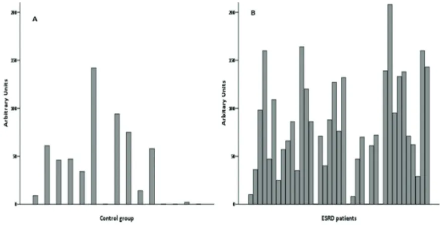
DNA damage presented as AU, for controls and patients, are displayed in Figure 2. We found that DNA damage (AU) was significantly higher in ESRD patients (71 [36–127]), when com-pared to controls (34 [0–61]). A significant increase was also observed for %T in ESRD patients, when compared to controls. We found a %T of 24.2 [17.2–35.4] and 16.8 [10.0–22.2] (expressed in pseudo percentage), and 10.12 [2.27–22.71] and 1.82 [−5.80–7.88] (using the conversion curve) for patients and controls, respectively (Figure 3). The conversion curve provides a better fitting between %T and AU [55, 60] and showed negative values of %T for AU below 26, indicating that %T was zero; above 400 AU, the %T was 84%, in accordance with others [55, 60]. Our data is in accordance with other studies reporting that the levels of DNA breaks and oxidative DNA lesions, measured by the comet assay, are higher in dialysis patients then in controls [77]. We also found that %T was negatively correlated (Spearman’s rank correlation) with CRP (r = −0.368; P = 0.021) and ferritin (r = −0.404; P = 0.011), in ESRD patients; no significant corre-lations were found between DNA lesions and the rhEPO dose used to treat anemia (r = 0.171; P = 0.306), or the time of HD treatment (r = −0.186; P = 0.256). In a cross-sectional study, the oxidative DNA lesions found in dialysis patients were inversely correlated with the duration of the dialysis sessions [77, 78].
Figure 1. Comet images of lymphocytes from end-stage renal disease (ERSD) patients showing different migration
patterns, according to the levels of DNA damage, from class 0 (undamaged) to class 4 (severe damage).
AQ3
Figure 2. DNA damage, presented in arbitrary units, for each of the 15 healthy controls (A) and for the 39 ESRD patients (B). 01 02 03 04 05 06 07 08 09 10 11 12 13 14 15 16 17 18
Figure 3. (A) Mean values in arbitrary units and the estimated percentage of DNA in the tail (E%T) calculated using two
equations: (B) the pseudo percentage score (ps = vs/5 + 10) and (C) the conversion curve = (AU − 25.87)/4.46, in controls and end-stage renal disease (ESRD) patients (differences between groups were tested using Mann-Whitney U test).
01
We did not find significant differences in DNA damage (comet tail length or tail intensity) for diabetics and nondiabetic ESRD patients, as reported by Ersson et al. [77]; however, our find-ings are in accordance with Mamur et al., reporting no difference in comet tail length or tail intensity between diabetic and non-diabetic ESRD patients on HD [78].
Our data suggest that long-term dialysis treatment or diabetes mellitus do not affect DNA damage, however there are still few studies and controversial data. Ersson et al. reported lower levels of DNA damage in salivary gland tissues of ESRD patients, as compared to con-trols, suggesting that ESRD might affect DNA in different ways, in peripheral tissues and in blood mononuclear cells [77].
Concerning cell-free DNA, we found that ESRD patients had a significantly higher value, compared to control; however, after statistical adjustment for age, the significance was lost (Table 2). Cell-free DNA correlated (Spearman’s rank correlation) significantly and positively with age in both groups (r = 0.342, P = 0.033; r = 0.589, P = 0.021; in patients and controls, respectively), and with CRP in ESRD patients (r = 0.483; P = 0.002). Our results are in accordance with others reporting increased levels of cfDNA in hemodia-lyzed patients [79].
To study the predictive risk of mortality associated with DNA damage, we recorded the num-ber of deaths that occurred along 1 year after the analytical study of the 39 ESRD patients; 9 out of the 39 ESRD patients died. We compared the analytical data from ESRD patients who were alive and from the patients who died in the 1 year follow-up period (the Mann-Whitney U test was used). The latter patients presented significantly higher (P = 0.006) cfDNA values (713 [415–809] ng/ml) than those who were still alive (337 [192–484] ng/ml). A trend towards (P = 0.149) higher CRP levels (7.5 [1.6–45.7] mg/l) in those who died, compared to those who were still alive (2.7 [1.7–9.3] mg/l), was also found.
The differences in DNA damage, observed between controls and ERSD patients, could be higher, if we were able to gather a gender matched population. It is known that DNA lesions are higher in women, both in healthy [43, 80–82] and in pathological conditions [83]. Divergent results have been reported for the levels of DNA damage and the time of dialysis treatment. Some studies showed a reduction of DNA damage on long-term maintenance HD [84, 85], while others showed an increase [8, 86, 87]. Recently, it was reported that online hemodiafiltration (OL-HDF) reduced the levels of DNA damage in these patients, as this approach provides a reduction of inflammation and oxidative stress [10]. In fact, a reduc-tion of binucleated cells with micronuclei in patients that changed from low-flux HD to post-dilution OL-HDF, as well as an increase in plasma antioxidant capacity, were shown [88]. Both single high-flux HD and OL-HDF remove circulating mitochondrial DNA, a pro-inflammatory agent, which has been correlated with the chronic pro-inflammatory grade of hemodialyzed patients [89]. Moreover, OL-HDF procedure has been associated with lower levels of the inflammatory markers, IL-6 and CRP, and with an improvement on endothe-lial (dys)function, in ESRD patients [90, 91]. Aberrant DNA hypermethylation has been also observed in dialysis patients and associated with the inflammatory state and with the
01 02 03 04 05 06 07 08 09 10 11 12 13 14 15 16 17 18 19 20 21 22 23 24 25 26 27 28 29 30 31 32 33 34 35 36 37 38 39 40
dialysis technique; patients under OL-HDF showed lower DNA methylation patterns than patients under HD, although higher than controls, suggesting a reduction in DNA hyper-methylation, with decreasing inflammation [92].
Dietary supplementation with folic acid [87, 93], vitamins A, B and B12 [93], zinc [94] and sele-nium [87] may also contribute to reduce/avoid genomic damage, once nutritional supplemen-tation has antioxidant effects, prevents cancer, increases DNA repair capacity, and improves CV and all-cause mortality rates [87].
The inverse correlation that we observed between %T and CRP in ESRD patients, suggests that as CRP (inflammation) levels increase, the damage in DNA also increases; however, it seems that for lower CRP values the damaged DNA is still within the cell, while at higher CRP values the increasing damaged DNA is released into plasma.
The increase of cfDNA in ESRD patients was also reported by others [5, 65, 66, 70, 79]. The slightly lower cfDNA values found in our study, compared with those found by others in HD patients [5], may be related with time of sample collection, as the levels of cfDNA increase during and after HD, returning to pre-HD levels half an hour post-HD [95].
We should notice that our study has some limitations, namely, the small sample size, the lack of age and gender matched controls. Thus, further studies in larger populations are needed to strengthen the value of cfDNA as a biomarker of inflammation and poor out-come in ESRD patients. A recent study showed that circulating free DNA, by favoring calcium phosphate precipitation and crystallization, may be involved in arterial calcifica-tion [96], a common feature in ESRD patients under HD. Thus, cfDNA, appears to be a biomarker for CVD risk, and a direct contributor for CV events, the main cause of death in ERSD patients.
5. Conclusions remarks
ESRD is characterized by a low-grade chronic inflammatory state, which favors the develop-ment of comorbidities. Genetic damage has been reported in ESRD patients, especially in those under HD. The higher degree of genomic damage in ESRD patients might be a consequence of inflammation and aging, and may contribute to increase the risk for cancer and cardiovascu-lar mortality. Several associations with DNA damage (evaluated by cfDNA and comet assay) have been reported and support this hypothesis; however, data is limited and controversial (Table 3).
Our studies showed that cell damaged DNA is increased in ESRD patients, and suggest that at lower CRP values the damaged DNA remains within the cell, while at higher CRP values damaged DNA is released into plasma and may contribute to further enhance inflammation in ESRD patients and increase mortality risk. Actually, we found that ESRD patients who died within the one follow-up period of the study, presented higher circulating damaged DNA
01 02 03 04 05 06 07 08 09 10 11 12 13 14 15 16 17 18 19 20 21 22 23 24 25 26 27 28 29 30 31 32 33 34 35 36
and inflammation. Moreover, our data suggest that the comet assay is more sensitive for low grade inflammatory conditions, while cfDNA appears as a good biomarker for more severe inflammatory conditions, as well as a biomarker for the outcome of ESRD patients. In sum-mary, the genomic damage in ESRD patients seems to result, at least in part, from inflamma-tion and aging, and may contribute to increase the risk for cancer and CV mortality.
Acknowledgements
This work received financial support from FCT/MEC through national funds and was co-financed by FEDER, under the Partnership Agreement PT2020 from UCBIO (UID/MULTI/04378/2013— POCI/01/0145/FEDER/007728) and North Portugal Regional Coordination and Development Commission (CCDR-N)/NORTE2020/Portugal 2020 (Norte-01-0145-FEDER-000024).
Comet assay
Positive association Negative association No association
Male gender [9] CRP* Gender [78]
Diabetes [9] Ferritin* Diabetes [78]*
Mortality [8, 97] Dialysis sessions duration [77] Duration of HD [78]*
Frequency of micronuclei [98] Ferritin [78] BMI > 25 kg/m2 [78] Age [78]
Intact PTH > 300 pg/ml [78] Hb [78] Leptin [99] Hypertension [78] Treatment modality [9] rhEPO dose*
cfDNA
Positive association No association
Age* TNF-α [70]
CRP [66]* IL-10 [70]
IL-6 [66, 70] Dialysis duration [100] All-cause mortality (post-dialysis) [5] WBC count (before HD) [100] Last 3-month mean: SBP, WBC, serum albumin, Cr, normalized protein catabolic
rate [101] Length of the HD session [95] In HD diabetic patients: SBP, Hb A1c, and serum albumin [101]
BMI, body mass index; Cr, creatinine; CRP, C-reactive protein; Hb, hemoglobin; IL, interleukin; PTH, parathyroid hormone; rhEPO, recombinant human erythropoietin; SBP, systolic blood pressure; TNF, tumor necrosis factor; WBC, white blood cell.
*According to our data.
Table 3. Associations reported for comet assay and cell-free (cf) DNA on hemodialysis (HD) patients [96–101]. 01 02 03 04 05 06 07 08 09 10 11
Author details
Susana Coimbra1,2, Alice Santos-Silva3*, Elísio Costa3 and Elsa Bronze-da-Rocha3
*Address all correspondence to: [email protected]
1 CESPU, Institute of Research and Advanced Training in Health Sciences and Technologies (IINFACTS), Gandra-PRD, Portugal
2 UCIBIO\REQUIMTE, Porto, Portugal
3 UCIBIO\REQUIMTE, Department of Biological Sciences, Laboratory of Biochemistry, Faculty of Pharmacy, University of Porto, Porto, Portugal
References
[1] Stevens PE, Levin A. Evaluation and management of chronic kidney disease: Synopsis of the kidney disease: Improving global outcomes 2012 clinical practice guideline. Annals of Internal Medicine. 2013;158:825-830
[2] Muslimovic A, Rasic S, Tulumovic D, Hasanspahic S, Rebic D. Inflammatory markers and procoagulants in chronic renal disease stages 1-4. Medical Archives. 2015;69:307-310 [3] do Sameiro-Faria M, Ribeiro S, Costa E, et al. Risk factors for mortality in hemodialysis
patients: Two-year follow-up study. Disease Markers. 2013;35:791-798
[4] Schupp N, Stopper H, Heidland A. DNA damage in chronic kidney disease: Evaluation of clinical biomarkers. Oxidative Medicine and Cellular Longevity. 2016;2016:3592042 [5] Tovbin D, Novack V, Wiessman MP, Abd Elkadir A, Zlotnik M, Douvdevani A.Circu-
lating cell-free DNA in hemodialysis patients predicts mortality. Nephrology, Dialysis, Transplantation. 2012;27:3929-3935
[6] Jeong JC, Kim JE, JY G, et al. Significance of the DNA-histone complex level as a predic-tor of major adverse cardiovascular events in hemodialysis patients: The effect of uremic toxin on DNA-histone complex formation. Blood Purification. 2016;41:64-71
[7] Sandoval SB, Pastor S, Stoyanova E, et al. Genomic instability in chronic renal failure patients. Environmental and Molecular Mutagenesis. 2012;53:343-349
[8] Corredor Z, Stoyanova E, Rodriguez-Ribera L, et al. Genomic damage as a biomarker of chronic kidney disease status. Environmental and Molecular Mutagenesis. 2015;56: 301-312
[9] Rangel-Lopez A, Paniagua-Medina ME, Urban-Reyes M, et al. Genetic damage in patients with chronic kidney disease, peritoneal dialysis and haemodialysis: A comparative study. Mutagenesis. 2013;28:219-225 01 02 03 04 05 06 07 08 09 10 11 12 13 14 15 16 17 18 19 20 21 22 23 24 25 26 27 28 29 30 31 32
[10] Corredor Z, Rodriguez-Ribera L, Silva I, et al. Levels of DNA damage in peripheral blood lymphocytes of patients undergoing standard hemodialysis vs on-line hemodi-afiltration: A comet assay investigation. Mutation Research, Genetic Toxicology and Environmental Mutagenesis. 2016;808:1-7
[11] Andrassy KM. Comments on 'KDIGO 2012 Clinical Practice Guideline for the Evaluation and Management of Chronic Kidney Disease'. Kidney International. 2013;84(3):622 [12] Inker LA. Albuminuria: Time to focus on accuracy. American Journal of Kidney Diseases.
2014;63:378-381
[13] Morton RL, Kurella Tamura M, Coast J, Davison SN. Supportive care: Economic consider-ations in advanced kidney disease. Clinical Journal of the American Society of Nephrology. 2016;11:1915-1920
[14] Kidney Disease: Improving Global Outcomes (KDIGO) CKD Work Group. KDIGO 2012 clinical practice guideline for the evaluation and management of chronic kidney disease. Kidney International Supplements. 2013;3:1-150
[15] Kidney Disease Outcomes Quality Initiative (K/DOQI). K/DOQI clinical practice guide-lines on hypertension and antihypertensive agents in chronic kidney disease. American Journal of Kidney Diseases. 2004;43:S1-290
[16] Cao Y, Li W, Yang G, Liu Y, Li X. Diabetes and hypertension have become leading causes of CKD in Chinese elderly patients: A comparison between 1990-1991 and 2009-2010. International Urology and Nephrology. 2012;44:1269-1276
[17] Levey AS, Coresh J. Chronic kidney disease. Lancet. 2012;379:165-180
[18] Levey AS, Eckardt KU, Tsukamoto Y, et al. Definition and classification of chronic kid-ney disease: A position statement from Kidkid-ney Disease: Improving Global Outcomes (KDIGO). Kidney International. 2005;67:2089-2100
[19] Vogelzang JL, van Stralen KJ, Noordzij M, et al. Mortality from infections and malig-nancies in patients treated with renal replacement therapy: Data from the ERA-EDTA registry. Nephrology, Dialysis, Transplantation. 2015;30:1028-1037
[20] Eknoyan G, Beck GJ, Cheung AK, et al. Effect of dialysis dose and membrane flux in maintenance hemodialysis. The New England Journal of Medicine. 2002;347:2010-2019 [21] Paniagua R, Amato D, Vonesh E, et al. Effects of increased peritoneal clearances on
mor-tality rates in peritoneal dialysis: ADEMEX, a prospective, randomized, controlled trial. Journal of the American Society of Nephrology. 2002;13:1307-1320
[22] Foley RN, Parfrey PS, Sarnak MJ. Clinical epidemiology of cardiovascular disease in chronic renal disease. American Journal of Kidney Diseases. 1998;32:S112-S119 [23] Tsai WC, HY W, Peng YS, et al. Risk factors for development and progression of chronic
kidney disease: A systematic review and exploratory meta-analysis. Medicine (Baltimore). 2016;95:e3013 01 02 03 04 05 06 07 08 09 10 11 12 13 14 15 16 17 18 19 20 21 22 23 24 25 26 27 28 29 30 31 32 33 34 35 36 37
[24] Webster AC, Nagler EV, Morton RL, Masson P. Chronic kidney disease. Lancet. 2016;
389:1238-1252
[25] Blacher J, Guerin AP, Pannier B, Marchais SJ, London GM. Arterial calcifications, arte-rial stiffness, and cardiovascular risk in end-stage renal disease. Hypertension. 2001;
38:938-942
[26] Carrero JJ, Stenvinkel P. Persistent inflammation as a catalyst for other risk factors in chronic kidney disease: A hypothesis proposal. Clinical Journal of the American Society of Nephrology. 2009;4(Suppl 1):S49-S55
[27] Stenvinkel P, Pecoits-Filho R, Lindholm B. Coronary artery disease in end-stage renal disease: No longer a simple plumbing problem. Journal of the American Society of Nephrology. 2003;14:1927-1939
[28] Ketteler M, Bongartz P, Westenfeld R, et al. Association of low fetuin-a (AHSG) concen-trations in serum with cardiovascular mortality in patients on dialysis: A cross-sectional study. Lancet. 2003;361:827-833
[29] Lee BT, Ahmed FA, Hamm LL, et al. Association of C-reactive protein, tumor necro-sis factor-alpha, and interleukin-6 with chronic kidney disease. BMC Nephrology. 2015;16:77
[30] Gupta J, Mitra N, Kanetsky PA, et al. Association between albuminuria, kidney function, and inflammatory biomarker profile in CKD in CRIC. Clinical Journal of the American Society of Nephrology. 2012;7:1938-1946
[31] Stenvinkel P, Ketteler M, Johnson RJ, et al. IL-10, IL-6, and TNF-alpha: Central factors in the altered cytokine network of uremia—The good, the bad, and the ugly. Kidney International. 2005;67:1216-1233
[32] Bazeley J, Bieber B, Li Y, et al. C-reactive protein and prediction of 1-year mortality in prevalent hemodialysis patients. Clinical Journal of the American Society of Nephrology. 2011;6:2452-2461
[33] Honda H, Qureshi AR, Heimburger O, et al. Serum albumin, C-reactive protein, inter-leukin 6, and fetuin a as predictors of malnutrition, cardiovascular disease, and mortal-ity in patients with ESRD. American Journal of Kidney Diseases. 2006;47:139-148 [34] Tripepi G, Mallamaci F, Zoccali C. Inflammation markers, adhesion molecules, and
all-cause and cardiovascular mortality in patients with ESRD: Searching for the best risk marker by multivariate modeling. Journal of the American Society of Nephrology. 2005;16(Suppl 1):S83-S88
[35] Allon M, Depner TA, Radeva M, et al. Impact of dialysis dose and membrane on infection-related hospitalization and death: Results of the HEMO study. Journal of the American Society of Nephrology. 2003;14:1863-1870
[36] Nassar GM. Preventing and treating inflammation: Role of dialysis access management. Seminars in Dialysis. 2013;26:28-30 01 02 03 04 05 06 07 08 09 10 11 12 13 14 15 16 17 18 19 20 21 22 23 24 25 26 27 28 29 30 31 32 33 34 35 36 37 38
[37] Glorieux GL, Dhondt AW, Jacobs P, et al. In vitro study of the potential role of guani-dines in leukocyte functions related to atherogenesis and infection. Kidney International. 2004;65:2184-2192
[38] Wang Y, Chen X, Song Y, Caballero B, Cheskin LJ. Association between obesity and kid-ney disease: A systematic review and meta-analysis. Kidkid-ney International. 2008;73:19-33 [39] Alix PM, Guebre-Egziabher F, Soulage CO. Leptin as an uremic toxin: Deleterious role
of leptin in chronic kidney disease. Biochimie. 2014;105:12-21
[40] Martinez Cantarin MP, Keith SW, Waldman SA, Falkner B. Adiponectin receptor and adiponectin signaling in human tissue among patients with end-stage renal disease. Nephrology, Dialysis, Transplantation. 2014;29:2268-2277
[41] Morena M, Delbosc S, Dupuy AM, Canaud B, Cristol JP. Overproduction of reactive oxy-gen species in end-stage renal disease patients: A potential component of hemodialysis-associated inflammation. Hemodialysis International. 2005;9:37-46
[42] Akchurin OM, Kaskel F. Update on inflammation in chronic kidney disease. Blood Purification. 2015;39:84-92
[43] Ribeiro S, Belo L, Reis F, Santos-Silva A. Iron therapy in chronic kidney disease: Recent changes, benefits and risks. Blood Reviews. 2016;30:65-72
[44] Gungor O, Unal HU, Guclu A, et al. IL-33 and ST2 levels in chronic kidney disease: Associ- ations with inflammation, vascular abnormalities, cardiovascular events, and survival. PLoS One. 2017;12:e0178939
[45] Tucker PS, Scanlan AT, Dalbo VJ. Chronic kidney disease influences multiple systems: Describing the relationship between oxidative stress, inflammation, kidney damage, and concomitant disease. Oxidative Medicine and Cellular Longevity. 2015;2015:806358 [46] Khan Z, Pandey M, Samartha RM. Role of cytogenetic biomarkers in management of
chronic kidney disease patients: A review. International Journal of Health Sciences. 2016;10:576-589
[47] Trachoo O, Assanatham M, Jinawath N, Nongnuch A. Chromosome 20p inverted dupli-cation deletion identified in a Thai female adult with mental retardation, obesity, chronic kidney disease and characteristic facial features. European Journal of Medical Genetics. 2013;56:319-324
[48] Zhou W, Otto EA, Cluckey A, et al. FAN1 mutations cause karyomegalic intersti-tial nephritis, linking chronic kidney failure to defective DNA damage repair. Nature Genetics. 2012;44:910-915
[49] Collins AR. The comet assay for DNA damage and repair: Principles, applications, and limitations. Molecular Biotechnology. 2004;26:249-261
[50] Collins AR. Measuring oxidative damage to DNA and its repair with the comet assay. Biochimica et Biophysica Acta. 2014;1840:794-800
01 02 03 04 05 06 07 08 09 10 11 12 13 14 15 16 17 18 19 20 21 22 23 24 25 26 27 28 29 30 31 32 33 34 35 36 37
[51] Ren N, Atyah M, Chen WY, Zhou CH. The various aspects of genetic and epigenetic toxi-cology: Testing methods and clinical applications. Journal of Translational Medicine. 2017;
15:110
[52] Azqueta A, Gutzkow KB, Brunborg G, Collins AR. Towards a more reliable comet assay: Optimising agarose concentration, unwinding time and electrophoresis conditions. Mutation Research. 2011;724:41-45
[53] Araldi RP, de Melo TC, Mendes TB, et al. Using the comet and micronucleus assays for genotoxicity studies: A review. Biomedicine & Pharmacotherapy. 2015;72:74-82 [54] Collins AR, Oscoz AA, Brunborg G, et al. The comet assay: Topical issues. Mutagenesis.
2008;23:143-151
[55] Moller P. Assessment of reference values for DNA damage detected by the comet assay in human blood cell DNA. Mutation Research. 2006;612:84-104
[56] Moller P, Loft S, Ersson C, Koppen G, Dusinska M, Collins A. On the search for an intel-ligible comet assay descriptor. Frontiers in Genetics. 2014;5:217
[57] Azqueta A, Collins AR. The essential comet assay: A comprehensive guide to measuring DNA damage and repair. Archives of Toxicology. 2013;87:949-968
[58] Azqueta A, Meier S, Priestley C, et al. The influence of scoring method on variability in results obtained with the comet assay. Mutagenesis. 2011;26:393-399
[59] Collins A, Dusinska M, Franklin M, et al. Comet assay in human biomonitoring stud-ies: Reliability, validation, and applications. Environmental and Molecular Mutagenesis. 1997;30:139-146
[60] Garcia O, Romero I, Gonzalez JE, et al. Visual estimation of the percentage of DNA in the tail in the comet assay: Evaluation of different approaches in an intercomparison exercise. Mutation Research. 2011;720:14-21
[61] Gyori BM, Venkatachalam G, Thiagarajan PS, Hsu D, Clement MV. OpenComet: An auto mated tool for comet assay image analysis. Redox Biology. 2014;2:457-465
[62] Ganapathy S, Muraleedharan A, Sathidevi PS, Chand P, Rajkumar RP. CometQ: An automated tool for the detection and quantification of DNA damage using comet assay image analysis. Computer Methods and Programs in Biomedicine. 2016;133:143-154 [63] Albert O, Reintsch WE, Chan P, Robaire B. HT-COMET: A novel automated approach for
high throughput assessment of human sperm chromatin quality. Human Reproduction. 2016;31:938-946
[64] Mondal M, Guo J. Comet-FISH for ultrasensitive strand-specific detection of DNA dam-age in single cells. Methods in Enzymology. 2017;591:83-95
[65] Korabecna M, Pazourkova E, Horinek A, Rocinova K, Tesar V. Cell-free nucleic acids as biomarkers in dialyzed patients. Journal of Nephrology. 2013;26:1001-1008
01 02 03 04 05 06 07 08 09 10 11 12 13 14 15 16 17 18 19 20 21 22 23 24 25 26 27 28 29 30 31 32 33 34 35 36
[66] Kohlova M, Ribeiro S, do Sameiro-Faria M, et al. Circulating cell-free DNA levels in hemodialysis patients and its association with inflammation, iron metabolism, and rhEPO doses. Hemodialysis International 2013; 17: 664-667.
[67] EI Tarhouny SA, Hadhoud KM, Ebrahem MM, Al Azizi NM. Assessment of cell-free DNA with microvascular complication of type II diabetes mellitus, using PCR and ELISA. Nucleosides, Nucleotides & Nucleic Acids. 2010;29:228-236
[68] Tempera I, Cipriani R, Campagna G, et al. Poly(ADP-ribose)polymerase activity is reduced in circulating mononuclear cells from type 2 diabetic patients. Journal of Cellular Physiology. 2005;205:387-392
[69] Szabo C. Roles of poly(ADP-ribose) polymerase activation in the pathogenesis of diabe-tes mellitus and its complications. Pharmacological Research. 2005;52:60-71
[70] Atamaniuk J, Kopecky C, Skoupy S, Saemann MD, Weichhart T. Apoptotic cell-free DNA promotes inflammation in haemodialysis patients. Nephrology, Dialysis, Transplantation. 2012;27:902-905
[71] Small DM, Coombes JS, Bennett N, Johnson DW, Gobe GC. Oxidative stress, anti-oxi-dant therapies and chronic kidney disease. Nephrology (Carlton). 2012;17:311-321 [72] Lisowska-Myjak B. Uremic toxins and their effects on multiple organ systems. Nephron.
Clinical Practice. 2014;128:303-311
[73] Goldshtein H, Hausmann MJ, Douvdevani A. A rapid direct fluorescent assay for cell-free DNA quantification in biological fluids. Annals of Clinical Biochemistry. 2009;46:488-494
[74] Czeiger D, Shaked G, Eini H, et al. Measurement of circulating cell-free DNA levels by a new simple fluorescent test in patients with primary colorectal cancer. American Journal of Clinical Pathology. 2011;135:264-270
[75] Coimbra S, Catarino C, Costa E, et al. Circulating cell-free DNA levels in Portuguese patients with psoriasis vulgaris according to severity and therapy. The British Journal of Dermatology. 2014;170:939-942
[76] Drueke TB, Parfrey PS. Summary of the KDIGO guideline on anemia and comment: Reading between the (guide)line(s). Kidney International. 2012;82:952-960
[77] Ersson C, Odar-Cederlof I, Fehrman-Ekholm I, Moller L. The effects of hemodialysis treatment on the level of DNA strand breaks and oxidative DNA lesions measured by the comet assay. Hemodialysis International. 2013;17:366-373
[78] Mamur S, Unal F, Altok K, Deger SM, Yuzbasioglu D. DNA damage in hemodialysis patients with chronic kidney disease; a test of the role of diabetes mellitus; a comet assay investigation. Mutation Research, Genetic Toxicology and Environmental Mutagenesis. 2016;800-801:22-27 01 02 03 04 05 06 07 08 09 10 11 12 13 14 15 16 17 18 19 20 21 22 23 24 25 26 27 28 29 30 31 32 33 34 35 36
[79] Cichota LC, Bochi GV, Tatsch E, et al. Circulating double-stranded DNA in plasma of Hemodialysis patients and its association with iron stores. Clinical Laboratory. 2015;
61:985-990
[80] Coskun M, Cayir A, Tok H. Evaluation of background DNA damage in a Turkish popula-tion measured by means of the cytokinesis-block micronucleus cytome assay. Mutapopula-tion Research. 2013;757:23-27
[81] Nefic H, Handzic I. The effect of age, sex, and lifestyle factors on micronucleus fre-quency in peripheral blood lymphocytes of the Bosnian population. Mutation Research. 2013;753:1-11
[82] Cho NY, Kim KW, Kim KK. Genomic health status assessed by a cytokinesis-block micronucleus cytome assay in a healthy middle-aged Korean population. Mutation Research. 2017;814:7-13
[83] Kiraz A, Acmaz G, Uysal G, Unal D, Donmez-Altuntas H. Micronucleus testing as a cancer detector: Endometrial hyperplasia to carcinoma. Archives of Gynecology and Obstetrics. 2016;293:1065-1071
[84] Stoyanova E, Sandoval SB, Zuniga LA, et al. Oxidative DNA damage in chronic renal failure patients. Nephrology, Dialysis, Transplantation. 2010;25:879-885
[85] Aykanat B, Demircigil GC, Fidan K, et al. Basal damage and oxidative DNA damage in children with chronic kidney disease measured by use of the comet assay. Mutation Research. 2011;725:22-28
[86] Stopper H, Boullay F, Heidland A, Vienken J, Bahner U. Comet-assay analysis identi-fies genomic damage in lymphocytes of uremic patients. American Journal of Kidney Diseases. 2001;38:296-301
[87] Zachara BA, Gromadzinska J, Palus J, et al. The effect of selenium supplementation in the prevention of DNA damage in white blood cells of hemodialyzed patients: A pilot study. Biological Trace Element Research. 2011;142:274-283
[88] Rodriguez-Ribera L, Pastor S, Corredor Z, et al. Genetic damage in patients moving from hemodialysis to online hemodiafiltration. Mutagenesis. 2016;31:131-135
[89] Cao H, Ye H, Sun Z, et al. Circulatory mitochondrial DNA is a pro-inflammatory agent in maintenance hemodialysis patients. PLoS One. 2014;9:e113179
[90] den Hoedt CH, Bots ML, Grooteman MP, et al. Online hemodiafiltration reduces systemic inflammation compared to low-flux hemodialysis. Kidney International. 2014;86:423-432 [91] Jia P, Jin W, Teng J, et al. Acute effects of hemodiafiltration versus conventional Hemo-dialysis on endothelial function and inflammation: A randomized crossover study. Medicine (Baltimore). 2016;95:e3440
[92] Ghigolea AB, Moldovan RA, Gherman-Caprioara M. DNA methylation: Hemodialysis versus hemodiafiltration. Therapeutic Apheresis and Dialysis. 2015;19:119-124
01 02 03 04 05 06 07 08 09 10 11 12 13 14 15 16 17 18 19 20 21 22 23 24 25 26 27 28 29 30 31 32 33 34 35 36 37
[93] Stopper H, Treutlein AT, Bahner U, et al. Reduction of the genomic damage level in haemodialysis patients by folic acid and vitamin B12 supplementation. Nephrology, Dialysis, Transplantation. 2008;23:3272-3279
[94] Guo D, Bi H, Wang D, Wu Q. Zinc oxide nanoparticles decrease the expression and activity of plasma membrane calcium ATPase, disrupt the intracellular calcium homeo-stasis in rat retinal ganglion cells. The International Journal of Biochemistry & Cell Biology. 2013;45:1849-1859
[95] Garcia Moreira V, de la Cera Martinez T, Gago Gonzalez E, Prieto Garcia B, Alvarez Menendez FV. Increase in and clearance of cell-free plasma DNA in hemodialysis quan-tified by real-time PCR. Clinical Chemistry and Laboratory Medicine. 2006;44:1410-1415 [96] Coscas R, Bensussan M, Jacob MP, et al. Free DNA precipitates calcium phosphate
apa-tite crystals in the arterial wall in vivo. Atherosclerosis. 2017;259:60-67
[97] Coll E, Stoyanova E, Rodriguez-Ribera L, et al. Genomic damage as an independent pre-dictor marker of mortality in hemodialysis patients. Clinical Nephrology. 2013;80:81-87 [98] Palazzo RP, Bagatini PB, Schefer PB, de Andrade FM, Maluf SW. Genomic instabil-ity in patients with type 2 diabetes mellitus on hemodialysis. Revista Brasileira de Hematologia e Hemoterapia. 2012;34:31-35
[99] Horoz M, Bolukbas FF, Bolukbas C, et al. The association of circulating leptin level with peripheral DNA damage in hemodialysis subjects. Clinical Biochemistry. 2006;
39:918-922
[100] Opatrna S, Wirth J, Korabecna M, Sefrna F. Cell-free plasma DNA during hemodialysis. Renal Failure. 2009;31:475-480
[101] Jeong DW, Moon JY, Choi YW, et al. Effect of blood pressure and glycemic control on the plasma cell-free DNA in hemodialysis patients. Kidney Research and Clinical Practice. 2015;34:201-206 01 02 03 04 05 06 07 08 09 10 11 12 13 14 15 16 17 18 19 20 21 22 23 24 25

![Table 3. Associations reported for comet assay and cell-free (cf) DNA on hemodialysis (HD) patients [96–101].](https://thumb-eu.123doks.com/thumbv2/123dok_br/15806551.1079935/15.722.119.579.117.476/table-associations-reported-comet-assay-cell-hemodialysis-patients.webp)
