UNIVERSIDADE DE LISBOA
FACULDADE DE CIÊNCIAS
DEPARTAMENTO DE BIOLOGIA ANIMAL
The relationship of ERG Potassium Channel with
programmed cell death in the vertebrate developing
limb
Rita Leiria Lopes Félix
Mestrado em Biologia Evolutiva e do Desenvolvimento
2010
UNIVERSIDADE DE LISBOA
FACULDADE DE CIÊNCIAS
DEPARTAMENTO DE BIOLOGIA ANIMAL
The relationship of ERG Potassium Channel with
programmed cell death in the vertebrate developing
limb
Rita Leiria Lopes Félix
Dissertação orientada por:
Professor Doutor Joaquín Rodríguez-León Professora Doutora Gabriela Rodrigues
Mestrado em Biologia Evolutiva e do Desenvolvimento
2010
I
Acknowledgments
Muitas são as pessoas a que tornaram a realização desta tese possível (até ao último minuto!) e a quem tenho de agradecer.
Um muito obrigada ao Joaquín! Sem ele toda esta experiência não poderia ter acontecido. Obrigada por toda a disponibilidade e conhecimentos transmitidos, sempre com muito humor e boa disposição! Obrigada por tudo, foste um chefe impecável!
À Professora Gabriela que aceitou ser minha orientadora interna e sempre demonstrou interesse pelo meu trabalho, estando sempre disponível para qualquer dúvida ou problema.
Ao Instituto Gulbenkian de Ciência, uma instituição com excelentes condições e espírito científico. À Catarina, pelo erg e todo o apoio nesta jornada! À Raquel, sempre incansável e paciente a ensinar e a aturar maluqueiras, mesmo fora de horas…
À Rita, que merece mais que duas páginas de agradecimentos por toda a ajuda ao longo do trabalho! À Joana e as suas manias, à Teresa, à Diana e ao Nando por toda a companhia!
Ao grupo do Moisés, que também sinto ser um pouco meu, em particular à Isabel, Sofia e Arnon, com quem a vida no laboratório não teria sido tão divertida.
Aos amigos e à família, em especial à mãe pelo apoio incondicional e “mãetrocínio” ao longo de todo este percurso!
III
Abstract
A proper rate of programmed cell death (apoptosis) is vital to maintain normal tissue homeostasis, limit organ growth, eliminate unnecessary cells and undergo normal development. During limb development the apoptotic areas are controlled by a tight interaction between different genes that are responsible for the specific localization of this domains in the limb bud. In here we show that ERG, an outward potassium channel belonging to the ether-a-go-go family of voltage-gated potassium channels, is expressed, in proper time and space, in the apoptotic areas. We have studied the relevance of erg during the apoptotic process during the formation of digits in species with a different pattern of interdigital cell death, namely duck and chicken. We have performed loss-of-function experiments through in ovo electroporation of a construct with specific RNAi against erg and by implantation of beads soaked in an ERG inhibitor. The phenotypes obtained show a decrease in apoptosis in the interdigital space. Moreover, inhibition of erg action is followed by downregulation of different molecules involved in the apoptotic process. We have also unraveled the relationship of erg with different signaling pathways known to be involved in the triggering of cell death during limb development like FGFs, BMPs and Retinoic Acid. Most importantly, our findings show that erg is a key player in the establishment of interdigital cell death during digit development. Our results support a model in which erg is responsible for detachment
V
Resumo
Uma taxa de morte celular programada (apoptose) adequada é vital para a manutenção da homeostase dos tecidos, limitação do crescimento celular, eliminação de células indesejadas e para o decorrer normal do desenvolvimento no organismo. O desenvolvimento do membro é uma situação paradigmática em que a morte celular programada tem um papel central na morfogénese (Saunders, 1966). Desta forma, o membro de vertebrados é um dos melhores modelos para estudar o mecanismo apoptótico.
Durante o desenvolvimento dos vertebrados, os primórdios das extremidades apresentam áreas específicas de morte celular programada. As zonas necróticas anterior e posterior (ANZ e PNZ) são responsáveis por definir a forma mais proximal da extremidade do membro - o estilopódio e o zeugopódio. Já o opaque-patch, na parte central do mesênquima, vai definir os elementos esqueléticos do zeugopódio, o rádio e a ulna. Por último, as áreas necróticas interdigitais (INZs) vão esculpir a forma final dos dedos (Zuzarte and Hurlé, 2005). O controlo e localização específica destas áreas de morte celular programada é feito através de uma delicada interacção entre diferentes genes durante o desenvolvimento do membro.
Das moléculas sinalizadoras que controlam a indução da apoptose, destacam-se as da família dos BMPs e FGFs (Ganan et al., 1996, Montero et al., 2001). De facto, para que a indução da morte celular programada seja eficaz, estas duas vias de sinalização devem actuar em conjunto (Montero et al., 2001). A actividade conjunta de BMPs e FGFs resulta na activação da expressão de genes específicos que são responsáveis, inicialmente, pela perda de comunicação celular no mesênquima, o que constitui um processo crucial para que a apoptose tenha lugar (Hurle and Fernandez-Teran, 1983; Martin-Bermudo et al., 1998). No final, a cascata das caspases é activada, o que culmina com a destruição controlada da célula (Zuzarte-Luis et al., 2004).
A morte celular programada partilha muitos mecanismos com outros processos celulares como a proliferação e diferenciação. No entanto, a resposta à mesma proteína pode resultar em diferentes efeitos, dependo do contexto celular. No mesênquima da extremidade do membro observa-se que os BMPs podem induzir tanto diferenciação condrogénica como morte celular. De forma semelhante, os FGFs são, inicialmente, responsáveis pela sobrevivência da célula enquanto que, mais tarde, induzem a sua apoptose (Ganan et al., 1996; Montero et al., 2001). Para além das numerosas cascatas genéticas que se sabem estar envolvidas na indução e desenvolvimento do membro de vertebrados existem também mecanismos epigenéticos que influenciam a padronização do mesmo. Desta forma, fecha-se o ciclo entre informação genética e o ambiente celular. Entre estes mecanismos epigenéticos destacam-se os fluxos iónicos (Altizer et al., 2001). Estes têm sido implicados em diferentes processos tais como a migração, morte e diferenciação celulares (Morokuma et al., 2008). Foram também observados durante o desenvolvimento embrionário, onde são responsáveis, por exemplo, pela correcta formação das extremidades (Altizer et al., 2001).
VI A acção de moléculas sinalizadoras na indução da apoptose tem sido principalmente estudada nas INZs. Resumidamente, a actividade do Ácido Retinóico activa a expressão de BMPs no espaço interdigital o que, em conjunto com a expressão de FGFs já existente nessa zona, induz a apoptose no interdígito (Rodriguez-Leon et al., 1999; Montero et al., 2001). No entanto, esta indução de morte celular programada deve ser precedida pela inibição da expressão de FGF8 na ectoderme distal do interdígito (Hernandez-Martinez et al., 2009). No final, a confluência de vários mecanismos nessas zonas resulta numa morte celular em massa nas áreas do interdígito.
Este trabalho procura mostrar a existência de um elo de ligação entre a actividade de ERG, um canal de saída de potássio, e a morte celular programada durante o desenvolvimento dos membros. ERG pertence à família ether-a-go-go de canais de potássio dependentes de voltagem, e é detectado nas áreas apoptóticas do autopódio de galinha (Gallus gallus) e pato (Anas platyrhynchos). O facto de estar presente em todo o espaço interdigital de espécies com dedos livres (galinha) e só na parte mais distal do interdígito em pato onde a morte fisiológica ocorre, sugere que a sua função pode estar conservada evolutivamente. O padrão de expressão deste canal de potássio é detectado nas INZs, de forma espacial e temporalmente conservada nos dois organismos.
Para analisar a função de ERG, realizaram-se estudos de perda de função através da electroporação in ovo de uma construção de RNAi específico para o gene em estudo. Outra abordagem usada para o mesmo fim foi a implantação de microesferas embebidas num inibidor químico do canal, de forma a possível bloquear funcionalmente a dinâmica de potássio intracelular no membro em desenvolvimento. A perda de função de ERG conduz a uma diminuição da morte celular programada nas áreas interdigitais, o que resulta em fenótipos de sindactilia (os dedos permanecem visivelmente unidos por uma membrana). A geração deste fenótipo é precedida pela inibição da expressão de genes envolvidos no processo apoptótico, tais como snail, msx2, bambi e fgf10, o que mostra que a actividade de ERG está directamente ligada à transdução de sinal nas vias de BMPs e FGFs. Assim, a acção de ERG é imprescindível no desencadear do processo apoptótico desde estádios muito iniciais da indução da morte celular.
A relação de erg com as diferentes vias de sinalização génica envolvidas no desencadeamento do processo apoptótico - tais como a de FGFs, BMPs e Ácido Retinoico (RA) - foi também clarificada. Experiências de ganho de função utilizando microesferas embebidas nestas moléculas mostram que estas três vias de sinalização regulam a expressão de erg. Em particular, a sinalização por BMPs activa a expressão do mesmo. Pelo contrário, o bloqueio destas moléculas não resulta na inibição de erg. Estes dados sugerem que adicionalmente à sinalização via BMPs, existirá uma outra cascata genética a induzir e/ou manter a expressão de erg. Mas, mais importante, este estudo mostra que erg é um intermediário fundamental no estabelecimento das áreas de morte interdigital durante o desenvolvimento dos dedos, precedendo o começo da apoptose. Em conjunto, os resultados deste trabalho apoiam um modelo no qual ERG é responsável pelo destacamento celular e consequente indução de morte celular programada nas áreas interdigitais.
VII Para além do controlo do destacamento celular através da activação do gene snail, uma outra função essencial do ERG na apoptose poderá ser a inactivação da expressão de fgf8 na ectoderme distal do interdígito, dado que a inibição da actividade do ERG leva à manutenção da expressão do fgf8 distalmente.
No entanto, é necessária a realização de mais experiências para clarificar, nomeadamente, a indução da expressão de erg através de BMPs e Ácido Retinóico. Uma possível abordagem seria a inibição conjunta de um ou vários elementos destas vias de sinalização de forma a esclarecer quais os elementos que actuam de forma conjunta para a indução e/ou manutenção da expressão de erg. Seria também importante melhorar a electroporação interdigital de RNAi para conseguir a perda de função na maior parte do mesênquima interdigital, tal como a realização de western blots de maneira a comprovar que a proteína não é traduzida. Ao mesmo tempo, seria de grande interesse a clonagem da sequência completa de erg para estudos de sobreexpressão no autopódio, confirmando-se assim se o aumento da sua actividade poderia induzir morte celular ectópica.
Palavras chave:
Canal de potássio ERG, apoptose, desenvolvimento dos membro, sinalização por FGF e BMPI
Table of Contents
Acknowledgments ... I Abstract ... III Resumo ... V I. Introduction ... 1Vertebrate limb development ... 1
Digit development ... 2
Molecular regulation of Interdigital Cell Death ... 4
Electric fields in development ... 7
Potassium channels and cell death ... 7
Biological activity of ERG ether-a-go-go (eag) family of voltage-dependent channels ... 7
Objectives ... 8
II. Materials and Methods ... 10
II.1 Animal models ... 10
II.2 Embryo collection, fixation and storage ... 10
II.3 Experimental manipulation of the limb ... 11
II.3.1 Bead Implantation ... 11
II.3.2 erg RNAi electroporation ... 12
II.4 Riboprobe preparation for whole-mount In situ hybridization ... 14
II.4.3 RNA/DNA precipitation with ethanol ... 16
II.4.4 In vitro DIG-labelled anti-sense RNA probe transcription ... 16
II.5 Whole-mount In situ hybridization ... 17
II.6 Histological analysis ... 18
II.6.1 Tissue processing and gelatin embedding ... 18
II.7 Limb morphological analysis ... 19
II.7.1 Alcian green staining ... 19
II.8 Cell death analysis ... 19
II.8.1 Acridine Orange ... 19
II.8.2 TUNEL analysis of dying cells ... 19
II
II.10 Statistic analysis ... 20
III. Results ... 22
Expression of erg during limb development correlates directly with apoptosis ... 22
Inhibition of erg activity during digit development ... 24
Regulation of erg expression through FGF signaling ... 30
RA signaling regulates erg expression ... 31
Regulation of erg expression by BMP signaling ... 32
IV. Discussion ... 34
erg expression co-localizes with apoptotic zones ... 34
erg gene triggers apoptosis ... 34
erg expression is regulated by apoptotic signals ... 35
ERG potassium channel involvement in cell detachment ... 36
Proposed model for erg activity ... 37
Concluding remarks ... 38
V. References ... 40
Appendix I – Buffers, Solutions and Media ... 45
Appendix II – Plasmid Maps ... 49
Appendix III – erg’s alignment ... 52
Appendix IV – erg cDNA cloned sequence ... 57
Introduction
1
I. Introduction
Vertebrate limb development
The vertebrate limb has been an important developmental model for the study of programmed cell death during embryogenesis since its apoptosis pattern is a consistent component in limb morphogenesis (Zuzarte-Luis and Hurle, 2005).
The early limb primordium of the amniota embryos is a simple structure. It starts as a core of mesenchymal cells encompassed by ectoderm and appears as a bud growing on restricted areas of the lateral plate mesoderm (LPM). These areas, called limb fields, are subjected to the influence of a cross-talk of genetic signaling cascades (wnt and fgf) between the LPM and ectoderm above. Wnt-stabilized FGF10 at LPM, activates FGF8 in the AER progenitors, which results in the establishment of an epithelial–mesenchymal feedback loop between AER-secreted FGF8 and mesenchymal FGF10 (Kawakami el al, 2001). The mesodermal cells of the early limb bud have skeletogenic potencial but are maintained undifferentiated and proliferating by the influence of a thickened region of the ectoderm at the distal margin of the bud, the apical ectodermal ridge (AER) (Kawakami et al., 2001; Montero and Hurle, 2010).
The vertebrate limb bud development is controlled by three signaling centers, the apical ectodermal ridge (AER) a specialized thickened region of the ectoderm at the distal margin of the limb bud, the polarizing region (ZPA) in the posterior mesenchyme and the non-ridge ectoderm. These signaling centers produce instructive signals that coordinately control proximo-distal (PD), anterior-posterior (AP) and dorsal-ventral (DV) limb axis formation. In particular, the ZPA produces Sonic Hedgehog (SHH), the dorsal ectoderm secretes WNT7a (Dealy et al., 1993; Parr et al., 1993), while the AER produces several fibroblast growth factors (FGF8, FGF4, FGF9, FGF17), FGF8 being the one of most importance. These signals from the AER act on the subjacent mesodermal cells that constitute the progress-zone (PZ), keeping them in a proliferative and undetermined state. According to the progress-zone model, the cells identities are determined along the PD axis by a ‘clock-type’ mechanism as they leave the progress zone, so the cell identities depend on the time of exit (Saunders J. W., 1948.; reviewed in (Zeller et al., 2009). Differentiation into cartilage, and cell death, occur when the cells of the PZ become displaced proximally into the core of the bud. More recently, the differentiation-front model assumes that PD identities are determined when the mesenchyme stops being under the AER-FGF signaling domain, when the proliferating mesenchyme leaves the undifferentiated zone (Tabin and Wolpert, 2007). On the other hand, it has been proposed the two-signal model whereas retinoic acid (RA) and FGF two-signaling induce proximal and distal cell fates in the limb bud mesenchyme, respectively. (Mercader et al., 2000). This hypothesis was analyzed during chicken limb bud development and has shown that RA induces proximal cell identity in the limb bud
2 mesenchyme by the induction of meis1 and meis2 expression, whereas AER-FGFs, which antagonize RA signaling, induce distal cell identity by the activation of hoxa11 and hoxa13.
During the progression of limb bud development, the three signaling centers are interlinked by an epithelial-mesenchymal (e-m) feedback loop that functions to propagate both AER-fgf and ZPA-shh expression. This e-m feedback loop is established by transcriptional upregulation of gremlin1 (the bone morphogenetic protein (BMP) antagonist) in a subpopulation of SHH responsive cells located in the dorsal and ventral part of the distal limb bud mesenchyme (Zeller, 2010).
Digit development
During limb bud development, cells that leave the PZ can undergo chondrogenesis and form the cartilage template for the skeleton or can die by apoptosis, defining regions of cell death around these chondrogenic aggregates (reviewed in Montero and Hurlé, 2010).
Physiological cell death is a key mechanism that ensures appropriate development and maintenance of tissues and organs in multicellular organisms. Cell death plays a role in sculpting the tetrapod limb that is crucial for the determination of its final shape and skeletal pattern. The involvement of cell death in vertebrate limb morphogenesis appears to be a characteristic of the amniotes only and exhibit significant differences between species (Hurle et al., 1996; Zuzarte-Luis and Hurle, 2002). The death (or lack of it) of specific cells in the vertebrate limb is genetically programmed and has been selected over the course of evolution (Gilbert, 2006). Cell death occurs in well-defined domains and sculpts the shape of the limb, eliminating the undifferentiated mesodermal cells (Zuzarte-Luis and Hurle, 2005) located between the differentiating cartilages (Hurle et al., 1996). Indeed, cell death is essential if joints are to form and fingers are to become separate (Gilbert, 2006). In the early stages of the avian limb development, the anterior (ANZ) and posterior (PNZ) necrotic zones eliminate the mesodermal cells located anterior and posterior to the zone of formation of the proximal skeletal components of the limb (Figure 1) (Montero et al., 2001). Another area of cell death is the opaque patch (OP) and takes place in the central mesenchyme of the limb, between the zeugopodial cartilages (Zuzarte-Luis and Hurle, 2002) (Figure 1). At more advanced stages of development, elimination of interdigital cells between the developing digits occurs in the interdigital necrotic zones (INZ) (Montero et al., 2001). In species with webbed digits, such the duck, apoptosis is limited to the distal part of the interdigit. Also, in species with autopods of singular morphology, for example the coot (Fulika atra) which have digits with lateral membranous lobulations or the splitted autopod of the chameleons, the pattern of interdigital cell death correlates closely with the specific phenotype of each species (Zuzarte-Luis et al., 2004). Although these zones are referred to as ‘necrotic’, this term is a holdover from the days when no distinction was made between necrotic death and apoptotic cell death (Gilbert, 2006).
Individualization of the digits is progressively achieved partly through the morphogenetic role of interdigital cell death (ICD) (Saunders and Gasseling, 1962). The current model supposes the
Introduction
3 formation of an interdigital tissue that subsequently degenerates and, as a result, digits individualize. Under this model, ICD would restrict the growth of the interdigital tissue. However, sculpturing the limb by interdigital tissue removal (as occurs in reptiles, birds, and mammals) is not the only way of making free digits, as exemplified by the amphibian forelimb, where the digits form by differential growth without participation of cell death (e.g., as demonstrated in Xenopus by (Fallon and Cameron, 1977)). In amniotes, differential growth also participates in the separation of the digits (Salas-Vidal et al., 2001). Therefore, both cell death and cell proliferation are crucial processes during the morphogenesis of the digits (Fernandez-Teran et al., 2006).
Figure 1 – Representation of the anterior necrotic zone (ANZ), posterior necrotic zone (PNZ), opaque patch (OP) and interdigital necrotic zone (INZ). Courtesy of Doctor Joaquín León.
In the chicken embryo, proper interdigital cell death at the INZ is first detected at stage 29–30HH (Hamburger and Hamilton, 1951) in the proximal part of the first, second, and third interdigital spaces, then in its distal part. Proximal and distal clusters of cell death in the interdigital spaces are first separated but later joined together and the whole interdigit exhibits massive cell death (Pautou, 1974). The INZ reaches its maximum at stage 31HH (day 7 and a half of incubation) and subsequently progressively declines (Fernandez-Teran et al., 2006).
Several experimental approaches have shown that the interdigital mesoderm contains cells with chondrogenic potential and when cell death is inhibited and chondrogenesis is activated, they are able to form an extra digit (Merino et al., 1999a).
The tissue components of the interdigit establish complex interactions through the production of a variety of secreted signaling molecules that control cell fate and tissue differentiation. Several molecules have been indentified that participate in the regulation of ICD; however, very little is known about their mechanism of action and how they interact and contribute to trigger interdigital cell death (Hernandez-Martinez et al., 2009).
4
Molecular regulation of Interdigital Cell Death
BMP, FGF, RA and Wnt signaling
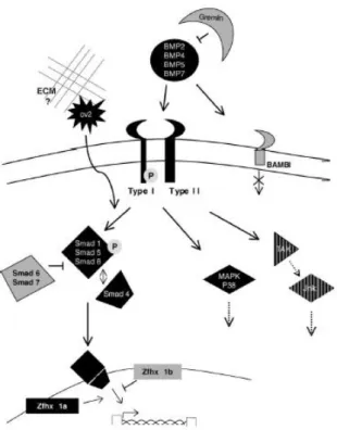
Programmed cell death during limb development shares many control mechanisms with proliferation and differenciation. Depending on mesodermal cells differentiation status, the response to the same protein may have opposite effects. The bone morphogenetic proteins (BMPs), members of the transforming growth factor superfamily, known to be involved in chondrogenesis were identified as the triggering apoptotic signals for both the ectoderm of the AER and mesodermal cells. The undifferentiated limb mesoderm undergoes apoptosis when is exposed to BMPs, but if the cells have initiated aggregation into the prechondrogenic blastemas, BMPs induce chondrogenesis (Ganan et al., 1996; Montero et al., 2001). It is interesting that factors responsible for the growth and differentiation of the skeleton are also the triggers for the apoptotic process (Zuzarte-Luis and Hurle, 2005). Several members of the BMP family (BMP-2, BMP-4, BMP-5 and BMP-7) are expressed in the undifferentiated limb mesoderm, AER and in the interdigital mesenchyme, coincident with apoptotic areas (Merino et al., 1999b). BMPs exert their function through serine/threonine receptor kinases composed of type I and type II receptors. The type IA and IB receptors mediate the chondrogenic effect of BMPs while the receptor implicated in the control of apoptosis awaits clarification (Figure 2) (Zuzarte and Hurle, 2005). There are at least two BMP intracellular signaling pathways through which BMPs exercise their apoptotic effect. Ligand binding of BMPs to their receptors activates members of the SMAD family. SMAD 1, 5 and 8 are phosphorylated coassembled with a cofactor, SMAD 4, and translocated into the nucleus where they activate gene transcription. The other pathway involved in apoptosis is the mitogen activated protein kinase (MAPK) pathway (Figure 2) (Montero et al, 2001; Zuzarte-Luis and Hurle, 2005). BMP activity is spatially and temporally fine-tuned by other factors during limb development such as antagonists of BMP function like Gremlin, Chordin and Noggin (Capdevila et al., 1999; Francis-West et al., 1999; Merino et al., 1998; Merino et al., 1999b). These antagonists share the functional property of binding specifically to BMPs, preventing their interaction with their receptors. This way, the function of BMPs is regulated specifically by different BMP antagonists that act in a complementary fashion rather than being redundant signals (Merino et al., 1999b). Gremlin for instance, is very important in limb patterning since it moderates BMP inhibition of FGFs, allowing the maintenance of the signaling loop between the ZPA (shh) and the AER (fgf) (fgf-shh feedback loop) (Khokha et al., 2003). Gremlin also regulates the regression of the interdigital tissue in the duck since it is expressed in a pattern covering the whole interdigital space except the most distal mesenchyme where cells will die by apoptosis (Merino et al., 1999b).
Introduction
5
Figure 2 – Schematic representation of BMP signaling pathway involved in interdigital cell death triggering. Inhibitory
molecules are colored in grey. Active signaling molecules are colored in black (From Zuzarte-Luis et al., 2005).
In this context, Montero and colleagues (2001) showed that BMPs alone are not sufficient to trigger the apoptotic process during limb development, since blockage of FGF signaling through application of its inhibitor SU5402 inhibited physiological and exogenous BMP mediated apoptosis. Delivery of exogenous FGFs strongly increased cell death 24 hours after the application. This shows that FGF signaling is also necessary for apoptosis and that the establishment of the areas of cell death is regulated by the convergence of FGF and BMP-mediated signaling pathways (Montero et al., 2001). It has been shown by Hernández-Martínez and colleagues that growth to form mouse and chick interdigital tissue occurs within a 12 hour time window. It has been proposed that most interdigital cells derive from the distal mesenchyme, which survival depends on ectodermal FGF8, and are the ones fated to die. According to this model, cell death is initiated in the distal region and as the limb grows and interdigital regression occurs, the dying cells acquire a more proximal position. This explains a progressive process rather than massive mechanism of cell death, through which mouse and distal chick ICD occurs. Chick’s most proximal ICD appears to follow a different mechanism dependent on interdigital BMP activity. Recently, it has been showed that modulation of Wnt/β-catenin signal in the limb ectoderm including the AER regulates interdigital apoptosis. They also demonstrated that ectodermal Wnt/β-catenin signaling can positively regulate fgf8 possibly antagonizing the BMP signaling within the AER (Villacorte et al., 2010).
6
Caspase activation
Apoptosis takes place by the action of cysteine-aspartic acid proteases, usually called caspases. There are at least two known pathways responsible for caspase activation, proteolysis and DNA fragmentation, and consequent apoptosis: the extrinsic pathway (death receptor pathway) and the intrinsic pathway (mitochondrial pathway). The extrinsic pathway requires binding of ligands (like CD95, TNF-α or Fas-ligand) to specific transmembrane receptors belonging to the superfamily of the tumor necrosis factor receptors (TNF-receptors), followed by the activation of caspase 8 (and/or caspase 10). The intrinsic pathway is triggered by modifications in the permeability of the mitochondrial membrane followed by the release of cytochrome c from de mitochondrial matrix into the cytosol. The apoptosome is the central element of the apoptotic machinery of the intrinsic pathway and is constituted by the Apaf-1 together with caspase 9 and cytochrome c. Both pathways involve the activation of initiator caspases (caspase 8 and caspase 9 specific of the extrinsic and intrinsic pathway respectively) by proteolytic cleavage of their pro-domain. After several intermediate steps, the two pathways converge in the activation of the effector caspase 3, caspase 6, and caspase 7, which are proteases responsible for cell degradation. Still, the pathways are also not mutually exclusive of each other, they can be interconnected at different levels. For example, the activation of caspase 8 in the death receptor pathway may induce the formation of proapoptotic proteins of the Bcl-2 family (Bax, Bag, Bak, Bcl-xs), which translocate to the outer mitochondrial membrane and facilitate the extrusion of cytochrome c, aggravating the apoptosis process. The apoptosis-inducing factor (AIF), a factor that is also released from the mitochondria, can also trigger apoptosis without caspases involvement (Burg et al., 2006; Zuzarte-Luis and Hurle, 2005). Also, it has been reported that even when caspases are fully inhibited, interdigital cell death in mouse embryos still occurs by a caspase-independent pathway (Chautan et al., 1999). Caspase 3 has been proposed to be the major effector caspase during physiological cell death in the developing limb. Even though, when the limb bud is treated with specific caspase 3 inhibitor peptides, apoptosis is only reduced and not blocked (Huang and Hales, 2002). Which of the pathways operates in the activation of caspase 3 by the dying cells of the developing limb is still a matter of debate (Zuzarte-Luis and Hurle, 2005). The molecular cascade responsible for the execution of apoptosis remains largely unknown (Zuzarte-Luis and Hurle, 2005). Besides the morphological features of apoptotic cells, apoptotic volume decrease (AVD) due to efflux of K+, Cl-, and H2O is an early hallmark of apoptosis that occurs within the 1st and
4th hour after apoptosis induction (Burg et al., 2006). This corresponds to the initial phase of AVD that takes place before the activation of the apoptosome. The late phase of AVD includes the subsequent caspase activation that leads to DNA fragmentation (Burg et al., 2006).
Introduction
7
Electric fields in development
In addition to the numerous genetic cascades known to be involved in the induction and outgrowth of the vertebrate limb, there are epigenetic mechanisms that influence the pattern, closing the loop between the nucleus and its environment. One of the epigenetic mechanisms suggested for limb bud initiation imply an endogenous electric field. Altizer and colleagues (2001) found an outwardly directed, steady, ionic current before and during the emergence of the mouse and chick limb bud. On the other hand, in the flank regions in the dorsal-ventral plane of the limb bud, the currents were usually inwardly directed (the direction of Na+ uptake by ectoderm), fulfilling a direct current circuit. The same type of mechanism was also observed in Borgens work regarding the amphibian limb development. When the endogenous ionic current of the limb field was reversed in stage 13HH (from Hamburger and Hamilton, 1951) chick embryos, for approximately 6 hours, limbs with skeletal pieces missing, shortened or thickened were formed. This abnormal limb development supports the existence of an early physiological (electrical) control of limb development, required for the normal development of the bud (Altizer et al., 2001). Deregulated transmembrane ion flow via ion channels has been implicated in the initiation and progression of apoptosis (Burg et al., 2006). After gene ontogeny analysis of limb bud transcriptome (Affymetrix DNA chips) of embryonic mouse limbs, ERG channel, an outward K+ channel, was found to be one of the most strongly expressed ion transporter in this tissue (Margarida Santos thesis).
Potassium channels and cell death
Potassium (K+) channels are one of the most diverse classes of membrane proteins and are ubiquitously found in many cells types (Felipe et al., 2006). Kv channels are characterized by their sensitivity to membrane potential. They open upon membrane depolarization, enabling K+ efflux based on its electrochemical driving force ([K+]in >> [K+]out), causing membrane hyperpolarization. The properties of Kv currents are diverse due to association of the pore-forming α subunits with modulatory βsubunits and alternative splicing of primary transcripts. There are twelve Kv subfamilies (Kv1-Kv12). The Kv channel α subunits have six transmembrane domains (S1–S6) and cytoplasmic N- and C- termini (Burg et al., 2006; Felipe et al., 2006). Besides the morphological features of apoptotic cells, apoptotic volume decrease (AVD) due to efflux of K+, Cl-, and H2O is an early hallmark of
apoptosis that occurs within the 1st and 4th hour after apoptosis induction (Burg et al., 2006).
Biological activity of ERG ether-a-go-go (eag) family of voltage-dependent channels
ERG channels are encoded by three different genes: erg1, erg2 and erg3 and belong to the ether-a-go-go (eag) family of voltage-dependent channels. They are known to be involved in the action potencial timing and have been well characterized as the molecular basis of the cardiac repolarizing current. Point mutations of the human erg1 gene (hERG also known as Kv11.1) cause the hereditary Long-QT syndrome or chronic arrhythmia (Greenwood et al., 2009; Scholz et al., 2009) It has been
8 proposed by Cherubini and colleagues a model for regulation of β1-integrin signaling by hERG channel activity, in which hERG channels are physically coupled to β1-integrin and represent a key step in integrin-regulated downstream signaling (Cherubini et al., 2005).
When limb fields start to appear (stage 18HH), erg expression pattern is restricted to the presumptive forelimb and hindlimb field region. At the anterior region of the embryo erg expression is seen in the myogenic precursors that derivate from the somites, and at the posterior region of the embryo to the medial part of the somites. As development continues, erg expression becomes stronger at the most distal part of the limbs, and by stage 23HH is restricted to the dorsal and distal regions of those. At stage 26HH, embryos express erg in both anterior and posterior necrotic zones, and by stage 30HH in the interdigital necrotic zone as well as in the medial part corresponding to the differentiating cells.
Objectives
Due to the strong co-expression of erg gene with apoptotic areas and previous evidence of the influence of ionic currents in limb induction and outgrowth (Altizer et al., 2001), we aim to further analyze the relationship of this gene with others gene cascades known to be involved in the control of apoptosis in limb development and this way understand the regulation of erg during digit patterning. For that we will study the expression pattern of erg during the establishment of the INZ during digit development. We will also depict the relationship of erg activity with different signaling molecules involved in the induction of programmed cell death. Also, we will study how erg controls the expression of key player in the apoptotic process. To understand if the role of erg is conserved in different organisms we will use two different species in this work, one with free digits, the chicken, and the other with webbed digits, the duck.
10
II. Materials and Methods
The composition of the solutions underlined is detailed in Appendix I – Buffers, Solutions and Media.
II.1 Animal models
In the field of experimental embryology, the avian embryos, namely the duck and chick, have been a good experimental models for studying developmental events because of their ready availability, they allow the manipulation of high numbers of embryos (fertilized eggs), and they are amenable to embryological and surgical manipulations at the desirable stages of development. Also, the chicken and duck are model organisms especially advantageous for this type of studies compared to other animal models due to the easiness to realize gain and loss-of-function experiments through transient transfection methods (Odani et al., 2008; Sauka-Spengler and Barembaum, 2008). These techniques allow transient spatiotemporally targeted gene alterations, making possible to study the effects of gene inactivation or overexpression on downstream transcriptional regulation and on embryonic derivatives (Sauka-Spengler and Barembaum, 2008). These species are also well characterized in terms of genes associated with programmed cell death and present a different apoptotic pattern in the interdigit, important in the ambit of this study (Merino et al., 1999b).
II.2 Embryo collection, fixation and storage
White legorn chicken (Gallus gallus) and duck (Anas platyrhynchos) fertilized eggs were acquired from Sociedade Agrícola da Quinta da Freiria, S.A.. Chicken embryos were staged according to the Hamburger and Hamilton developmental table (Hamburger and Hamilton, 1951) (see Appendix V). We used chick embryos between 68-72 hours and 9 days of incubation, stage 19HH (just before the appearance of the apoptotic areas during limb development) to stage 35HH (when the interdigital mesodermal cells regression ceases) and duck embryos between 7 and 10 days of incubation. The fertilized eggs are incubated at 40% humidity and 37.5-38°C until the required embryonic stage. To ensure that the embryos were maintained in an RNAse free environment and in aseptic conditions, all microsurgery instruments were sterilized at 120⁰C. Chicken and duck eggs were windowed when embryos reached the desired stage of development. The embryos were dissected from the yolk/viteline sac, and transferred to a sterilized petri dish containing Phosphate buffer saline (PBS). From stage 26HH onwards only the hindlimbs were collected for further analysis. Then the embryos were fixed in 4% paraformaldehyde (PFA) to preserve embryo structures and to prevent mRNA degradation. This fixation occurred at 4⁰C for 2 hours or overnight (ON) depending on the following protocol, immunostaining or in situ hybridization assay, respectively. Next, embryos were washed twice in a PBT solution in order to permeate the tissues and were dehydrated through a crescent series of methanol in PBT (25%, 50%, 75% and 100%) and stored at -20ºC in order to stabilize RNAses and prevent transcript degradation. It is important that the embryos are stored in
Materials and Methods
11 absolute methanol to avoid water crystals that could compromise the tissues integrity. (with exception of the embryos for immunostaining in sections, that are not dehydrated and follow the histological protocol described bellow (see section II.6)
II.3 Experimental manipulation of the limb
Before starting any manipulation process, eggs were withdrawn from the incubator about 1 hour in advance in order to slow down embryo heart beating and decrease their blood pressure. This was done to induce a faster healing process in case of vessel injury during manipulation.
Both bead implantation and electroporation experiments were performed in ovo, and for that we incubated eggs with their blunt pole up. This way, the air chamber localized at the top of the egg and the embryo was accessed performing a window in the eggshell without touching the embryo. After that, the vitelline membrane was carefully opened at the site of manipulation using fine forceps. In all cases, the right hindlimb was manipulated and left hindlimb was employed as a control.
II.3.1 Bead Implantation
The function of the erg gene and its regulation by other molecules known to be involved in the apoptotic process was studied by analyzing the effects of local application of different molecules through bead implantation.
Beads were implanted at stage 28-29HH (Hamburger and Hamilton, 1951) chicken embryos, into the third interdigital space at its most distal tip, subjacent to the AER. For this purpose the eggs were windowed, the vitelline membrane was opened with fine forceps and the right limb bud was exposed. Next, a little incision was made on the distal part of the third interdigital space and the bead was inserted into the limb mesoderm. For this experiment we used heparin acrylic beads (Sigma, H5263) soaked in BMP2 (0.5μg/μl), BMP4 (0,1 μg/μl), Noggin (1μg/μl), Gremlin (1μg/μl), FGF8 (1μg/μl), FGF10 (1μg/μl) (all from R&D systems) or ERG inhibitor (M5060 E-4031 Sigma) (10mM) and ion exchange (AG1-X2, Bio-Rad) beads soaked in SU5402 (2 μg/μl, Calbiochem), retinoic acid (50μg/μl) or Citral (25% in DMSO, Fulka). In all the experiments PBS-soaked beads were also implanted as a control. We selected beads ranging between 100 and 200µm in diameter. The heparin acrylic beads were washed in PBS and incubated for 1 hour at room temperature in 2µl of the selected protein solution. The AG1-X2 beads used for RA delivery followed a different protocol, in the dark. Beads were added to a microtube with 50-100µl of the retinoid solution and were vortexed for 30min. After that, the beads were spin down and the retinoic solution was substituted by 100µl of Dulbecco’s medium and mixed for 10min. Finally the supernatant was removed and the beads were resuspended in 20µl of cell medium.
After bead implantation the egg was sealed with tape to prevent contaminations and re-incubated until the embryo reached the desired stage.
12 In order to continuously deliver the ERG inhibitor, several beads were implanted in the same embryo at different times. The first bead was implanted at stage 28-29HH, and 6 to 7 hours later a second bead was implanted distally. In the next 12 hours period a third bead was implanted. Embryos were then fixed at different time points for ulterior processing.
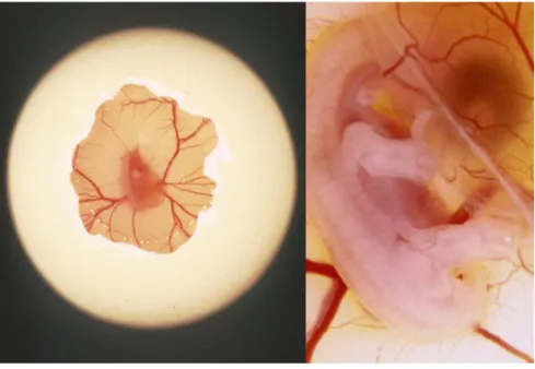
Figure 3 – Experimental manipulation of a chicken embryo, in ovo. (A) In ovo manipulation of a chicken embryo by opening
a window in the eggshield. (B) Application of beads in the third interdigital space in ovo. Courtesy of Doctor Joaquín León.
II.3.2 erg RNAi electroporation
The electroporation method involves the application of an electric field to a tissue that transiently disrupts the stability of the cell plasma membrane. This results in the formation of small reversible pores through which entrance of DNA is allowed.
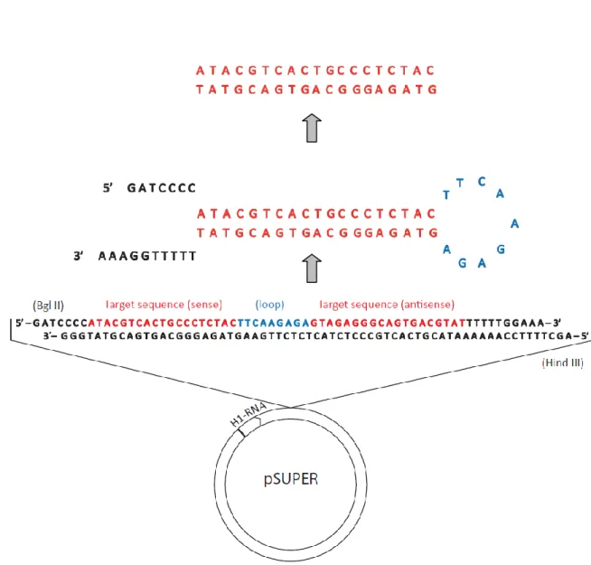
We used the pSUPER vector to drive the synthesis of RNAi transcripts that target specific mRNAs and suppress endogenous RNA activity. The RNAi against erg was cloned with the forward primer 5’ – GAT CCC CAT ACG TCA CTG CCC TCT ACT TCA AGA GAG TAG AGG GCA GTG ACG TAT TTT TTG GAA A – 3’ and reverse primer 5’ – AGC TTT TCC AAA AAA TAC GTC ACT GCC CTC TAC TCT CTT GAA GTA GAG GGC AGT GAC GTA TGG G – 3’ (Figure 4).
Materials and Methods
13 Electroporation was done in ovo, at stage 28-29HH chick embryos. We microinjected into the third interdigit mesoderm a pSUPER-erg RNAi plasmid solution (5-6μg/μl) together with a pCAGGS-AGFP (0.5-1μg/μl) one that will be used as readout for cell transfection (Figure 3A). The solution also includes 5% of Fast green to contrast. Single electroporation of pCAGGS-AGFP at 5-6μg/μl was carried out as a control. Electroporation was performed using an Intracel TSS20 Ovodyne electroporator (Intracel LTD) using 5 pulses of 60 ms length (8 V each) with 50 ms interval. We placed the electrodes (cathode and the anode) on both dorsal and ventral sides of the right hindlimb, at the third interdigit level. Then we repeated the procedure with the electrodes in the inverse order to allow DNA transfection into the whole interdigit. (Figure 3B,C). After electroporation, eggs were sealed and re-incubated until the desired stage was reached.
14
Figure 5 - Schematic representation of the electroporation technique. (A) injection of the DNA solution at the interdigital
space. (B) and (C) positioning of the electrodes to perform electroporation.
II.4 Riboprobe preparation for whole-mount In situ hybridization
A chicken and duck erg cDNA fragments were available at the laboratory cloned into pGEM-T easy plasmid. RNA probes were synthesized from those plasmids using the restriction and RNA polymerase enzymes that are described in Table I.
First, the plasmid vector containing the cDNA of the gene of interest was linearized and purified. Linearization was done in two different settings in order to generate templates for both antisense (positive probe) and sense (negative control probe) probes. After digestion the DNA was purified by phenol:chloroform extraction (see next item in this section), and precipitated with ethanol (see below in this section). After drying, the pellet was resuspended in milliQ water.
Sequencing reaction
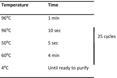
After plasmid purification, all sequences of cDNA of interest were checked. The primers used in the sequencing reaction annealed to RNA polymerase promoters T3, T7 or SP6 that flank the multiple cloning site of the vector (see Table I). Each 10μL reaction contained 2μL of BigDye® terminator sequencing buffer (5X), 2μL BigDye® terminator ready reaction mix, 500ng of template DNA and 5pmol of primers. The PCR conditions were the following:
Materials and Methods 15 Temperature Time 96⁰C 1 min 96⁰C 10 sec 50⁰C 5 sec 60⁰C 4 min
4⁰C Until ready to purify
After the PCR, the reaction product was precipitated. After mixing, the tubes were incubated at room temperature for 30min and centrifuged at 14000rpm for another 30min at 4ºC. The supernatant was removed and the pellet was washed in 250μL of 70% ethanol. The tubes were centrifuged at 14000 rpm for 15min at 4ºC. The supernatant was again removed and the pellet was air-dried. The samples were then sent to the IGC sequencing service. The resulting output sequences were analyzed in detail by the combined use of the BLAST (http://www.ncbi.nlm.nih.gov/BLAST/), Bioedit, Chromas and Sequence Analysis software.
cDNA Plasmid Restriction enzyme RNA polymerase
sense antisense sense antisense
c erg pGEM-t easy NcoI Sal I Sp6 T7
d erg pGem-t easy NcoI BamH I Sp6 T7
smad8 pCRII-TOPO EcoRV HindIII/BamH I T3 T7 msx2 pBluescriptII SK (+/-) HindIII/EcoRV Spe I T3 T7 snail pBluescriptII SK (+/-) XhoI HindIII T7 T3 bambi pBluescriptII SK (+/-) SacI XbaI T3 T7
Table I - Plasmids for the synthesis of riboprobes and the enzymes used for digestion and RNA polymerization.
II.4.1 Plasmid Linearization
Plasmidic DNA was digested with a unique restriction enzyme upstream in order to create a linear fragment. To obtain the template cDNA for probe transcription, we digested 10μg of the plasmidic DNA containing the cDNA of interest using 1μL of the appropriate restriction enzyme (see Table I),
16 2μL of the respective enzyme buffer (10x) and milliQ water for a final volume of 20μL. The reaction occurred for about 3 hours at 37⁰C. Completed digestion was confirmed by running 1µl of digestion product in a 0.8% agarose gel and observing a linear band with the total plasmid size. The linear plasmid was then purified by phenol:chloroform extraction and precipitated with ethanol.
Agarose gel electrophoresis
Agarose was dissolved in 1X TAE, usually at a concentration of 0.8%. Ethidium bromide was added in order to visualize the DNA with the use of a UV light to a final concentration of 0.2μg/mL. Loading buffer was added to each sample to a 1x final concentration and a DNA ladder was used to estimate the size of the DNA fragments. An electric current of 80-110V was applied to the gel immersed in 1X TAE buffer.
II.4.2 Phenol:Chloroform extraction
When the digestion reaction was completed the DNA was purified by phenol-chloroform extraction. This was performed to remove proteins from the nucleic acid solution. For this, milliQ water was used to make a final volume of 100μL to reduce the loss of DNA during the process and an equal volume of phenol-chloroform was added. The sample was mixed by strong vortexing and centrifuged for 5min at 14000 rpm. The DNA was recovered from the aqueous phase and transferred into a clean microtube and precipitated with ethanol.
II.4.3 RNA/DNA precipitation with ethanol
The nucleic acids were precipitated with 0.1 volumes of 3M sodium acetate (pH5.2) in the case of DNA or the same amount of lithium chloride for RNA samples, and 2.5 volumes of absolute ethanol for 30min at -80⁰C. The precipitated RNA/DNA was recovered by centrifugation at 14000rpm for 30min at 4⁰C. Then supernatant was disposed and the pellet was washed with 5 volumes of 70% ethanol by centrifugation at 14000rpm for 15min at 4⁰C. Finally, the supernatant was discarded and the RNA/DNA pellet was air-dried. The precipitated RNA/DNA was resuspended in 20μL of milliQ water and stored at -20⁰C.
II.4.4 In vitro DIG-labelled anti-sense RNA probe transcription
Riboprobes were synthesized by in vitro transcription with an adequate RNA polymerase and a mixture of dNTPs that contains DIG-labelled dUTPs. The synthesis of DIG-labelled anti-sense RNA probes was carried out by in vitro transcription at 37°C for 2h 30 min in a 20 μl reaction. The reaction contained 1X transcription buffer, 20U of RNase inhibitor, 1X DIG RNA labelling mix (Roche), 20 U of the appropriate RNA polymerase and 1 μg of the linearized template. After the generation of the riboprobe, 1 μl of the mixture was run on an agarose gel to estimate the amount of the probe.
Materials and Methods
17 Afterwards, the RNA was precipitated with ethanol, resuspended in 20 μl of milliQ water and stored at - 20°C.
II.5 Whole-mount In situ hybridization
The in situ hybridization technique was used to detect specific messenger RNA (mRNA) sequences in intact cells or tissues. It consists in the use of a single-stranded gene-specific RNA probe (riboprobe) labelled with an epitope, in this case digoxigenin (DIG). The riboprobe will only bind to its complementary mRNA transcripts, allowing the detection at the target gene expression sites. This allows great specificity considering that RNA/RNA hybrids are thermodynamically very stable and that hybridization is performed at stringent conditions (high temperature and increased content of formamide). The DIG-labeled RNA was detected by an anti-DIG antibody coupled to alkaline phosphatase (AP) and hybridization was visualized by a permanent dye that precipitates using BM-purple. (Veeck and Dahl, 2010).
The protocol was carried through for at least 3 days and all the washing and incubation steps were performed with gentle agitation using a roller, except when the opposite was mentioned.
On the first day, fixed embryos were rehydrated through graded methanols in PBT (100%, 75%, 50% and 25% methanol) and washed twice in PBT for about 10min each step. Then the embryos were bleached in 6% hydrogen peroxide in PBT for 1hour, at room temperature to block endogenous peroxidase activity. After that, they were washed three times for 5min in PBT. In order to improve probe penetration into the tissue during hybridization, the embryos were permeabilized by treatment with 10µg/ml proteinase K at room temperature (without agitation). The incubation time with proteinase K varied with the stage of the embryo and the level of penetration that we wanted to achieve. To stop the reaction the embryos were washed with fresh prepared glycine in PBT. Since the embryos are fragile after proteinase K treatment, embryos were fixed 20 min in PBT containing 4% PFA and 0.2% of glutaraldehyde at room temperature. To remove the fixative the embryos were washed twice with PBT for 5min. Then, the embryos were pre-hybridized with hybridization solution for 2 to 3 hours at 68-70⁰C. The high temperature and content of formamide in the hybridization solution increase the hybridization reaction stringency and therefore probe-target mRNA specificity, preventing non-specific hybridizations. Embryos were incubated overnight with hybridization solution with the probe (400-700 ng/ml) at 68-70°C.
On the second day, the probe was recovered and stored at -20⁰C for further use. Embryos were washed in stringent solutions (at hybridization temperature, without agitation) to remove non-hybridized RNA. First were washed twice with post-hybridization solution I for 1 hour, followed by two 30min washes with solution III. The embryos were further washed in MABT and then incubated in the blocking solution for 2 to 3 hours at room temperature. Finally, the embryos were incubated in the blocking solution with an anti-DIG antibody conjugated with AP in a 1:2000 ratio, overnight at 4⁰C to allow specific antibody binding to Digoxigenin.
18 During the third day of protocol, the embryos were washed in MABT with levamisole (an endogenous alkaline phosphatase inhibitor) at room temperature, renewing the solution every hour. Washing at this point is of great importance since unbound antibody has to be removed to obtain an efficient signal-to-noise ratio. In our case, we wash the embryos for 1 to 3 days, to reduce background as much as possible. Embryos were then washed at room temperature three times with NTMT for 10 minutes to create an alkaline medium that allows phosphatase activity present in the antibody. AFter that, embryos were incubated in the developing solution BM-purple (Roche) in the dark and at room temperature. BM-purple reacts with the antibody AP, resulting in the formation of a purple precipitate in the cells where target transcripts are localized. The appearance of the signal depended on the probe used and was checked by observing the embryos under the stereoscope. When a clear signal was observed the reaction was stopped with PBT. The embryos were fixed in 4% PFA overnight and stored in PBT until further analysis. As a control we performed the same protocol using a sense probe that didn’t present precipitation. Results were imaged in a stereoscope attached to a digital camera.
Some of the embryos after in situ hybridization protocol were sectioned in a cryostat. For that, embryos previously soaked in sucrose solution were embedded in mounting medium for frozen samples and sectioned at 20 µm (see section II.6).
II.6 Histological analysis
II.6.1 Tissue processing and gelatin embedding
After proper embryo fixation (in 4% PFA at 4⁰C for 2 hours for immunostaining/TUNEL assay, or after the whole-mount in situ protocol final fixation), the embryos were rinsed in PBS and soaked in 10% sucrose in PBS overnight at 4⁰C. In the next day, embryos were washed in a fresh 10% sucrose-PBS solution and incubated in PBS with 10% gelatin and 10% sucrose at 37⁰C for an hour. After this, embryos were transferred to a mould with an already solidified gelatin layer, covered with 37⁰C 10% gelatin and oriented in the desired position. The mould was removed after gelatin solidification (at room temperature or 4⁰C) and the gelatin block was cut in the appropriate size and orientation. The samples were fixed in a slice of cork with Tissue Tek O.C.T.™ Compound, frozen and stored at -80⁰C until cryostat sectioning.
Samples were sectioned at 12-14μm for future immunostaing/TUNEL assay or at 20-24μm for embryos after in situ hybridization. Sections were collected in SuperFrost Plus slides for better adhesion, and stored at -20⁰C.
Materials and Methods
19
II.7 Limb morphological analysis
II.7.1 Alcian green staining
The morphology of the limbs was initially analyzed in specimens stained for cartilage following alcian green protocol (Ganan et al., 1996). The embryos were sacrificed, fixed in 5% trichloroacetic acid overnight and stained with 0.1% Alcian green during the next night. In the following day, embryos were washed in an acid alcohol solution and dehydrated with absolute ethanol (2 washes of one hour each). Finally, the embryos were cleared in methyl salicylate.
II.8 Cell death analysis
The pattern of cell death was analyzed by using the TUNEL assay performed in tissue sections. Acridine orange was employed as an alternative to the TUNEL method in whole autopods.
II.8.1 Acridine Orange
The vital dye acridine orange permeates dying cells to bind chromatin and interacts with DNA and RNA by intercalation or electrostatic attraction respectively. DNA intercalated acridine orange fluoresces green (525nm). It has been shown in drosophila embryos that acridine orange, is specific for apoptotic forms of cell death and does not significantly label cells undergoing necrotic death provoked by injury (Abrams et al., 1993). The treated embryo legs were rinsed in 1:1000 acridine orange (1mg/ml stock solution) in preheated PBS and incubated at 37⁰C for 20-30min. Next, the embryos were washed twice in preheated PBS for 5min each. After a final PBS wash at room temperature, interdigital cell death was visualized with a fluorescent stereoscope with a camera attached.
II.8.2 TUNEL analysis of dying cells
Cryopreserved tissue sections of autopods were analyzed for apoptotic DNA fragmentation by the terminal deoxynucleotidyl transferase-mediated dUTP-TRIC nick end labeling (TUNEL) assay, using the in situ cell death detection kit (Roche). Labeling of DNA strand breaks by Terminal deoxynucleotidyl transferase (TdT) which catalyzes polymerization of labeled nucleotides to free 3’-OH DNA ends in a template-independent manner (TUNEL-reaction). TMR red labeled nucleotides, incorporated in nucleotide polymers, are detected and quantified by fluorescence microscopy. To evaluate the rate between living and dead cells, TUNEL was combined with DAPI (and/or TO-PRO3) staining and immunostaining for phospho histone H3 to detect proliferating cells. For that, we used longitudinal 14μm sections of the third interdigit. To remove the gelatin from the sections, the slides were washed/emerged in preheated (37⁰C) TBS for 30-60min. The slides were washed three times for 5min in TBS and incubated for 30min in block buffer. This is important to inhibit the
20 unspecific ligation of the primary antibody to the cells. After blocking, the slides were incubated with 1:100 of the primary antibody against H3 (anti-H3, rat Sigma h9908) in TBS++, overnight at 4⁰C. In the next day, sections were washed three times for 5min in TBS++, followed by the incubation of the respective secondary antibody (goat anti-rat Alexa 488) for one hour, at 37⁰C. Next, slides were further washed with TBS three times, and incubated with DAPI (1:10000) for 10min to label the nucleus. After this, samples were washed with TBS once and incubated with TO-PRO3 (1:1000) for 15’ and washed again with TBS. The sections were then fixed with 4%PFA for 50min at room temperature. After several TBS washes for about 2hours, sections were incubated with 0.2%triton 0.05%tween TBS for 30min and washed in TBS twice after. Next, slides were washed in 10mM Tris-HCl + 5mM EDTA for 5min and permeated with 20μg/ml proteinase K in 10mM Tris-Tris-HCl + 5mM EDTA for 15min at room temperature. Then, the slides were washed twice with 5mM EDTA for 5min followed by a 10min TdT buffer wash. Finally the slides were incubated with the TUNEL TMR kit (Roche) for 2 hours at 37⁰C. After incubation, slides were washed twice in SSC + EDTA for 10min and washed three times in TBS for 5min. Sections were once again contrasted with DAPI, washed in TBS for 5min and finally mounted in vectashield. The slides were stored at 4⁰C until further analysis in the fluorescent microscope.
II.9 Confocal microscopy
Samples were examined with a laser confocal microscope (Zeiss LSM 510 META) by using a Plan-Neofluar 10x or Plan-Apochromat 20x objectives, and argon ion laser (488nm) to excite FITC fluorescence and a HeNe laser (543nm) to excite Texas Red. For stacks digitalization and image processing, we used ImageJ software. The images shown in this study are the most representative of the experiment.
II.10 Statistic analysis
Interdigits of hindlimbs injected with erg RNAi+AGFP (n=75) or AGFP (n=38) constructs, and their respective control legs were measured using ImageJ software. Data was grouped in two sets, one for erg RNAi+AGFP and the other for AGFP controls, and two subsets, manipulated and controls. Sets were analyzed using the GraphPad Prism 5 software and statistic significance of the experimental procedure was evaluated using the non-parametric 2-tailed Wilcoxon matched-pairs signed rank test, with a 95% confidence interval.
22
III. Results
Expression of erg during limb development correlates directly with apoptosis
The expression pattern of erg was studied in chicken (Gallus gallus) embryos during digit development, between stages 28 to 35HH (Hamburger and Hamilton, 1951, see Appendix V).
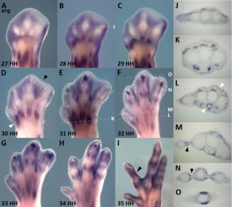
Erg exhibited a dynamic pattern of expression in the limb mesoderm throughout all the studied stages (Figure 6 and 7). We have observed that interdigital mesenchymal cells showed a high level of erg transcripts at stages immediately preceding cell death. Well-defined domains of erg expression were detected in a pattern that correlates with the areas of limb cell death, in particular the interdigital necrotic zones (INZ). In the interdigital tissue, erg exhibits a pattern of expression prefiguring the areas of interdigital cell death (ICD) (Figure 6A-D and Figure 7) beginning at stage 27HH. Between stages 27HH and 30HH, erg transcripts were concentrated in the most proximal interdigital mesoderm (Figure 6A-D). From stage 31HH onwards, the most central interdigital expression of erg was gradually lost, becoming restricted to the tissue around the digits (Figure 6E-I and Figure 7). At these advanced stages of limb development, erg was progressively expressed in the perichondrium of the developing digits, surrounding the diaphysis (Figure 6I, black arrowhead), (Figure 6I, N and O) and in the remaining interdigit (Figure 6I).
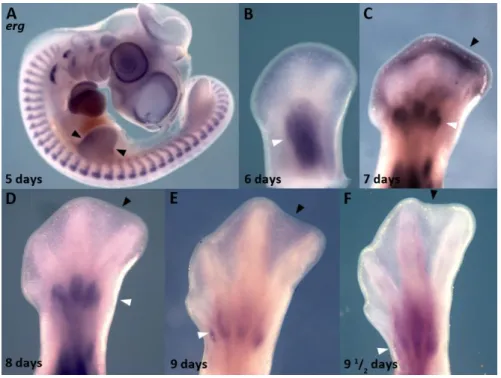
This correlation between erg gene expression and ICD was also observed in the webbed digits of duck embryos, in which cell death is restricted to the most distal portion of the interdigital tissue (Hurle and Colvee, 1982) (Figure 8C, black arrowhead). During chick limb development, erg is also expressed in the anterior and posterior necrotic zones (Margarida Santos thesis) and the same expression pattern is observed in duck embryos (Figure 8A, black arrowheads). These findings point to a conserved role of erg during the apoptotic process. Throughout the whole period studied, erg expression showed a direct relationship with the distribution of the programmed cell death areas in different species, in particular with INZ.
In addition to its expression in the apoptotic areas, erg expression was also detected in the dermomyotome (shown in Margarida Santos thesis). Furthermore erg transcripts appeared at the hindlimb medial part, corresponding to the myogenic condensations present at the limb (Figure 6 A-C), in myogenic cells of the differentiating muscles (Figure 6, E and K) and surrounding the dorsal and ventral tendons (Figure 6L).
Results
23
Figure 6 – erg expression during digit development. In situ
hybridization for erg in developing autopods at stages 27HH (A), 28HH (B), 29 HH (C), 30HH (D), 31HH (E), 32HH (F), 33HH (G), 34HH (H) and 34HH (I). J, K and L-O are transverse sections of B, E and F, respectively.
In situ hybridization for erg shows
that it is expressed in the interdigital areas prior the physiological cell death initiation (A-C and J). At stage 30, erg is still expressed at the entire interdigital space (D, black arrowhead), but as development proceeds and ICD begins, erg expression becomes progressively restricted to the side of the digits (E-I and N, black arrowhead). At stage 35 erg is expressed surrounding the phalanxes and the remaining interdigit (I). Throughout digit development, erg is also present in muscular masses (A-H, D and L, white arrowheads) and around the tendons (L).
Figure 7 –Schematic representation of erg expression pattern during digit development. erg begins to be expressed
throughout the interdigital areas (28HH) and in the myogenic cells. As ICD initiates, erg transcripts become progressively restricted to digit sides and to the differentiating muscles.
24
Figure 8 – erg expression pattern in duck. In situ hybridization for erg in duck embryos shows that during limb
development this gene is expressed at the anterior and posterior necrotic zones (A, arrowheads). As shown in chick limbs,
erg is expressed in myogenic masses (B-F white arrowheads). Between 7-8 days of incubation, erg is expressed at the distal
tip of the interdigit (C, black arrowhead) where cell death will occur. Note that the interdigital domains of erg become restricted distally coincidently with the zones of interdigital cell death in the duck. At later stages (E-F) erg interdigital expression is lost (black arrowheads) but remains expressed in the muscle masses (white arrowheads).
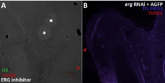
Inhibition of erg activity during digit development
Since chick and duck erg expression patterns suggest that this gene would be a good candidate to be involved in interdigital cell death (ICD) control, we performed loss-of-function studies to further understand its possible role in the apoptotic process. To approach that, we used chick embryos between stage 28HH and 29HH and implanted beads soaked in 10mM ERG Inhibitor (M5060 E-4031 Sigma), a specific ERG activity blocker, to functionally challenge the intercellular dynamics of potassium. In order to continuously deliver the drug, several beads were implanted in the same interdigital space at different time points. The first bead was implanted into the third interdigit of the right hindlimb, and 6 to 7 hours later a second bead substituted or was implanted distally to the first one when cell death has not yet started in the interdigits. A third bead was implanted 12h later and when embryos reached stage 30-31HH they were processed for cell death analysis by acridine orange vital staining (Figure 9). There was a visible reduction of cell death in the INZ after ERG inhibitor-soaked beads implantation (Figure 9A) when compared with the control interdigit (Figure 9A’) and PBS bead treated limbs (Figure 9B). Note the inhibition of cell death in the area close to the ERG inhibitor bead (Figure 9A). The interdigit apoptotic pattern was not altered after a PBS-bead implantation (Figure 9B) when compared to the control leg (Figure 9B’). Interdigital longitudinal sections of treated limbs were also analyzed for apoptotic DNA fragmentation by TUNEL assay, together with the immunolabeling for phospho-histone H3 (Figure 11). As showed for the acridine orange staining, interdigital apoptosis was reduced, but apoptosis at joint formation was still visible