Faculdade de Ciências
Departamento de Biologia Vegetal
Transfer and accumulation of
Paralytic and Amnesic Shellfish
Toxins in secondary consumers of the
marine trophic chain
Sandra Lage
Mestrado em Ecologia Marinha
Faculdade de Ciências
Departamento de Biologia Vegetal
Transfer and accumulation of Paralytic and
Amnesic Shellfish Toxins in secondary consumers
of the marine trophic chain
Sandra Lage
Mestrado em Ecologia Marinha
2010
Dissertation supervised by:
Dr Pedro Costa
Instituto de Investigação das Pescas e do Mar – IPIMAR
Professora Dr.ª Vanda Brotas
Faculdade de Ciências da Universidade de Lisboa – FCUL
Centro de Oceanografia – CO
Resumo --- 6
Abstract --- 11
1. Introduction 1.1 Harmful algal blooms: causes, impacts and detection --- 13
1.2 Paralytic shellfish poisoning --- 14
1.3 Amnesic shellfish poisoning --- 16
1.4 Marine toxins in the food chain --- 17
1.5 Objectives --- 20
2. Temporal variability of paralytic and amnesic shellfish toxins in horse mackerel (Trachurus Trachurus) 2.1 Introduction --- 21
2.2 Methods 2.2.1 Collection and preparation of horse mackerel samples --- 23
2.2.2 Toxin extractions and HPLC analysis --- 24
2.2.2.1 Paralytic shellfish toxins --- 25
2.2.2.2 Amnesic shellfish toxin --- 26
2.3 Results --- 29
2.4 Discussion --- 32
3. Uptake and elimination of paralytic shellfish toxins in fish exposed to toxin – contaminated cockles 3.1 Introduction --- 34
3.2 Methods 3.2.1 Preparation of toxic diet --- 34 __________________________________________________________________________________
3.2.3 Toxin extraction and HPLC analysis --- 35
3.3 Results 3.3.1 Toxin profile of Cerastoderma edule --- 36
3.3.2 Accumulation and elimination of PSTs by Diplodus sargus sargus --- 39
3.4 Discussion --- 42
4. Detection and sub - cellular distribution of the amnesic shellfish toxin in the digestive gland of Octopus vulgaris during periods of toxin absence 4.1 Introduction --- 44
4.2 Methods 4.2.1 Collection and preparation of octopus and bivalve samples --- 45
4.2.2 Sub - cellular fractionation --- 46
4.2.3 Toxin extraction and HPLC analysis --- 46
4.2.4 Statistics --- 46
4.3 Discussion --- 49
5. Concluding remarks --- 52
6. References --- 54
1. Introduction
1.1 Gymnodinium catenatum --- 15 1.2 Pseudo-nitzschia australis; a) 1400x; b) 2800x; c) 10 000x --- 17 1.3 Toxins transfer in the marine trophic chain --- 19 2. Temporal variability of paralytic and amnesic shellfish toxins in horse mackerel (Trachurus Trachurus)
2.1 Molecular structure of Paralytic shellfish toxins --- 21 2.3 Molecular structure of domoic acid --- 22 2.3 PSTs concentration (µg STXeq. kg-1) in the gastrointestinal tract (median, 25 and 75 quartiles, minimum and maximum, total n = 33) of Trachurus trachurus captured in Septmber-2009 --- 26 2.4 HPLC-FLD chromatograms of (a) fish tissues homogenate (1:10 dilution) (b) PSTs standards solution --- 27 2.5 Toxin composition profile of Trachurus trachurus --- 28 2.6 HPLC-UV chromatogram (a) fish tissues homogenate (b) DA standard solution
--- 28 3. Uptake and elimination of paralytic shellfish toxins in fish exposed to toxin – contaminated cockles
3.1 a) Toxin profile (molar ratio) of cockles (Cerastoderma edule) supplied to fish; b) Contribution of each PSTs to the total toxicity (3807 µg STXeq kg-1) ---- 36 __________________________________________________________________________________
feeding experiment (Day 1-5, exposure; Day 6-15, elimination). Mean ± S.D. (n = 3) --- 37 3.3 Changes in concentration of (a) B1 and (b) dcSTX in viscera of Diplodus
sargus sargus during the whole feeding experiment (Day 1-5, exposure; Day
6-15, elimination). Mean ± S.D. (n = 3) --- 38 Table 3.1 Concentration of C1+2 in gastrointestinal tract of Diplodus sargus sargus during the whole feeding experiment (Day 1-5, exposure; Day 6-15, elimination) in each replicate (ND – not detected; NQ – not quantified) --- 38 4. Detection and sub - cellular distribution of the amnesic shellfish toxin in the digestive gland of Octopus vulgaris during periods of toxin absence
Table 4.1 Octopus vulgaris: Sampling date (2009); number of individuals (n); total weight (mean ± SD) and digestive gland weight (mean ± SD) --- 45
4.1 Seasonal variation of domoic acid concentration (µg g-1) in mussels, Mytilus
galloprovinciallis, collected from Peniche and adjacent fishing area (NW
Portuguese coast) during January and November of 2009 --- 47 4.2 Domoic acid concentration (µg g-1) in the digestive gland (median, 25 and 75 quartiles, minimum and maximum, total n = 32) of Octopus vulgaris in 4 sampling dates (23- Sep; 19 and 27-Oct and 18 -Nov 2009). Significant differences at p < 0.05 are marked with *, after applying the Mann-Whitney test. --- 47 4.3 Relative abundance (mean ± SD) of domoic acid in digestive gland cell
fractions of Octopus vulgaris naturally contaminated with 3 levels of toxin (10.0 ±0.5, 15.0 ±0.5 and 22.0 ± 0.5 µg DA g-1): a) soluble fraction (cytosol)
O fitoplâncton é o principal produtor primário dos oceanos, representando uma relevante fonte de matéria orgânica que suporta o funcionamento da cadeia alimentar marinha. Contudo a proliferação relativamente repentina de determinadas espécies de fitoplâncton tóxico e/ou nocivo, atingindo elevadas concentrações de células na água do mar, conduz a graves impactos na cadeia trófica marinha, na ecologia dos oceanos, na integridade dos ecossistemas, na saúde humana e nas indústrias e economias apoiadas nos recursos marinhos. Estima-se que existam cerca de 4000 espécies de fitoplâncton marinho, das quais 200 são consideradas nocivas e apenas 80, essencialmente dinoflagelados, têm a capacidade para produzir toxinas.
Organismos marinhos que obtêm o alimento por mecanismos de filtração, como é o caso dos moluscos bivalves, podem concentrar um elevado número de células de fitoplâncton tóxico acumulando concentrações elevadas de toxinas marinhas nos seus tecidos e provocar intoxicações no Homem. Em Portugal, as biotoxinas com propriedades neurotóxicas mais comuns são as toxinas paralisantes (PST) e as toxinas amnésicas (AST).
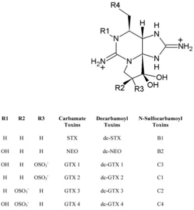
Até ao momento foram identificadas pelo menos 24 toxinas paralisantes, com diferentes estruturas químicas e toxicidades. Os compostos PST podem ser divididos em três grupos, conforme apresentado a seguir e por ordem crescente de toxicidade: N - sulfocarbamoil (B1, B2, C1 - C4), decarbamoil (dcGTX1 - dcGTX4, dcNeo, dcSTX) e carbamoil (GTX1 - GTX4, Neo, STX). A composição de toxinas das espécies produtoras de PSTs é variável, dependendo da zona geográfica e dos respectivos
Resumo
factores ambientais que caracterizam essas zonas. Em Portugal, os compostos PST, são principalmente produzidos pelo dinoflagelado Gymnodinium catenatum, sendo o seu perfil constituído maioritariamente pelas toxinas N - sulfocarbamoil e decarbamoil. Este perfil é normalmente detectado na fauna marinha exposta aos florescimentos deste dinoflagelado. Todavia algumas das espécies contaminadas apresentam alterações nos seus perfis de toxinas, devido a possuírem mecanismos de acumulação, biotransformação e eliminação das toxinas no seu organismo.
O aminoácido ácido domóico (DA) é a principal AST e apresenta uma estrutura química semelhante ao neurotransmissor excitatório ácido glutâmico e à neurotoxina excitatória cainato. Esta toxina amnésica é produzida por diatomáceas marinhas do género
nitzschia spp, sendo na costa portuguesa essencialmente produzido por Pseudo-nitzschia australis.
As toxinas marinhas entram na cadeia alimentar a partir de moluscos bivalves e outros consumidores primários tais como zooplâncton e peixes plânctivoros, que se alimentam de microalgas tóxicas. Crustáceos braquiúros e cefalópodes são um exemplo de consumidores secundários, com uma dieta composta essencialmente por marisco e pequenos peixes pelágicos e bentónicos, em que elevadas concentrações de toxinas foram já reportadas. Por via de relações tróficas, as toxinas são transferidas até aos predadores de topo, podendo nos episódios agudos conduzir à mortalidade de mamíferos e aves marinhas. Estas consequências ecológicas fomentam a necessidade de avaliar o potencial dos consumidores secundários como vectores e os impactos das toxinas marinhas na cadeia trófica.
O carapau, Trachurus trachurus, é um consumidor secundário de elevada importância em termos de abundância na costa portuguesa. Este peixe semi - pelágico ocupa uma posição central na cadeia trófica podendo ser um potencial vector de toxinas para os predadores de topo. De modo a avaliar a sua capacidade para acumular PSTs, indivíduos desta espécie foram amostrados quinzenalmente em Peniche e Portimão, costa noroeste e sul portuguesa, entre Setembro de 2009 e Março de 2010.
Concentrações elevadas de PST foram determinadas em todos os indivíduos capturados em Peniche em Setembro 2009. O que possibilita que o carapau transfira as toxinas para os predadores de topo, mamíferos e aves marinhas, que são sensíveis às PST e que consequentemente pode provocar a sua mortalidade. Contudo nas amostras de carapau colhidas em Peniche nos meses seguintes e todas as amostras colhidas em Portimão as PST não foram detectadas.
Ao se verificar que os peixes, no meio natural, acumulam PSTs a partir do consumo de presas contaminadas, surge a necessidade de compreender a dinâmica de acumulação e eliminação destas toxinas. Para tal o sargo, Diplodus sarugus sargus foi exposto, em ambiente controlado, a berbigão, Cerastoderma edule, contaminado com PSTs durante um período de 5 dias. Seguidamente realizou-se um período de eliminação, de 10 dias, em que o sargo se alimentou de berbigão não tóxico.
Durante o período de exposição ao berbigão contaminado, a concentração das PSTs revelou um aumento cumulativo nos tecidos gastrointestinais do sargo. Contudo das 10 PSTs presentes no perfil de toxinas do berbigão, apenas: C1+2, B1 e dcSTX foram detectadas no sargo. C1+2 foi esporadicamente detectado nos primeiros dias da
experiência, enquanto B1 e dcSTX foram detectados continuamente. Durante o período de exposição as concentrações de B1 foram sempre superiores às de dcSTX, o que se encontra de acordo com as respectivas proporções no perfil de PSTs do berbigão. A eliminação lenta de B1 e dcSTX comparativamente à eliminação das outras PSTs sugere que o sargo apresenta um mecanismo de eliminação específico. Tendo em conta que ambas as toxinas foram igualmente dominantes no perfil de toxinas do carapau naturalmente contaminado, é possível extrapolar-se que uma eliminação específica destas toxinas possa ocorrer em várias espécies de peixes.
Na costa portuguesa, as contaminações de bivalves por AST ocorrem maioritariamente nos meses de Verão, embora fenómenos ocasionais possam ocorrer entre Fevereiro e Agosto. Os espécimes de carapau capturados na costa noroeste e sul portuguesa durante Setembro de 2009 e Março de 2010 não apresentaram DA nos seus tecidos. Esta ausência da toxina pode significar que as presas do carapau não estiveram expostas a DA. Contudo, em todos os espécimes de polvo capturados entre Setembro e Novembro de 2009, na costa noroeste, foi detectado DA na glândula digestiva. Este resultado demonstrou, pela primeira vez, que os polvos acumulam DA durante períodos de ausência das algas produtoras. O que não era previsto, pois sendo o DA um composto hidrofílico e o polvo um organismo com elevada taxa metabólica, a toxina deveria ser rapidamente eliminada. Além disso, após o fraccionamento da glândula digestiva verifica-se que a grande parte do DA se encontra livre no citosol. O que implica um mecanismo de retenção de DA na glândula digestiva do polvo e/ou uma ligação a uma proteína, como previamente verificado para um toxina quimicamente semelhante ao DA.
Em síntese, nos presentes estudos verificou - se que os consumidores secundários podem conter PST e AST nos seus tecidos digestivos após se alimentarem de presas contaminadas. O que incrementa a necessidade de avaliar a dinâmica de transferência, acumulação e eliminação destas toxinas na cadeia alimentar, pois a presença de toxinas nos recursos marinhos pode desencadear distúrbios ecológicos no ecossistema marinho e a mortalidade nos predadores de topo (mamíferos e aves marinhas). Além disso, devido ao elevado valor comercial de algumas das espécies em causa, um risco para a saúde pública pode estar subjacente.
Palavras-chave
Paralytic and amnesic shellfish toxins can enter in the marine trophic chain by filter-feeding and benthic organisms. Then due to trophic interrelationships the toxins are transferred to theirs predators. In the last instance, top predators (marine mammals and sea birds) can be intoxicated after fed on primary and secondary consumers.
The wild horse mackerel (Trachurus trachurus) was collected in NW and S Portuguese coast between September of 2009 and March of 2010. The results show for the first time that horse mackerel can accumulate high concentrations of PSTs in theirs gastrointestinal tract. Which increased the requirement of evaluated the dynamics of accumulation and elimination of PSTs in secondary consumer fish.
Thus aquaculture fishes, white sea breams, were fed with toxin-contaminated cockles. After 5 days of toxin exposure, the fishes were fed with non toxic cockles during 10 days. In this feeding experiment, an increasing of toxin concentration over the exposure period was noticed in the fish gastrointestinal tract. B1 and dcSTX were the only toxins continuously detected during the exposure/elimination period, which reveal a slower elimination than the others PSTs. These two PSTs were also the main toxins in the mackerel toxin profile, which may suggest a specific PSTs elimination by fish species.
Although AST were not detected in any mackerel specimen, in octopus (Octopus
vulgaris) specimens captured in the same fishing area, domoic acid (DA) was detected
in the digestive glands. This accumulation of DA in the octopus was previously identified. However this is the first time that DA was detected during periods of absence
Abstract
of ASP events. Plus DA was predominantly found in the cytosol which increased the evidence of a retention mechanism. Since DA as a hydrophilic compound should be easily release.
This thesis supplies relevant data to the growing knowledge on the dynamics of accumulation and elimination of PSTs and AST in secondary consumers and on the toxin transfer in the marine food web.
Key words
1.1 . Harmful algal blooms: causes, impacts and detection
Phytoplankton are crucial to marine and freshwater ecosystems. They play a critical role in the trophic chain, being the most important biomass producer. Blooms of phytoplankton are increasingly frequent in marine, brackish and fresh water environments. They occur in the upper layer of water - the epipelagic zone- which is rich in oxygen, penetrated by sunlight, and warmer than water at lower levels. Blooms reveal a season dependency, which is mainly determined by seawater temperature and sunlight. The hydrographic conditions are also relevant, especially the presence of a thermocline, which is indirectly influenced by windforce, turbulency in the water and by climatological events such as the North Atlantic Oscillation (NAO). The amount of nutrients in seawater also needs to be adequate to fulfil the requirements of the organisms, which can be provided by the nutrient- rich waters from upwelling and freshwater runoff. Although algal blooms are natural phenomena, it’s current increasing in frequency and geographic distribution can be attributed to anthropogenic activities such as fertilisers, discharge of human waste and ballast waters (Masó and Garcés, 2006).
When these phytoplankton blooms includes species capable to produce toxic substances, or that are noxious or even promoting a nuisance impact in other aquatic organisms, they are named as Harmful Algal Blooms (HAB) (Smayda, 1997). Of the 4000 of marine planktonic microalgae species, just 200 can be harmful, and of these,
1. Introduction
only around 80 (mainly dinoflagellates) have the potential to produce toxins (Smayda and Reynolds, 2003).
Filter-feeding organisms such as shellfish can accumulate high levels of marine toxins and cause severe human poisonings. In Portugal, a regular monitoring programme for marine toxins was implemented in 1986 (Sampayo et al., 1997). The main objective of the monitoring program is the public health protection through supporting the quality control of the marine products from fishery and aquaculture.
Marine toxins are functionally categorized according to their clinical symptoms, namely neurological or hepatic. In Portugal, the main toxins causing neurological symptoms are responsible for the human syndromes Paralytic Shellfish Poisoning (PSP) and Amnesic Shellfish Poisoning (ASP).
1.2 . Paralytic Shellfish Poisoning
Paralytic Shellfish Poisoning (PSP) is a human illness that typically results from the consumption of shellfish that have been exposed to the toxigenic dinoflagellates
Alexandrium spp, Gymnodinium catenatum or Pyrodinium bahamense (Shumway,
1990). These plankton species are known producers of paralytic shellfish toxins (PSTs). Their mode of action involves reversible and highly specific block of ion transport by the sodium channel and then confine signal transmission between excitable membranes (nerve and muscle fibers) (Narahashi, 1988; Easthaugh and Shepherd, 1989). Therefore
muscle paralysis death and cardio- respiratory arrest are the last effects in mammals (Price et al., 1991).
Fig. 1.1 Gymnodinium catenatum
(Adapt from National Institute of Fisheries and Sea Research – IPIMAR)
Saxitoxin (STX) is the main PST, but at least 24 analogues have been identified (Etheridge, 2010). They differ in chemical structure and toxicity, and can be grouped into three classes in increasing order of toxicity: N-sulfocarbamoyl (B1, B2, C1-C4), decarbamoyl (dcGTX1- dcGTX4, dcNeo, dcSTX) and carbamoyl toxins (GTX1- GTX4, Neo, STX) (Oshima et al., 1993).
Each phytoplankton species has its specific toxin composition, which can even vary with geographic region and environmental factors (Etheridge and Roesler, 2005). In Portugal, G. catenatum is the main PST producer (Fig. 1.1). This species has been
recurrently responsible for shellfish closures to harvesting since 2005, when the first evident bloom occurred after a 10 year break period (Moita et al., 2005).
The toxin composition of G. catenatum is mostly constituted by N-sulfocarbamoyl and decarbamoyl toxins (Negri et al., 2007). This toxin profile is also detected in marine fauna exposed to G. catenatum blooms. Nevertheless changes in toxin profiles are expect to occur due to inter-specific differences in PSTs accumulation, biotransformation and elimination (Bricelj and Shumway, 1998). Considering that these processes occur simultaneously and the toxin composition can vary greatly among species and time, it becomes clear that predicting accumulation of PSTs is a complex task.
1.3 . Amnesic Shellfish Poisoning
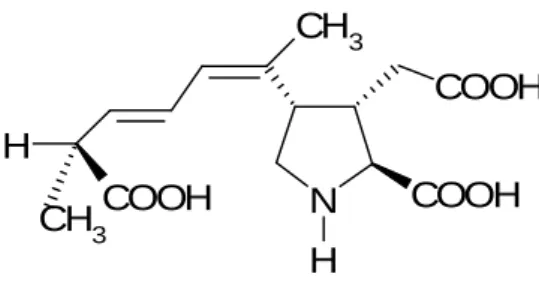
Human consumption of shellfish contaminated with domoic acid results in the Amnesic Shellfish Poisoning (ASP). Domoic acid is a heat-stable amino acid that is structurally similar to the excitatory neurotransmitter glutamic acid and the excitatory neurotoxin kainic acid. DA has a much stronger receptor affinity, being 3 times more potent than kainic acid and up to 100 more potent than glutamic acid (Todd, 1993). This structural similarity allows the binding to glutamate receptors in the central nervous system causing neurones’ depolarization, depletion of energy, neuronal swelling and cell death (Berman and Murray, 1997, Debonnel et al., 1989).
The first documented case of ASP occurred in 1987, when three people died and more than 100 became ill after eating mussels from Prince Edward Island, Canada (Bates et al., 1998). The victims revealed neurological symptoms such as confusion, nausea and selective short- term memory loss (Todd, 1993). DA was then found to be produced by the marine pennate diatom Pseudo- nitzschia multiserries (Bates et al., 1989). The
Pseudo-nitzschia australis is the main DA producer in the Portuguese coast with their
blooms highly associated to upwelling events that occur during spring and summer (Costa and Garrido, 2004, Palma et al., 2010) (Fig. 1.2 a, b, c).
Fig. 1.2 Pseudo-nitzschia australis; a) 1 400x; b) 2 800x; c) 10 000x (Adapt from Phycologycal Society of America)
1.4 . Marine toxins in the food chain
Marine toxins can enter in the marine food chain by bivalve molluscs and other primary consumers such as zooplankton and planktivorous fish (Bargu et al., 2002; Costa and Garrido, 2004). Due to trophic interrelationships the marine toxins (domoic acid and saxitoxins) can be transferred to theirs predators. High toxin concentrations have been
b) c)
found in crabs and cephalopods whose diet is mostly constituted by shellfish and small pelagic and benthic fish (Costa et al., 2003; Costa et al., 2004). Subsequently through consumption of primary and secondary consumers, top predators can be intoxicated (Fig. 1.3). This could result in toxic episodes with massive deaths of marine mammals and sea birds (Sierra-Beltran et al., 1997; Scholin et al., 2000).
Toxin transference through the food chain may be similar between the several marine toxins. However the toxin-producing diatoms, due to its morphology, have a propensity to become less buoyant and to sink on the sediment, and thus providing benthic pathways for contamination of soft bottom dwelling invertebrates (Trainer et al., 2000; Costa et al., 2005). Contamination of the marine food web and acute intoxications of top predators may promote changes on the ecosystem stability. Besides the sporadic highly toxic episodes, organisms are seasonally exposed to small doses of marine toxins without apparent harm. Nevertheless, little is known regarding the chronic exposure of wild populations to marine toxins.
There is a growing concern on the transfer of DA and PSTs through the marine food web. They can reach several groups of animals and hence pose potential risks to ecosystem stability.
Fig. 1.3 Toxins transfer in the marine trophic chain
1.5 . Objectives
PSP and ASP outbreaks result from the consumption of toxin-contaminated shellfish. Those vector species that are edible and have economic interests are the most studied. However the recent identification of more marine species as vectors of PSTs and of DA, stresses the ecological importance of evaluating the impacts of harmful algal blooms on wildlife resources and on the food webs that support them. Baseline measurements of toxin occurrence in fish and cephalopods, studies on toxin kinetics of accumulation and depuration, and knowledge of toxin tissue distribution and its biotransformation are important for understanding the transfer of marine toxins in the food web.
Hence, the main objectives of this thesis are:
i) To study the temporal variability of PSTs and DA in fish collected from NW and S Portuguese coast (Chapter 2)
ii) To assess the uptake, distribution and elimination of PSTs in fish orally exposed to marine toxins under controlled conditions (Chapter 3)
iii) To study the accumulation and the sub-cellular distribution of domoic acid in octopus during periods of toxin absence in the environment (Chapter 4)
2.1. Introduction
Paralytic Shellfish Toxins (PST) and Amnesic Shellfish Toxins (AST) are the most common neurotoxins produced by marine microalgae in the Portuguese coast. PSTs originating from blooms of the dinoflagellates Alexandrium minutum or Gymnodinium
catenatum occurred since the 1940s in the NW Portuguese coast (Franca and Almeida,
1989) (Fig. 2.1). However, after 1995 neither the PSTs were detected in shellfish, nor evident blooms of toxigenic species were observed in seawater. By 2005, the first bloom of G. catenatum seen in 10 years occurred.
Fig. 2.1 Molecular structure of Paralytic shellfish toxins (Adapt from Etheridge, 2010)
2. Temporal variability of Paralytic and
Amnesic
Shellfish
Toxins
in
horse
mackerel (Trachurus trachurus)
Domoic acid (DA), which is the main AST, was detected for the first time in shellfish of the Portuguese coast in 1995 (Sampayo et al., 1997) (Fig. 2.2). The Pseudo-nitzschia
australis was identified as the dominant diatom species responsible for DA production
in this coast, when high cell densities (> 7x10⁶ cells g-1) were found in the stomach content of contaminated planktivorous fish (Costa and Garrido, 2004). The presence of neurotoxins in Portuguese coastal waters is therefore a relative recent reported phenomenon. N H COOH COOH CH3 COOH H CH3
Fig. 2.2 Molecular structure of domoic acid
By consuming toxic algae, filter-feeding and planktivorous organisms, such as sardines, may accumulate high toxin levels and act as vectors to their predators. There are several reports of acute toxic episodes that have conducted to massive fish-kills (White, 1981; 1984; Saito et al., 1985; Montoya et al., 1996) and even to the mortality of sea birds and marine mammals after ingestion of contaminated fish (Geraci et al., 1989; Lefebvre et al., 1999; Scholin et al., 2000). Moreover, little is known on the dynamics of accumulation and elimination of marine toxins in fish and on toxins biokinetics via trophic transfer to fish.
Sardine (Sardina pilchardus) and horse mackerel (Trachurus trachurus) are, in terms of abundance, important fish species of the Portuguese waters. Sardines have been found to ingest high amounts of Pseudo-nitzschia diatoms and their use as sentinels of
phytoplankton blooms occurring offshore has been suggested (Costa and Garrido 2004). Horse mackerel is not a filter-feeder but a secondary consumer. They have a central position in the trophic chain. Their diet is mainly composed by teleosts, cephalopods, larger decapod crustaceans and smaller crustaceans such as copepods, mysids, and euphausiids (Cabral and Murta, 2002). They are in turn, important food items to bony fishes, rays, seals and sea lions (Castley et al., 1991; Ebert, 1991; Silva, 1999).
The goal of this study is to assess the temporal and spatial variability of PST and AST in a secondary consumer, such as the horse mackerel. For this purpose fish samples were obtained between September 2009 to March 2010 in the fishing areas of Peniche (NW Portuguese coast) and Portimão (S Portuguese coast), where seasonal blooms of
Pseudo-nitzschia australis and Gymnodinum catenatum have been reported (Moita et
al., 1998; Costa and Garrido, 2004) .
2.2. Methods
2.2.1 Collection and Preparation of Horse mackerel samples
Horse mackerel (Trachurus trachurus) were fortnightly captured in the NW and S Portuguese coast (Peniche and Portimão, respectively) between September of 2009 and March of 2010.
The specimens were measured, weighted and dissected. Each composite sample was constituted by tissues from the gastrointestinal tract of three fish specimens of the same length class. Samples were homogenized and stored at – 20 ºC for toxin analysis.
2.2.2 Toxin extraction and HPLC analysis 2.2.2.1 Paralytic Shellfish Toxins
The frozen samples were thawed at room temperature and 5g homogenized with acetic acid 1%. Toxins were heat extracted in boiling water for 5 min and centrifuged at 3500 x g for 15 min. One millilitre of the supernatant was transferred to pre-conditioned Supelco LC18 cartridges, for purification. Afterwards a peroxide and periodate oxidation was carried out and the non-hydroxylated toxins (STX, C1+2, B1, dcSTX, GTX2+3, dcGTX2+3) quantified by high performance liquid chromatography with fluorescence detection (HPLC–FLD). An additional purification with a SPE ion exchange cartridges with caraboxylic acidsiliane (COOH) bonded to silica gel (Bakerbond COOH, 3mL, J. T. Baker, USA) were applied to the samples containing N-1-hydroxylated PST (Neo, dcNeo, GTX1+4, C3+4 and B2).
PST oxidation products were separated using a reversed-phase Supelcosil LC-18, 15 x 4.6, 5 µm column (Supelco, USA). The mobile phase gradient consisted of 0–5% B (0.1 M) ammonium formate in 5% acetonitrile, (pH = 6) in the first 5 min, 5–70% B for the next 4 min and back to 0% B in the next 2 min. Subsequently 100% mobile phase A (0.1 M ammonium formate, pH = 6) was used for 3 mim before next injection. Flow rate was 1 mL/ min and detection wavelength was set to 340 nm for excitation and 395 nm for emission. The equipment consisted of a Hewlett– Packard/Agilent Model 1050 quaternary pump, Model 1100 in-line degasser, autosampler, column oven, and Model 1200 fluorescence detector. The limits of detection (LOD) ranged from 0.07 ng STX equiv. g-1 for C1+2 and B1 to 4 ng STX equiv. g-1 for dcNeo. The toxicity factors stated
in the ‘‘Supplemental Information for PSP toxins CRMs” were used for calculation of PSTs in terms of saxitoxin dihydrochloride equivalents (Quilliam, 2007). The above units were chosen to express PSTs concentrations in the fish matrices.
All solvents and chemical reagents were HPLC or analytical grade. Certified calibration solutions for PSTs were obtained from the Certified Reference Materials Program of the Institute for Marine Biosciences, National Research Council, Canada (CRM-STX-e, GTX2&3-b, dcSTX, dcGTX2&3, GTX5-b (B1) and CRM-C1&2).
2.2.2.2 Amnesic Shellfish Toxin
A 4 g aliquot of homogenate was extracted with 50% MeOH (ratio 1:4, v/v). After 10 min of centrifugation at 2200 x g, five millilitres of the supernatant was transferred to SAX cartridges, washed with 50% MeOH and eluted with Formic acid 0.1M, for purification (Quilliam et al., 1995). Afterwards the sample was filtered into a screw-cap autosampler vial with a nylon (0.22 µm) disposable syringe filter. The soluble fraction, after filtered was injected into the HPLC system.
Liquid chromatography (LC) was performed on a Hewlett-Packard (HP) model 1100, equipped with in-line degasser, quaternary pump, autosampler, oven and diode-array detector (DAD); data collection and treatment of the results were performed by the HP ‘Chemstation’ software. The column used was a Nucleosil 100-5C-18 (125 x 3 mm, 5 µm), with a guard-column (4 x 4 mm, 5 µm), both heated to 40 ºC. The flow rate was set at 0.45 ml min-1 of acetonitrile: 0.1% formic acid (10:90, v/v) throughout the run.
The injection volume was 5 µL, and the analysis time was set at 10 min. Detection wavelength was set at 242 nm with a 10 nm bandwidth, and reference wavelength was set at 450 nm with a 100 nm bandwidth. Calibration was performed with a full set of calibration standards of DA (0.5, 2, 4 and 10 µg mL-1). Under these conditions the detection limit was 0.06 µg mL-1, which corresponded to 0.3 µg g-1 in tissue.
Solvents used for the HPLC analysis were methanol, acetonitrile and formic acid of LC grade supplied by Merck. Certified DA standard was purchased from the National Research Council of Canada (NRC).
2.3. Results
Paralytic Shellfish Toxins (PST) were found in the gastrointestinal tract of horse mackerel (201.6 ± 30.9 g of body weight and 28.7 ± 1.5 cm of total length) captured in NW Portuguese coast in September of 2009. Toxin concentrations ranging from 110 to 4700 µg STXeq.kg-1 were determined(Fig. 2.3). However PSTs were not detected in any specimen from the following months, neither in any specimen collected in S Portuguese coast.
Fig.2.3 PSTs concentration (µg STXeq. Kg-1) in the gastrointestinal tract (median, 25 and 75 quartiles, minimum and maximum, total n = 33) of Trachurus trachurus captured in
Septmber-The typical HPLC profiles of samples of the gastrointestinal tract (1:10 dilution) from September-2009 are illustrated in the chromatogram shown in figure 2.4a. This chromatogram demonstrates that the retention times of the peaks identified as C1+2, dcSTX and B1 match with the peaks of the chromatogram of the certified PST standard mixture (Fig.2.4b).
The toxin composition of the fish was mainly composed by dcSTX and B1, with molar ratios of 56.2% and 31.1% respectively, (Fig.2.5). The minor toxins were C1+2 (5.4%), dcGTX2+3(5.1%), GTX2+3 (0.1%) and STX (0.3%). After the periodate oxidation peaks matching with the retention times of the N-1-hydroxylated B2 and C3+4 were
Fig.2.4 HPLC-FLD chromatograms of (a) Fish tissues homogenate (1:10 dilution) (b) PSTs standard solution a) C1+2 dcSTX B1 B1 b) C1+2 dcGTX2+3 dcSTX GTX2+3 STX
0 20 40 60 80 dcG TX 2+3 C1+2 dcS TX GTX 2+3 B1 STX M o la r ra ti o (% )
Fig.2.5 Toxin composition profile of Trachurus trachurus captured in September - 2009
also detected. Nevertheless its presence was not confirmed since there are not standards for these PST analogues.
Amnesic shellfish toxin (AST) was not reported in any fish sample from NW and S Portuguese coast between September of 2009 and March of 2010. A typical chromatogram of horse mackerel matrices and a chromatogram of domoic acid in a calibration standard were showed in figure 2.6 a, b.
Fig.2.6 HPLC - UV chromatograms of (a) Fish tissues homogenate (b) DA standard solution DA
a)
2.4. Discussion
The occurrence of paralytic and amnesic shellfish toxins (PST and AST) in the digestive tissues of planktivorous fish has been reported by several studies (e.g. White, 1981; Montoya et al., 1998; Lefebvre et al., 1999, Costa and Garrido, 2004). However the horse mackerel is a carnivorous fish which its capacity for accumulate PST and AST was never been studied before.
In the present work, high concentrations of PSTs were detected in the gastrointestinal tract of horse mackerel captured in the NW Portuguese coast. However only the specimens collected in September of 2009 were contaminated, which can be due to the seasonality of the G. catenatum blooms. Since these events are almost confined to summer (Moita et al., 2003) which match with the season of horse mackerel intense feeding (Garrido et al., 2008; Cabral and Murta, 2002).
The continuous feeding in contaminated preys can also suggested that mackerel specimens were not neurological affected by PSTs, which indicates that toxins are not easily absorbed from the gastrointestinal tract and subsequently do not reach excitable membranes. The high amounts of PSTs in the mackerel gastrointestinal tract would increment toxin transfer to piscivorous organisms and then could lead to the mortality of top predators, which are sensitive to PSTs.
A previous study with Atlantic mackerel (Scomber scombrus) suggested that fish can retain PST year-round and progressively exhibit a cumulative toxicity with the fish aging (Castonguay et al., 1997). However, our data do not support those findings. After the high levels determined in September, no PSTs were detected in samples captured 15
days later. In fact, PSTs are water-soluble molecules that can be easily excreted from fish tissues.
Samples from the S Portuguese coast were toxin free. This can be due to an absence of
G. catenatum blooms and subsequently a toxin clearance of the fish preys during the
sampling dates.
Although the fish PST profile was composed by several PST analogues (dcSTX, B1, C1+2, dcGTX2+3, GTX2+3, STX, B2, C3+4), dcSTX and B1 were the main toxins comprising 87.3% of the toxin composition. By comparing this PST profile with the profile of G. catenatum described in the literature (Ordás et al., 2004; Negri et al., 2007), several differences in the toxin proportions are noticed. Since in the Portuguese coast the G. catenatum PST profile is mainly composed by C1+2 and B1, being dcSTX a minor toxin. In several organisms the change of the ingested toxin profile has been related with specific accumulation, elimination and/or biotransformation mechanisms (e.g. Cembella et al., 1994; Bricelj and Shumway, 1998). This may have also occurred in the horse mackerel and /or in its preys.
AST were not detected in any of the fish samples from NW and S Portuguese coast, which can be due to absence of blooms of Pseudo-nitzschia spp. .
This is the first report of PST occurrence in the horse mackerel. The restricted time period of contamination suggests a high linkage with the PST events and the absence of toxin retention mechanisms. However the high toxin concentration values detected in the fish samples increase the probability of becoming potential PST vectors.
In conclusion, the present data are the first step for evaluate PST and AST temporal variability in horse mackerel and subsequently a relevant contribution for the growing knowledge on toxins route in the marine trophic chain.
3.1 . Introduction
Most humans who experience Paralytic Shellfish Poisoning (PSP) have consumed toxic bivalves but occasionally, non traditional vectors such as toxic gastropods, crustaceans and fish are implicated (Deeds et al., 2008). Large-scale mortalities of marine fauna are also occasionally associated with harmful algal blooms (Landsberg, 2002). Fish have been shown to be impacted by Paralytic Shellfish Toxins (PSTs) and fish kills associated with PST-producing dinoflagellates have been reported in several regions (White, 1981; 1984; Montoya et al., 1996; Sephton et al., 2007). Fish can accumulate PSTs by direct ingestion of toxic phytoplankton, by the consumption of zooplanktonic organisms that have fed on toxic algae or by consumption of other secondary consumers that have been indirectly exposed to toxins. Mortality of aquaculture salmons during blooms of PST- producing dinoflagellate have also been reported (Cembella et al., 2002)
In addition to highly visible toxic events in fish, such as those described above involving mass mortalities, fish can accumulate PSTs without outward signs of toxicity. Under these conditions, fish can play a vectorial role and spread PSTs to piscivorous predators, leading to poisoning events in top predators such as marine mammals (Geraci et al., 1989; Castonguay et al., 1997; Reyero et al., 1999).
3. Uptake and elimination of Paralytic
Shellfish Toxins in fish exposed to toxin –
contaminated cockles
At least 24 compounds have been identified as PSTs (see Chapter 2). The most common PSTs can be divided in three groups of increasing toxicity as follows: N-sulfocarbamoyl (B1, B2, C1-C4), decarbamoyl (dcGTX1- dcGTX4, dcNeo, dcSTX) and carbamoyl toxins (GTX1- GTX4, Neo, STX). Each toxigenic dinoflagellate species has its characteristic toxin profile. While the toxin composition of Alexandrium species are usually constituted by carbamoyl toxins, the N-sulfocarbamoyl and the decarbamoyl toxins are dominant in the Gymnodinum catenatum from the Portuguese and Galician coast (Ordás et al., 2004; Negri et al., 2007). Organisms feeding on G. catenatum may initially present a very similar toxin profile to the toxigenic plankton. However, inter-specific differences on accumulation, metabolism and elimination lead to changes in the toxin profiles (e.g. Bricelj et al., 1991). In certain cases, the less potent PSTs (N-sulfocarbamoyl toxins) have been reported to convert into its more potent analogues (carbamoyl toxins), which increases the potential toxicity to humans (Cembella et al., 1994). Different toxin profiles including different toxin composition have been determined in fish tissues and in its preys (Montoya et al., 1996; Kwong et al., 2006).
Considering the high levels of PSTs found in fish caught in the Portuguese coast (Chapter 2) and their important position in the trophic chain, studies characterizing the PST kinetics into the fish body are required. This work aims to quantify the accumulation, transformation and elimination of the main PSTs, occurring in the Portuguese coast, in a juvenile white sea bream (Diplodus sargus sargus) orally exposed to toxin-contaminated cockles (Cerastoderma edule) under controlled conditions. This fish species was selected because its life cycle is completely known and dominated in marine aquaculture and bivalve molluscs arise in its natural diet (Leitão et al., 2007; Osman and Mahmoud, 2009).
3.2 Methods
3.2.1 Preparation of toxic diet
Cockles Cerastoderma edule were collected from a natural bed in Aveiro Lagoon (NW coast of Portugal), and checked for the presence of PSTs by means of liquid chromatography. Shellfish tissues (400 g) were dissected and stored at -30 ºC till its use in the feeding experiment.
3.2.2 Feeding experiment
Sixty juvenile white sea bream, Diplodus sargus sargus (37.3 ± 2.1 g of body weight and 12.9 ± 0.4 cm of total length) were divided for three tanks containing 300 L of seawater with controlled salinity, temperature and pH.
Previously to the exposure/elimination experiment, fish were fed with non toxic cockles (soft tissues) to minimize disturbance from dietary changes. The feeding experiment lasted 15 days. In the first 5 days the fish fed the toxic cockles – exposure period, and in the following 10 days its diet were constituted by non-toxic cockles – elimination period. To facilitate PSTs exposure, the fish fed roughly 3.0 % of its wet weight.
Fish were fed with cockles during the morning of each experiment day. To assure that toxic cockles were completely digested fish specimens were removed from tanks in the following morning. Fish samples were taken out for toxin analysis in each day of the exposure period, and at days 6 – 8, 10 and 15 of elimination period. At each sampling
time, two fish were arbitrarily removed from each tank for composite samples (n = 3). Afterwards fish were measured, weighed and dissected into two parts, viscera and muscles. Specimens were dissected cautiously and under partly defrosted conditions to minimize contamination across tissues.
3.2.3 Toxin extraction and HPLC analysis
Toxin extractions and high-performance liquid chromatography with fluorescent detection (HPLC-FLD) analysis were accomplished according to the Association of Official Analytical Chemists (AOAC) approved HPLC-FLD method for determination of PSTs (Lawrence et al., 2005). Details of PSTs extraction and subsequent analysis are shown in the chapter 2.
3.3 Results
3.3.1 Toxin profile of Cerastoderma edule
The toxin concentration, in terms of STX equivalents per kg, determined in the cockles soft tissues, was 3807 µg STXeq kg-1. The toxin profile (Fig.3.1a) consisted of the following compounds in decrease order of relative abundance (molar ratio): C1+2 (83 %) > B1 (7.6 %) > dcGTX2+3 (4.8 %) > dcNeo (2.0 %) > dcSTX (1.6 %) > GTX2+3 (0.2 %) > STX (0.1%). The contribution of each PSTs for the total toxicity of the cockles in descending order are: C1+2 (65.1%) > dcGTX2+3 (14.6%) > dcNeo (8.4%) > dcSTX (6.6%) > B1 (3.9 %) > GTX2+3 (0.9%) > STX (0.5%) (Fig.3.1 b).
dcGTX2+3 C1+2 dcNeo dcSTX GTX2+3 B1 STX C1+2 STX dcSTX GTX2+3 B1 dcNeo dcGTX2+3 a) b)
Fig.3.1 a) Toxin profile (molar ratio) of cockles (Cerastoderma edule) supplied to fish; b) Contribution of each PSTs to the total toxicity (3807 µg STXeq kg-1)
3.3.2 Accumulation and elimination of PSTs by Diplodus sargus sargus
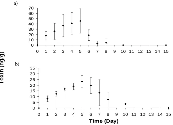
Cockles soft tissues were quickly consumed by fish. The fish stomach content was empty when specimens were removed from tanks. Nevertheless, PSTs were detected in the fish viscera throughout the exposure and elimination period. Despite the high PST content in cockles, the concentrations determined in fish were low. Toxins concentration increased over time during the exposure period reaching the maximum value of 19.6 ± 8.5 µg STXeq kg-1 in the last day of exposure (day 5) (Fig.3.2).
Toxin concentration decreased when fish were fed non-toxic cockles. The complete elimination of PSTs was achieved at day 15.
The PST profile determined in cockles differed markedly from the profile determined in fish viscera. While 10 PST analogues were found in cockles, only B1, dcSTX and C1+2 were detected in the fish viscera. B1 and dcSTX were detected throughout the
0 5 10 15 20 25 30 0 1 2 3 4 5 6 7 8 9 10 11 12 13 14 15
Time (Day)
m
g
S
T
X
e
q
k
g
-1Fig.3.2 Toxicity changes in the viscera of the fish Diplodus sargus sargus in the whole feeding experiment (Day 1-5, exposure; Day 6-15, elimination). Mean ± S.D. (n = 3).
experiment. B1 was the dominant compound, reaching 45.3 ± 23.0 ng g-1 in the last day of exposure (Fig.3.3a). High variable B1 concentrations were determined between replicates during the exposure period. The dcSTX concentrations also increased during the exposure period reaching 23.0 ± 5.2 ng g-1 (Fig.3.3b). C1+2 was sporadically detected and its occurrence were restricted to the first 6 days of the experiment (Table 3.1).
B1 concentration have a remarkably decrease in the first depuration day, having approximately 40 % of the toxin been eliminated. At day 10, B1 was completely eliminated. Contrary to B1, highly variable levels of dcSTX were detected during the depuration day, and a slower elimination was observed to dcSTX. At day 8, dcSTX concentration was still 7.4 ± 6.5 ng g-1. This compound was only completely eliminated in the last day of the experiment.
0 10 20 30 40 50 60 70 0 1 2 3 4 5 6 7 8 9 10 11 12 13 14 15 0 5 10 15 20 25 30 35 0 1 2 3 4 5 6 7 8 9 10 11 12 13 14 15 Time (Day)
PSTs were not detected in fish muscle throughout the experiment.
a)
b)
Fig.3.3 Changes in concentration of (a) B1 and (b) dcSTX in viscera of Diplodus sargus sargus during the whole feeding experiment (Day 1-5, exposure; Day 6-15, elimination). Mean ± S.D. (n = 3).
Table 3.1. Concentration of C1+2 in gastrointestinal tract of Diplodus sargus sargus during the whole feeding experiment (Day 1-5, exposure; Day 6-15, elimination) in each replicate (ND – not detected; NQ – not quantified).
Concentration of C1+2 (ng/g)
Replicates Day1 Day2 Day3 Day4 Day5 Day6 Day7 Day8 Day10 Day15 1 102.12 82.40 204.74 ND NQ NQ ND ND ND ND 2 ND ND NQ ND ND 74.97 ND ND ND ND 3 ND ND 79.63 ND 119.07 ND ND ND ND ND
3.4. Discussion
T
he previous chapter highlighted that the fish is capable of accumulating high PSTs concentrations from their prey. The present experiment aimed to understand the uptake/elimination dynamics of PSTs in fish through dietary exposure.While feeding on contaminated cockles, the PSTs concentration increased in fish viscera. However, the maximum concentration detected was two orders of magnitude lower than the PSTs concentration determined in the toxic dietary cockles. Fish specimens were collected for analysis in the following morning after being exposed to contaminated shellfish. Therefore, the toxin concentration detected corresponds to the remaining amount that was not eliminated. While C1+2 were the main PSTs found in cockles toxin profile, only small concentrations were detected in the fish samples. The main PSTs accumulated in the fish gastrointestinal tract were B1 and dcSTX. This result suggests that C1+2, which are the most soluble analogues of the PSTs group (Samsur et al., 2006), is rapidly excreted from fish tissues and thus contributing to the lower toxin concentration found in fish.
From the 10 compounds identified in cockles, only B1 and dcSTX showed a parallel increase of their concentration with the supply of contaminated food. On the contrary to C1+2, B1 and dcSTX are not rapidly eliminated from the gastrointestinal tract. Nevertheless, they were not found in the muscle tissue.
During the exposure period, the concentrations of B1 were consistently higher than dcSTX, which is in concordance with their proportion in cockles. Highly variable values were determined for B1, which is in contrast with the low variable values
determined for dcSTX. This result suggests that in addition to the uptake of B1 there was simultaneously a relevant elimination of this compound. When the toxic food supplied to the fishes was replaced by the non toxic cockles, B1 sharply decreased. During the first day of the depuration period, concentration of B1 decreased 40 % and by day 10 of the experiment (day 5 of depuration phase) B1 was completely eliminated. In contrast to B1, higher differences between replicates were found for dcSTX during the elimination period and not during the exposure period. These results indicate that dcSTX would be easier accumulated in fish tissues and B1 easier eliminated. In fact, dcSTX did not show an initial remarkable decrease when the non toxic diet was supplied to fishes. The complete elimination of dcSTX was only achieved in the last day of the experiment.
The uptake and elimination of PSTs is extensively studied for bivalve molluscs (Bricelj et al., 1991; Chebib et al., 1993; Bricelj and Shumway 1998). The ability to eliminate the accumulated PSTs varies markedly between species. Nevertheless, in most cases elimination patterns follow a negative exponential model (Bricelj and Shumway 1998). In our experiment, B1 followed this pattern, but not dcSTX. The uptake and elimination dynamics have been only studied recently in fish (Kwong et al., 2006). In their experiments, Kwong and colleagues found that although N-sulfocarbamoyl (C1+2) and carbamate (STX, neoSTX, GTX1, 2, 3 and 4) toxins were present in the prey (clams) tissues only C1+2 were detected in the fish, which suggested a possible differential accumulation or toxin degradation in the fish gut, as we noticed in the present study. The toxin concentration increase for the consecutive days of toxin exposure was also very similar. However our study presents the first data for the suite of toxins commonly found in the Portuguese and Galician coast.
This study provides relevant data for assessing the health risk of PSTs in seafood and the ecological repercussions of the fish as a toxin vector.
4.1. Introduction
Domoic acid (DA), known as the amnesic shellfish toxin, is in the Portuguese coast, mainly produced by Pseudo-nitzschia australis (Costa and Garrido, 2004).
Filter feeding organisms, such as mussels, concentrate high DA levels without apparent harm, which render them unsafe for human consumption. Domoic acid is a tricarboxylic aminoacid that binds irreversibly to glutamate receptor sites, causing destructive neuronal depolarisation and permanent short-term memory loss in mammals (Debonnel et al., 1989; Perl et al., 1990; Todd, 1993). For human health protection, shellfish harvesting is always banned when DA is detected above the regulatory limit of 20 µg DA g-1 (EU Directive 2004). In average, closures of shellfish harvesting do not last more than two weeks in the Portuguese coast (Vale et al., 2008). Domoic acid is rapidly eliminated from tissues of most shellfish species. In mussels, depuration rates of 0.49-0.99 day-1 and 0.40-0.58 day-1 have been observed for Mytilus edulis and M.
galloprovincialis, respectively (Blanco et al., 2002a; Novaczeck et al., 1992). However,
a small number of organisms have been shown to retain this neurotoxin for long periods of time. This is the case of the king scallop (Pecten maximus), sea scallop (Placopecten
4. Detection and sub - cellular distribution
of the Amnesic Shellfish Toxin in the
digestive gland of Octopus vulgaris during
periods of toxin absence
magellanicus) and razor clam (Siliqua patula). Low DA depuration rates ranging from
0.004 to 0.009 day-1 have been reported for them (Blanco et al., 2002b; Douglas et al., 1997; Horner et al., 1993). The mechanism beneath DA retention is still unknown.
Blooms of Pseudo-nitzschia diatoms, which are associated to upwelling events, occur preferentially during spring and early summer in the Portuguese coast (Palma et al., 2010). Detection of DA in bivalve molluscs is usually restricted to these seasons. Although DA is traditionally searched in bivalve molluscs, accumulation of DA in organisms such as planktivorous fish, crustaceans and cephalopods have also been reported (Costa et al., 2003, 2004; Lefebvre et al., 2002). These organisms can act as DA vectors and through feeding interactions in marine food webs they can be responsible for acute intoxications of marine mammals and seabirds (e.g. Scholin et al., 2000).
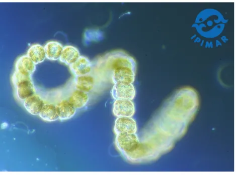
The common octopus (Octopus vulgaris) has been shown to accumulate DA in their tissues during spring (Costa et al., 2004). The highest levels of DA were detected in the digestive gland (storage organ) followed by the branchial hearts and the kidneys (excretory organs), which suggests a route for DA elimination (Costa et al., 2004). These authors detected DA in 90 specimens caught in the NW and S Portuguese coast between February and May 2003, and suggested that as all specimens showed DA contamination, octopus may not completely eliminate this toxin as most shellfish species do.
In the present study, to test whether octopus is one of the few species with the ability to retain DA, specimens were caught between September and November 2009 for DA
analysis and compared to values determined in bivalve molluscs obtained from the same fishing area. Having consistently found DA contamination in the octopus digestive gland, the sub-cellular distribution of the toxin was investigated through a sequential centrifugation to obtain insoluble (organelles) and soluble (cytosol) fractions.
Splitting the whole is a route towards understanding. The study of association of DA in cell compartments will provide valuable information to better understand octopus low DA depuration, possible toxic effects and DA trophic availability.
4.2. Methods
4.2.1. Collection and preparation of octopus and bivalve samples
Thirty two octopus (Octopus vulgaris) specimens were captured in four sampling dates between September and November 2009 in Peniche, NW Portuguese coast (Table 4.1). The specimens were weighed and measured. The digestive gland was removed, gently homogenized and a 5 g aliquot was weighted separately for DA determination.
Bivalve molluscs, Mytilus galloprovincialis, were obtained within the national monitoring program for marine toxins. Composite samples of at least 30 individuals or 1.5 Kg were weekly collected from Peniche and fortnightly in the adjacent fishing areas. A homogenate of shellfish soft tissue was prepared according the monitoring program and stored at -20 ºC for subsequent analysis.
Table 4.1 Octopus vulgaris: Sampling date (2009); number of individuals (n); total weight (mean ± SD) and digestive gland weight (mean ± SD).
4.2.2. Sub-cellular fractionation
Two digestive glands of 3 different levels of DA concentrations (22.0 ± 0.5 , 15.0 ± 0.5and 10.0 ± 0.5 µg g-1) were selected for sub-cellular analyses. Digestive glands were homogenized in triplicate at a dilution of 1:5 (wet weight: volume of buffer) of 0.1 M Tris pH 7.6 and 0.15 M ammonium formate buffer solution, while refrigerated by maintaining in a ice bath (Mauriz and Blanco, 2009). Homogenization was carried out during short periods of time and at low speed to minimize organelle breakage.
Fractionation of digestive glands was performed by differential centrifugation (adapted from Campbell et al., 2005; Raimundo et al., 2008). The homogenate was first fractionated at 700 x g for 15 min to separate the nucleus; the supernatant was centrifuged at 9000 x g for 20 min and 30 000 x g for 25 min for the mitochondrial and lysosomal fraction respectively. The last centrifugation at 100 000 x g for 40 min,
Sampling date
(day- month) N Total weight (g)
Digestive gland weight (g) 23- Sep 9 2322 ± 308 115 ± 26 19- Oct 8 2479 ± 406 141 ± 20 27- Oct 4 2904 ± 737 111 ± 41 18- Nov 11 1433 ± 571 46 28
separated the microsomes from the supernatant cytosol. Each centrifugation was performed at 4ºC.
4.2.3. Toxin extraction and HPLC analysis
The protocols for DA extraction and for the subsequent analysis with high performance liquid chromatography with ultraviolet detection (HPLC-UV) were show in Chapter 2.
4.2.4. Statistics
After verifying the non-guassian distribution of the data, the non parametric Kruskal– Wallis test was used in order to detect differences (p < 0.05) of DA concentrations between sampling dates and among sub-cellular fractions (octopus digestive glands). Having demonstrated significant differences among groups (sampling dates), we applied the Mann-Whitney test to find out where those differences were.
4.3. Results
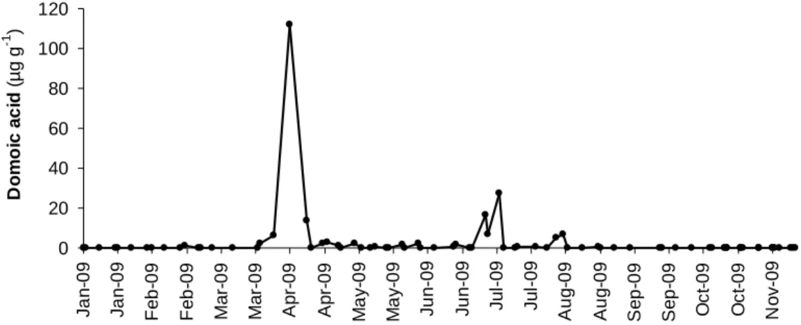
Seasonal variation of DA in mussels showed two main episodes of toxin occurrence, one in spring and a second one in summer (Fig.4.1). DA was firstly detected in February, reaching the highest concentration of 112 µg DA g-1 in early April. A sharp decrease to 3.1 µg DA g-1 was observed after two weeks. A second peak, with concentrations above the regulatory limit, was detected in July. DA was not found in mussels collected after early August. However, DA was detected in each of the 32 octopus specimens caught between September and November (Fig. 4.2).
0 20 40 60 80 100 120 J a n -0 9 J a n -0 9 F e b -0 9 F e b -0 9 M a r-0 9 M a r-0 9 A p r-0 9 A p r-0 9 M a y -0 9 M a y -0 9 J u n -0 9 J u n -0 9 J u l-0 9 J u l-0 9 A u g -0 9 A u g -0 9 S e p -0 9 S e p -0 9 O c t-0 9 O c t-0 9 N o v -0 9 D o m o ic a c id ( µg g -1 )
Fig.4.1 Seasonal variation of domoic acid concentration (µg g-1) in mussels, Mytilus galloprovinciallis, collected from Peniche and adjacent fishing area (NW Portuguese coast) during January and November of 2009.
Fig. 4.2 Domoic acid concentration (µg g-1) in the digestive gland (median, 25 and 75 quartiles, minimum and maximum, total n = 32) of Octopus vulgaris in 4 sampling dates (23- Sep; 19 and 27-Oct and 18 -Nov 2009). Significant differences at p < 0.05 are marked with *, after applying the Mann-Whitney test.
Concentration of DA determined in the digestive gland ranged from 1.0 to 26.6 µg DA g-1. A high intra-specific variability of DA (CV > 75 %) was found in three of the four sampling dates. Toxin levels were significantly different between sampling dates (H3=13,1 p < 0.05). Although toxin concentrations determined in specimens caught at
October 19th were significantly higher than specimens caught in the remaining dates, a *
decrease of toxin concentration in function of time (between September and November) was not evident. In fact, the highest DA concentration was determined for a specimens caught in November (Fig 4.2).
80 85 90 95 100 <10 14 -16 >20 R e la ti v e D o m o ic A c id A b u n d a n c e ( % ) 0 1 2 3 4 5 <10 14 -16 >20 R e la ti v e D o m o ic A c id A b u n d a n c e ( % )
Cytosol Nuclei Mitochondria Lysosome Microsome
Fig. 4.3 Relative abundance (mean ± SD) of domoic acid in digestive gland cell fractions of Octopus
vulgaris naturally contaminated with 3 levels of toxin (10.0 ±0.5, 15.0 ±0.5 and 22.0 ± 0.5 µg DA g-1): a) soluble fraction (cytosol) and b) four insoluble fractions (nuclei, mitochondria, lysosome and microsome).
Sub-cellular partitioning of DA in the soluble and insoluble fractions (nuclei, mitochondria, lysosome, microsome) showed that nearly all DA (92 to 94 %) is found in cytosol (Fig. 4.3 a). The distribution of the remaining DA in each fraction was found in the following decreasing order: nuclei > lysosomes > mitochondria > microsomes (Fig.4.3 b). No significant differences were found between DA concentrations (H2=4.57, p > 0.05). Nevertheless, an increase of DA relative abundance in organelles
fractions, namely in the nuclei and mitochondria, with decreasing in the whole digestive DA Concentration (µg g-¹)
b) a)
gland was observed. On the contrary, the lysosome fraction showed the highest DA relative abundance in the most toxic samples.
4.4. Discussion
Domoic acid is recurrently detected in shellfish during the spring-summer season, when blooms of Pseudo-nitzschia diatom occur associated with upwelling events (Palma et al., 2010; Vale and Sampayo 2001). Not surprisingly, DA was detected in mussels collected from February to August in the NW Portuguese coast during 2009. Mussels have been used as bioindicator organisms within the monitoring program for marine toxins (Vale et al., 2008). They are good indicators of the occurrence of
Pseudo-nitzschia blooms, in particularly, and of the contamination of the marine environment in
general. Another ecological role of bivalve molluscs is their importance as a food item in the octopus diet (Anderson et al., 2008; Rosa et al., 2005).
Although not being detected in mussels since early August, DA was detected in the digestive gland of each octopus specimen caught from September to November of 2009. Concentrations up to 26.6 µg DA g-1 were determined in these specimens while concentrations up to 166 and 124 µg DA g-1 were recorded in octopus samples collected during blooms of toxic plankton in May 2003 and June 2007, respectively (Costa et al., 2004; Costa and Pereira, 2010). Considering that octopuses are active predators with high growth and metabolic rates and DA is a hydrophilic compound, a fast DA elimination from octopus tissues would be reasonably expected. Moreover, a time-course decrease of DA concentration due to their degradation and elimination from octopus tissues would also be expected to be observable in the samples collected in
September-November 2009. These results suggest that a mechanism for retention of DA leading to slow toxin elimination lies beneath.
The sub-cellular fractionation of digestive glands showed that nearly all DA is found in the soluble fraction (cytosol) and not bound to any organelle. Residual retention of DA in the octopus digestive gland is not unique. Most of the DA retained in digestive gland of king scallops is in the free form in the cytosol and not bound to any substance, as reported by Mauriz and Blanco (2009). These authors also suggest that for zwitterion compounds, such as DA, a membrane transporter would be required to effectively eliminate DA from the cell. For tetrodotoxin (TTX), which is a zwitterion compound with neurotoxic properties that accumulates in the liver of certain puffer fish species, a binding protein has been purified (Matsui et al., 2000; Yotsu-Yamashita et al., 2002). Nevertheless, most of TTX may remain unbound (Matsumoto et al., 2010). Sub-cellular distribution of TTX in fish liver cells showed an identical distribution pattern to DA in digestive gland of octopus and scallops (Nagashima et al., 1999). Irrespectively to toxin levels, TTX was predominantly found in the cytosol.
In our study no significant differences (p > 0.05) were found on the sub-cellular distribution between levels of DA. Nevertheless, the relative DA abundance in fractions containing organelles with vital functions, such as nuclei and mitochondria, decreased with increasing toxin concentrations in the whole digestive gland. Lysosomes, which have functions of storage, degradation and elimination of toxic and waste compounds appear to be the dominant fraction when DA reaches higher levels in octopus digestive gland. DA toxicity to cell compartments is not known. In fact, the toxicity of DA to marine invertebrates has been scantily studied. Increasing concentrations of DA
intra-muscular injected to mussels suggested genotoxic responses of digestive gland cells that rapidly repaired after few days of incubation (Dizer et al., 2001).
This is the first report showing that octopus can retain DA for long periods after diatom bloom events. Nearly all DA remains in the soluble fraction of digestive gland cells. If similar to contaminants such as metals, this intracellular disposition may favour the trophic transfer of DA from prey to predators (Wallace and Lopez, 1997; Wallace and Luoma, 2003). Furthermore, the octopus central position in the marine food web and their low toxin depuration suggests octopus as highly potential toxin vector.