UNIVERSIDADE DE LISBOA
FACULDADE DE CIÊNCIAS
Departamento de Biologia Vegetal
NON-VIRAL GENE DELIVERY TO HUMAN MESENCHYMAL
STEM CELLS USING CATIONIC LIPOSOMES
Rui Daniel Martins Nunes Mendes
Mestrado em Biologia Molecular Humana
UNIVERSIDADE DE LISBOA
FACULDADE DE CIÊNCIAS
Departamento de Biologia Vegetal
NON-VIRAL GENE DELIVERY TO HUMAN MESENCHYMAL
STEM CELLS USING CATIONIC LIPOSOMES
Rui Daniel Martins Nunes Mendes
Mestrado em Biologia Molecular Humana
Dissertação orientada por:
Doutora Cláudia Alexandra Martins Lobato da Silva
(Instituto Superior Técnico, Universidade Técnica de Lisboa)
Doutora Maria Gabriela Gomes de Figueiredo Rodrigues
(Faculdade de Ciências da Universidade de Lisboa)
Table of Contents
List of figures ... i
List of tables ... ii
List of Abbreviations ... iii
Acknowledgements ... iv
Abstract ... v
Resumo ... vi
Resumo alargado... vii
Keywords ... xi
Palavras-Chave ... xii
I. Introduction ... 1
I.1 Stem Cell Basics ... 1
I.2 An overview on Mesenchymal Stem Cells ... 2
I.2.1 Mesenchymal Stem Cell Features ... 2
I.2.2 Mesenchymal Stem Cell Characterisation ... 3
I.2.3 Mesenchymal Stem Cells in Cell Therapy ... 3
I.3 Mesenchymal Stem Cells in Gene Therapy ... 5
I.3.1 Mesenchymal Stem Cells as a potential therapeutic ... 5
I.3.2 Application of Genetically-engineered Mesenchymal Stem Cells ... 6
I.3.3 Gene Delivery to Mesenchymal Stem Cells ... 7
II. Aim of Studies... 9
III. Materials and methods ... 10
III.1 Plasmid ... 10
III.2 Isolation of pDNA ... 10
III.3 Real-Time PCR for determination of MSC plasmid content ... 10
III.4 Cell samples ... 11
III.5 Mesenchymal Stem Cell Thawing and Expansion ... 11
III.6 Lipofection ... 12
III.7 Detection of eGFP expression and cell sample preparation for RT- PCR ... 12
III.8 Immunophenotype Staining for flow cytometry analysis ... 12
III.9 Evaluation of Differentiative Potential ... 13
III.10 Colony- Forming Units (CFU) Assay ... 13
IV. Results and Discussion ... 14
IV.1 Real-Time PCR method establishment ... 14
IV.1.1 Cell Number and Lipofectamine effects on Real Time-PCR amplification efficiency ... 15
IV.1.2 Standard Curve and Limits of Real-Time PCR detection ... 17
IV.2 Transfection of Human Mesenchymal Stem Cells ... 18
IV.2.1 Optimization of Transfection protocol using Lipofectamine ... 18
IV.2.1.1 Effect of Cell Plating Density on Transfection Efficiency ... 19
IV.2.1.2 Effect of the Ratio Lipofectamine/pDNA on transfection efficiency assessed by Flow Cytometry and Real-Time PCR analysis ... 20
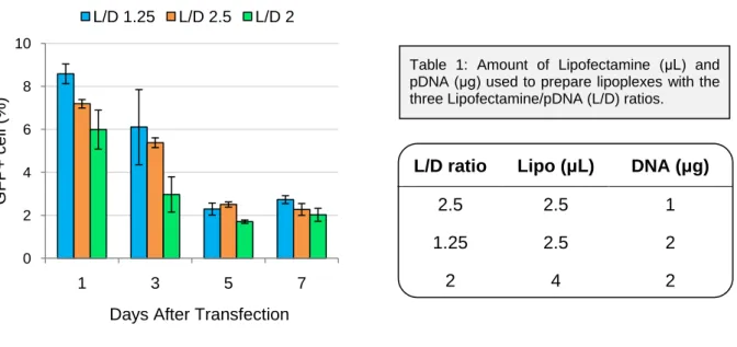
IV.2.2 Analysis of eGFP expression using different Lipofectamine/ pDNA ratios ... 21
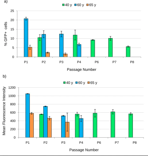
IV.2.3 Effect of Passage Number on Transfection Efficiency assessed by Flow Cytometry and Real-time PCR ... 22
IV.3 Maintenance of Multipotency of Mesenchymal Stem Cell after Transfection ... 26
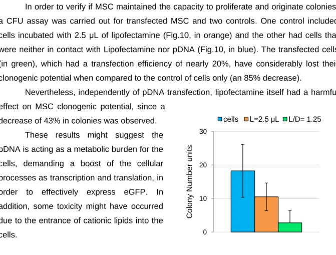
IV.3.1 MSC Clonogenic potential ... 26
IV.3.2 MSC Differentiation potential ... 27
IV.3.3 MSC Immunophenotypic Analysis ... 28
V. Conclusions and Future Trends ... 29
i
List of figures
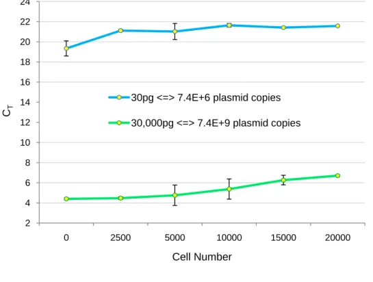
Figure 1: Analysis of the effect of MSC number on the threshold cycle (CT). These analyses were
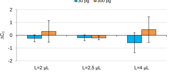
carried out for both 30 and 30,000 pg of pDNA. Results are the mean of two independent experiments with standard deviation. ... 15 Figure 2: Influence of Lipofectamine on the threshold cycle (CT), for amplification of 30 and 300
pg of pDNA. The CT values represent mean and standard deviation values of 4 different assays.
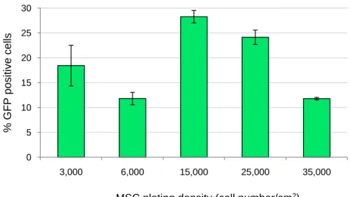
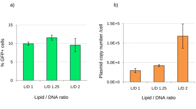
... 16 Figure 3: Calibration curve obtained from 0.5 pg to 100 ng of plasmid DNA spiked in 10,000 MSC. Values from 30 to 30,000pg represent mean and standard deviation values of 11 different assays. Linear range was observed between 3 pg to 30,000 pg with Ct= -1.87ln (plasmid copy number) + 49.215, R2=0.9993. ... 17 Figure 4: Phase contrast (left) and fluorescence (right) microscopic view of lipofectamine-mediated pVAX-GFP transfer in MSC, 24 hours after transfection. Images obtained by fluorescence optical microscopy (100x). ... 18 Figure 5: Transfection efficiency evaluation with respect to different cell plating densities. Cells plated at lower densities (3,000 and 6,000 cell/cm2) were grown in culture for 3 days before transfection. After that, cells plated both at low and high densities were transfected. The transfection efficiency was determined by the number of GFP positive cells. MSC were transfected at passage 7. Results represent mean and standard deviation values of duplicates. 19 Figure 6: Differences between the percentage of eGFP positive cells assessed by flow cytometry analysis (a) and intracellular plasmid copy number determined by RT-PCR (b). MSC were transfected at passage 6 with three different Lipofectamine/pDNA (L/D) ratios: 1, 1.25 and 2. Results are the mean of four independent experiments with standard deviation. ... 20 Figure 7: Evaluation of eGFP expression along 7 days after transfection. MSC were transfected at passage 4 with three different Lipofectamine/pDNA (L/D) ratios: 1.25, 2 and 2.5. eGFP positive cells were analysed by flow cytometry and the mean values ± standard deviation are represented. ... 21 Figure 8: Analysis of the cell passage number effect on eGFP expression. MSC from 3 different donors (40, 60 and 65 years old) were transfected from passage 1 to 8. Results were analysed by the a) % eGFP + cells and b) mean fluorescence intensity through flow cytometry. Mean values are represented ± standard deviation of duplicates. ... 23 Figure 9: Percentage of eGFP positive cells assessed by flow cytometry analysis (a) and intracellular plasmid copy number determined by RT-PCR (b) at different cell passages. MSC from two different donors (60 and 65 years old) were transfected from passage 1 to 4. Results are displayed as mean ± standard deviation of duplicates. ... 24 Figure 10: CFU assay of MSC after transfection (in green). Two different controls were assayed, cells which have not been in contact with lipofectamine and pDNA (blue), and cells which have only been in contact with lipofectamine (L=2.5 μL – orange). MSC were transfected at passage 4 with a transfection efficiency of nearly 20%. The assay was performed in duplicate. Results are presented as mean ± standard deviation of two independent experiments. ... 26 Figure 11: Evaluation of transfected MSC differentiation potential through observation of the standard staining tests. Images obtained through fluorescence optical microscopy (200x). a) Oil red-O staining and b) Alkaline Phosphatase and Von Kossa stainings were tested in order to analyse the maintenance of adipogenic and osteogenic differentiation potential after transfection. Controls with cells and cells incubated with lipofectamine (L=2.5μL) were also performed. MSC were transfected at passage 4 with a transfection efficiency of nearly 20%. The assay was performed in duplicate. ... 27
ii
List of tables
Table 1: Amount of Lipofectamine (μL) and pDNA (μg) used to prepare lipoplexes with the three Lipofectamine/pDNA (L/D) ratios...21 Table 2: MSC viability and recovery at different passages (from 1 to 8), 24 hours after transfection. Controls are non transfected cells. Results presented as mean ± standard deviation of duplicates...24 Table 3: Immunophenotype evaluation of transfected MSC through flow cytometry analysis. The identification of MSC characteristic phenotype was assessed by fluorescent-conjugated monoclonal antibodies against CD73, CD105 and CD 90. Controls with non-transfected cells were also performed as well as the appropriate isotype controls IgG γ1 and IgG γ2. Results are presented as mean... 28
iii
List of Abbreviations
∆CT Threshold variation
BDNF Brainderived neurotrophic factor
BM Bone marrow
BMP Bone morphogenetic protein
bp base-pair
CAc Number of non-transfected cells alive
CAt Number of transfected cells alive
CD Cluster of differentiation
CFU Colony-forming units
CMV Cytomegalovirus
ColE1 E.coli plasmid vector Colicin E1
CR Cell recovery
CT Threshold cycle
DC Dendritic cell(s)
DMEM Dulbecco’s Modified Eagle’s Medium
dsDNA Double-stranded DNA
eGFP Enhanced green fluorescent protein
ESC Embryonic stem cell(s)
FACS Fluorescence-activated cell sorting
FBS Fetal bovine serum
GFP Green fluorescent protein
GVHD Graft-versus-host disease
HGF Hepatocyte growth factor
HSC Hematopoietic stem cell(s)
hTERT Human telomerase reverse transcriptase
IFN-β Interferon-beta
IFN-γ Interferon-gamma
Ig Immunoglobulin
IL Interleukin
IMDM Iscove’s modified Dulbecco’s medium
L/D ratio Lipofectamine/ DNA ratio
MHC Major histocompatibility complex
MNC Mononuclear cells
MSC Mesenchymal stem cell(s)
NK Natural killer
PBS Phosphate buffered saline
PCR Polymerase chain reaction
PD Population doubling
pDNA Plasmid deoxyribonucleic acid
PE R-phycoerythrin
PFA Paraformaldehyde
RT-PCR Real-time polymerase chain reaction
Th1 Type 1 helper T
TNF Tumor necrosis factor
TRAIL Factor-related apoptosis-inducing ligand
iv
Acknowledgements
This thrilling cycle is ending up ... I cannot finish without being thankful to some of you ...
First, I would like to thank to Professor Joaquim Cabral for the opportunity of performing my Master Thesis at Institute for Biotechnology and Bioengineering (IBB), Instituto Superior Técnico (IST).
Cláudia thank you very much for receiving me at the Stem Cell Bioengineering Laboratory (IBB-IST), allowing me to born for Science! I am very proud and grateful for the opportunity to embrace this challenging project in this great team. Your availability, whenever possible, and your exceptional guiding were the main key for this successful project! I hope I was somehow useful to the lab, to which I wish a good fortune!
Catarina and Sofia I sincerely don´t know how to thank you for all your support! You were truly amazing! I really appreciate all your supervision where a good mood was the hallmark. You were tireless, being always up for guiding and helping me, even when you were hard working. I will never forget being part of the transfection team!
Greetings to all the Stem Cell Bioengineering team for all your help, support, advices and companionship.
I want to take the opportunity to deeply thank my friends for your best friendship, which gave me a special force during this long journey! An enormous hug to each of you for your special peculiarity!
For my brother, my mother and my father I still don´t know how to thank you, since your completely dedication and love is overly long! You´re my fortune, huge thanks for your unconditional care!
Finally, you know how I´m wholly grateful to find you in this giant world! I cannot imagine living without you, my angel! All your love, all your friendship, all your company and dedication give me the smile every morning I wake up! I just want to keep the plane and start our new life. I love you forever!
v
Abstract
Mesenchymal Stem Cells (MSC) hold a great promise for application in several therapies due to their unique biological characteristics. MSC can be easily isolated from bone marrow (BM) based on its adherence to plastic, and can be expanded in culture while maintaining the capacity to differentiate into mesoderm-lineage cell types. These cells have demonstrated immunosuppressive properties, allowing their escape from host allogeneic responses. Importantly, MSC secrete a large spectrum of bioactive molecules that provide a regenerative microenvironment for a variety of injured tissues. Despite being successfully used for the treatment of many diseases, it might be essential to enhance some of their features through gene delivery strategies to harness their full potential in cell or gene-based therapies.
In this context, the main goal of this work was to develop an efficient and safe methodology to genetically-engineer mesenchymal stem cells, enhancing their therapeutic efficacy in Regenerative Medicine settings. To this end the delivery of plasmid DNA (pDNA) encoding the gene of a green fluorescent protein (pVAX-GFP) was optimized for BM MSC using Lipofectamine, a cationic liposomes-based reagent. Importantly, it was also established a Real-time Polymerase Chain Reaction (RT-PCR) method for quantification of pVAX-GFP copy numbers in lipofected MSC.
The new RT-PCR method, which was considerably useful for transfection optimization, showed good reproducibility and high sensitivity, covering a wide linear range from 74 to 7.4×10⁵ copies pDNA/cell. MSC demonstrated better transfection efficiencies when plated both at earlier passages and high cell densities (15,000-25,000 cell/cm2) with an L/D 1.25 ratio. The obtained values of transfection efficiency ranged from 1.6% to 28.3% and eGFP was expressed during seven days. As transfected MSC have shown high cell viabilities and cell recoveries while maintained their multipotency, this is an advantageous transfection strategy when it is desirable to efficiently express the therapeutic gene in a safe and transient way.
vi
Resumo
As características biológicas típicas das células estaminais do mesênquima (Mesenchymal Stem Cells – MSC) têm despertado o interesse para a sua potencial aplicação em diversas terapias. As MSC podem ser isoladas da medula óssea e depois expandidas, mantendo a capacidade de diferenciação em células de linhagens mesodérmicas. Estas células demonstraram propriedades imunosupressivas, permitindo a evasão a respostas alogénicas no hospedeiro. As MSC excretam um vasto espectro de moléculas bioactivas, fornecendo um microambiente regenerativo em diversos tecidos lesados. Apesar de terem sido utilizadas com sucesso no tratamento de várias doenças, torna-se necessário melhorar algumas das suas características através de estratégias de transfecção, aproveitando todo o seu potencial em terapias celulares e genéticas.
Neste contexto, o objectivo principal deste trabalho foi o desenvolvimento de uma metodologia eficiente e segura para modificar as MSC geneticamente, melhorando a sua eficácia terapêutica em Medicina Regenerativa. Com este propósito foi optimizada a transfecção de DNA plasmídico (DNAp), contendo um gene que codifica para a proteína fluorescente verde (pVAX-GFP), em MSC da medula óssea usando Lipofectamina, um reagente à base de lipossomas catiónicos. Foi também estabelecido um método através de Real-Time PCR para a quantificação do número de cópias de pVAX-GFP dentro das células lipofectadas.
O novo método desenvolvido por RT-PCR, demonstrou ser bastante útil na optimização da transfecção, apresentou boa reprodutibilidade e elevada sensibilidade, tendo sido detectada uma ampla gama de quantidades DNAp/célula (entre 74 e 7,4×10⁵ cópias). As MSC apresentaram melhores eficiências de transfecção quando plaqueadas a passagens celulares baixas e densidades elevadas (15.000-25.000 céls./cm2), para uma razão L/D de 1,25. Os valores obtidos variaram de 1,6% a 28,3% e a expressão de GFP durou pelo menos sete dias. Uma vez que as MSC transfectadas apresentaram boas viabilidades e recuperação celulares, mantendo a sua capacidade de multipotência, esta estratégia de transfecção é vantajosa nos casos em que se pretende uma expressão eficiente, segura e transiente de um gene terapêutico.
vii
Resumo alargado
Devido às suas propriedades únicas de auto-renovação e diferenciação, o uso de células estaminais para fins terapêuticos tem despoletado um enorme interesse para inúmeras aplicações em Medicina Regenerativa. Neste campo, as células estaminais do mesênquima (mesenchymal stem cells - MSC) têm assumido um papel de destaque, uma vez que são providas de uma panóplia de características que as colocam como uma promissora alternativa no tratamento de diversas doenças genéticas ou adquiridas.
Esta população de células adultas que foi originalmente obtida a partir da medula óssea pode também ser isolada de outros tecidos como o tecido adiposo e o cordão umbilical. Apesar de existirem em pequenas quantidades na medula óssea (0.01-0.001% do total de células mononucleadas), estas células podem ser facilmente isoladas e expandidas in vitro.
Durante o seu estudo, algumas características únicas das MSC desde logo despertaram a atenção da comunidade científica. Em primeiro lugar, as MSC têm a capacidade de diferenciação em diversas linhagens mesodérmicas dando origem a células do tecido ósseo, adiposo, cartilaginoso e muscular. Em segundo, estas células são dotadas de um surpreendente conjunto de mecanismos, baseados em propriedades imunomodulatórias e hipo-imunogénicas que permitem escapar a respostas alogénicas após transplantação. Além disso, as MSC apresentam uma actividade secretória intrínseca, libertando um vasto espectro de macromoléculas bioactivas e imunoreguladoras que estabelecem um microambiente regenerativo em tecidos lesados ou destruídos. Esta capacidade regenerativa das MSC pode ser explicada através da sua actividade secretora, nomeadamente pela libertação de factores bioactivos que, para além de promoverem a angiogénese e a mitose de células estaminais presentes no próprio tecido, são também capazes de inibir a apoptose. Finalmente, alguns autores defendem uma capacidade migratória destas células para os tecidos lesados após administração intravenosa.
Deste modo, ainda que os mecanismos promotores da regeneração tecidular e de imunoregulação por parte das MSC careça de uma explicação mais detalhada, os dados disponíveis apoiam distintamente a ideia de utilizar MSC autólogas e alogénicas como agentes terapêuticos para aplicação em Terapia Celular e Terapia Génica.
Actualmente, existem diversos estudos de transplantação, nos quais MSC expandidas ex-vivo e transplantadas num tecido lesado, como sendo osso ou cartilagem, foram capazes de diferenciar e reparar o tecido, recuperando parte da sua função normal.
Devido à notável eficácia dos tratamentos com recurso ao transplante de MSC alogénicas e à ausência de uma resposta imunitária do hospedeiro após administração local
viii ou sistémica, as MSC são vistas como um transportador ideal para a expressão de genes terapêuticos em tecidos de interesse. Deste modo, a utilização de MSC para terapia génica tem ganho cada vez mais adeptos, uma vez que a alteração genética de MSC pode ser aplicada segundo diferentes estratégicas terapêuticas. Estas células podem ser utilizadas como agentes imunosupressivos ou como células modificadas para excretar diferentes proteínas terapêuticas (in vitro ou in vivo) para terapia celular de substituição enzimática, ou em doenças genéticas e adquiridas, como desordens ao nível da medula óssea, osso, cartilagem e até mesmo cancro.
As grandes vantagens da utilização de terapia génica em relação aos fármacos convencionais são a possibilidade de um efeito de longa duração após uma única intervenção e a possibilidade de expressão dos genes de interesse apenas no local alvo, enquanto a difusão sistémica de medicamentos poderá ter efeitos secundários adversos.
Um extenso número de estudos revelou que as MSC podem ser eficientemente transfectadas com genes terapêuticos, incluindo colagénio I para tratamento de osteogenesis imperfecta, factor de coagulação VIII e IX para hemofilia, interferão beta (IFN-β) e IL-2 para neoplasias e BMP-2 e 4 para uma série de problemas músculo-esqueléticos.
No entanto, as MSC não têm sido apenas modificadas para terapia génica, mas também para melhorar o seu potencial terapêutico. Uma vez que um dos principais mecanismos que contribuem para a regeneração de tecidos é a sua actividade parácrina, estas células podem ser alteradas com vista a fomentar a sua capacidade de migração para o tecido lesado e secreção de factores bioactivos.
Tendo em vista a alteração genética de MSC, diferentes sistemas de modificação têm sido utilizados. Grande parte dos métodos de modificação de MSC utiliza vírus defectivos como, lentivírus e retrovírus. Apesar dos sistemas virais serem particularmente eficientes no transporte de DNA para as MSC e na consequente expressão proteica, alguns testes clínicos realizados alertaram para o perigo de segurança relacionado com o uso destes vectores, nomeadamente, a sua imunogenicidade, elevada toxicidade e possibilidade de induzir mutagénese. Por outro lado, os vectores não-virais oferecem vantagens relacionadas com a segurança como uma baixa imunogenicidade e toxicidade, são facilmente produzidos e conseguem transportar maiores quantidades de material genético. Os lipossomas catiónicos, por exemplo, estão descritos na literatura como um dos sistemas não-virais mais eficientes na transfecção de MSC, associados a baixos níveis de toxicidade para a célula.
Neste contexto, o objectivo deste trabalho foi o desenvolvimento de uma metodologia segura e eficiente para alterar geneticamente MSC, tendo em vista a sua futura aplicação num vasto espectro de terapias clínicas. Deste modo, pretendeu-se optimizar a transfecção de MSC provenientes da medula óssea utilizando um reagente comercial (Lipofectamina
ix 2000) constituído por lipossomas catiónicos. Estes vectores são constituídos por lípidos de natureza catiónica que interagem electrostaticamente com moléculas de DNA carregadas negativamente, permitindo a passagem do DNA através da membrana celular por endocitose. Utiliza-se também na sua formulação um lípido neutro com propriedades fusogénicas que promove a libertação do DNA do endossoma para o citoplasma evitando a sua degradação no lisossoma.
Tendo em vista a optimização da transfecção, foi estabelecido um método de Real-time PCR para quantificar o número de cópias de DNA plasmídico (DNAp) que entra em cada célula. Para determinar o número de cópias de DNAp foi utilizado um sistema de detecção de fluorescência, emitida por um agente que se intercala no DNA de cadeia dupla, o SYBR Green I. Este sistema consegue detectar o ciclo de amplificação no qual se observa um aumento significativo da fluorescência, designado de CT. Através deste valor é então
possível calcular a quantidade de DNAp existente numa amostra de células transfectadas. Este novo procedimento tem a vantagem de não necessitar de passos de lise celular e purificação do DNAp, o que permite uma maior precisão na quantificação. Neste estudo foi demonstrado não só a importância de definir um número de células constante (10.000 MSC por reacção) na construção da curva de calibração e na posterior análise de cada amostra, mas também que a lipofectamina não influencia o CT. Verificou-se que o método
estabelecido através de RT-PCR é sensível e reprodutível, detectando uma vasta gama de quantidades de DNAp (74 - 7,4×10⁵ cópias/célula).
Uma vez estabelecido o método, procedeu-se então à optimização da transfecção do plasmídeo pVAX-GFP, que codifica o gene repórter para a proteína fluorescente verde (enhanced green fluorescent protein - eGFP). Através de citometria de fluxo e Real-Time PCR foram analisados parâmetros importantes na obtenção de melhores eficiências de transfecção como a densidade celular a que as células são plaqueadas e as razões de lipofectamina (L) e DNAp (D) usadas na preparação de lipoplexos. Verificou-se que a melhor densidade celular inicial é de 15.000 células por cm2 (30.000 MSC por poço numa placa de 24 poços) e a melhor razão L/D é 1,25 (L=2,5 μL, DNAp=2 μg). Seguidamente observou-se que a expressão do plasmídeo é de curta duração (aproximadamente 7 dias) e que as melhores eficiências de transfecção são obtidas nas passagens celulares mais baixas. Os níveis de toxicidade detectados na transfecção com lipofectamina foram inferiores a 20%.
Por último, foi analisada a manutenção das características de multipotência das MSC após lipofecção, tendo-se verificado que estas células mantêm o seu potencial diferenciativo multilinhagem (osso e gordura). Observou-se apenas um decréscimo (inferior a 9%) na expressão dos marcadores CD73, CD105 e CD90 quando comparados com células não transfectadas.
x Em suma, demonstrou-se que a quantificação do número de cópias de DNAp por célula pode ser uma ferramenta relevante para a optimização de protocolos de transfecção, especialmente no que diz respeito à relação entre a expressão e o número de cópias de plasmídeo numa população de células transfectadas. Considerando as eficiências de transfecção obtidas, conclui-se que os resultados foram bastante satisfatórios, uma vez que atingiram-se níveis de expressão elevados (≈30%) comparativamente com a maioria da literatura existente (2-10%). Uma vez que este sistema é seguro, apresenta moderadas eficiências de transfecção e baixos níveis de toxicidade, é considerado uma alternativa promissora para alteração genética de MSC, tendo em vista terapias onde é desejável a expressão transiente, de curta-duração, de um gene.
xi
Keywords
Mesenchymal Stem Cells Gene Therapy
Non-Viral Vectors Cationic Liposomes Real-Time PCR Plasmid Copy Number
xii
Palavras-Chave
Células Estaminais do Mesênquima Terapia Génica
Vector não-viral
Lipossomas catiónicos Real-Time PCR
1
I. Introduction
I.1 Stem Cell Basics
A stem cell is a special type of cell that has a unique capacity to self-renew and to give rise to specialized cell types. Unlike the majority of cells in our body which are committed to conduct a specific function, such as skin, blood, muscle, and nerve cells, a stem cell is uncommitted and remains in this state, until it receives a signal to specialize and differentiate into a tissue-specific cell type. Due to their proliferative capacity and ability to become specialized, these cells hold an enormous promise to Regenerative Medicine for the repair and regeneration of cells and tissues that are damaged or dead [1].
Stem cells can be found since the early stage of human development until the end of life and can be classified as embryonic or adult, depending on their tissue of origin. Both embryonic and adult stem cell types may be useful for biomedical research, but each one has both advantages and limitations. An embryonic stem cell (ESC) is derived from a group of cells called the inner cell mass, which is part of the blastocyst (early 4 to 5-day embryo). ESC are pluripotent since they can give rise to cells from the three germ layers (mesoderm, endoderm, and ectoderm) from which all the cells of the body arise [2]. The potential use of ESC in human treatments is controversial not only for ethical reasons, but also because there is some evidence, based on clinical findings, that the use of ESC-derived cells in long-term therapies may carry risks due to a neoplastic potential of these cells [3].
An adult stem cell is an undifferentiated cell type found in specialized tissues, which has the ability to self renew and differentiate, giving rise to all the mature cell types, with characteristic morphologies and specialized functions, that maintain the integrity and functionality of a specific tissue, such as muscle or skin. Although quite rare, adult stem cells are found in a number of different tissues of fully developed humans like bone marrow, eye, fat, brain, gastrointestinal tract, epidermis, and liver [4].
The most well characterized adult stem cell type is the hematopoietic stem cell (HSC) [5], which give origin to all blood cell types. Nevertheless, another bone marrow stem cell type rapidly became well known, by their unique and interesting properties, the Mesenchymal Stem Cells (MSC).
2
I.2 An overview on Mesenchymal Stem Cells
I.2.1 Mesenchymal Stem Cell Features
Human bone marrow (BM) contains a rare population of stem cells, named Mesenchymal Stem Cells (MSC), which represent only about 0.01-0.001% of the total mononuclear cell (MNC) fraction, a proportion that further decreases with age [6]. Though originally isolated from BM, MSC have been found in several tissues, including adipose tissue, umbilical cord, peripheral blood, liver and fetal tissues among others [7].
MSC are capable of multilineage differentiation into mesoderm-type cells, namely fibroblasts, osteoblasts, chondroblasts, adipocytes and myoblasts [8]. However, some reports suggest that these cells can also give rise to cells with neuroectoderm and endoderm characteristics in vitro [9].
MSC are among the first stem cell types to be used in Regenerative Medicine due to several attractive biological characteristics. Firstly, the large interest in MSC relies on their ease isolation from several tissues and their extensive capacity for in vitro expansion without compromising its genetic stability [10]. Secondly, there is emerging evidence that MSC display an astonishing array of mechanisms based on MSC hypoimmunogenic and immunomodulatory properties, which allow their escape from host allogeneic responses. Their characteristic phenotype indicates that MSC express intermediate levels of MHC class I, lacking expression of MHC class II and co-stimulatory molecules CD40, CD80 and CD86. As a result, MSC class I antigens may stimulate alloreactive T cells, but since co-stimulatory molecules are not expressed a secondary signalling cannot proceed [11]. In addition, MSC hold an intrinsic secretory activity, releasing a broad spectrum of bioactive macromolecules, which are immunoregulatory and establish a regenerative microenvironment at sites of injured or damaged tissue. The immunoregulatory effect is characterized by a strong inhibition of host T-cell recognition and expansion owing to the inhibition of Tumor Necrosis Factor-alpha (TNF-α) secretion by dendritic cells (DC) and Interferon-gamma (IFN-γ) production by Type 1 helper T (Th1) cells or Natural Killer (NK) cells. Furthermore, MSC pre-induce a state of immune tolerance increasing IL-10 levels that stimulate regulatory T cells.
In fact, MSC regenerative capacity has been explained based on its secretory activity, i.e. bioactive factors, that can not only inhibit scarring and apoptosis, but also stimulate angiogenesis and mitosis of tissue-intrinsic stem cells [1], allowing the repair and regeneration of injured or damaged tissue.
3 Finally, it may be possible that upon intravenously administration MSC have the ability to migrate to damaged tissues. Although, this migratory capacity is not well understood, it has been observed in many studies with animal injured models [12].
Overall, while the mechanisms underlying those effects are yet to be verified, the data available clearly support the concept of using allogeneic MSC as therapeutic agents.
I.2.2 Mesenchymal Stem Cell Characterisation
The noteworthy therapeutic potential of MSC has definitely increased the interest in a wide variety of biomedical research. Regarding this fact, different methods for their isolation and expansion, as well as different approaches for MSC characterization were determined.
Conventionally, MSC are isolated from a low-density mononuclear fraction of BM aspirates by their capacity for adherence to plastic surfaces; nevertheless, MSC cultures established by this method are heterogeneous containing not only MSC but also other progenitor cell populations [13]. Different efforts were made to describe MSC phenotype through their expression of many CD markers [12] but since a MSC specific marker is yet to be found, it is difficult to identify an homogeneous MSC population. In order to deal with an increasing difficulty to compare and contrast study outcomes, the Mesenchymal and Tissue Stem Cell Committee of the International Society for Cellular Therapy proposed, in 2006, the minimal criteria to define MSC based on phenotypic and functional characteristics [14]. First MSC must be plastic-adherent when cultured in standard conditions. Second, cells must express a specific pattern of surface antigen expression, namely CD73, CD90, CD105, and not express CD45, CD34, CD14 (hematopoietic markers) and MHC class II. Third, MSC must demonstrate multipotent differentiation potential into at least three different lineages: osteogenic, adipogenic and chondrogenic.
Subsequently, this minimal set of standard criteria has simplified the exchange of data between investigators.
I.2.3 Mesenchymal Stem Cells in Cell Therapy
Due to their multipotency and capacity for extensive self-renewal, MSC have shown great potential for cell and gene therapy applications. Nowadays, there are several transplantation studies, in which MSC expanded ex vivo were able to differentiate into cells of the residing tissue and could repair damaged tissue caused by trauma or disease, restoring part of its normal function. For instance, MSC were used for the treatment of a large
4 number of skeletal and cardiovascular diseases [12]. In a series of clinical reports locally injected autologous MSC have successfully contributed for the treatment of large bone defects in patients with defective fracture healing and early stages of osteonecrosis [15]. Moreover, when systemic allogenic MSC transplantation was used to treat osteogenesis imperfecta (a genetic defect in type I collagen that causes bone fragility and increased risk of fracture), very interesting improvements in children health condition were observed [12]. In particular, it was demonstrated the engraftment of donor MSC into the bone, and MSC capacity to differentiate into osteoblast in vivo; in addition an increase in children height and in total bone mineral content was detected.
Initial studies with MSC have also demonstrated some beneficial effects in cardiac muscle repair. After myocardial infarction, patients who were intracoronary injected with ex-vivo expanded autologous MSC, considerably improved left ventricular functionality and myocardial contractility [12]. Initially, it was expected that MSC differentiate into cardiomyocytes, but this hypothesis was recently excluded. Thus, although the mechanisms responsible for these effects are still unclear, it is possible that MSC enhance angiogenesis in the ischemic tissues by secreting paracrine factors, such as angiogenic cytokines and anti-apoptotic factors.
Since MSC have the capacity to enhance immunosuppressive effects and produce a large number of cytokines, which support hematopoiesis, these cells have been used in the prevention and treatment of hematopoietic diseases, particularly in the graft-versus-host disease (GVHD) and improvement of transplanted HSC functional recovery. In patients with leukemia, co-infusing expanded autologous MSC in an allogenic HSC transplantation setting, decreased the possibility of GVHD and showed a remarkable hematopoietic recovery [16]. It is also reported a quicker hematopoietic recovery after infusion of autologous HSC together with culture-expanded MSC, in patients exposed to radiation therapies [12, 17].
The ability of MSC to modulate immune responses also implies their potential role in cellular immunoregulatory therapy by facilitating engraftment in organ transplantation and reintroducing tolerance in autoimmune diseases [18, 19].
5
I.3 Mesenchymal Stem Cells in Gene Therapy
I.3.1 Mesenchymal Stem Cells as a potential therapeutic
Considering MSC efficacy in multiple tissue repair and regeneration settings, and in cell therapy applications, MSC transplantation has been proven to be an efficient method to treat a large spectrum of diseases. It is noteworthy that both autologous and allogeneic MSC have not induced host immunoreactivity upon local transplantation or systemic administrations, therefore, MSC are an ideal carrier to deliver genes into the tissues of interest for gene therapy applications [17].
Genetically manipulated MSC can be used in different therapeutic strategies, either as immunosuppressive agents or as engineered cells to secrete a variety of different proteins in vitro and in vivo that could potentially treat a variety of serum protein deficiencies and other genetic or acquired diseases, such as bone, cartilage and BM disorders, or even cancer. In addition, the ability to genetically modify these MSC would further contribute to Tissue Engineering settings enabling the selective enhancement of specific differentiation pathways [4].
As MSC are not immunologically rejected and possibly home to damaged tissues, they represent an opportunity to deliver therapeutic proteins. The advantages of MSC gene therapy over pharmaceutical agents are the potential of long-term effects after a single intervention and the local expression of the desired gene where systemic delivery would have adverse effects [20]. In this way, some scientists have previewed a potential revolution in medicine, since gene therapies are aimed at treating or eliminating the causes of disease, whereas most current drugs only treat the symptoms [21].
To deliver a therapeutic gene to MSC, a carrier molecule called vector can be used. Currently, there are several types of vectors available including plasmids, which have been used extensively for gene delivery and expression. In this approach, the desired gene is inserted into the plasmid, and after the plasmid is amplified, for example in E.coli cultures, it is transferred to MSC through gene delivery systems [22].
One of the limitations of cell transplantation is that the majority of grafted cells do not survive, even if from an autologous or syngenic source. Gene therapy can increase survival of engrafted stem cells when transgenes are inserted into the cell to prevent or reduce apoptosis and inflammatory injury. Overall, by using plasmids one can modify genes or introduce new ones to make the cell undergo apoptosis or survive longer, secrete proteins or switch off genes, differentiate or not differentiate and even proliferate [23].
6
I.3.2 Application of Genetically-engineered Mesenchymal Stem Cells
A large number of studies have revealed that MSC can be successfully transfected with therapeutic genes, including collagen I for osteogenesis imperfecta, coagulation factor VIII and IX for haemophilia, IFN-β and IL-2 for malignancies and BMP-2 and 4 for a variety of musculoskeletal defects [11]. In fact, MSC were not only engineered for gene therapy settings, but have also been improved to enhance their therapeutic potential. Concerning both capacities of homing to the local of injury and the secretion of bioactive factors, MSC can be engineered to overexpress a growth factor or a cytokine to accelerate the healing process. In this way, it was shown that engineered MSC, overexpressing hepatocyte growth factor (HGF), which is a recognized angiogenic factor and endothelial cell chemoattractant, can be used successfully for treatment of myocardial ischemia [24]. Likewise, gene-modified MSC are useful as therapeutic tools for brain tissue damage, such as brain infarction, and malignant brain neoplasms. For instance, MSC expressing brain-derived neurotrophic factor (BDNF) have promoted functional recovery and reduced infarct size in the rat middle cerebral artery occlusion model, as well as the expression of therapeutic cytokines have increased the antitumor effect and prolonged the survival of tumour-bearing animals [25].
Due to MSC apparent capacity to home tumour sites, these cells are being studied as a potential shield and vehicle for a tumouricidal gene therapy [26]. Indeed, MSC engineered to produce and deliver TNF-related apoptosis-inducing ligand (TRAIL), a transmembrane protein that causes selective apoptosis of tumour cells, have caused lung (A549), breast (MDAMB231), squamous (H357), cervical (HeLa) cancer cell apoptosis and death in co-culture experiments [27]. TRAIL-expressing MSC have also induced caspase-mediated apoptosis in both established glioma cell lines and CD133-positive primary glioma cells in vitro [28].
Despite their unquestionable potential, a very few number of MSC can be obtained from the available donors, whereas a significant larger number of cells is needed for instances as adjuvant in BM transplantation.
Unlike embryonic stem cells, MSC, which lack telomerase activity [29], showed a defined in vitro proliferation potential, reaching senescence and losing multilineage differentiation potential after 34-50 population doublings (PD) in culture. Therefore, it is crucial to develop novel approaches to extend the proliferative capacity of MSC without impairing their multipotency [11]. Several studies have shown that engineered MSC expressing human telomerase reverse transcriptase (hTERT) could prolong their life span to more than 260 population doublings, while maintaining their osteogenic, chondrogenic, adipogenic, and stromal differentiation potential up to at least 40 PD [30]. Despite having a normal karyotype, after a large number of doublings, these immortalized MSC showed loss
7 of contact inhibition, anchorage independence and tumour formation [31]. Furthermore hTERT-engineered MSC express higher levels of osteogenic lineage specific genes when compared to normal MSC, which could potentially compromise their ability to differentiate into other cell lineages [32].
I.3.3 Gene Delivery to Mesenchymal Stem Cells
Despite the promise of stem cell-based gene therapy to have an impact on human health, technical challenges remain to be solved in order to harness the full potential of stem cells. Presently, the widely used method to transfer genes to MSC is performed through defective viruses, such as adenovirus, lentivirus and retrovirus [33]. When MSC are used to compensate or correct a genetic pathology and must express the therapeutic gene for the duration of a patient’s life (permanent expression), integrating viruses, such as lentivirus or retrovirus are preferred because of their well-known capacity for long-term expression. On the contrary, when MSC are used to treat non-inherited diseases and are only required to express the therapeutic gene for a short period of time (transient expression), non-integrating vectors including adenoviruses and non-viral gene delivery systems are preferred [34].
Although these cells can be more efficiently modified using viral methods, safety issues, including mutagenesis, toxicity and the immunogenicity of the virus itself, remain considerable concerns [35]. Alternatively, and despite its less efficiency compared to viral methods, the advantage of using non-viral methods resides on its safety, demonstrating no immunogenicity, negligible toxicity and easier preparation, having the ability to carry large therapeutic genes [35]. For these reasons, there is an increased interest in the development of a safe and efficient non-viral gene delivery system that can overtake the limitations seen with the viral approach. Importantly, for in vitro analysis and subsequent use for transplantation, the used system should not affect MSC proliferation and differentiation after transfection. Among the current non-viral methods, liposome carriers and electroporation-based gene transfer techniques were determined most efficient for transfecting MSC [36]. Electroporation, while effective in transfecting stem cells, is rather harsh and leads to excessive cell death [37, 38].
In a few reports, some commercial lipofection reagents were described to successfully introduce transgenes and small interfering RNAs (siRNAs) into MSC [34, 39, 40], while these cells have maintained their proliferation capacity and ability to differentiate into different mesodermal lineages (bone, cartilage and fat) without loss of transgene expression [24]. These lipid-based non-viral carriers are typically cationic in nature, and electrostatically interact with negatively charged DNA molecules to make possible the DNA
8 passage through cell membranes. Then, these DNA complexes, which are called lipoplexes, can enter the cell through the endocytotic pathway and deliver the DNA into lysosomal compartments. DNA molecules can escape this compartment with the help of a fusogenic lipid and consequently can enter into the nucleus [41].
The main reason why cationic liposomes have demonstrated lower transfection efficiencies compared to viral vectors is that these non-viral vectors are not provided with specific devices for controlling intracellular gene trafficking, turning its optimization essential [42, 43]. So far, the development of liposomal vectors has been an empirical process taking into account the measurement of liposomal transfection by the percentage of cells expressing a reporter protein encoded by the plasmid.
Although gene expression is the main goal of transfection, it depends on several factors including plasmid uptake, intracellular plasmid stability, plasmid access to nucleus, and finally transcription and translation efficiency. In this context, the more promising enhanced vectors might be developed considering all barriers that delivered DNA must traverse in its journey from the outside to the nucleus of target cells. Furthermore, the mechanism of transport of the delivered gene to the nucleus, as well as the relationship between the amounts of gene delivered to the nucleus and gene expression, is unclear at the present. Thus, a quantitative understanding of the intracellular trafficking of plasmids delivered by these vectors is required to understand the factors governing the efficiency of gene expression [42].
To achieve this goal the quantification of the number of plasmid molecules that enter the cells can not only contribute to understand the underlying mechanisms of liposomal gene delivery but it can also be a helpful tool to optimize the liposomal vector protocols, increasing their transfection efficiencies. This approach will allow both the determination of the optimal amount of delivered DNA required for best transgene expression and the comparison of delivery efficiency through various gene transfer strategies.
9
II. Aim of Studies
This Master thesis Project was developed within the Stem Cell Bioengineering area, combining Stem Cell Biology and Animal Cell Bioprocessing methodologies in a synergistic way, towards the application of human Mesenchymal Stem Cells (MSC) in Regenerative Medicine.
Overall, the main goal of this study is to develop an efficient and safe methodology to genetically-engineer MSC, aiming at their application in a wide range of clinical therapies. Different approaches were used in order to accomplish this goal, namely:
Optimization of transfection protocol for bone marrow (BM)-derived MSC using commercial cationic liposomes (Lipofectamine™ 2000 Transfection Reagent);
Establishment of a method based on Real-Time Polymerase Chain Reaction for quantification of plasmid copy number in lipofected MSC;
Study of MSC features maintenance upon lipofection.
Indeed, MSC transplantation combined with these enhanced gene therapy tools can dramatically improve its therapeutic efficacy in Regenerative Medicine in a safe and efficient way. Importantly, MSC transfection can also boost Tissue Engineering applications since it could be possible engineer MSC to promote their proliferation or differentiation into a specific cell lineage.
10
III. Materials and methods
III.1 Plasmid
pVAX-GFP (3697 bp) plasmid was constructed at IBB-IST by modification of the commercial plasmid pVAX1lacZ (6050 bp, Invitrogen), by replacement of the β-galactosidase reporter gene by the enhanced Green Fluorescent Protein (eGFP) gene. It contains the human cytomegalovirus (CMV) immediate-early promoter, a ColE1 type origin of replication and the kanamycin resistance gene for bacterial selection.
III.2 Isolation of pDNA
Plasmid DNA (pDNA) was obtained by growing E.coli cultures (harbouring pVAX-GFP) overnight, in 2 L shake-flasks containing 250 mL LB medium and antibiotics (30μg/mL of kanamycin). Plasmid purification was performed according to the Endotoxin-free Plasmid DNA Purification Kit protocol (Macherey-Nagel). The concentration of purified pDNA solutions was assayed by spectrophotometry at 260 nm (Nanodrop) and DNA integrity was confirmed by DNA agarose gels stained with ethidium bromide.
III.3 Real-Time PCR for determination of MSC plasmid content
Quantitative Real-Time PCR was performed by amplification of a 108 bp sequence within the eGFP gene (forward primer: 5´-TCG AGC TGG ACG GCG ACG TAA A-3´; reverse primer: 5´-TGC CGG TGG TGC AGA TGA AC-3´). PCR reactions were carried out in a Roche LightCyclerTM detection system using the FastStart DNA Master SYBR Green I kit (Roche) as recommended by the manufacturer. Each 20 μL of final reaction volume contained 2.0 μL of the 10x SYBR Green mixture, 0.4 μL of each primer (0.4 μM final concentration), 1.6 μL of MgCl2 solution (3.0 mM final concentration), 2-7 μL of our sample
(corresponding volume to 10,000 MSC) and the remaining volume was fulfilled by PCR grade water. Reactions were incubated at 95ºC for 10 min to activate FastStart DNA polymerase and lyse cells, followed by 40 cycles of 10 s at 95ºC, 5 s at 55ºC and 7 s at 72ºC.
Calibration curves were constructed by adding serial dilutions of pDNA standards (pVAX-GFP) to a suspension of non-transfected MSC cells (10,000 cells per reaction). These samples were then mixed with the other PCR reagents as described above. Two negative
11 controls were always included; one containing the same amount of non-transfected cells, exposed to the pDNA, but not to lipofectamine, and in the other, PCR grade water was used instead of control cells to detect undesired contamination.
III.4 Cell samples
Bone marrow (BM) aspirates were obtained from volunteer donors, after informed consent. Low density BM mononuclear cells (MNC) were separated by a Ficoll density gradient (1.077 g/ml) (GE Healthcare) and then washed twice in Dulbecco’s Modified Essential Medium (DMEM, Gibco) with 10% Fetal Bovine Serum (FBS, MSC qualified, GibcoBRL). BM MNC were then plated at a density of 2x10⁵ cells/cm2 on plastic culture flasks (BD Falcon) in DMEM with 10% FBS at 37ºC and 5% CO2 in a humidified atmosphere.
Medium was changed twice a week. BM mesenchymal stem cells (MSC) were isolated based on adherence to plastic, and near cell confluence (70-80%) exhausted medium was removed from the flasks, cells were washed with phosphate buffered saline (PBS, Gibco) and detached from the flask by adding Accutase solution (Sigma) for 7 minutes at 37ºC.Upon isolation, MSC were expanded for 2-4 passages (see III.5) and kept frozen in liquid nitrogen.
III.5 Mesenchymal Stem Cell Thawing and Expansion
Cells were thawed in a 37ºC water bath during approximately 1 minute and resuspended in 5 mL of pre-warmed Iscove´s modified Dulbecco´s medium (IMDM, Gibco) supplemented with 20% Fetal Bovine Serum (FBS). Then the suspension was centrifuged at 1250 rpm for 7 min and the pellet resuspended in 1 mL of pre-warmed (37ºC) DMEM-10% FBS, 1% (v/v) penicillin (10,000 U/mL)/ streptomycin (10,000 g/mL) and 0.1% (v/v) Fungizone (Gibco). After determination of the cell number and cell viability using the Trypan Blue (Gibco) dye exclusion test, the cells were plated in 75 cm2 T-flasks at a density of 3000-6000 cells/cm2, and kept at 37ºC in a humidified atmosphere containing 5% CO2. The
medium was changed every 3-4 days. When cultures reached approximately 80-90% confluence, cells were washed with phosphate buffered saline (PBS) and harvested by enzymatic treatment as previously described (III.4). After discarding the supernatant, the cells were resuspended in 1 mL DMEM. Finally, cells were counted using a Neubauer chamber with the Trypan Blue (Gibco) dye exclusion test and seeded on T-flasks or 24 well-plates for the transfection protocol.
12
III.6 Lipofection
A total number of 50,000 cells was plated per well in a 24-well plate. After 24 hours, Lipofectamine™ 2000 (1 mg/mL) mediated transient transfection was performed according to the protocol given by the supplier (Invitrogen), varying transfection reagent volumes and the amount of DNA (GFP) in ratios lipofectamine/pDNA from 1 to 2.5 . Dilutions of pVAX-GFP and Lipofectamine™ 2000 were carried out in OPTIMEM 1 (Gibco), without serum or antibiotics. Before the transfection mixture has been added to the adherent MSC, the culture medium was changed to serum- and antibiotics-free DMEM. Five hours after transfection the medium was replaced with fresh 500 μL DMEM supplemented with serum and antibiotics.
III.7 Detection of eGFP expression and cell sample preparation for RT- PCR
Determination of transfection efficiency was performed 24 hours after transfection through fluorescence microscopy and flow cytometry analysis. Culture medium was removed and 500 μL of PBS were added to each well. Cells were observed immediately under a fluorescence optical microscope (Leica DMI 3000B) and digital images were taken with a digital camera (Nikon DXM 1200 F).Cells were then harvested by enzymatic treatment and cells suspension was then collected into a conical tube, centrifuged at 1250 rpm for 7 min, and after discarding the supernatant, the cells were resuspended in 1mL PBS. Thereafter, 500 μL of this cell resuspension were collected into FACS tubes for eGFP expression analysis using a FACScalibur equipment (BD Biosciences) and the CellQuest software (BD Biosciences). The other 500 μL were used to determine the cell number and cell viability using a Neubauer counting chamber with the Trypan Blue dye exclusion test. Finally, cells were centrifuged again at 2500 rpm for 6 min, the supernatant solution was removed, and pellets were stored at –80ºC for subsequent Real-Time PCR analysis.
III.8 Immunophenotype Staining for flow cytometry analysis
Approximately 50,000 transfected and non-transfected (control) MSC per FACS tube were incubated in the dark, with phycoerythrin conjugated monoclonal antibodies anti-CD73PE (BD Biosciences), CD105PE (Invitrogen) and CD90PE (RD Systems), for 15 min at room temperature. Then the cells were washed in PBS and fixed with 2% (w/v) paraformaldehyde (PFA, Sigma). Appropriate isotype controls IgG γ1 and IgG γ2 (BD Biosciences) were also considered.
13
III.9 Evaluation of Differentiative Potential
When MSC in culture reached total confluence, adipogenic and osteogenic differentiation was induced through replacement of expansion medium by adipogenic differentiation medium or osteogenic differentiation medium (both from Gibco). The medium was changed every 4 days and after 14 days on culture the differentiative potential of MSC was checked by observation of the standard staining tests.
Oil red-O staining (adipogenesis) - Cells in culture were washed with PBS, fixed with 2% (w/v) PFA for 45 min, washed again with distilled water and incubated with 0.3% Oil Red-O solution (Sigma) for 1 hour. After a second wash with distilled water, cells were observed under the microscope.
Alkaline Phosphatase and Von Kossa Stainings (osteogenesis) - Cells in culture were washed with PBS, fixed with 2% (w/v) PFA for 45 min and washed again with distilled water. Then cells were incubated in a solution of 1:3 Naphtol AS-MX phosphate (Sigma) and Fast Violet (Sigma) for 45 min, washed three times with distilled water and observed under the microscope. Cells were then stained for Von Kossa by incubation with 2.5% (w/v) Silver Nitrate solution (Fluka) for 30 min and were finally observed.
III.10 Colony- Forming Units (CFU) Assay
MSC were plated on 25 cm2 T-flasks with 5 mL DMEM containing 10% FBS (MSC qualified) at a density of 10 cells/ cm2 (250 cells per T-flask). After 14 days in culture, without medium replacement, the cells were washed with PBS, and a solution of 0.5% (w/v) crystal violet (Sigma) was added and the cells were kept at room temperature for 30 min. After crystal violet removal, the cells were washed 4 times with PBS and 1 time with distilled water. Finally, all the freestanding water was pipetted off and when the T-flasks were dry, the number of colony-forming units (CFU) was counted. The colonies, coloured in violet, were counted according to standard criteria.
III.11 Cell-Recovery
For each transfected sample (t), Cell Recovery (CR) was determined using the equation:
100
(%)
c tCA
CA
t
CR
where CAt is the number of transfected cells alive and CAc is the number of non-transfected cells alive (control).
14
IV. Results and Discussion
IV.1 Real-Time PCR method establishment
Advances in fluorescent labelling of both DNA and carrier molecules have allowed for intracellular visualization of non-viral vectors in live cells. However, it is important to consider that those fluorescence experiments require modification of delivered DNA, fact that could alter their intracellular trafficking and should be complemented with experiments that do not require labelling. This is particularly important for quantitative experiments in which it is difficult to ensure that the fluorescent signal has not been cleaved from the DNA or carrier.
In an attempt to quantify the number of plasmid copies entering BM MSC by lipofection it was established a method based on real-time polymerase chain reaction (RT-PCR) using the SYBR Green I fluorescent detection system. SYBR Green I is an intercalant fluorescent dye with the ability to bind to all double-stranded (ds)DNA molecules. Once SYBR Green I binds to dsDNA in a PCR reaction, a fluorescence signal is detected, thereby, an increase in DNA product during PCR leads to an increase in fluorescence intensity measured at each cycle, allowing DNA concentrations to be quantified. The threshold cycle (CT value) is the cycle in which there is the first detectable significant increase in
fluorescence. Therefore the CT serves as a tool for calculation of starting template amount in
each sample. In addition, a standard curve based on serial dilutions of standards is required to match a given CT to a DNA concentration.
Aiming at quantifying the average number of pVAX-GFP copies for transfected MSC, a quantitative Real-Time PCR was accomplished by amplification of a 108 bp sequence within the eGFP gene. Using this new RT-PCR quantification method it was possible to develop an experimental procedure in which cell lysis and DNA purification steps were eliminated. Indeed, this is of utmost importance since the common approach for quantitative PCR analysis usually relies on previous cell lysis and plasmid purification, causing a considerable variability in copy number determination due to different efficiencies of plasmid extraction. In order to avoid those time consuming and error prone steps, whole MSC was used for quantification, which allowed multiple sample analysis.
Moreover, some other parameters, namely cell number and lipofectamine concentration could interfere with the RT-PCR sensitivity. Hence, in order to get a reliable quantification of the plasmid copies it was analysed to what extension those parameters could affect RT-PCR amplification efficiency.
15
IV.1.1 Cell Number and Lipofectamine effects on Real Time-PCR amplification
efficiency
Initially, to verify in which extent the number of cells may affect the RT-PCR amplification efficiency, two pure plasmid samples of pVAX-GFP (30 and 30,000 pg) were mixed with 2,500 to 20,000 MSC. The threshold cycle (CT) observed in each different
reaction was compared to those obtained without cells (Fig.1). It was observed that the presence of MSC affected in a different way the CT, depending on the cell number and
plasmid concentration. This effect is more evident for 30 pg than for 30,000 pg where a gradual increase in CT is observed when more cells are added to the reaction. This can be
explained by the easy access of the PCR reagents to a higher plasmid copy in the reaction (103 copies). As it can be observed in Fig.1, for 30 pg of plasmid DNA the difference in the CT between the amplification without cells and 10,000 cells can be up to 2 cycles while for
30,000 pg the difference was roughly 1 cycle. Moreover, an abrupt increase in CT is earlier
detected for 30pg, indicating that the cellular content is considerably affecting the amplification efficiency for low plasmid quantities. This result caught our attention to the importance of spiking the same number of cells with pure pDNA in the construction of our calibration curves. 2 4 6 8 10 12 14 16 18 20 22 24 0 2500 5000 10000 15000 20000 CT Cell Number
30pg <=> 7.4E+6 plasmid copies 30,000pg <=> 7.4E+9 plasmid copies
Figure 1: Analysis of the effect of MSC number on the threshold cycle (CT). These analyses were carried out for both 30 and 30,000 pg of pDNA. Results are the mean of two independent experiments with standard deviation.
16 -2 -1 0 1 2 L=2 μL L=2,5 μL L=4 μL ∆C T 30 pg 300 pg
Afterwards, it was analysed the possible influence of lipofectamine on the amplification efficiency to be sure that the plasmid copy number inside the cell was not under- or overestimated. To this end 30 or 300 pg of pVAX-GFP were spiked with 10,000 cells previously incubated with 2, 2.5 and 4 μL of lipofectamine (volumes used for transfection). The CT values obtained were compared with those obtained from control cells
that were not incubated with lipofectamine, represented in Fig.2 as the baseline (∆CT =0) of
the graph. Although the results have shown an oscillatory variation of CT values in relation to
the baseline for 30 and 300 pg, this variation seems insignificant since it varies less than 0.3 for 2 and 2.5 μL of lipofectamine and less than 0.6 for 4 μL of the transfection agent. These small variations can be related to errors in plasmid quantification, cell counting and concentrations of reagents in the amplification mixture.
Figure 2: Influence of Lipofectamine on the threshold cycle (CT), for amplification of 30 and 300 pg of pDNA. The CT values represent mean and standard deviation values of 4 different assays.
17 0 5 10 15 20 25 30
1E+05 1E+06 1E+07 1E+08 1E+09 1E+10 1E+11 CT
Log 10 Plasmid Copy Number
1pg 30pg 3pg 300pg 1,500pg 3,000pg 30,000pg 100,000pg 0,5pg 50,000pg
IV.1.2 Standard Curve and Limits of Real-Time PCR detection
Finally, since there were evidences that the numbers of cells affect the CT (Fig.1)
calibration curves were performed by adding a constant number of 10,000 MSC (non-transfected) per reaction to serial dilutions of pVAX-GFP, containing from 0.5 pg to 100 ng of plasmid DNA (Fig.3). Different cell and plasmid batches were used. The method showed a wide linear range covering from 3 pg (74 copies/cell) to 30,000 pg (7.4x10⁵ copies/cell), values which are represented through a standard curve, CT= -1.87ln (plasmid copy number)
+ 49.215, with a remarkably high correlation coefficient of R2= 0.9993. Therefore, it can be assumed that a given CT of a sample corresponds to a determined number of plasmid copies.
Figure 3: Calibration curve obtained from 0.5 pg to 100 ng of plasmid DNA spiked in 10,000 MSC. Values from 30 to 30,000pg represent mean and standard deviation values of 11 different assays. Linear range was observed between 3 pg to 30,000 pg with Ct= -1.87ln (plasmid copy number) + 49.215, R2=0.9993.
18
IV.2 Transfection of Human Mesenchymal Stem Cells
Once the RT-PCR quantification method was established the optimization of a transfection protocol for human (BM) bone marrow derived mesenchymal stem cells (MSC) using Lipofectamine™ 2000 (lipofectamine) was performed. This RT-PCR quantification methodology is especially useful for transfection optimization since it gives the average number of plasmids inside MSC. This strategy allows both comparing the MSC delivery efficiency in different transfecting conditions and investigating the correlation between expression and number of intracellular plasmids within a population of transfected cells. However, since the major interest in MSC transfection in this project is the expression of specific genes inserted in a plasmid vector, the evaluation of transfection efficiency would be mostly based on the quantification of the number of cells expressing a fluorescent reporter protein encoded by the plasmid through flow-cytometry quantification. In Figure 4 are shown two images of transfected MSC. By phase contrast image (left) is possible to verify that no relevant changes have occurred in MSC morphology upon lipoplexes contact. Moreover, cells expressing eGFP (Fig. 4, image on right) also maintained the characteristic spindle-shaped morphology of MSC.
IV.2.1 Optimization of Transfection protocol using Lipofectamine
Regarding the transfection efficiencies based on the percentage of cells expressing enhanced Green Fluorescent Protein (eGFP), coded in the plasmid used (pVAX-GFP), and the plasmid uptake by quantitative real-time PCR, different strategies were tested to enhance and optimize the transfection protocol using lipofectamine. To obtain the highest transfection efficiency and low cytotoxicity, transfection conditions were optimized by varying cell density, as well as pDNA (D) and Lipofectamine (L) amount.
Figure 4: Phase contrast (left) and fluorescence (right) microscopic view of lipofectamine-mediated pVAX-GFP transfer in MSC, 24 hours after transfection. Images obtained by fluorescence optical microscopy (100x).