2 Sílvia Raquel Filipe da Silva
Comparative study of ascorbic acid and
derivatives with interest in anti-aging
cosmetics
Master in Pharmaceutical Technology
Work carried out under the supervision of Professor Isabel Almeida and co-supervision of Professor Emília Sousa
4 IT IS NOT PERMITED TO REPRODUCE ANY PART OF THIS DISSERTATION.
DE ACORDO COM A LEGISLAÇÃO EM VIGOR, NÃO É PERMITIDA A REPRODUÇÃO DE QUALQUER PARTE DESTA DISSERTAÇÃO.
_____________________________________________________ (Assinatura do autor)
6
Author’s declaration:
Under the terms of the Decree-Law nº 216/92, of October 13th, is hereby declared that the author afforded a major contribution to the conceptual design and technical execution of the work and interpretation of the results included in this dissertation.
Under the terms of the referred Decree-Law, is hereby declared that the following articles/communications were prepared in the scope of this dissertation. The results presented in this dissertation are part of the following scientific works:
Article in preparation:
Sílvia Silva, Ana S. Oliveira, Catarina Magalhães, Maria E. Sousa, Madalena M. M. Pinto, José M. Sousa-Lobo, Isabel F. Almeida. Trends in the use of antioxidants in anti-aging cosmetics. (in preparation)
Poster communications
S Silva, A Oliveira, C Magalhães, ME Sousa, JM Sousa Lobo, IF Almeida. Trends in use of antioxidants in anti-aging cosmetics. Skin Forum, London, UK, 23 of June 2016.
8
Acknowledgments
No final desta dissertação, marco de uma aventurosa viagem académica, pretendo agradecer às pessoas cuja vontade convergiu na concretização do meu projecto. Por isso, a todas as pessoas, que de algum modo me auxiliaram neste itinerário, que fique expresso o meu reconhecimento e gratidão.
De modo particular dirijo-me com votos de agradecimento à minha orientadora, a Professora Doutora Isabel Almeida, pela sua colaboração, compreensão, disponibilidade e auxílio ao construir críticas, opiniões, vias e soluções que foram fundamentais para o encaminhamento deste trabalho. Agradeço a sua ambição neste projeto que me levou a desenvolver-me em diversas vertentes bem como em participar em atividades que me fizeram crescer enquanto profissional. À minha co-orientadora Professora Doutora Emília Sousa agradeço o apoio incondicional no encaminhamento das tarefas experimentais e pelo ânimo nas situações de angústia e cansaço.
Igualmente, ao Professor Doutor Domingos Ferreira, que acompanhou todo o meu trajeto no mestrado em tecnologia farmacêutica; ao Professor Doutor Paulo Costa que pela sua destreza na operação com os equipamentos e pelo auxílio quando mais necessitava. À Professora Doutora Helena Amaral pela compressão demonstrada no decorrer dos trabalhos.
Expresso igualmente uma mensagem de gratulação a todo o núcleo de professores do departamento que de alguma forma, pela mais simples ajuda, permitiram que hoje tenha o meu trabalho concluído. À D. Conceição e restantes funcionários do departamento que tornam possível o desenvolvimento da parte experimental. À Dra. Sara Cravo e à Gisela Adriano, pela assistência nas técnicas laboratoriais e pelo fornecimento de conhecimentos práticos durante o período da realização da dissertação.
Ao meu namorado João Santos, pelo apoio e pelos bons momentos…
Às minhas amizades, da terra, que me desanuviaram nos tempos mais conturbados, Salomé, Ranzal, André, Vítor, Dani; e, às do departamento, e em especial, um agradecimento à Ana Daniela que se tornou de uma forma espontânea a pessoa mais peculiar para mim com palavras de apoio e vontade de entreajuda.
Aos meus colegas de trabalho referindo-me com toda a gratidão à minha colega de trabalho, Sara Silva, pelos momentos de desabafo e encorajamento. À restante equipa, Patrícia, Cristiano e Marisa, que apreciaram o meu gosto pelo encaminhamento dos
9 meus estudos. Agora, ao Bruno Rocha e à Doutora Eva Pereira, meus patrões, que me presentearam com a sua confiança, disponibilidade, flexibilidade e presunção neste projeto e enquanto colaboradora da então farmácia.
Agradeço ao meu irmão, Miguel Silva, pelo apoio especial demonstrado ao longo destes anos, principalmente nos momentos dos apertos em que nada nos parece ajudar. Á Sónia e à minha pequenina afilhada que pela sua ingenuidade, simplicidade, e imensas brincadeiras, me encoraja e me dá vontade de fazer mais e melhor.
Ao meu pai que pelas palavras enlaçadas e marcadas pela subtilidade e espontaneidade, que me ajudou a tornar o sonho possível e me deu ao mundo numa forma mais feliz, Adão Silva. À minha mãe que me deu ao mundo, Alzira Filipe, e que mesmo não confiando nesta minha opção, hoje, acredite e fique feliz por mais uma etapa cumprida. A todos que fazem parte do meu dia-a-dia e me fazem feliz 😃!
10
Inscription
Ao meu avô que partiu na imensidão do seu partir e me deixou a contar as ondas Até não haver mais mar seu para contar. Prendo-me com nostalgia, num tom muito denso de saudades, das suas mãos molhadas de palavras.
Ao seu último olhar que me traçou a alma das suas convicções e que me fazem acreditar que é possível…
À minha afilhada que por mil fantasias do seu olhar arrasta desenfreadamente a luz que brilha à sua passagem… o seu sorriso.
ii Índex Index of figures iv Abbreviations x Abstract xii Resumo xiv
Outline of this dissertation xvii
Chapter I: Introduction
Vitamin C Discovery
Sources
Vitamin C: physical and chemical properties Ascorbic acid biological functions and metabolism Skin effects
i. Ascorbic acid as skin antioxidant
ii. Ascorbic acid in photoprotection and skin damage iii. Collagen synthesis
iv. Depigmenting agent
v. Anti-inflammatory action of vitamin C Topical pharmaceutical formulation aspects Ascorbyl palmitate
Ascorbic acid-2-glucoside Magnesium ascorbyl phosphate Sodium ascorbyl phosphate 3-O-Ethylascorbic acid
Charpter II: Aims
Chapter III: Materials and Methods
III.1 General reagents III.2 General equipments
III.3. Solubility in water and glycerol III.4. Stability III.4.1. pH III.4.2. Temperature 2 4 4 4 5 8 10 10 11 12 13 13 14 15 18 21 24 26 31 35 35 36 37 38 38 39
iii III.4.3. Metals
III.4.4. Photostability
III.5. Compatibility with excipients III.6. Antioxidant activity
Chapter IV: Results and Discussion
IV.3.1. Solubility
IV.3.1.1. Solubility in water IV.3.1.2. Solubility in glycerol IV.3.2. Stability
IV.3.2.1. pH
IV.3.2.2. Temperature IV.3.2.3. Metals IV.3.3. Photostability
IV.3.4. Compatibility with excipients of topical formulations IV.3.5. Antioxidant activity
Chapter V: Conclusions
Chapter VI: Bibliography
39 40 41 43 47 47 47 52 55 56 63 68 74 78 103 107 112
iv
Index of figures
Figure 1 Ascorbic acid: structural formula.
Figure 2 Antioxidant mechanism of Ascorbic acid (AA) and its metabolism. AA
protects organism due to oxidation of ascorbate to (semydehydroascorbic acid) SDA and then to DHA (dehydroascorbic acid) and has diverse functions to maintain the normal physiological state in humans.
Figure 3 Skin lightning mechanism of vitamin C. Figure 4 Ascorbyl palmitate structural formula.
Figure 5 Ascorbic acid-2-glucoside structural formula. Figure 6 Magnesium ascorbyl phosphate structural formula. Figure 7 Sodium ascorbyl phosphate structural formula. Figure 8 3-O-Ethylascorbic acid structural formula.
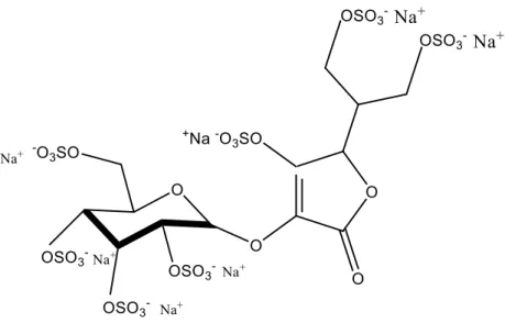
Figure 9 Sulphate ascorbic acid-2-glucoside structural formula.
Figure 10 Saturated solutions in water of each antioxidant in study at 25ºC. By the
following order from left to right: AA2G, AA, 3OAA, MAP, SAP and AP.
Figure 11 UV/VIS spectrum of saturated AA concentration at 25ºC. Figure 12 UV/VIS spectrum of saturated AP concentration at 25ºC. Figure 13 UV/VIS spectrum of saturated AA2G concentration at 25ºC. Figure 14 UV/VIS spectrum of saturated MAP concentration at 25ºC. Figure 15 UV/VIS spectrum of saturated SAP concentration at 25ºC Figure 16 UV/VIS spectrum of saturated 3OAA concentration at 25ºC.
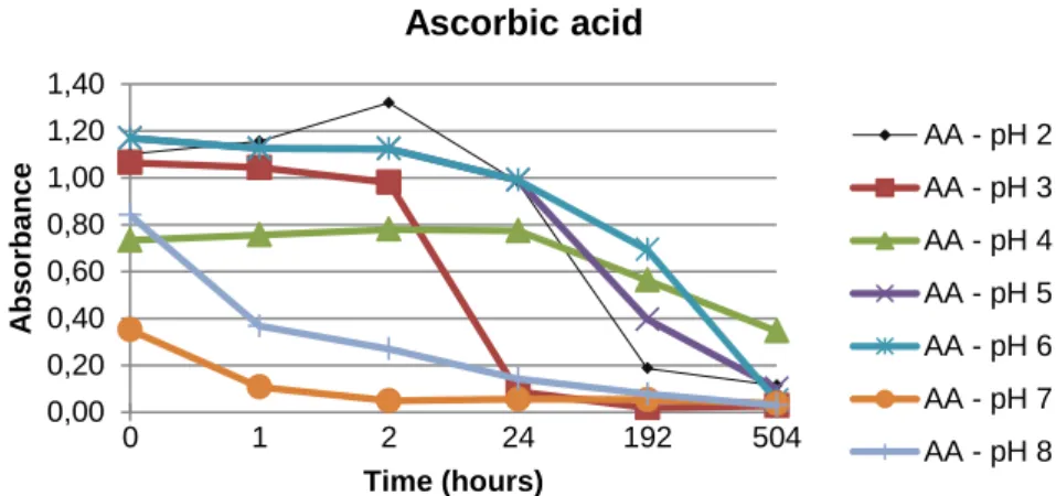
Figure 17 The relative AA stability given by variation in pH buffers over the time of
analysis (0, 1, 2 hours and 1, 8 and 21 days) at 25ºC.
Figure 18 The relative AP stability given by variation in pH buffers over the time of
analysis (0, 1, 2 hours and 1, 8 and 21 days) at 25ºC.
Figure 19 The relative MAP stability given by variation in pH buffers over the time
of analysis (0, 1, 2 hours and 1, 8 and 21 days) at 25ºC.
Figure 20 The relative SAP stability given by variation in pH buffers over the time
of analysis (0, 1, 2 hours and 1, 8 and 21 days) at 25ºC.
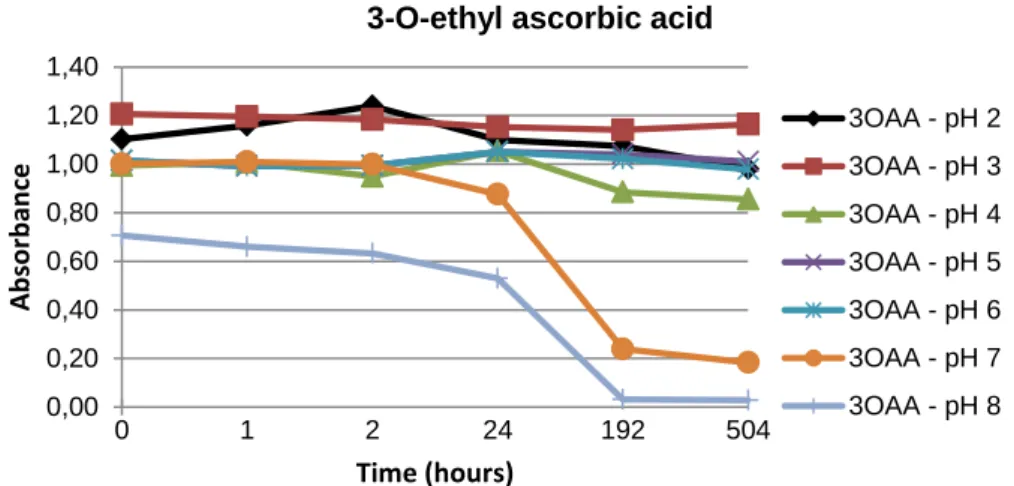
Figure 21 The relative 3OAA stability given by variation in pH buffers over the time
of analysis (0, 1, 2 hours and 1, 8 and 21 days) at 25ºC.
Figure 22 The relative AA2G stability given by variation in pH buffers over the time
of analysis (0, 1, 2 hours and 1, 8 and 21 days) at 25ºC
Figure 23 The relative AA2GS stability given by variation in pH buffers over the
time of analysis (0, 1, 2 hours and 1, 8 and 21 days) at 25ºC
5 9 13 15 18 21 24 26 28 48 49 49 49 49 49 49 57 57 57 57 58 58 58
v
Figure 24 The relative AA stability given by variation in temperature over the time
of analysis (0, 1, 2 hours and 1, 8 and 21 days) buffered at pH 5.
Figure 25 The relative AP stability given by variation in temperature over the time
of analysis (0, 1, 2 hours and 1, 8 and 21 days) buffered at pH 5.
Figure 26 The relative MAP stability given by variation in temperature over the time
of analysis (0, 1, 2 hours and 1, 8 and 21 days) buffered at pH 5.
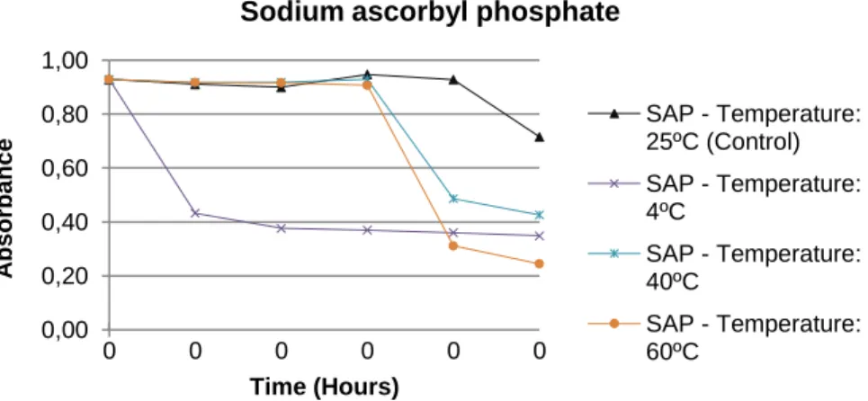
Figure 27 The relative SAP stability given by variation in temperature over the time
of analysis (0, 1, 2 hours and 1, 8 and 21 days) buffered at pH 5.
Figure 28 The relative 3OAA stability given by variation in temperature over the
time of analysis (0, 1, 2 hours and 1, 8 and 21 days) buffered at pH 5.
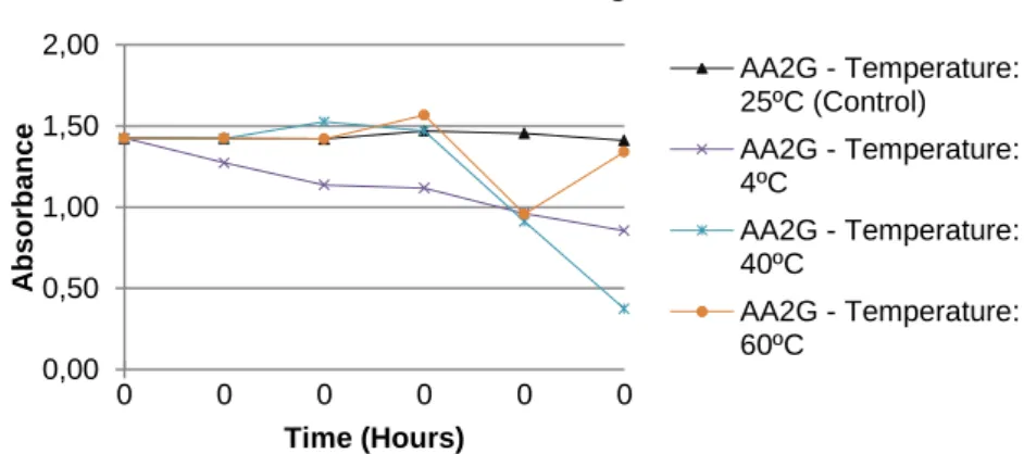
Figure 29 The relative AA2G stability given by variation in temperature over the
time of analysis (0, 1, 2 hours and 1, 8 and 21 days) buffered at pH 5.
Figure 30 The relative AA2GS stability given by variation in temperature over the
time of analysis (0, 1, 2 hours and 1, 8 and 21 days) buffered at pH 5.
Figure 31 Antioxidants solutions in presence of 50 μM FeCl3 solution at different
periods of time (0, 1 and 2 hours and 1, 8 and 21 days).
Figure 32 Antioxidants solutions in presence of 50 μM FeSO4 solution at different
periods of time (0, 1 and 2 hours and 1, 8 and 21 days).
Figure 33 Antioxidants solutions in presence of 50 μM CuSO4 solution at different
periods of time (0, 1 and 2 hours and 1, 8 and 21 days).
Figure 34 Antioxidants solutions in presence of 50 μM MgSO4 solution at different
periods of time (0, 1 and 2 hours and 1, 8 and 21 days).
Figure 35 Antioxidants solutions in presence of 50 μM Ca(OH)2 solution at different
periods of time (0, 1 and 2 hours and 1, 8 and 21 days).
Figure 36 Photostability of AA at 0, 5, 10 and 15 minutes under exposure of UV
light.
Figure 37 Photostability of AP at 0, 5, 10 and 15 minutes under exposure of UV
light.
Figure 38 Photostability of SAP at 0, 5, 10 and 15 minutes under exposure of UV
light.
Figure 39 Photostability of MAP at 0, 5, 10 and 15 minutes under exposure of UV
light.
Figure 40 Photostability of 3OAA at 0, 5, 10 and 15 minutes under exposure of UV
light.
Figure 41 Photostability of AA2G at 0, 5, 10 and 15 minutes under exposure of UV
light. 64 64 64 64 65 65 65 70 70 70 70 70 75 75 75 75 75 75 75
vi
Figure 42 Photostability of AA2GS at 0, 5, 10 and 15 minutes under exposure of
UV light.
Figure 43 AA curve of DSC in heating rate of 10ºC min-1 Figure 44 AP curve of DSC in heating rate of 10ºC min-1 Figure 45 MAP curve of DSC in heating rate of 10ºC min-1 Figure 46 SAP curve of DSC in heating rate of 10ºC min-1 Figure 47 3OAA curve of DSC in heating rate of 10ºC min-1 Figure 48 AA2G curve of DSC in heating rate of 10ºC min-1 Figure 49 AA2GS curve of DSC in heating rate of 10ºC min-1 Figure 50 AA + AEE curve of DSC in heating rate of 10ºC min-1 Figure 51 AP + AEE curve of DSC in heating rate of 10ºC min-1 Figure 52 MAP + AEE curve of DSC in heating rate of 10ºC min-1 Figure 53 SAP + AEE curve of DSC in heating rate of 10ºC min-1 Figure 54 3OAA + AEE curve of DSC in heating rate of 10ºC min-1 Figure 55 AA2G + AEE curve of DSC in heating rate of 10ºC min-1 Figure 56 AA2GS + AEE curve of DSC in heating rate of 10ºC min-1 Figure 57 AA + CP curve of DSC in heating rate of 10ºC min-1 Figure 58 AP + CP curve of DSC in heating rate of 10ºC min-1 Figure 59 MAP + CP curve of DSC in heating rate of 10ºC min-1 Figure 60 SAP + CP curve of DSC in heating rate of 10ºC min-1 Figure 61 3OAA + CP curve of DSC in heating rate of 10ºC min-1 Figure 62 AA2G + CP curve of DSC in heating rate of 10ºC min-1 Figure 63 AA2GS + CP curve of DSC in heating rate of 10ºC min-1 Figure 64 AA + EDTA curve of DSC in heating rate of 10ºC min-1 Figure 65 AP + EDTA curve of DSC in heating rate of 10ºC min-1 Figure 66 MAP + EDTA curve of DSC in heating rate of 10ºC min-1 Figure 67 SAP + EDTA curve of DSC in heating rate of 10ºC min-1 Figure 68 3OAA + EDTA curve of DSC in heating rate of 10ºC min-1 Figure 69 AA2G + EDTA curve of DSC in heating rate of 10ºC min-1 Figure 70 AA2GS + EDTA curve of DSC in heating rate of 10ºC min-1 Figure 71 AA + HEC curve of DSC in heating rate of 10ºC min-1 Figure 72 AP + HEC curve of DSC in heating rate of 10ºC min-1 Figure 73 MAP + HEC curve of DSC in heating rate of 10ºC min-1 Figure 74 SAP + HEC curve of DSC in heating rate of 10ºC min-1 Figure 75 3OAA + HEC curve of DSC in heating rate of 10ºC min-1 Figure 76 AA2G + HEC curve of DSC in heating rate of 10ºC min-1
76 80 80 80 80 81 81 81 83 83 83 83 84 84 84 85 85 85 85 86 86 86 87 87 87 87 88 88 88 89 89 89 89 90 90
vii
Figure 77 AA2GS + HEC curve of DSC in heating rate of 10ºC min-1 Figure 78 AA + MIG curve of DSC in heating rate of 10ºC min-1 Figure 79 AP + MIG curve of DSC in heating rate of 10ºC min-1 Figure 80 MAP + MIG curve of DSC in heating rate of 10ºC min-1 Figure 81 SAP + MIG curve of DSC in heating rate of 10ºC min-1 Figure 82 3OAA + MIG curve of DSC in heating rate of 10ºC min-1 Figure 83 AA2G + MIG curve of DSC in heating rate of 10ºC min-1 Figure 84 AA2GS + MIG curve of DSC in heating rate of 10ºC min-1 Figure 85 AA + PCP curve of DSC in heating rate of 10ºC min-1 Figure 86 AP + PCP curve of DSC in heating rate of 10ºC min-1
Figure 87 MAP + PCP curve of DSC in heating rate of 10ºC min-1 Figure 88 SAP + PCP curve of DSC in heating rate of 10ºC min-1 Figure 89 3OAA + PCP curve of DSC in heating rate of 10ºC min-1 Figure 90 AA2G + PCP curve of DSC in heating rate of 10ºC min-1 Figure 91 AA2GS + PCP curve of DSC in heating rate of 10ºC min-1 Figure 92 AA + TP curve of DSC in heating rate of 10ºC min-1 Figure 93 AP + TP curve of DSC in heating rate of 10ºC min-1 Figure 94 MAP + TP curve of DSC in heating rate of 10ºC min-1 Figure 95 SAP + TP curve of DSC in heating rate of 10ºC min-1 Figure 96 3OAA + TP curve of DSC in heating rate of 10ºC min-1 Figure 97 AA2G + TP curve of DSC in heating rate of 10ºC min-1 Figure 98 AA2GS + AEE curve of DSC in heating rate of 10ºC min-1.
90 91 91 91 91 92 92 92 93 93 93 93 94 94 94 95 95 95 95 96 96 96
viii
Index of tables
Table 1 Physical and chemical properties of ascorbic acid. Table 2 Physical and chemical properties of ascorbyl palmitate. Table 3 Physical and chemical properties of ascorbic acid-2-glucoside. Table 4 Physical and chemical properties of magnesium ascorbyl phosphate.
Table 5 Physical and chemical properties of sodium ascorbyl phosphate. Table 6 Physical and chemical properties of 3-O-ethylascorbic acid.
Table 7 Physical and chemical properties of sulphate ascorbic acid-2-glucoside. Table 8 Solubility criteria European Pharmacopea
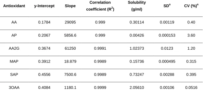
Table 9 Linearity data for ascorbic acid and its derivatives quantification using a
spectrophotometric methodology (280nm)
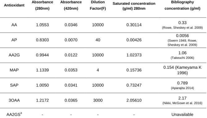
Table 10 Comparison the values at ultraviolet and visible light; and, comparison
obtained values in experimental analyses with available bibliography data.
Table 11 Linearity data for ascorbic acid and its derivatives quantification using a
spectrophotometric methodology (280nm)
Table 12 Comparison between the experimental values of solubility obtained with
bibliography data available.
Table 13 Stability classification criteria of antioxidants in coordination with tested
variants.
Table 14 Stability over time of AA, AP, MAP, SAP, 3OAA, AA2G and AA2GS in
different buffered solution at pH 2, 3, 4, 5, 6, 7 and 8.
Table 15 Stability over the time of AA, AP, MAP, SAP, 3OAA, AA2G and AA2GS
buffered solution at 5 at 4, 25, 40 and 60ºC.
Table 16 Summary of antioxidants solutions in presence of metallic solutions at
different times of measurement.
Table 17 Raw materials in compatibility study between antioxidants and excipients. Table 18 DSC thermoanalytical data of antioxidants in the heating rate of 10ºC
min1.
Table 19 DSC thermoanalytical data of excipients in the heating rate of 10ºC min-1.
Table 20 DSC thermoanalytical data of binary mixtures 1:1 (w/w) between AA, AP,
MAP, SAP, 3OAA, AA2G and AA2GS plus excipients in the heating rate of 10ºC min-1.
Table 21 Antioxidant activity of each antioxidant given by EC50 values (μM).
6 16 19 22 25 27 28 48 50 51 52 53 59 60 66 71 79 82 97 98 103
ix
Index of equations
Equation 1 DPPH reduction in presence of antioxidant 43 Equation 2 Calculation of antioxidant activity percentage in DPPH assay. 43
x
Abbreviations
3OAA 3-O-Ethyl-L-ascorbic acid AA Ascorbic acid AA2GAscorbic acid 2-O-α-glucoside AA2GS
Sulphate ascorbic acid-2-glucoside ADME
Absorption, distribution, metabolism and excretion AEE Stearyl alcohol AP Ascorbyl-6-palmitate CP Carbopol DHA Dehydroascorbic acid DNA Deoxyribonucleic acid DPPH 2,2-diphenyl-1-picrylhydrazyl DSC
Differential Scanning Calorimeter FTIR
Fourrier transformed infrared spectroscopy GSH Glutathione HEC Hydroxyethyl cellulose IL Interleukin LAA L-ascorbic acid MAP Magnesium ascorbyl-2-phosphate MIG Miglyol 812 NFkB
Nuclear transcription factor kappa B, NMF
Natural moisturizing factor PCP
Potassium cetyl phosphate ROS
Reactive oxygen species SAP Sodium ascorbyl-2-phosphate SDA Semydehydroascorbic acid T Temperature TNF-α
Tumour Necrosis Factor α TP Tocopherol UV Ultraviolet light Vis Visible ΔH Enthalpy
xi
Abstract
Skin health and beauty are cornerstones of general well-being in humans. Anti-aging cosmetics are used to provide a healthy and youthful appearance. Among different cosmetic actives, antioxidants have been incorporated in anti-aging products. Ascorbic acid and its derivatives are being consistently the most used antioxidants in anti-aging formulations.
Ascorbic acid possesses a variety of cutaneous benefits, besides antioxidant activity, including photoprotection from UVA and B radiation, neocollagenesis, inhibition of melanogenesis and improvement of a variety of inflammatory skin disorders. One of the main challenges with using vitamin C in pharmaceutical and cosmetic formulations is its chemical instability, which fostered the development of derivatives with higher stability. The main aims of this dissertation were to investigate the solubility in water and glycerol, at different pH values (2, 3, 4, 5, 6, 7, 8), the thermal stability (4ºC, 25ºC, 40ºC, 60ºC), the stability in presence of metal ions (Ca2+, Mg 2+, Fe2+, Fe3+, Cu2+) and the photostability of ascorbic acid (AA) and five derivatives commercially available - ascorbyl palmitate, magnesium ascorbyl phosphate, sodium ascorbyl phosphate, 3-O-ethyl ascorbic acid, ascorbic acid-2-glucoside – and one new derivative - sulphate ascorbic acid-2-glucoside. The compatibility of these antioxidants with common excipients of topical formulations (stearilic alcohol, carbopol, hydroxyethylcellulose, tocopherol, potassium cetyl phosphate, EDTA and miglyol) was evaluated with DSC. Additionally, their antioxidant activity was also compared by the DPPH reduction assay.
The stability of ascorbic acid and its derivatives studied in dependence on the pH of their aqueous solution revealed that at pH 5 the majority of antioxidants are stable. The derivatives of AA demonstrated to be more stable than AA in the range investigated. Exposure to high temperatures revealed that AA derivatives were also relatively stable. Preferentially, the storage temperatures recommended are 4ºC and 25ºC.
In the presence of Mg2+, AA does not suffer any decrease in its concentration, suggesting that magnesium ions do not affect AA stability. The stability of MAP, SAP and 3OAA against Fe2+ and Fe3+ ion was shown to be high with 90% of their concentration maintained even after 24 h in 100 µM of Fe2+ and Fe3+ ion, whereas AA was almost completely degraded under these conditions. Likewise, compound 3OAA was more stable
xii than AA and MAP and SAP, although 3OAA stability was relatively lower in the presence of Fe3+ ion.
The results of photostability evaluation showed that, after 15 min of UV exposure, a 40% decrease of AA absorption was noted and AP suffers bathochromic alterations in spectrum. The other antioxidants showed better stability than AA. With respect to thermal analysis of AA, AP, MAP, SAP and AA2G one exothermic peak was detected. In case of AA2GS an endothermic peak was observed. After analysing the thermograms from binary mixtures, strong interactions between the antioxidants and EDTA, and PCP can be assumed because the active ingredient melting peak almost disappeared and/or Tpeak is
not possible to be precisely determined. AEE, CP, EDTA, PCP, TP, MIG, HEC it can be assumed that some excipients did not affect some antioxidants thermal behaviour without any apparent physical-chemical change or incompatibility. The antioxidants with higher compatibility with the tested excipients were SAP, AA2G and AA2GS. AP and 3OAA was the antioxidant with more incompatibilities.
The highest antioxidant activity, as evaluated with DPPH reduction assay, was obtained for AA followed by 3OAA, SAP, AA2G and AA2GS.
Overall, the most stable derivative of AA was found to be 3OAA. The new compound has a similar behaviour as its parent AA2G and is highlighted by its highest compatibility with a variety of typical excipients of semi-solid formulations.
xiii
Resumo
A saúde da pele e a sua beleza são pedras angulares no bem-estar geral dos humanos. Os cosméticos anti-envelhecimento são utilizados de forma a fornecer uma aparência saudável e jovem. Entre os diferentes ingredientes activos dos cosméticos, os antioxidantes têm sido incorporados nos produtos anti-idade. Topicamente aplicados os antioxidantes exercem os seus benefícios ao oferecer protecção contra os radicais livres. Os demais produtos cosméticos incorporam antioxidantes são os mais populares produtos anti-idade. O ácido ascórbico e os seus derivados têm sido consistentemente os antioxidantes mais utilizados nas formulações anti-idade. Os resultados de um estudo prévio demonstraram que de entre os demais antioxidantes, durante os últimos cinco anos, o ácido ascórbico e os seus derivados são amplamente utilizados. Em 2015, dois derivados incluídos nesta dissertação, o ácido ascórbico glicosilado e o palmitate de ascorbilo apareciam no ―top ten‖ dos antioxidantes mais utilizados. Em 2011 e 2013, o ácido ascórbico e os seus derivados – o fosfato de magnésio e fosfato de sódio do ácido ascórbico permaneceram no ―top ten‖.
O ácido ascórbico tem sido amplamente utilizado, por possuir uma variedade de benefícios cutâneos, além da sua actividade antioxidante, incluindo a fotoproteção contra a radiação UVA e B, a promoção da neocolagénese, a inibição da melanogénese e a melhoria de uma variedade de desordens inflamatórias da pele. Um dos principais desafios com o uso de vitamina C em formulações farmacêuticas e cosméticas é sua instabilidade química, que promoveu o desenvolvimento de derivados com maior estabilidade.
Os objectivos principais desta dissertação foram investigar a solubilidade em água e glicerina, em valores de pH diferentes (2, 3, 4, 5, 6, 7, 8), a estabilidade térmica (4ºC, 25ºC, 40ºC, 60ºC), a estabilidade na presença de iões metálicos (Ca2+, Mg 2+, Fe2+, Fe3+, Cu2+) e a fotoestabilidade do ácido ascórbico (AA) e de cinco derivados disponíveis comercialmente - palmitato de ascorbilo, fosfato de ascorbilo e magnésio, fosfato de ascorbilo e sódio , ácido etil-3-O- ascórbico, ácido ascórbico-2-glicosídeo – e um novo derivado – o sulfato do ácido ascórbico-2-glicosídeo. A compatibilidade destes antioxidantes com excipientes comuns em formulações tópicas (álcool estearílico, carbopol, hidroxietilcelulose, tocoferol, fosfato de potássio cetílico, EDTA e miglyol) foi
xiv avaliada por DSC. Além disso, a sua actividade antioxidante foi também comparada pelo ensaio de redução do DPPH.
A estabilidade do ácido ascórbico e seus derivados foi estudada em relação ao pH das suas soluções aquosas e revelou-se que, a pH 5, a maioria dos antioxidantes é estável. Os derivados de AA demonstraram ser mais estáveis do que AA na faixa de pH investigados. A exposição a temperaturas elevadas revelou que os derivados de AA também eram relativamente estáveis. Preferencialmente, as temperaturas de armazenamento recomendadas são 4ºC e 25ºC.
Na presença de Mg2+, o AA não sofre qualquer diminuição da sua concentração, sugerindo que os iões de magnésio não afectam a estabilidade do AA. A estabilidade de compostos MAP, SAP e 3OAA na presença de Fe2+ e Fe3+ é elevada com 90% da sua concentração mantida mesmo após 24 h em 100 µM iões Fe2+ e Fe3+, enquanto o AA foi quase completamente degradado nestas condições. O composto 3OAA demonstrou ser mais estável que o AA, o MAP e o SAP, embora a sua estabilidade ter sido relativamente menor na presença de iões de Fe3+.
Os resultados da avaliação da fotoestabilidade mostraram que, após 15 min de exposição aos raios UV uma diminuição de 40% da absorção de AA e, por seu turno, o AP sofre alterações batocrómicas no espectro. Os outros antioxidantes mostraram melhor estabilidade do que AA. No que diz respeito a análise térmica de AA, AP, MAP, SAP e AA2G apresentam um pico exotérmico. No caso de AA2GS, observou-se um pico endotérmico. Depois de analisar os termogramas de misturas binárias, com AEE, CP, EDTA, PCP, TP, MIG, HEC, pode-se supor que estes excipientes não afectam o comportamento térmico dos antioxidantes não tendo sido observado qualquer mudança físico-química aparente ou incompatibilidade. Os antioxidantes com maior compatibilidade com os excipientes estadas foram AA2G e AA2GS.
A actividade antioxidante mais elevada, conforme avaliado com ensaio de redução de DPPH, obteve-se para AA seguido por 3OAA, SAP e AA2G e AA2GS.
No geral, o derivado mais estável da AA é o 3OAA. O novo composto (AA2GS) apresenta um comportamento semelhante ao seu análogo AA2G e é destacado pela sua elevada compatibilidade com os excipientes utilizados nas preparações semi-sólidas.
Palavras-chave: Ácido ascórbico, derivados do ácido ascórbico, estabilidade,
xvi
Outline of this dissertation
CHAPTER 1 – INTRODUCTION
Chapter 1 includes a brief introduction to ascorbic acid and its derivatives as anti-aging active ingredients with interest in formulation of cosmetics. Their physical and chemical properties, pharmaceutical and technologic aspects are also presented.
CHAPTER 2 – AIMS
In this chapter, the main objectives are described. CHAPTER 3 - MATERIAL AND METHODS
In this chapter, the general methods including the materials used, reagents and also characterization methods are described as well as the experimental procedures and conditions used in each study.
CHAPTER 4 - RESULTS AND DISCUSSION
Results and discussion are divided into four topics. The first topic describes the studies towards the solubility in water and glycerol. The second topic concerns the chemical stability of all compounds. This topic is also subdivided in evaluation of pH stability, thermal stability, and stability in presence of metals and photostability. The third topic deals with the compatibility of compounds with broadly employed excipients in cosmetic formulations. Finally, the fourth topic describes the results of the antioxidant capacity of each investigated compound.
CHAPTER 5 – CONCLUSIONS
This chapter summarizes the main conclusions concerning this dissertation. CHAPTER 6 – REFERENCES
In this chapter, the references cited throughout the dissertation are presented. The main bibliographic search engines/databases were ISI Web of Knowledge, Scopus, PubMed and Google Scholar.
I
2
Chapter I: Introduction
Aging is defined by some authors as an universal progressive irreversible intrinsic process which all living things suffer from, as an expression of the interaction between genetics of the individual and their environment aggressors (Reynolds D. et al, 2002; Murkheerjee P. et al., 2011). Avoid aging has been one of the greatest ambitions of human beings and, consequently, to combat aging is a challenge for modern medicine. In modern society, there is a great increase in the search for eternal youth (Silva, JA et
al. 2010; Murkheerjee P. et al., 2011). Over time, most of the functions of the various
organs and tissues in the body decrease their activity, either by alterations in cellular metabolic activity or by processes that affect these cells. With the aging associated with the chronological age, a proteolytic degradation of the fiber network at a cellular level occurs, which leaves visible signs on the surface of the skin (Murkheerjee P. et
al., 2011). All individual systems exhibit this process, but each one develops differently,
what characterizes old age of great biological variability (Reynolds D. et al, 2002; Murkheerjee P. et al., 2011).
There are many cosmetic ingredients that are claimed to have anti-aging effects when used topically. These products are geared in the prevention and treatment of broad term anti-aging, which derives mainly from photoaging that is characterized by clinical signs including irregular dryness, dark/light pigmentation, sallowness, deep furrows or severe atrophy, telangiectases, premalignant lesions, laxity, and a leathery appearance (Burke K, 2004; Silva JA et al. 2010). Other signs include elastosis (a coarse, yellow, cobblestoned effect of the skin) and actinic purpura (easy bruising related to vascular wall fragility in the dermis) (Bissett D., 2005). Therefore, the anti-aging cosmetics are based on the prevention of these signs in support of improving the wide range of signals, in which they can bring benefits such as wrinkles, sagging, texture, pallor, hyperpigmentation, etc. (Bissett D., 2005; Burke K, 2004; Murkheerjee P. et al., 2011; Silva, JA et al. 2010).
Burke et al. (2006) defends that topical application of antioxidants can give far higher concentrations in the skin than maximal oral intake. However, the correct formulation is of utmost importance to attain efficacy. The challenge is to use the correct form of the antioxidant molecule, to keep the antioxidant active to attain a reasonable shelf-life for the product, and to achieve effective transcutaneous absorption that delivers effectively high concentrations of the active antioxidant to the dermis as well as the epidermis.
3 The results of a previous study showed that among antioxidants, over five years AA and derivatives are widely used. In 2015, two derivatives included in this dissertation, ascorbyl glucoside and ascorbyl palmitate reached the top ten antioxidants. In 2011 and 2013, ascorbic acid and its derivatives: magnesium and sodium ascorbyl phosphate reached the top ten. For this reason it is compiled in this dissertation a comparative study of ascorbic acid and derivatives: magnesium ascorbyl phosphate (MAP), ascorbyl palmitate (AP), sodium ascorbyl phosphate (SAP), ascorbic acid-2-glucoside (AA2G), sulphate ascorbic acid-2-acid-2-glucoside (AA2GS) and 3-O-ethyl ascorbic acid (3OAA) to analyse the best physical and chemical characteristics that justifies a widely use in anti-aging cosmetics.
4
Vitamin C
Discovery
Following the outbreak of scurvy during World War I, it was shown that germinating, but not dry, cereals and legumes are effective against scurvy in monkeys and guinea pigs (Bissett DL, 2005). Although, it was not known which compound present in these fresh foods reduced the expression of scurvy. However, people linked the appearance of scorbutus with an insufficient ingestion of vegetables and fruits. Like this, scorbutus was common in discoveries epoch and in greatest wars where food resources are limited (Nakamura S, 2009; Takebayashi J et al. 2006; Santos M, 2014). In 1928, Albert Szent-Gyorgyi isolated a six-carbon reducing substance from oranges and cabbages. In 1932, he and C.C. King showed this substance to be the antiscorbutic principle when identifying its role in scurvy (Cadenas E, Packer L., 2007; Telang P, 2013). Albert Szent-Gyorgyi named it ascorbic acid (AA) and was awarded the Nobel Prize in 1937 (Bissett DL, 2005; Cadenas E, Packer L., 2007).
Sources
Most plants and animals can synthesise ascorbic acid (AA), in vivo from glucose. Animals synthesize AA from glucose in the liver (in mammals) or kidneys (birds and reptiles). Several species of animals, distributed through the evolutionary tree, are unable to synthetize vitamin C. These include human and nonhuman primates, guinea pigs, indian fruit bats, bulbus, and some fish (Cadenas E, Packer L., 2007; Pinnell SR, 2003).
In the human, the gene encoding gulonolactone oxidase enzyme has extensive mutations, so that there is no protein product (Cadenas E, Packer L., 2007; Pinnell SR, 2003). Humans and animal‘s incapable to synthetize vitamin C usually obtain from exogenous sources sufficient amounts from their largely plant diet acquired from citrus fruit, green leafy vegetables, strawberries, papaya and broccoli where this antioxidant is present in its reduced form: L-ascorbic acid (LAA) and in its oxidation product dehydroascorbic acid (DHA), both with vitamin activity (Telang P, 2013, Santos M, 2014). Vitamin C is synthetized by plants from several precursors and is abundant in leaves, and, in particular, the chloroplast. It may play a role in photosynthesis, stress resistance, and plant growth and development (Cadenas E, Packer L., 2007).
5
Vitamin C: physical and chemical properties
AA is a natural water-soluble vitamin (Stamford NP, 2012). This non-enzymatic antioxidant acts by scavenging and reducing reactive species to less reactive species (type 2 mechanism) (Telang P, 2013; Takebayashi J et al. 2002).
AA corresponding to a ketolactone with 6 carbons and the molecular formula C6H8O6
(Figure 1) is very unstable and easily oxidized in aqueous solutions and cosmetic formulations. It can be used with α-tocopherol acting as a co-antioxidant with a synergic effect (Telang P, 2013; Santos M, 2014).
Hence, vitamin C or ascorbic acid or even ascorbate is a low molecular weight six-carbon lactone with hydrophilic, acidity and power reducing character (Cadenas E, Packer L., 2007; Pinnell SR, 2013, Telang P, 2013; Santos M, 2014; Oroian et al. 2015). Table 1 summarizes and highlights the physical and chemical properties of AA.
6 Table 1 Physical and chemical properties of ascorbic acid (Rowe et al. 2009).
Property Ascorbic acid specifications
IUPAC name (2R)-2-[(1S)-1,2-dihydroxyethyl]z-3,4-dihydroxy-2H-furan-5-one
Appearance White, odourless, crystalline solid with sharp acidic state
Formula C6H8O6 Molecular mass 176.13 Density 1.65 g/cm3 pH ≈ 3 pKa pKa1=4.17 pKa2=11.57 Solubility (g/ml) Water 0.33 Glycerol (USP) 0.01
Fats and oils
solvents Insoluble
As mentioned above, AA has a 5-hydrocarbon ring similar to that of glucose. If in the ascorbic acid a hydrogen ion is associated, L-ascorbic acid (LAA) becomes a weak sugar acid, similar to other α-hydroxyacids used in dermatology (Davey M et al. 2000). With a metal ion, it forms a mineral ascorbate (Telang P, 2013; Takebayashi J, 2014).
In solution, delocalisation of the -electrons over the C2-C3 conjugated enediol system stabilises the molecule and causes the hydrogen of the C3 hydroxyl to become highly acidic, and to dissociate with a pKa of 4.13. AA is stable at a pH values below the first pKa. At physiological pH, LAA exists as a monovalent anion (L-ascorbate). Dissociation of the second hydroxyl takes place at pH 11.6 (Cadenas E, Packer L., 2007; Telang P, 2013; Takebayashi J, 2014; Austria R. et al. 1997).
7 L-Ascorbic acid is the chemically active form of vitamin C. In nature, ascorbic acid is found in equal parts as L-ascorbic acid (LAA) and D-ascorbic acid (DHA). These are essentially isomeric molecules and are mutually interchangeable as mentioned previously. Nevertheless, only LAA is biologically active and thus useful in pharmaceutical and cosmetic formulations (Humbert PG et al. 2003; Telang P, 2013; Shalmashi A et al. 2008; Hacisekvi A, 2009; Sheraz M et al. 2011). AA in powder is relatively stable in air. In the absence of oxygen and other oxidizing agents it is also heat stable (Rowe R et al. 2009).
AA oxidation is generated by its ionization in aqueous solution (Austria R. et al. 1997; Stamford NP, 2012). In solution, AA readily undergoes oxidation; however, other factors increase the rate of degradation of vitamin C particularly alkaline solution (pH>7), Ultraviolet light (UV) exposure and heat, metal ions in amounts that catalyse its oxidation (e.g. traces of copper and iron and other oxidants) and presence of dissolved oxygen (Tipson R, Horton D, 1997; Davey M et al. 2000; Stamford NP, 2012). Ascorbic acid solutions exhibit maximum stability at about pH 5.4 (Rowe R et al. 2009; Stamford NP, 2012).
This vitamin is usually used at 1 to 20% of concentration in topical formulations containing an acidic pH (around 3.5) to effectively penetrate the skin (Sheraz M et al. 2011). Vitamin C is extremely reactive and unstable in dispersions due to its fast oxidation and further irreversible chemical transformation. Indeed, the degradation by oxidation of AA most often accompanied by a yellowish discolouration (Shalmashi A et
al. 2008; Stamford NP, 2012).
8
Ascorbic acid biological functions and metabolism
Besides its involvement in metabolic functions such as fighting bacterial infections, detoxifying reactions and formation of collagen, AA exerts several functions on the skin such as collagen synthesis, depigmentation and antioxidant activity, being widely used in anti-aging skin care formulations (Sheraz M et al. 2011; Silva GM, Maia Campos PM, 2000; Smart RC, Crawford CL, 1991; Stojiljkovic D et al. 2014).
AA is a cofactor for numerous enzymes participating in the post-translational hydroxylation of collagen, in the production of L-carnitine, in bioconversion of some neurotransmitters, and tyrosine metabolism (Hacisekvi A, 2009; Oresajo C et al. 2012; Pandel R et al. 2013; Pillai S et al. 2005; Pinnell SR, 2003).
AA has three main types of distinct biological activities in animals as an enzyme co-factor; as a direct physiological radical scavenger and, finally as a donor/acceptor in electron transport in plasma membrane (Masaki H, 2010; Mukherjee PK et al. 2011; Oresajo C et al. 2012; Smart RC, Crawford CL, 1991). In this manner, AA is a water soluble radical scavenger widely distributed in aerobic organisms that plays an essential role in defence of cellular components against oxidative damage by free radicals and oxidants that are involved in the progress of a chronic diseases such as cancer, brain dysfunction, aging (Masaki H, 2010; Pinnell SR, 2003), heart disease (Lynch SM et al. 1996), inflammation, and stroke (Telang P, 2013). Ascorbate is an electron donor, and this property accounts for its known and postulated functions (Sies H, 1997; Cadenas E, Packer L, 2007; Poljak B et al. 2011; Telang P, 2013). It is believed that AA is effective in scavenging superoxide radical anions, hydroxyl radicals, hydrogen peroxide, reactive nitrogen species and singlet oxygen (Oroian M et al. 2015). AA has, at a structural level, 4 hydroxyl groups which can donate hydrogen to an oxidizing system (Oroian M et al. 2015). So, AA has its redox potential are strictly associated with the electron rich C2-C3-enediol moiety of its five-membered lactone ring (Olabisi A, 2004). When it donates two electrons from mentioned bonds, the intermediate free radical semydehydroascorbic acid (SDA) – ascorbate free radical is formed - Figure 2 (Olabisi A, 2004; Nimse S, Pal D, 2015).
The ascorbate free radical is unstable, but is much less reactive than other free radicals provided by other compounds that have potential to form harmful free radicals (Cadenas E, Packer L, 2007; Oroian M et al. 2015). Besides, SDA can be reversibly reduced to ascorbate (Oroian M et al. 2015). These properties make AA an ideal
9 electron donor (Poljak B et al. 2011; Puvabanditsin P, Vongtongsri R, 2016; Stojiljkovic D et al. 2014).
SDA, being unstable, undergoes further oxidation to form the more stable product, DHA which can exist in more than one structural form, but only a few minutes at physiological pH (Cadenas E, Packer L, 2007; Nimse S, Pal D, 2015).
DHA can be reduced back to ascorbate by enzyme dehydroascorbic acid reductase in the presence of glutathione (GSH) (Pinnell SR, 2003; Telang P, 2013), with formation of glutathione disulphide or by enzymatic reduction (Cadenas E, Packer L, 2007). If not reduced, DHA decay as the lactone ring irreversibly opens and it is hydrolysed to 2,3-diketogulonic acid (Hacisekvi A, 2009; Humbert PG et al. 2003; Lynch SM et al. 1996;Nimse S, Pal D, 2015). Diketogulonic acid in turn is metabolized to xylose, xylonite, lyxonate, and oxalate with oxalate being a clinically significant end product of ascorbate metabolism(Cadenas E, Packer L, 2007). Molecular oxygen, with or without trace metals (iron, copper), superoxide, hydroxyl radicals, and hypochlorous acid, all can oxidize AA to DHA in biological systems (Cadenas E, Packer L, 2007; Colven RM, Pinnell SR, 1996).
Figure 2 Antioxidant mechanism of Ascorbic acid (AA) and its metabolism. AA protects organism due to oxidation of ascorbate to (semydehydroascorbic acid) SDA and then to DHA (dehydroascorbic acid) and has diverse functions to maintain the nor mal physiological state in humans. Adapted from Cadenas & Packer,200 7.
10
Skin effects
i. Ascorbic acid as skin antioxidant
Ascorbic acid (AA), the most abundant antioxidant in human skin, forms a part of the complex group of enzymatic and non-enzymatic antioxidants that co-exist to protect the skin from reactive oxygen species (ROS) (Cadenas E, Packer L, 2007; Telang P, 2013). As AA is water soluble, it functions in the aqueous compartments of the cell, it becomes the major aqueous phase reductant (Pinnell SR, 2003; Santos M, 2014). As previously described, AA has a reducing agent behaviour. Hence, AA donates two electrons sequentially, and when it loses these electrons becomes oxidized vitamin. Another substance is reduced. In this way, vitamin prevents oxidation of the reduced substance (Pinnell SR, 2003; Nimse S, Pal D, 2015).
Bendich, Machlin, Scandurra, Burton, and Wayner proposed the antioxidant mechanism of AA when it interacts with reactive specie (Oroian M et al. 2015). At physiological pH, the ascorbate anion is the predominant form present, due to the acidic nature of AA. This compound can undergo a reversible oxidation process and forms DHA, with ascorbyl radical formation. The ascorbyl radical is relatively unreactive and may react with other free radicals and the propagation of free radical reactions may be stopped (Pham-Huy L et al. 2008; Segal A, Moyano A, 2008; Pinnell SR, 2013; Stojiljkovic D et al. 2014; Pisoschi AM, 2015).
However, vitamin C is not able to act in lipophilic compartments (Olabisi 2004, Telang 2013). Therefore, AA cannot scavenge lipophilic radicals directly within the lipid compartments. In this case, this hydrophilic antioxidant acts as a synergist with tocopherol (TP) for the reduction of lipid peroxide radicals (Cadenas E, Packer L, 2007; Stamford NP, 2012; Pinnell SR 2013).
11
ii. Ascorbic acid in photoprotection and skin damage
The topical application of AA increase cutaneous levels of this vitamin, and these is associated with protection of the skin from UVB-induced oxidative damage as measured by decrease in UVB erythema and sunburn cell formation (Burke KE, 2004; Stamford NP, 2012). The level of AA diminishes with exposure of UV light; nevertheless, its ability in neutralization the ROS (superoxide anion, peroxide and singlet oxygen) formed due to UV exposure was demonstrated, being equally effective against both UVB (290-320 nm) and UVA (320-400 nm) (Telang P, 2013). These ROS present harmful potential to start chain or cascade reactions that damage the cells, resulting in direct chemical alterations of the cellular DNA, the cell membrane and the cellular proteins, including collagen (Pinnell SR, 2003; Telang P, 2013).
Sunscreens when properly applied prevent UV-induced erythema and thymine dimer mutations that contribute to cutaneous carcinogenesis. Similar sunscreens preventing photoageing to decrease UV-induced erythema, sunburn cell formation and inducing collagen repair. Thus, UV protection can be optimized when combined filters with a topical antioxidant (Lopez-Torres M et al. 1988; Saral Y et al. 2002; Puvabanditsin P, Vongtongsri R, 2016; Almeida M et al. 2010). AA does not absorb UV light but in sunscreen exerts an UV-protective effect by neutralizing free radicals. As already suggested, AA alone can provide photoprotection, still works best in conjunction with TP, the vitamin E, which potentiates the photoprotection action of ascorbate against UV-induced erythema and diminished the number of sunburn cells as compared to that seen for each antioxidant individually which underlines the synergistic effects against UV-induced oxidative damage (Colven RM, Pinnell SR, 1996; Pinnell SR, 2003; Puvabanditsin P, Vongtongsri R, 2016; Stamford NP, 2012).
12
iii. Collagen synthesis
AA is essential for collagen biosynthesis. It has been proposed that this vitamin influences quantitative collagen synthesis in addition to stimulating qualitative changes in the collagen molecule (Telang P, 2013). Once, ascorbate is evident in connective tissue and during collagen formation, acting as cofactor for the enzymes prolysyl and lysyl hydroxylase, both of which are required for the post-translational processing of types I and III collagen (Bisset DL, 2009; Masaki H, 2010, Stamford NP, 2012; Telang P, 2013). Additionally these enzymes are responsible for stabilizing and cross-linking the collagen molecules (Telang P, 2013; Santos M, 2014).
Another mechanism by which AA influences the collagen synthesis is by stimulation of lipid peroxidation, and the product of this process, malondialdehyde, in turn stimulates collagen gene expression (Bisset DL, 2009; Telang P, 2013).
AA also directly activates the transcription of collagen synthesis by increasing fibroblast proliferation resulting in greater collagen production and stabilizes procollagen mRNA (Stamford NP, 2012; Telang P, 2013). This role of AA in connective tissue is known at long time, but only since XVI century this evidence gained recognition, associated with signs and symptoms of scurvy (e.g. in blooding gums) (Telang P, 2013; Santos M, 2014). By these facts, AA might be presumed that possesses the potential to also increase collagen production for wrinkle appearance reduction (Stamford NP, 2012).
13
iii. Depigmenting agent
AA interrupts the key steps of melanogenesis (Pinnell SR, 2003) resulting in a depigmentation action. AA is a depigmenting agent due to its inhibitory effect on tyrosinase (Masaki H, 2010). Vitamin C interacts with copper ions at the tyrosinase-active site and inhibits action of the enzyme tyrosinase, thus decreasing the melanin formation (Smaoui S et al. 2013; Telang P, 2013). So, vitamin C supresses melanin synthesis when entering skin cells and reduces significantly the production of melanin by inhibition tyrosinase activity. This results in a reduction of dopaquinone, an intermediate of melanin synthesis. Additionally, it converts exiting dopaquinone to L-dopa. Vitamin C can also reduce existing melanin exerting lightener of black melanin by a reduction reaction as schematized in Figure3 (Baertschi S, 2006; Hidrata L et al. 2014; Lorencini M et al. 2014; Olabisi A, 2004).
Figure3 Skin lightning mechanism of vitamin C.
v. Anti-inflammatory action of vitamin C
AA inhibits nuclear transcription factor kappa B (NFkB), which is responsible for the activation of a number of pro-inflammatory cytokines such as TNF-α, IL1, IL6 and IL8. Hence, AA has a potential anti-inflammatory activity and can be used in conditions like acne vulgaris and rosacea. Still, AA can promote wound healing and prevent post-inflammatory hyperpigmentation (Bisset DL, 2009; Stamford NP, 2012; Smaoui S et al. 2013; Telang P, 2013).
14
Topical pharmaceutical formulation aspects
Orally, AA is absorbed at gut level but when given by supplementation in high doses since this process is limited by an active transport mechanism (Coronado MH et al. 2015; Lobo V et al. 2010; Lorencini M et al. 2014; Telang P, 2013). Accordingly, bioavailability of AA in the skin is inadequate when it is administered per os. So, topical application of this antioxidant is the only way to increase skin concentrations. Therefore, its use is favoured in the practice of dermatology (Colven RM, Pinnell SR, 1996, Haftek M et al. 2008, Sheraz M et al. 2011, Pinnell SR, 2003, Telang P, 2013). Conversely, due to inherent hydrophilicity, penetration of AA across the skin is poor (Stamford NP, 2012). To increase penetration of the epidermal barrier, aqueous formulations of AA must be at a pH below the pKa of AA itself (pKa 4.2), thus reducing the charge density on the molecule (Sheraz M et al. 2011). At pH lower than 3.5, the stability of AA is controlled and the ionic charge on the molecule is removed and its well transported across the stratum corneum (Stamford NP, 2012; Pinnell SR, 2013). AA is available in the market as a variety of creams, serums and transdermal patches. Of these, only the serums contain active AA in an almost colourless form. It is unstable and, on exposure to light, gets oxidized to DHA, which imparts a yellow colour (Rowe
et al. 2009, Stamford NP, 2012, Sui et al. 2014). Accordingly with its typical instability,
the bulk material should be stored in an oxygen-impermeable non-metallic container, endangered from light, in a cool and dry place at low pH and minimum of water (Rowe, Sheskey et al. 2009, Stamford 2012, Sui et al. 2014).
Although AA is widely used in skin products to achieve clinical improvements, its poor skin penetration and its instability in formulations reduce its clinical efficacy as already described. To overcome these disadvantages, several AA derivatives, such as magnesium ascorbyl-2-phosphate (MAP), ascorbic acid 2-O-α-glucoside (AA2G), sodium ascorbyl phosphate (SAP), ascorbyl-6-palmitate (AP), and tetra-isopalmitoyl ascorbic acid, among others, have been synthetized and evaluated for their potential as pro-ascorbic acid derivatives (Austria R et al. 1997, Masaki H, 2010).
15
Ascorbyl palmitate
Ascorbyl palmitate (AP – Figure4), also known as vitamin C palmitate, L-ascorbyl-6-palmitate and 3-oxo-L-gulofuranolactone 6-L-ascorbyl-6-palmitate is a synthetic ester comprised of 16-carbon chain saturated fatty acid palmitic acid and LAA. The ester linkage is at the carbon 6 of AA (Mauludin R et al. 2014). AP is prepared by condensing palmitoyl chloride and AA in the presence of a dehydrochlorinating agent such as pyridine (Godic A et al. 2014).
Therefore, AP is a fat-soluble synthetic ester of AA with lipophilic properties because of its chemical structure (Austria R et al. 1997; Meves A et al. 2012). Accordingly, the molecules of AP are orientated with the palmitic residue in the lipophilic phase and the cyclic ring in the aqueous phase (Segal A, Moyano A, 2008). Hydrolysis of ascorbyl palmitate yields AA and palmitic acid (Lupo MP, 2001).
Figure4 Ascorbyl palmitate structural formula.
The AP lipophilic character seems to be an advantage to penetrate the stratum
corneum and also AP is pH neutral, making it non-irritating to the skin (Swern D, 1949;
Segal A, Moyano A, 2008). In the following table – Table 2 – are summarized physical and chemical properties of this AA derivative (Mauludin R et al. 2014; Segal A, Moyano A, 2008; Smart RC, Crawford CL, 1991).
16 Table 2 Physical and chemical properties of ascorbyl palmitate (Swern 1949, Rowe, Sheskey
et al. 2009).
Property Ascorbyl palmitate specifications
IUPAC name [2-(3,4-dihydroxy-5-oxo-2H-furan-2-yl)-2-hydroxyethyl] hexadecanoate
Appearance Odourless, white to yellowish powder
Formula C22H38O7
Molecular mass 414.54
Density 1.15 g/cm3
pH Neutral pH (non-acidic) molecule
pKa 4.45
Melting Point 107oC-117o C
Solubility (g/ml)
Water Practically insoluble: 0.0062 mg/m l Glycerol (USP) 0.01 mg/ml
Meves et al. 2002, was investigated the antioxidant effects of AP at cellular membranes. The antioxidant effect of AP has been recognized, because in its presence intracellular levels of ROS decreased. However, the same authors verified that lipid part of AP promotes UVB induced lipid peroxidation and formation of potentially toxic oxidized lipids that induces substantial cellular damage (Smart RC, Crawford CL, 1991; Perricone N, 1993; Meves A et al. 2012).
Other authors was found that AP is thirtyfold more effective than AA after topical application as an inhibitor of some of the biochemical parameters associated with tumour production in mice skin (Smart RC, Crawford CL, 1991). Likewise, AP when applied after UV burning reduces redness rather than patient that were untreated. The suspected mechanism is that AP acts as both an antioxidant and anti-inflammatory agent (Smart RC, Crawford CL, 1991; Perricone N, 1993; Lupo MP, 2001)
17 In terms of technological aspects, the cyclic ring of AP structure is sensitive to oxidation and can influence the formulation. In oil in water (O/W) emulsions, the cyclic ring (hydrophilic part of the molecule) is in the external aqueous phase, and with this, the stability of AP can be compromised (Segal A, Moyano A, 2008).
Other technological aspect is the fact that the combination of AP with α-tocopherol (TP) shows marked synergism, which increases the effect of the components and allows the amount, used to be reduced (Rowe et al. 2009). The solubility of AP in alcohol permits it to be used in non-aqueous and aqueous systems and emulsions.
Ascorbyl palmitate is touted as being good antioxidant to be incorporated in emulsions, creams, lotions, and oils (Lupo MP, 2001; Rowe et al. 2009; Pinnell SR, 2013).
Moreover, AP due to its amphiphilic character is able to penetrate the skin better and was shown to have better stability than AA (Smart RC, Crawford CL, 1991; Perricone N, 1993; Meves A et al. 2012; Mauludin R et al. 2014). Furthermore, AP appears to remain on the extracellular surface of cells and may not be readily converted to LAA (Smart RC, Crawford CL, 1991; Perricone N, 1993; Meves A et al. 2012; Mauludin R et
al. 2014).
The main problem of AP is, its oxidation mediated by transition metal ions presented in traces. Besides oxygen, light can also accelerate oxidative degradation of AP (Meves A
et al. 2012). AP is stable in the dry state, but is gradually oxidized and becomes
discoloured when expose to light and high humidity. In an unopened container, stored in a cool place, it has a shelf life of at least 12 months (Meves A et al. 2012; Mauludin R et al. 2014).
18
Ascorbic acid-2-glucoside
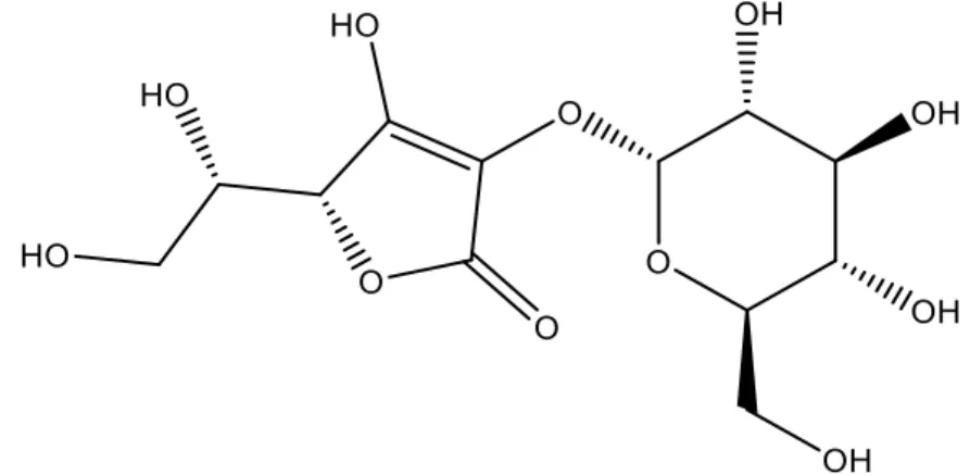
Ascorbic acid-2-O-glucoside (AA2G– Figure5), is a glycoside of disaccharide in which glucose and AA are bound with an 1,2-linkage (Nakamura S, Oku T, 2009).
Figure5 Ascorbic acid-2-glucoside structural formula.
Ascorbic acid-2-glucoside or 2-O-α-D-glucopyranosyl-L-ascorbic acid is a stable derivative of AA which is efficiently synthesized by regioselective transglucosylation with α-glucosidase and cyclodextrin glucosyltransferase (CGTase) (Nakamura S, Oku T, 2009; Nakazawa H et al. 2012; Nayama S et al. 1999). Glycosylation of ascorbate at the 2-O position improves the stability and its feasibility as a radical scavenger has been reported (Nakamura S, Oku T, 2009). The hydroxyl group of the second carbon in AA is highly reactive and plays an important role in the biologic activities. However, it can also be the site of inactivation and decomposition. AA2G is produced from AA and starch by use of enzymes that reversibly bind glucose to the reactive hydroxyl group to protect the site from destruction (Nakamura S, Oku T, 2009).
Table 3 summarizes the main physical and chemical properties of ascorbic acid-2-glucoside.
19 Table 3 Physical and chemical properties of ascorbic acid -2-glucoside (Takeuchi 2006, 2016).
Property Ascorbic acid-2-glucoside specifications
IUPAC name
(2R)-2-[(1S)-1,2-dihydroxyethyl]-3-hydroxy-4- [(2R,3R,4S,5S,6R)-3,4,5-trihydroxy-6-(hydroxymethyl)oxan-2-yl]oxy-2H-furan-5-one Appearance White to yellowish white powder, or
crystalline powder Formula C12H18O11 Molecular mass 338.265 Density 1.83g/cm 3 pH 2.3-2.4 pKa N/A
Melting Point N/A
Solubility (g/ml)
Water 1.25 Glycerol
(USP) N/A
N/A – Information not available
In vivo, AA2G is easily hydrolysed by α-glucosidase, an enzyme present in the
membrane of skin cells, (Tagawa M, Tabata Y, 1988; Takebayashi J et al. 2002; Stamford NP, 2012) exhibiting biological activities as an antiscorbutic effect, collagen synthesis-enhancing effect melanin synthesis-inhibiting effect, antioxidative as free radical scavenging activities also with metal chelating activity and reducing ability and anti-aging effects (Huang WY et al. 2013; Nakamura S, Oku T, 2009). AA2G is hydrolysed over a prolonged period, resulting in consistent and sustained beneficial physiological effect on the skin (Tai A et al. 2014; Tai A et al. 2003; Takebayashi J et
al. 2008; Takebayashi J et al. 2006; Takebayashi J et al. 2002).
It has showed that even though AA2G has a higher stability its antioxidative capacity was lower than the activity in vivo of AA and AP. This can be explained by poor penetration of this compound through human stratum corneum due to its high hydrophilicity (Tai A et al. 2003; Takebayashi J et al. 2008).
AA2G has no cytotoxicity becoming an excellent AA derivative for topical application (Tai A et al. 2003; Takebayashi J et al. 2008). Accordingly, AA2G has been widely
20 used in cosmetic products practically used as a skin lightening cosmetic ingredient (Tai A et al. 2003; Takebayashi J et al. 2008). AA2G has superior formulation stability being able to resist discolouration and degradation, while retaining all biologic activities that provide lightening, UV damage protection and anti-aging properties (Tai A et al. 2003; Takebayashi J et al. 2008). AA2G is highly stable in solution even at high temperatures, low pH and in presence of metal ions. These facts help maintaining the final product quality (Takebayashi J et al. 2008; Takebayashi J et al. 2006; Takebayashi J et al. 2002). However, the degradation of this derivative is triggered by the temperature change and pH variation. The stability of this antioxidant as previously described is influenced by pH. For this reason, leaving it under prolonged conditions of strong acidity or alkalinity (pH 2-4 or pH 9-12) should be avoided (Takebayashi J et al. 2008; Takebayashi J et al. 2006; Takebayashi J et al. 2002). These variations were very critical for determining its bio functionality of AA2G in skin. The optimal condition of retaining AA2G with the highest stability was determined to be 55.3ºC and pH 6.4 (Takebayashi J et al. 2006; Takebayashi J et al. 2002).
21
Magnesium ascorbyl phosphate
Magnesium-L-ascorbyl-2-phosphate (MAP – Figure 6) is a stable water-soluble ascorbyl ester as inorganic precursor of vitamin C that ensures delivery of vitamin C to the skin (Austria R et al. 1997; Smaoui S et al. 2013; Telang P, 2013) exhibiting antioxidative effects (Tagawa M, Tabata Y, 1988).
Figure 6 Magnesium ascorbyl phosphate structural formula.
MAP is obtained by the esterification reaction of a hydroxyl group of AA with an inorganic chain, which protects the enediol ring from the degradation reactions (Tagawa M, Tabata Y, 1988). The hydroxyl group is modified by an inorganic phosphoric ester group introduced in position 2.
In Table 4 are shows physical and chemical properties of magnesium ascorbyl phosphate.
22 Table 4 Physical and chemical properties of magnesium ascorbyl phosphate (Silva and Campos 2000, Smaoui 2013).
Property Magnesium ascorbyl phosphate specifications
IUPAC name trimagnesium (2R)-2-[(1S)-1,2-dihydroxyethyl]-3,4-dihydroxy-2H-furan-5-one; diphosphate Appearance White or yellowish powder
Formula C6H8Mg3O14P2 Molecular mass 759.22 Density N/A pH 7 pKa N/A Melting Point >300ºC Solubility (g/ml) Water 1.54 Glycerol (USP) N/A
N/A – Information not available
MAP presents instability in presence of metal ions. This antioxidant is unstable under UV light, and is stable at 50ºC at least for 10 weeks (in aqueous solution) and 80ºC at least for 20 hours (Austria R et al. 1997, Stamford NP, 2012). When compared with AA and AP, MAP showed better stability after 60 days confirming that the phosphoric group has the capability of protect the enediol system from hydrolysis (Kameyama K et
al. 1996; Silva GM, Maia Campos PM, 2000; Smaoui S et al. 2013; Tagawa M, Tabata
Y, 1988). Unlike AA and AP that are sensitive to high pH and have a better stability at pH 3, MAP is stable at pH 7, like SAP. One study showed that this compound is highly unstable at acid pH (Austria R et al. 1997; Stamford NP, 2012).
MAP also demonstrates an inhibitory effect on melanogenesis, suppressing melanin formation by tyrosinase activity and melanoma cells and, thus, decrease the skin pigmentation, resulting in spots removal and whitening effect (Tai A et al. 2014; Tagawa M, Tabata Y, 1988). Thus, MAP has been mainly utilized as an active ingredient of whitening cosmetics (Kameyama K et al. 1996; Silva GM, Maia Campos PM, 2000; Smaoui S et al. 2013; Tagawa M, Tabata Y, 1988).